Article

Clinical Imaging and Dosimetry of a Pan-Cancer Targeting Alkylphosphocholine Analog, [124I]I-NM404

1
Department of Radiology, University of Wisconsin—Madison, Madison, WI 53705, USA
2
Department of Radiology and Imaging Sciences, Emory University, Atlanta, GA 30322, USA
3
University of Wisconsin Carbone Cancer Center, University of Wisconsin—Madison, Madison, WI 53705, USA
4
Department of Medical Physics, University of Wisconsin—Madison, Madison, WI 53705, USA
5
Neuroscience and Brain Disease Center, Graduate Institute of Biomedical Sciences, China Medical University, North District, Taichung City 404333, Taiwan
6
Department of Neurosurgery, Dell Medical School, The University of Texas at Austin, Austin, TX 78712, USA
*
Author to whom correspondence should be addressed.
Radiation 2022, 2(2), 215-227; https://doi.org/10.3390/radiation2020015
Submission received: 11 April 2022 / Revised: 3 May 2022 / Accepted: 11 May 2022 / Published: 17 May 2022
(This article belongs to the Topic Cancer Biology and Radiation Therapy)

Abstract

:

Simple Summary

Nearly 10 million people die from cancer each year. Diagnosing cancer as early as possible gives patients have the best chance for successful treatment. [124I]I-NM404 is an alkylphosphocholine (APC) analog that has the unique ability to seek out and enter cancer cells in a wide variety of solid primary and metastatic malignancies anywhere in the body. The difference in signal between tumor and normal organs provides physicians the necessary contrast to visualize the cancer. The aim of this study was to characterize the imaging and dosimetric characteristics of [124I]I-NM404 in the clinical setting.

Abstract

The purpose of this study was to assess organ dosimetry and clinical use of [124I]I-NM404, a radiotheranostic alkylphosphocholine (APC) analog, for accurate detection and characterization of a wide variety of solid primary and metastatic malignancies anywhere in the body. Methods: Patterns of [124I]I-NM404 uptake were quantitatively analyzed and qualitatively compared with [18F]FDG PET/CT in 14 patients (median age, 61.5 years; 7 males, 7 females) with refractory metastatic cancer who were enrolled in one of two Phase I imaging studies. Primary cancer types included bronchogenic (n = 7), colorectal (n = 1), prostate (n = 1), triple-negative breast (n = 1), head and neck (n = 2), pancreatic (n = 1) carcinoma, and melanoma (n = 1). Patients were administered [124I]I-NM404 and imaged via PET/CT at 1–2, 4–6, 24, and 48 h and at 5–10 days post injection, from top of the skull to mid-thigh. Volumes of interest were drawn over lungs, heart, liver, kidneys, and whole body for dosimetry estimation using OLINDA 1.1 Representative metastatic index lesions were chosen when applicable for each case with active sites of disease to calculate maximum and mean tumor-to-background ratios (TBRmax, TBRmean), using the adjacent normal organ parenchyma as background when possible. Results: Administrations of [124I]-NM404 were safe and well-tolerated. The organs with the highest estimated absorbed dose (mean ± SD) were the lungs (1.74 ± 0.39 mSv/MBq), heart wall (1.52 ± 0.29 mSv/MBq), liver (1.28 ± 0.21 mSv/MBq) and kidneys (1.09 ± 0.20 mSv/MBq). The effective dose was 0.77 ± 0.05 mSv/MBq. Preferential uptake within metastatic foci was observed with all cancer subtypes, TBRmax ranged from 1.95 to 15.36 and TBRmean ranged from 1.63 to 6.63. Robust sensitive imaging of lesions was enhanced by delayed timing (2–6 days after single injection of [124I]I-NM404, respectively) due to persistent tumor retention coupled with progressive washout of background activity. NM404 uptake was evident in pulmonary, nodal, skeletal, CNS, and other metastatic sites of disease. Radiation related injury or necrosis were NM404 negative, whereas certain small number of metastatic brain lesions were false negative for NM404. Conclusions: In addition to being well tolerated, selective tumor uptake of NM404 with prolonged retention was demonstrated within a broad spectrum of highly treated metastatic cancers.

1. Introduction

Many of the current standard options for cancer therapy, such as surgery, external beam radiation, and cytotoxic chemotherapy are generically blunt in action but can be applied to a wide variety of tumors. Given the relative lack of specificity for cancer cells, these therapies often exert a deleterious effect on normal non-target tissues. In contrast, newer, more selective therapeutic agents may focus on a specific target highly expressed in cancer cells, but typically can be applied to only a limited range of tumor types. A wide variety of tumor-targeting vehicles are being developed (e.g., based on viruses, antibodies, peptides, and nanoparticles) to selectively deliver an even broader array of therapies. However, in general, it is difficult to ascertain prior to treatment follow-up whether a given therapeutic agent will be efficacious. Pre-therapy tracer imaging can be used to confirm that an agent will concentrate within the targeted cancer cells and spares normal tissues. Using the same agent for both diagnostic and therapeutic purposes (“theranostics”) ensures selective tumor uptake, personalized dosing, and a relevant mechanism for imaging surveillance [1].
Certain synthetic alkylphosphocholine (APC) analogs have recently demonstrated tumor-selective properties that are gaining attention. After basic investigation into structure-activity relationships of an array of phospholipid ether and APC analogs, CLR1404 (NM404) [18-(p-iodophenyl) octadecylphosphocholine] was identified as a potentially optimal cancer-specific agent for combining imaging and treatment [2]. Extensive preclinical in vivo testing in over 60 cancer models has demonstrated prolonged tumor-selective retention in a wide variety of tumors, as well as washout from normal tissues and a favorable toxicity profile [3]. The selective uptake and prolonged retention of antitumor APCs is attributed in large part to the selective insertion of these types of compounds into distinct areas of cell membranes that contain large accumulations of sphingolipids and cholesterol known as lipid rafts [4,5,6,7,8]. Malignant cells have been demonstrated to have greatly increased amounts (6-10-fold) of membrane lipid rafts compared to normal cells [9,10,11], and the selective uptake and prolonged retention of APCs in malignant cells compared to normal cells has been demonstrated to be nearly universal, regardless of anatomic location [3]. Furthermore, the nearly universal tumor targeting moiety of this molecule does not appear to be affected by the particular diagnostic or therapeutic reporter attached to the “theranostic” moiety, be it 124I for PET imaging, 131I(4,5) or 125I [6] for radiotherapy, or even fluorescent compounds for real-time optical imaging [7,8,9,10]. In addition, uptake of NM404 within cancer stem cells, an increasingly recognized source for treatment failures, raises hope for a more durable response [3]. Preliminary clinical experience with the universal tumor targeting moiety, NM404, has shown early success in low- and high-grade gliomas with [124I]I-NM404 [12,13] and relapsed or refractory solid tumors with [131I]I-NM404 [14,15].
The primary aims of this study are to estimate organ dosimetry and assess the use of [124I]I-NM404 for accurate detection and characterization of a wide variety of solid primary and metastatic malignancies anywhere in the body.

2. Materials and Methods

2.1. Inclusion and Exclusion Criteria

Patients were enrolled in the imaging studies between March 2013 and August 2014. The imaging protocols were approved by the Health Sciences Institutional Review Board at the University of Wisconsin-Madison, WI. Signed informed consent was obtained from all participating patients.

2.2. Patients

For the purposes of describing this imaging investigation of [124I]I-NM404, subjects from two IRB-approved early phase human trials have been combined, a Phase I study with subjects with non-small cell lung cancer (NCT00582283) and a Phase I/II Study of subjects with advanced solid malignancies (NCT01662284). The primary inclusion criterion for the clinical trials consisted of adults with biopsy-proven advanced solid malignancy, defined as having locoregional and/or systemic metastatic disease. Table 1 shows the patient demographic for the imaging study.

2.3. Synthesis of Radioiodinated NM404

Non-radioactive CLR1404 was subjected to isotope exchange conditions as described previously [2]. The 124I PET radioisotope used in the labeling reaction was obtained from IBA (Richmond, VA, USA). CLR1404 (10 µg/37 MBq) was labeled with 124I via isotope exchange, purified by HPLC (>95% radiochemical purity), formulated in aqueous 0.4% polysorbate-20 and sterile filtered through a 0.22-micron filter prior to intravenous injection for PET/CT scans. Pyrogenicity and sterility were also assessed. [124I]I-NM404 was synthesized and radiolabeled under a GMP protocol with extensive release acceptance criteria.

2.4. PET/CT Imaging Protocols

It is important to note that the NM404 imaging studies from these descriptive, early phase feasibility trials were not yet optimized in terms of dose, acquisition, and timing of imaging post injection, as these parameters itself were under investigation along with basic dosimetry and safety measures.
[124I]I-NM404 PET/CT scans were acquired on a 64-slice PET/CT scanner (Discovery VCT, GE Healthcare, Waukesha, WI, USA) at [124I]I-NM404 CT/PET scan at 1–2, 4–6, 24, and 48 h and at 5–10 days. Following the injection of 111, 185, or 277.5 MBq of [124I]I-NM404 using a 90 min 2D dynamic acquisition sequence (9 frames @ 8 min each, VIP list mode on) with image reconstruction (Advantage workstation version AW4.4, GE, 30 cm DFOV, 128 × 128, OS-EM Vue Point, 10 subsets with 2 iterations, standard Z-axis (i.e., 1–4-1 filter applied post-recon on the images in the axial direction), attenuation correction and dead time, scatter, and decay correction). No correction for the 124I cascade gammas was employed. Low-dose non-contrast multi-detector CT for attenuation correction and lesion localization was performed with a technique of 140 kVp and tube current modulation (70 mA average).
Concurrent [18F]FDG PET/CT imaging was performed as part of the study protocol for all patients. Following injection of approximately 5.18 MBq/kg of [18F]FDG, PET/CT images were obtained (Discovery VCT, GE Healthcare) after an uptake period of 60 min. Body images included a low-dose CT that was performed for attenuation correction followed by PET images taken in 3D mode at 3 min per bed position. FDG PET images were reconstructed with CT attenuation correction using a 3D OS-EM Vue Point iterative method.

2.5. Semiquantitative and Qualitative Image Analysis

All PET imaging studies were reviewed on a standard PACS workstation (McKesson Technology Solutions; Alpharetta, GA, USA.) equipped with image viewing and analysis software (Mirada XD3; Oxford, UK) that allows for fusion of PET imaging studies with CT. Any additional relevant cross-sectional imaging studies performed as part of routine patient care were also reviewed. Biodistribution of uptake and activity of NM404 was qualitatively compared with FDG PET and cross-sectional imaging for metastatic disease.
Representative metastatic index lesions were chosen when applicable for each case with active sites of disease to calculate maximum and mean tumor-to-background ratios (TBRmax, TBRmean), using the adjacent normal organ parenchyma as background whenever possible.

2.6. Biodistribution and Dosimetry

Maximum intensity projections (MIP) of standard uptake value normalized by body weight (SUVbw) at 1, 4, 24, 48 and 120 h were displayed for a patient with unsuspecting brain metastases. The mean SUVbw for brain tumor, normal brain, liver, lungs, kidneys, heart contents, and spleen was plotted (Inveon Research Workplace 4.2; Siemens Medical Solutions, Malvern, PA, USA).
Radiation dose estimates for [124I]I-NM404 were estimated using the longitudinal PET/CT biodistribution data obtained in human subjects. Imaging data were analyzed with the MIPAV [16] visualization framework to estimate the fraction of the injected activity (FIA) in visible organs. Volumes of interest (VOI) were drawn over the kidneys, liver, spleen, heart, lungs, bone marrow, intestines, stomach, and whole body to determine FIA in each organ. The FIA for each organ at each time point was fit to one or two exponential terms using the SAAM II software [17]. Time integrals of activity were calculated and converted to numbers of disintegrations in the source organs [18]; these values were entered into the OLINDA/EXM software [19] using the adult male model and the MIRD schema. Excretion of the activity from the body (i.e., difference between assuming only physical decay and residence time in the whole body) was assumed to be solely through the urinary tract, using the dynamic bladder model in OLINDA/EXM, using a 3.5-h bladder voiding interval.

2.7. Statistical Analysis

All descriptive statistics are reported as mean ± SD. Coefficient of variation (CV) was reported for the organ dosimetry. All statistical analysis was performed using GraphPad Prism version 7.00 for Windows, GraphPad Software, La Jolla, CA, USA.

3. Results

3.1. Safety of [124I]I-NM404 Administration

To date, [124I]I-NM404 has been well tolerated. Our initial experience (single doses ranging from 111 to 277.5 MBq) has identified only mild to moderate (Grades 1–2) adverse events that were possibly associated with [124I]I-NM404 administration.

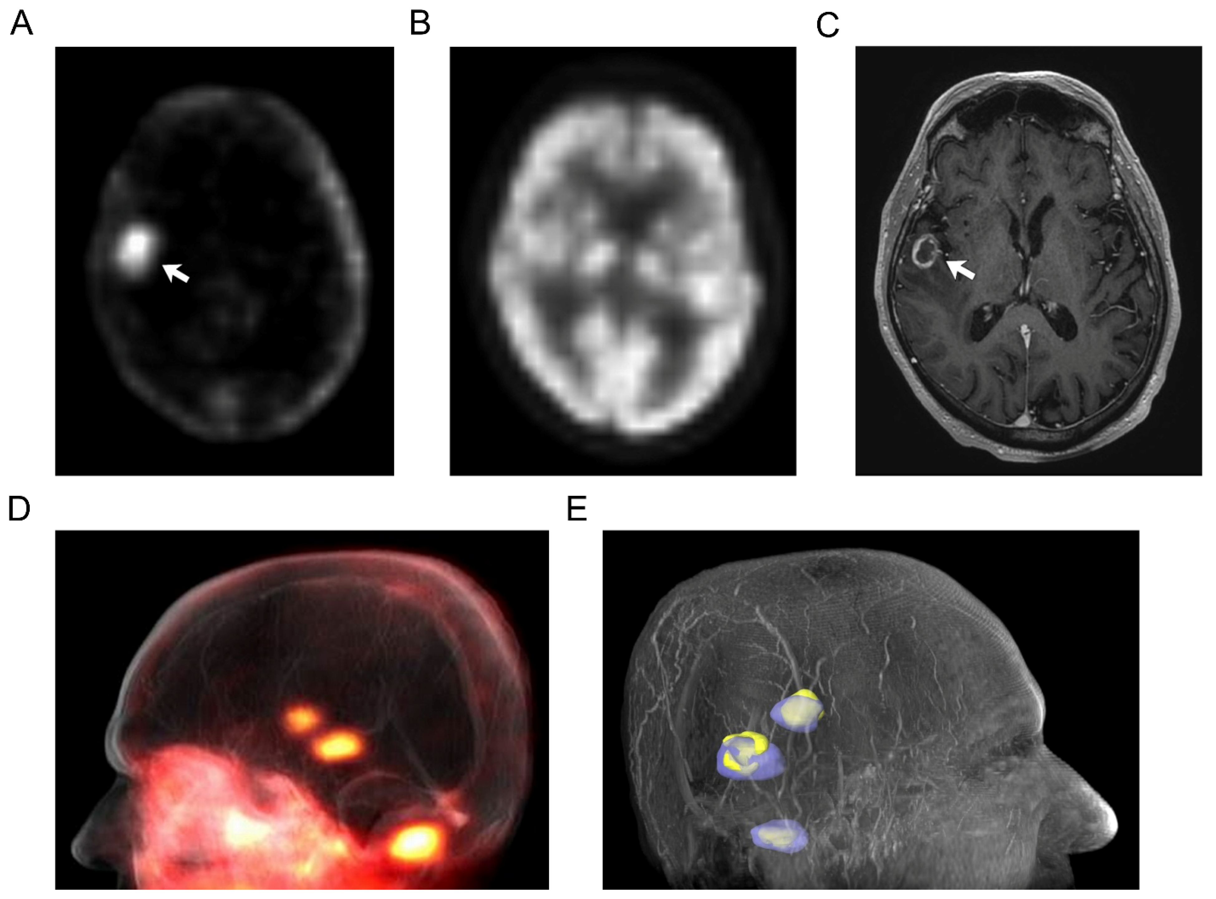
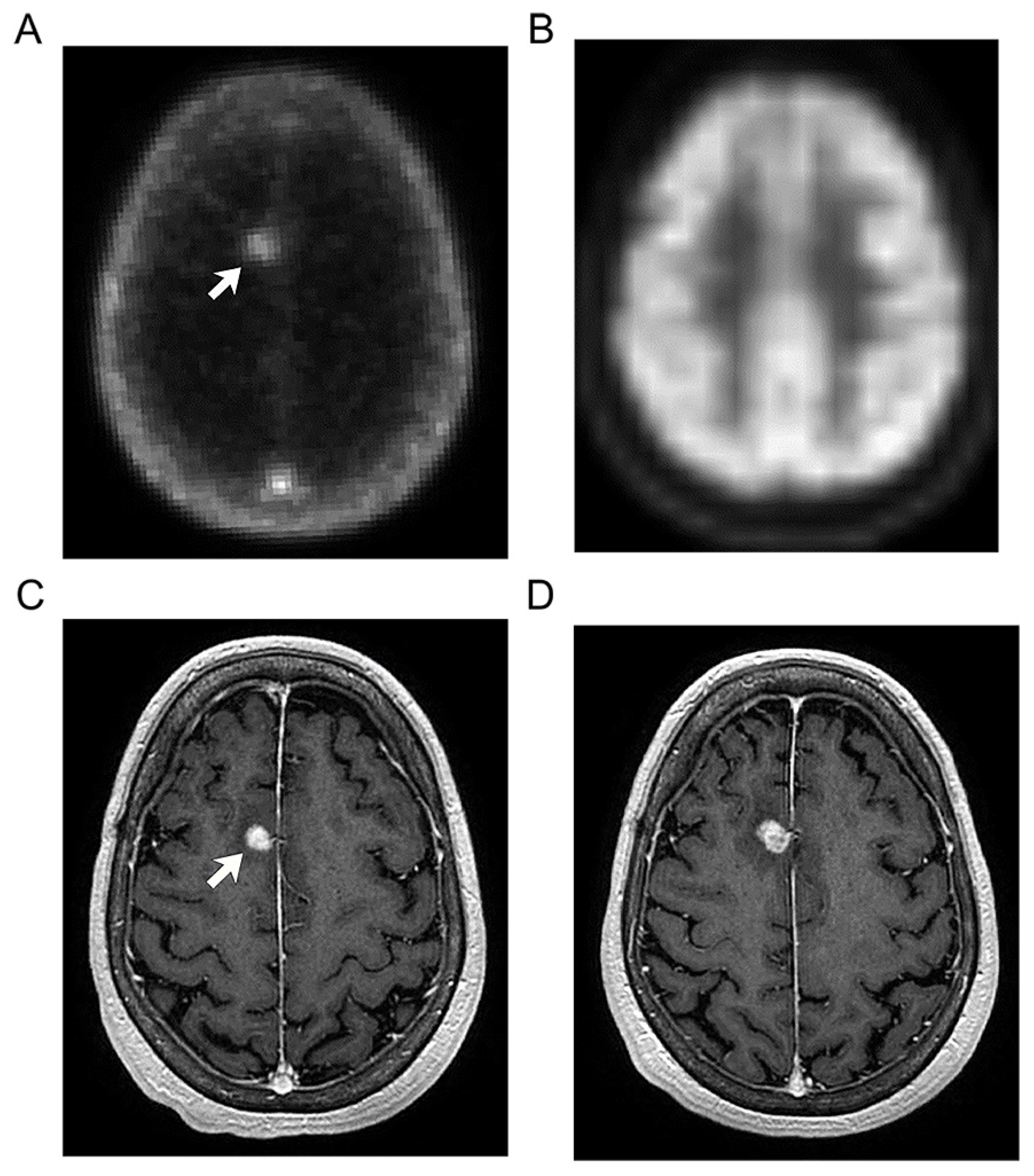
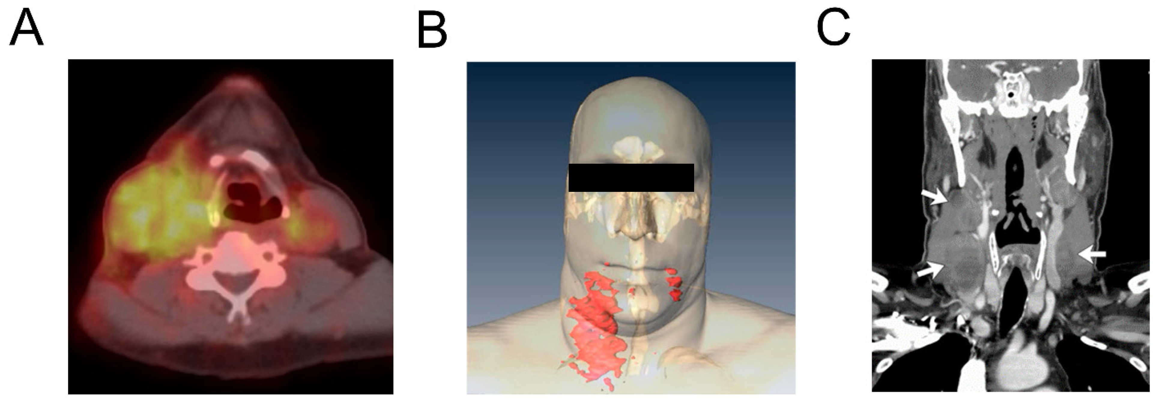
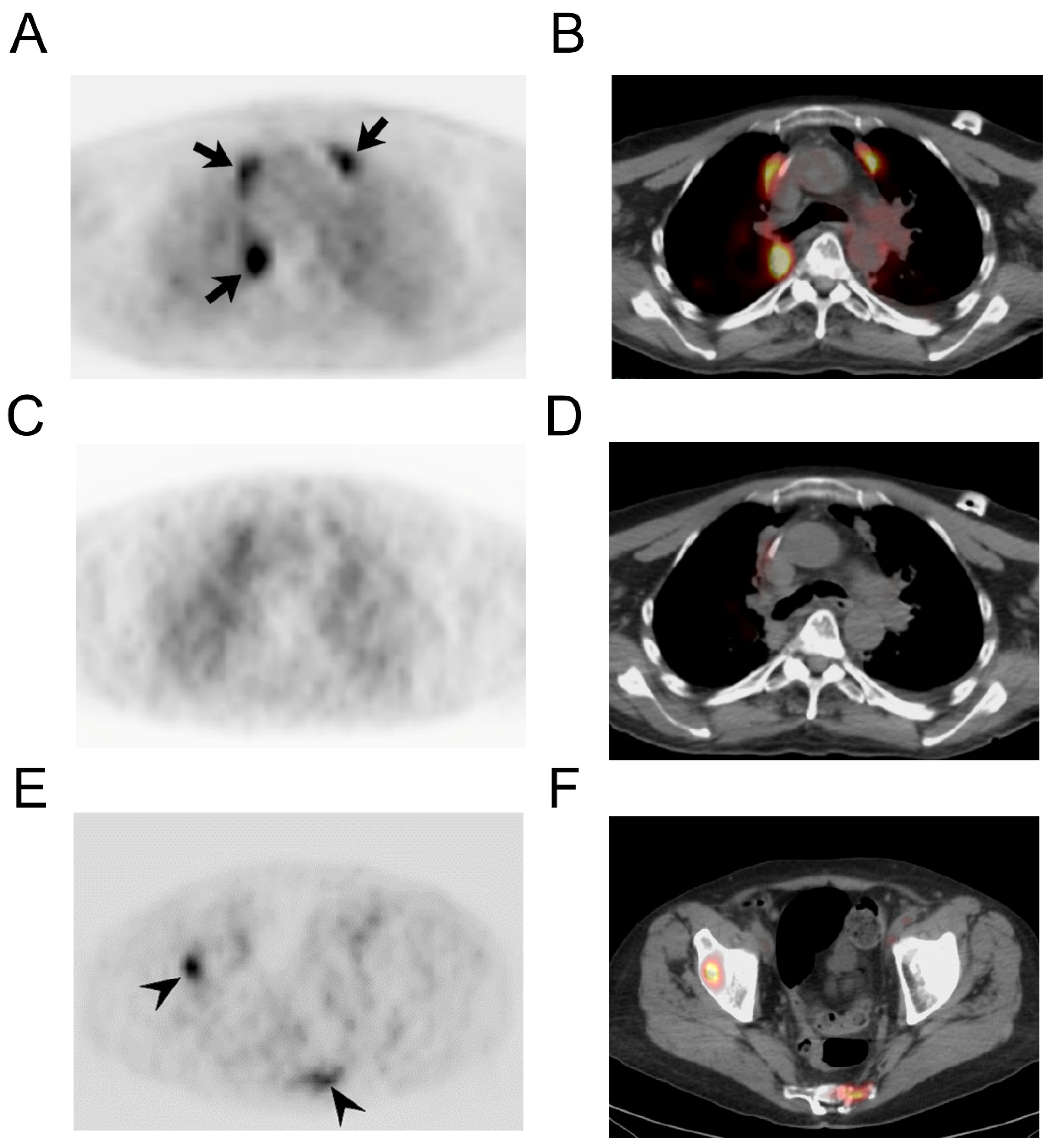
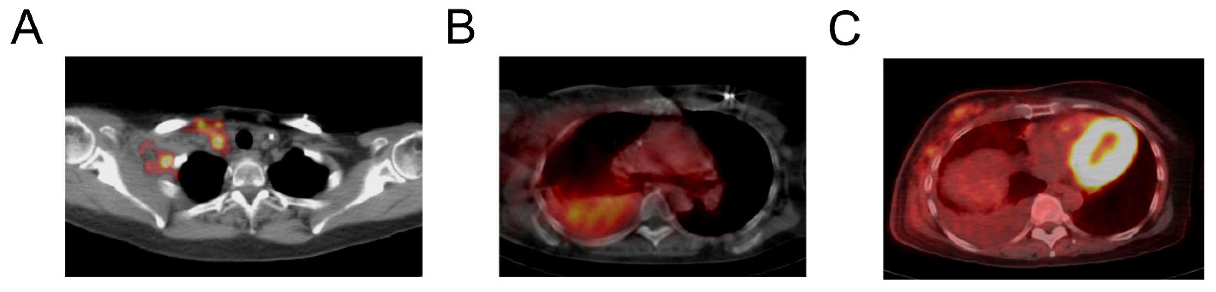
3.2. Imaging Findings

Whole body PET/CT imaging with [124I]I-NM404 was performed in 14 patients. We report the patterns of uptake of NM404 on delayed imaging according to metastatic involvement of the various organ systems: CNS, pulmonary, nodal, skeletal, and other sites. Although multiple imaging series with NM404 were obtained in each case, emphasis was generally placed on the most delayed phase given the progressive increase in tumor-to-background activity that was observed. Table 2 shows the tumor-to-background ratios (TBR) for NM404 activity of index lesions. Preferential uptake within metastatic foci was observed with all cancer subtypes, with the TBRmax for active index lesions ranged from 1.95 to 15.36 and TBRmean ranged from 1.63 to 6.63. For some cases, TBR data could not be derived, due to lack of suitable active index lesions (see Table 2).
In contrast to standard clinical nuclear imaging, brain lesions were well depicted with [124I]I-NM404 PET imaging due to the high tumor-to-background activity (Figure 1) with [18F]FDG PET imaging, brain metastases are generally not detectable, largely due to the high level of [18F]FDG avidity for normal brain tissue (Figure 1B). However, at subsequent follow-up MR imaging (Figure 1C), the nonspecific enhancing lesion has continued to enlarge, associated with progressive perilesional edema, more concerning for metastatic disease, and prompting close clinical follow up. In this patient with primary bronchogenic carcinoma, the detection of clinically unsuspected, [18F]FDG PET occult, brain metastases by [124I]I-NM404 PET altered the treatment plan.
In another case, increased [124I]I-NM404 uptake matched with a small focus of abnormal enhancement in the right frontal lobe on MR in a patient who previously underwent resection and subsequent stereotactic radiosurgery for metastatic melanoma (Figure 2). No abnormal uptake was seen in this area on the concurrent clinical [18F]FDG PET study (Figure 2B). Based on the clinical MR and [18F]FDG PET findings, distinction between radiation necrosis and tumor recurrence was not possible. However, at subsequent follow-up MR imaging (Figure 2D), the nonspecific enhancing lesion has continued to enlarge, associated with progressive perilesional edema, more concerning for metastatic disease.
Cervical lymphadenopathy in a 46-year-old man with metastatic squamous cell carcinoma that originated from the tongue base (Figure 3).
At this preliminary stage of investigation, the size threshold for CLR1404 detection of metastatic pulmonary nodules remains uncertain. In one patient with micronodular (miliary) pulmonary metastases, neither [18F]FDG nor [124I]I-NM404 PET imaging showed detectable abnormal activity. Post-treatment changes in the lung from radiation or chemotherapy are a recognized cause of false-positive uptake on [18F]FDG PET (Figure 4) However, NM404 showed avid uptake within sites of active osseous metastatic disease. The activity within lytic bone metastases from a variety of primary cancers corresponded with hypermetabolic FDG foci. In one case, a lytic vertebral lesion representing a solitary focus of osseous metastatic disease was identified by [124I]I-NM404 PET but missed on the clinical CT interpretation (Figure 4). In some cases, with longstanding blastic or sclerotic osseous lesions, [124I]I-NM404 uptake was not appreciated, which appeared to be discordant with FDG PET and [99mTc]Tc-MDP skeletal scintigraphy in some cases. It is unclear whether this discordance corresponds to [124I]I-NM404 false-negative detection or false-positive reactive activity from the clinical exams related to treatment response or “flare” phenomenon from bone remodeling.
Uptake of [124I]I-NM404was observed within metastatic lymph nodes in a variety of primary tumors (Figure 5). CT correlation was useful for localization of [124I]I-NM404activity within nodal disease. Examples of nodal uptake were seen with [124I]I-NM404 PET imaging. Beyond brain, lung, lymph node, and bone metastases, several other sites of active metastatic disease were demonstrated with [124I]I-NM404 in this heterogeneous patient cohort. Failed detection of hepatic metastatic disease with [124I]I-NM404 in one case appeared to be related to a lack of delayed imaging, although the low injected dose (185 MBq) may have also contributed. Increased uptake was also seen within body wall soft tissue involvement (Figure 5A). Increased [124I]I-NM404 activity was observed in two patients with cytology-proven malignant pleural effusions, one with triple-negative breast cancer and the other with bronchogenic carcinoma. Neither of these malignant pleural collections showed increased [18F]FDG uptake, and increased [124I]I-NM404 uptake was not observed in cases of benign pleural effusions, suggesting utility for [124I]I-NM404 PET with malignant pleural involvement.

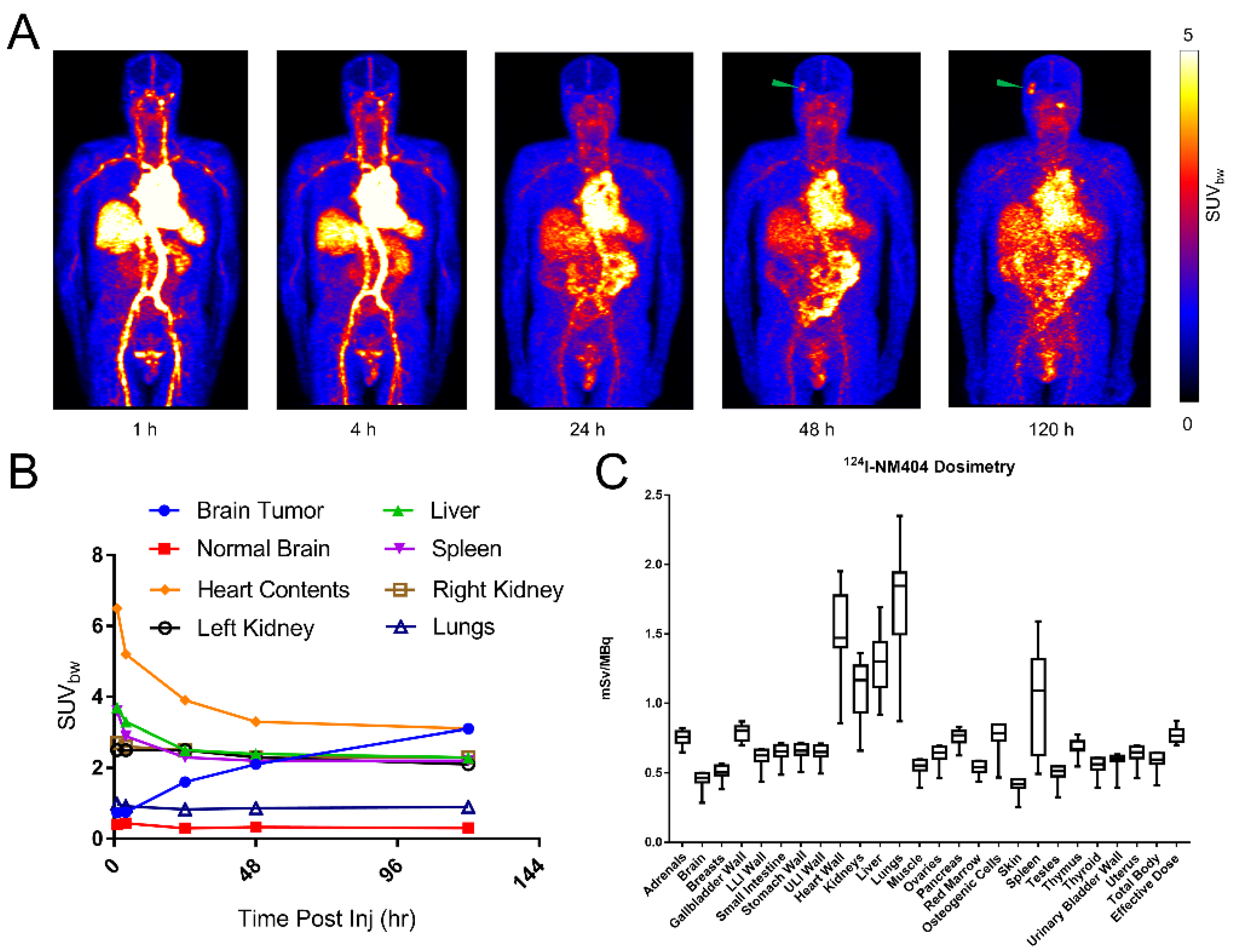
3.3. [124I]I-NM404 Biodistribution and Dosimetry Estimation

Because the biological half-life of [127I]I-NM404 is on the order of hundreds of hours [14], the effective half-life of [124I]I-NM404 (t1/2 = 4.18 days) is heavily weighted by its physical half-life. Due to this prolonged retention within the blood, shown in Figure 6A,B, [124I]I-NM404 is clearly detected within the contents of the heart during the first 6 days after injection with a mean SUVbw as high as 6.5 at 1 h post injection and 3.1 at 120 h. Since NM404 is predominantly cleared via the hepatobiliary system, the liver and spleen also show uptake within the first hour, a mean SUVbw of 3.7 and 3.6, respectively, but experience considerable clearance thereafter, with a mean SUVbw of 2.3 and 2.2, respectively, at 120 h post injection. The continual tumor uptake and protracted blood clearance leads to a ratio of mean SUVbw of the brain tumor to normal brain greater than 10 at 120 h. The highest dose (mean ± SD) was to the lungs 1.74 ± 0.39 mSv/MBq, next was heart wall 1.52 ± 0.29, followed by liver 1.28 ± 0.21, kidneys 1.09 ± 0.20, and the effect dose is 0.77 ± 0.05 mSv/MBq. The only two organs whose coefficient of variation (CV%) was greater than 20% were the lungs and spleen, which were 22% and 37%, respectively. A summary of dosimetry results is shown in Figure 6C and Table 3.

4. Discussion

NM404 [18-(p-iodophenyl) octadecylphosphocholine] is the first of a new class of synthetic alkylphosphocholine analogs designed for tumor-selective diagnosis and treatment in a broad spectrum of human cancers. Our early investigations have demonstrated selective uptake and prolonged retention of this novel molecular agent within sites of metastatic disease in a wide range of clinically important human cancers in this and other studies [15,16,20,21].
Prior to human testing, NM404 was subjected to intensive preclinical evaluation. Exquisite cancer-selective uptake and retention has been observed with dozens of in vivo rodent cancer models, including both transgenic and human tumor xenograft models [3]. Tumor uptake appears to be independent of anatomic location, with little or no clearance over time. The cellular mechanism for uptake appears to be related to lipid rafts, which are more abundant on the cell membrane of cancer cells [3,10,22,23]. In addition to imaging and semiquantitative analysis with [124I]I-NM404 PET, the therapeutic versatility of this molecule has been demonstrated with the beta-emitting 131I radioisostere, with efficacy in a wide range of cancer models, often after a single IV dose. Beyond the radioiodinated analogs of NM404, fluorescent derivatives of this molecule have demonstrated similar cancer-selective properties. The fluorescent analogs have further elucidated the intracellular localization of this molecule (primarily within membrane-bound cytosolic vesicles and organelles) and also allow the opportunity for real-time optical imaging [12,23].
[18F]FDG PET/CT is currently in widespread clinical use for the initial staging, evaluation of treatment response, and surveillance for a broad array of malignancies. Although quite useful, FDG is not a cancer-specific PET imaging agent, with avidity seen in a wide variety of inflammatory and reactive conditions. In the setting of post-treatment oncologic evaluation, false-positive PET diagnosis related to FDG-avid changes may be caused by interventional and surgical procedures, radiation therapy, and chemotherapy, in addition to infection and inflammation [24,25]. In addition, FDG PET has recognized limitations in the evaluation of brain metastases and primary CNS tumors primarily due to the high background in normal brain tissues, leading to false-negative PET diagnosis, even when combined with CT [26]. Preliminary investigation into [124I]I-NM404 has shown that this novel PET agent may be more cancer specific, with potential advantages over FDG for oncologic PET imaging. Preclinical studies have demonstrated that, unlike FDG, NM404 is not taken up in inflammatory lesions or benign neoplasms [3]. In our early clinical experience described herein, we have observed potential false-negative and false-positive results with [18F]FDG that may be rectified by [124I]I-NM404. However, the discordant findings we have observed between NM404 imaging and standard clinical imaging, such as FDG PET and gadolinium-enhanced MRI, require further investigation to determine their significance. Potential pitfalls with [124I]I-NM404 PET could include regions of interest with high background activity due to blood pool (if scans are not sufficiently delayed) and hepatic excretion into bowel. Although the more prolonged kinetic profile of [124I]I-NM404 could present new challenges in terms of PET scheduling, the fact that the agent can be synthesized to GMP standards at one site and shipped for use at other sites without an on-site or nearby cyclotron can be seen as an advantage. Post-surgical efficacy can also be assessed with a follow-up scan without the need for a second injection.
As evidence in Figure 6A,B, NM404 undergoes prolonged retention within organs, which is marked by a very long blood half-life. However, this protracted blood circulation seems to contribute to the continually uptake of tumors, which is clearly shown qualitatively (Figure 6A) and semi quantitatively (Figure 2B). Furthermore, the dosimetry estimates for [124I]I-NM404 (Figure 6C and Table 3) are comparable to a 124I-labeled small molecule, MIP-1095, which targets the prostate-specific membrane antigen (PSMA) for prostate cancer therapy [27]. Although the effective dose is less for [124I]I-MIP-1095, 0.58 vs. 0.77 mSv/MBq, the kidney and liver estimates for [124I]I-MIP-1095 are greater: 1.39 vs. 1.28 and 1.66 vs. 1.09 mSv/MBq, respectively.
If successful, the diapeutic paradigm offered by the NM404 platform has several potential advantages over existing approaches in both oncologic imaging and therapy. Importantly, the imaging agent and therapeutic agent are essentially the same molecule, providing direct evidence of where treatment will be directed, and providing information on the therapeutic ratio. This also paves the way for whole-body quantitative 4-D mapping of the in vivo biodistribution of this agent. Therefore, PET/CT-based dosimetry could allow for [131I]I-NM404 treatment planning. The PET agent also allows for treatment-specific post-therapy surveillance. The nearly universal profile of uptake of NM404 within solid human cancers may eventually provide a more uniform and robust approach to oncologic therapy.
We acknowledge limitations to our study. First and foremost, this is a largely descriptive early phase investigation seeking proof of concept that combines observations from several distinct Phase 1 clinical trials with heterogeneous patient cohorts and imaging protocols. The absence of optimized imaging parameters, including dose of injection and timing of image acquisition, preclude a more formal detailed quantitative analysis. For [124I]I-NM404 PET imaging, a correction method for cascade gamma radiation coincidences using 3D acquisition was not yet available. Therefore, a 2D acquisition mode was utilized, which lowers the count statistics because the thin septa of lead or tungsten that separate each crystal ring allows only coincidences that are between detectors within the same ring or lying in closely neighboring rings. Going forward, image quality will be further improved by employing this correction with 3D acquisition. In some cases, imaging was likely not sufficiently delayed, allowing for adequate clearance of background activity, and non-optimized dose was likely delivered. The complex prior treatment history for this heterogeneous patient cohort also complicates analysis and likely renders this cohort suboptimal for primary investigation. Therefore, more focused are homogeneous clinical trials are needed.

5. Conclusions

In conclusion, NM404 is a promising new molecular imaging agent that has the theranostic potential to integrate diagnostic oncologic imaging and therapy for a wide spectrum of human cancers. [124I]I-NM404 exhibits a similar dosimetric profile to that of [124I]I-MIP-1095 and has potential advantages over the current FDG approach for PET, in that it exhibits low brain background and is a theranostic analog which could potentially impact oncologic diagnosis and surveillance.

Author Contributions

Conceptualization, L.T.H. and J.W.; methodology, A.T. and G.L.; software, B.B.; formal analysis, J.J.G., L.T.H., S.C. and M.H.L.; investigation, L.T.H., A.T. and G.L.; resources, M.L. and A.P.; data curation, C.J.; writing—original draft preparation, J.J.G.; writing—review and editing, J.S.K. and L.T.H.; visualization, J.J.G.; project administration, L.T.H.; funding acquisition, J.W. and L.T.H. All authors have read and agreed to the published version of the manuscript.

Funding

National Institutes of Health, NIH 1R01CA158800-01, NIH R21CA092412.

Institutional Review Board Statement

The study was conducted in accordance with the Declaration of Helsinki. For studies involving humans. all human subjects research protocols were approved by the Health Sciences IRB of the University of Wisconsin-Madison. NCT00582283-PI was Anne Traynor(thoracic). NCT01662284 was Glenn Liu(GU).

Informed Consent Statement

Written informed consent from each subject was obtained at screening, prior to the performance of any study-specific procedures.

Data Availability Statement

Data available on request due to restrictions, e.g., privacy or ethical. The data presented in this study are available on request from the corresponding author. The data are not publicly available due to industry sponsored trial.

Acknowledgments

The authors would like to acknowledge Michael Stabin for his dosimetry consulting.

Conflicts of Interest

J.W. was a cofounder of Cellectar Biosciences.

References

  1. Herrmann, K.; Schwaiger, M.; Lewis, J.S.; Solomon, S.B.; McNeil, B.J.; Baumann, M.; Gambhir, S.S.; Hricak, H.; Weissleder, R. Radiotheranostics: A roadmap for future development. Lancet Oncol. 2020, 21, e146–e156. [Google Scholar] [CrossRef]
  2. Pinchuk, A.N.; Rampy, M.A.; Longino, M.A.; Skinner, R.W.; Gross, M.D.; Weichert, J.P.; Counsell, R.E. Synthesis and structure-activity relationship effects on the tumor avidity of radioiodinated phospholipid ether analogues. J. Med. Chem. 2006, 49, 2155–2165. [Google Scholar] [CrossRef] [PubMed]
  3. Weichert, J.P.; Clark, P.A.; Kandela, I.K.; Vaccaro, A.M.; Clarke, W.; Longino, M.A.; Pinchuk, A.N.; Farhoud, M.; Swanson, K.I.; Floberg, J.M.; et al. Alkylphosphocholine Analogs for Broad-Spectrum Cancer Imaging and Therapy. Sci. Transl. Med. 2014, 6, 240ra75. [Google Scholar] [CrossRef] [PubMed] [Green Version]
  4. Kirschner, A.S.; Ice, R.D.; Beierwaltes, W.H. Radiation dosimetry of 131I-19-Iodocholesterol. J. Nucl. Med. 1973, 14, 713–717. [Google Scholar] [PubMed]
  5. Carrasco, M.P.; Jiménez-López, J.M.; Ríos-Marco, P.; Segovia, J.L.; Marco, C. Disruption of cellular cholesterol transport and homeostasis as a novel mechanism of action of membrane-targeted alkylphospholipid analogues. Br. J. Pharmacol. 2010, 160, 355–366. [Google Scholar] [CrossRef] [PubMed] [Green Version]
  6. Gajate, C. The antitumor ether lipid ET-18-OCH3 induces apoptosis through translocation and capping of Fas/CD95 into membrane rafts in human leukemic cells. Blood 2001, 98, 3860–3863. [Google Scholar] [CrossRef] [Green Version]
  7. Reis-Sobreiro, M.; Roué, G.; Moros, A.; Gajate, C.; de la Iglesia-Vicente, J.; Colomer, D.; Mollinedo, F. Lipid raft-mediated Akt signaling as a therapeutic target in mantle cell lymphoma. Blood Cancer J. 2013, 3, e118. [Google Scholar] [CrossRef] [Green Version]
  8. van der Luit, A.H.; Budde, M.; Ruurs, P.; Verheij, M.; Van Blitterswijk, W.J. Alkyl-lysophospholipid accumulates in lipid rafts and induces apoptosis via raft-dependent endocytosis and inhibition of phosphatidylcholine synthesis. J. Biol. Chem. 2002, 277, 39541–39547. [Google Scholar] [CrossRef] [Green Version]
  9. Lasserre, R.; Guo, X.-J.; Conchonaud, F.; Hamon, Y.; Hawchar, O.; Bernard, A.-M.; Soudja, S.M.; Lenne, P.-F.; Rigneault, H.; Olive, D.; et al. Raft nanodomains contribute to Akt/PKB plasma membrane recruitment and activation. Nat. Chem. Biol. 2008, 4, 538–547. [Google Scholar] [CrossRef]
  10. Li, Y.C.; Park, M.J.; Ye, S.-K.; Kim, C.-W.; Kim, Y.-N. Elevated levels of cholesterol-rich lipid rafts in cancer cells are correlated with apoptosis sensitivity induced by cholesterol-depleting agents. Am. J. Pathol. 2006, 168, 1107–1118; quiz 1404–1405. [Google Scholar] [CrossRef] [Green Version]
  11. Mollinedo, F.; Gajate, C. Lipid rafts and clusters of apoptotic signaling molecule-enriched rafts in cancer therapy. Future Oncol. 2010, 6, 811–821. [Google Scholar] [CrossRef] [PubMed]
  12. Hall, L.T.; Fourzali, Y.; Hernan, H.; Weichert, J.P.; Traynor, A.M. First in human use of 124I-NM404 PET/CT in primary and metastatic brain tumors. Mol. Imaging Biol. 2012, 14, S1644. [Google Scholar]
  13. Hall, L.T.; Titz, B.; Robins, H.I.; Bednarz, B.P.; Perlman, S.B.; Weichert, J.P.; Kuo, J.S. I-CLR1404 in high and low-grade brain tumors. Am. J. Nucl. Med. Mol. Imaging 2017, 7, 157–166. [Google Scholar] [PubMed]
  14. Grudzinski, J.J.; Titz, B.; Kozak, K.; Clarke, W.; Allen, E.; Trembath, L.; Stabin, M.; Marshall, J.; Cho, S.Y.; Wong, T.Z.; et al. A Phase 1 Study of 131I-CLR1404 in Patients with Relapsed or Refractory Advanced Solid Tumors: Dosimetry, Biodistribution, Pharmacokinetics, and Safety. PLoS ONE 2014, 9, e111652. [Google Scholar] [CrossRef] [PubMed]
  15. Lubner, S.J.; Mullvain, J.; Perlman, S.; Pishvaian, M.; Mortimer, J.; Oliver, K.; Heideman, J.; Hall, L.; Liu, G.; Joseph, S.; et al. A Phase 1, Multi-Center, Open-Label, Dose- Escalation Study of I-CLR1404 in Subjects with Relapsed or Refractory Advanced Solid Malignancies I-CLR1404 in Subjects with Relapsed or Refractory Advanced Solid Malignancies. Cancer Investig. 2015, 33, 483–489. [Google Scholar] [CrossRef] [PubMed]
  16. McAuliffe, M.J.; Lalonde, F.M.; McGarry, D.; Gandler, W.; Csaky, K.; Trus, B.L. Medical image processing, analysis & visualization in clinical research. In Proceedings of the IEEE Symposium on Computer-Based Medical Systems, CBMS, Bethesda, MD, USA, 26–27 July 2001; Volume 2001, pp. 381–386. [Google Scholar]
  17. Barrett, P.H.; Bell, B.M.; Cobelli, C.; Golde, H.; Schumitzky, A.; Vicini, P.; Foster, D.M. SAAM II: Simulation, Analysis, and Modeling Software for Tracer and Pharmacokinetic Studies. Metabolism 1998, 47, 484–492. [Google Scholar] [CrossRef]
  18. Stabin, M.G.; Siegel, J.A. Physical Models and Dose Factors for Use in Internal Dose Assessment. Health Phys. 2003, 85, 294–310. [Google Scholar] [CrossRef]
  19. Stabin, M.G.; Sparks, R.B.; Crowe, E. OLINDA/EXM: The second-generation personal computer software for internal dose assessment in nuclear medicine. J. Nucl. Med. 2005, 46, 1023–1027. [Google Scholar]
  20. Pickhardt, P.J.; Hall, L.T.; Lee, M.; Longino, M.; Pinchuk, A.; Banash, M.; Grudzinski, J.; Titz, B.; Jaskowiak, C.; Kuo, J.S.; et al. A novel “diapeutic” molecular imaging agent for combined oncologic diagnosis and therapy in a broad spectrum of human cancers: Preliminary clinical experience with CLR1404. J. Clin. Oncol. 2014, 32, 11000. [Google Scholar] [CrossRef]
  21. Hall, L.T.; Titz, B.; Baidya, N.; van der Kolk, A.G.; Robins, H.I.; Otto, M.; Perlman, S.B.; Weichert, J.P.; Kuo, J.S. [124I]CLR1404 PET/CT in High-Grade Primary and Metastatic Brain Tumors. Mol. Imaging Biol. 2020, 22, 434–443. [Google Scholar] [CrossRef]
  22. Swanson, K.I.; Clark, P.A.; Zhang, R.R.; Kandela, I.K.; Farhoud, M.; Weichert, J.P.; Kuo, J.S. Fluorescent Cancer-Selective Alkylphosphocholine Analogs for Intraoperative Glioma Detection. Neurosurgery 2014, 90, 115–124. [Google Scholar] [CrossRef] [Green Version]
  23. Lingwood, D.; Simons, K. Lipid rafts as a membrane-organizing principle. Science 2010, 327, 46–50. [Google Scholar] [CrossRef] [Green Version]
  24. Shreve, P.D.; Anzai, Y.; Wahl, R.L. Pitfalls in oncologic diagnosis with FDG PET imaging: Physiologic and benign variants. Radiographics 1999, 19, 61–77. [Google Scholar] [CrossRef] [PubMed]
  25. Lee, T.S.; Ahn, S.H.; Moon, B.S.; Chun, K.S.; Kang, J.H.; Cheon, G.J.; Choi, C.W.; Lim, S.M. Comparison of 18F-FDG, 18F-FET and 18F-FLT for differentiation between tumor and inflammation in rats. Nucl. Med. Biol. 2009, 36, 681–686. [Google Scholar] [CrossRef]
  26. Chao, S.T.; Suh, J.H.; Raja, S.; Lee, S.Y.; Barnett, G. The sensitivity and specificity of FDG PET in distinguishing recurrent brain tumor from radionecrosis in patients treated with stereotactic radiosurgery. Int. J. Cancer 2001, 96, 191–197. [Google Scholar] [CrossRef] [PubMed]
  27. Zechmann, C.M.; Afshar-Oromieh, A.; Armor, T.; Stubbs, J.B.; Mier, W.; Hadaschik, B.; Joyal, J.; Kopka, K.; Debus, J.; Babich, J.W.; et al. Radiation dosimetry and first therapy results with a (124)I/(131)I-labeled small molecule (MIP-1095) targeting PSMA for prostate cancer therapy. Eur. J. Nucl. Med. Mol. Imaging 2014, 41, 1280–1292. [Google Scholar] [CrossRef] [PubMed] [Green Version]
Figure 1. Unsuspected brain metastases in 70-year-old man with primary bronchogenic carcinoma. Transverse PET image (A) obtained 6 days after injection of 185 MBq [124I]I-NM404 shows a right hemispheric focus of activity (arrow) that was not detectable on [18F]FDG PET (B) but confirmed on subsequent contrast-enhanced MR ((C), arrow). Fused volume-rendered [124I]I-NM404 PET-MR image (D) shows a total of three unsuspected brain metastases (2 cerebral and 1 cerebellar) that were identified on [124I]I-NM404 PET and confirmed on MR, which altered the treatment strategy for this patient. Additional fused volume-rendered [124I]I-NM404 PET-MR image (E) with segmentation of the brain metastases shows the regions of [124I]I-NM404 uptake (purple), which exceed the regions of abnormal MR contrast enhancement (yellow). The clinical significance of this uptake-enhancement discordance within the tumor is uncertain.
Radiation 02 00015 g001
Figure 2. Recurrent brain metastasis in 60-year-old woman with malignant melanoma. [124I]I-NM404 (A) and [18F]FDG (B) PET images 8 months after stereotactic radiosurgery for tumor recurrence of a right frontal falcine metastasis shows a focus of abnormal activity with NM404 (arrow) but no detectable abnormality with [18F]FDG. Corresponding enhancing focus on MR imaging ((C), arrow) was interpreted as radiation necrosis versus possible recurrence. Subsequent MR imaging (D) showed further increase in size of the nonspecific enhancing lesion, coupled with increased perilesional edema. Patient remains in close-interval imaging surveillance.
Radiation 02 00015 g002
Figure 3. Cervical lymphadenopathy in a 46-year-old man with metastatic squamous cell carcinoma that originated from the tongue base. (A) Displays an axial slice of [124I]I-NM404 PET overlaid on the CT volume. (B) Shows an isosurface representation of the tumor volume. (C) Coronal CT slice with white arrows indicating squamous cell carcinoma.
Radiation 02 00015 g003
Figure 4. Radiation change and osseous metastatic disease in a 65-year-old man with bronchogenic carcinoma. PET (A) and fused PET/CT (B) images using [18F]FDG PET shows abnormal linear paramediastinal activity (arrows), characteristic of radiation therapy. Corresponding PET (C) and fused PET/CT (D) images using [124I]I-NM404 do not show florid uptake in this reactive process. However, [124I]I-NM404 PET did demonstrate multifocal osseous metastatic disease (E,F), arrows.
Radiation 02 00015 g004
Figure 5. A 53-year-old woman with metastatic triple-negative breast cancer imaged with both [124I]I -CLR1404 and [131I]I-CLR1404. Fused PET/CT image (A) obtained 6 days after 124I-CLR1404 administration shows abnormal uptake within the right axillary and cervical region, corresponding to metastatic involvement of the body wall and lymph nodes. Fused SPECT/CT image (B) obtained 21 days after injection of [131I]I-CLR1404 shows increased activity within a moderate-sized right pleural effusion, which was proven malignant by cytology after thoracentesis. Increased pleural uptake was also seen on [124I]I-CLR1404 PET (not shown) but not on [18F]FDG PET (C). Note also prominent myocardial uptake with FDG, which is generally not seen with CLR1404.
Radiation 02 00015 g005
Figure 6. Longitudinal biodistribution and dosimetry estimations of [124I]I-NM404. (A) Maximum intensity projections (MIP) of standard uptake value normalized by body weight (SUVbw) at 1, 4, 24, 48 and 120 h for a patient with unsuspecting brain metastases (same as patient depicted in Figure 1). The green arrows at 48 h and 120 h indicate intense signal in the head region indicative of brain metastases. (B) The mean SUVbw of the brain metastases and various organs of interest are plotted over time to show the biodistribution of [124I]I-NM404within this patient. Notice how the SUVbw of the brain metastases increases continually during the longitudinal imaging. (C) Semiquantitative organ dosimetry estimations (mSv/MBq) are shown as box plots for [124I]I-NM404 in patients with a wide variety of solid primary and metastatic malignancies (n = 14). Whiskers indicate the minimum and maximum values.
Radiation 02 00015 g006
Table 1. Demographic and imaging data for the [124I]I-NM404 PET.
Patient Age (Dx)Age (Im)GenderWeight (kg)Primary TumorDose
16364F57Pancreas185 MBq
25153F65Breast, triple-negative185 MBq
34646M93SCC tongue base185 MBq
45661M131Colorectal185 MBq
56675M98Prostate185 MBq
66161M132SCC tonsil185 MBq
75560F83Melanoma185 MBq
86062M75Bronchogenic185 MBq
95661F74Bronchogenic185 MBq
106870M77Bronchogenic185 MBq
116263F88Bronchogenic111 MBq
126363F93Bronchogenic111 MBq
136465M88Bronchogenic277.5 MBq
148080F71Bronchogenic277.5 MBq
Table 2. The tumor-to-background (TBR) of NM4040 in index lesions.
PatientIndex LesionMaximum TBRMean TBR
1No active focal index lesionn/an/a
2Right axillary lymph node1.951.63
3Right cervical node6.394.21
4No active focal index lesionn/an/a
5Right iliac lymph node4.312.39
6Left cervical lymph node3.952.63
7Right frontal lobe brain lesion5.734.07
8No active focal index lesionn/an/a
9Left upper lobe lung lesion3.322.31
10Right temporal lobe brain lesion15.366.63
11No active focal index lesionn/an/a
12T2 vertebral bone lesion6.223.55
13Right scapular lesion8.403.36
14Right lung lesion4.642.97
6.033.38Mean
3.741.40SD
Table 3. Tabulated organ dose estimates for [124I]I-NM404 are presented in units of mSv/MBq. The mean, standard deviation (SD), and coefficient of variation (%) for the patients (n = 14) is shown.
mSv/MBq
MeanSDCOV (%)
Adrenals7.55 × 10−15.06 × 10−26.70
Brain4.50 × 10−15.79 × 10−212.86
Breasts5.11 × 10−14.93 × 10−29.65
Gallbladder Wall7.86 × 10−15.57 × 10−27.09
LLI Wall6.08 × 10−16.52 × 10−210.72
Small Intestine6.47 × 10−16.29 × 10−29.73
Stomach Wall6.56 × 10−15.71 × 10−28.70
ULI Wall6.47 × 10−16.07 × 10−29.38
Heart Wall1.52 × 1002.88 × 10−119.00
Kidneys1.09 × 1002.04 × 10−118.71
Liver1.28 × 1002.12 × 10−116.56
Lungs1.74 × 1003.88 × 10−122.29
Muscle5.43 × 10−15.57 × 10−210.24
Ovaries6.31 × 10−16.55 × 10−210.37
Pancreas7.60 × 10−15.50 × 10−27.24
Red Marrow5.37 × 10−14.45 × 10−28.29
Osteogenic Cells7.63 × 10−11.00 × 10−113.11
Skin4.09 × 10−15.22 × 10−212.77
Spleen1.05 × 1003.55 × 10−133.70
Testes4.95 × 10−16.03 × 10−212.19
Thymus6.87 × 10−16.00 × 10−28.74
Thyroid5.52 × 10−15.96 × 10−210.81
Urinary Bladder Wall5.86 × 10−16.08 × 10−210.37
Uterus6.33 × 10−16.55 × 10−210.35
Total Body5.89 × 10−16.16 × 10−210.45
Effective Dose7.72 × 10−15.49 × 10−27.11
Publisher’s Note: MDPI stays neutral with regard to jurisdictional claims in published maps and institutional affiliations.

