Communication

Magnetic Resonance Diffusion-Weighted Imaging for Detecting Fundal Intracholecystic Papillary Neoplasm inside Rokitansky-Aschoff Sinuses: A Comparison of Two Cases and a Literature Review

1
Unit of Radiology, Department of Clinical, Surgical, Diagnostic and Pediatric Sciences, University of Pavia, 27100 Pavia, Italy
2
Department of Radiology, I.R.C.C.S. Policlinico San Matteo, 27100 Pavia, Italy
3
Department of General Surgery, University of Pavia and I.R.C.C.S. Policlinico San Matteo, 27100 Pavia, Italy
4
Unit of Anatomic Pathology, Department of Molecular Medicine, University of Pavia and I.R.C.C.S. Policlinico San Matteo, 27100 Pavia, Italy
5
Center for Computer Vision and Imaging Biomarkers, UCLA Brain Tumor Imaging Laboratory (BTIL), University of California Los Angeles, Los Angeles, CA 90024, USA
6
Medical Scientist Training Program, David Geffen School of Medicine, University of California Los Angeles, Los Angeles, CA 90024, USA
7
Department of Radiological Sciences, David Geffen School of Medicine, University of California Los Angeles, Los Angeles, CA 90024, USA
8
Department of Bioengineering, Henry Samueli School of Engineering and Applied Science, University of California Los Angeles, Los Angeles, CA 90024, USA
*
Author to whom correspondence should be addressed.
Radiation 2022, 2(1), 52-61; https://doi.org/10.3390/radiation2010004
Submission received: 13 November 2021 / Revised: 27 December 2021 / Accepted: 28 December 2021 / Published: 31 December 2021

Abstract

:

Simple Summary

Intracholecystic papillary neoplasm, an in situ malignant lesion, may be challenging to detect when involving adenomyomatous mucosa or the Rokitansky-Aschoff sinuses, for wall thickening can be present in these regions in the absence of malignancy as a result of chronic inflammation. We hereby compare two cases advocating for a role of diffusion imaging to diagnose malignant wall thickening within Rokitansky-Aschoff sinuses. In addition, we review previously published studies reporting cases of intracholecystic papillary neoplasm arising from Rokitansky-Aschoff sinuses and articles assessing the value of diffusion imaging in gallbladder tumors. Considering our presented cases along with other evidence in the literature, we conclude that the visualization of T2-hyperintense cystic spaces representing RAS should not be used to rule out an underlying malignancy, and MR-DWI restriction should be employed as a primary MRI biomarker for detecting malignant tissue (i.e., ICPN or adenocarcinoma) within the RAS mucosa.

Abstract

Rokitansky-Aschoff sinuses (RAS) are a common imaging finding in gallbladder adenomyomatosis (ADM), often presenting as fundal cystic spaces. Intracholecystic papillary neoplasm (ICPN) is a relatively uncommon pre-invasive tumor of the gallbladder epithelium that rarely involves RAS mucosa. We compare two cases that showed similar fundal cystic spaces resembling RAS, in which Magnetic Resonance Diffusion-Weighted Imaging (MR-DWI) was valuable for detecting (or ruling out) an underlying malignant ICPN. Evidence from the literature overall supports the role of MR-DWI for detecting intracholecystic malignant tissue.

Graphical Abstract

1. Introduction

Gallbladder adenomyomatosis (ADM) is a benign, acquired condition induced by chronic inflammation and characterized by a thickened muscular layer and by Rokitansky-Aschoff sinuses (RAS), which are intramural diverticula of the hypertrophic mucosa evaginating in the muscular wall [1]. RAS can be visualized with several imaging techniques (ultrasound, magnetic resonance, computed tomography) as bile-filled cystic spaces within the gallbladder wall [2], and their distribution can present three different topographic patterns: segmental, fundal, or diffuse [1].
Intracholecystic papillary neoplasm (ICPN) is a neoplastic lesion of the gallbladder epithelium [3] representing the same histopathological type as the intraductal papillary neoplasm of the bile duct (IPNB). IPNB was proposed for the first time in 2010 as an independent oncologic entity by the World Health Organization [4], representing the bile duct counterpart of the pancreatic intraductal papillary-mucinous neoplasm (IPMN) [5], consequently showing either malignant or pre-malignant features [3]. ICPN arises from variable sites of the gallbladder mucosa and can also be located inside RAS, either arising from the RAS mucosa or extending onto it with a papillary growth pattern, similarly to what had been previously described in cases of gallbladder adenocarcinoma [6]. According to some authors, mural neoplasms of RAS mucosa with a papillary appearance usually arise from pre-existing ADM and may represent a distinct clinicopathologic entity [7]. Overall, when involving RAS mucosa, ICPN may be challenging to detect with imaging techniques, as a certain degree of wall thickening can be expected even in the typical benign ADM as a result of inflammatory events.
Magnetic Resonance Diffusion-Weighted Imaging (MR-DWI) is a non-invasive imaging technique providing useful in vivo information regarding Brownian motion of water molecules within biological tissue [8]. One quantitative metric derived from MR-DWI is the Apparent Diffusion Coefficient (ADC), which reflects the degree of water motion within the voxel. ADC is particularly relevant in oncologic imaging, as low ADC values have been proven reliable biomarkers of high cellular density in numerous tumor types [9,10,11,12].
We hereby present two cases in which, in the presence of ADM, MR-DWI was valuable for detecting and ruling out, respectively, an underlying malignant ICPN co-localizing with RAS.
These two patients gave their written informed consent to have their images used for research purposes.

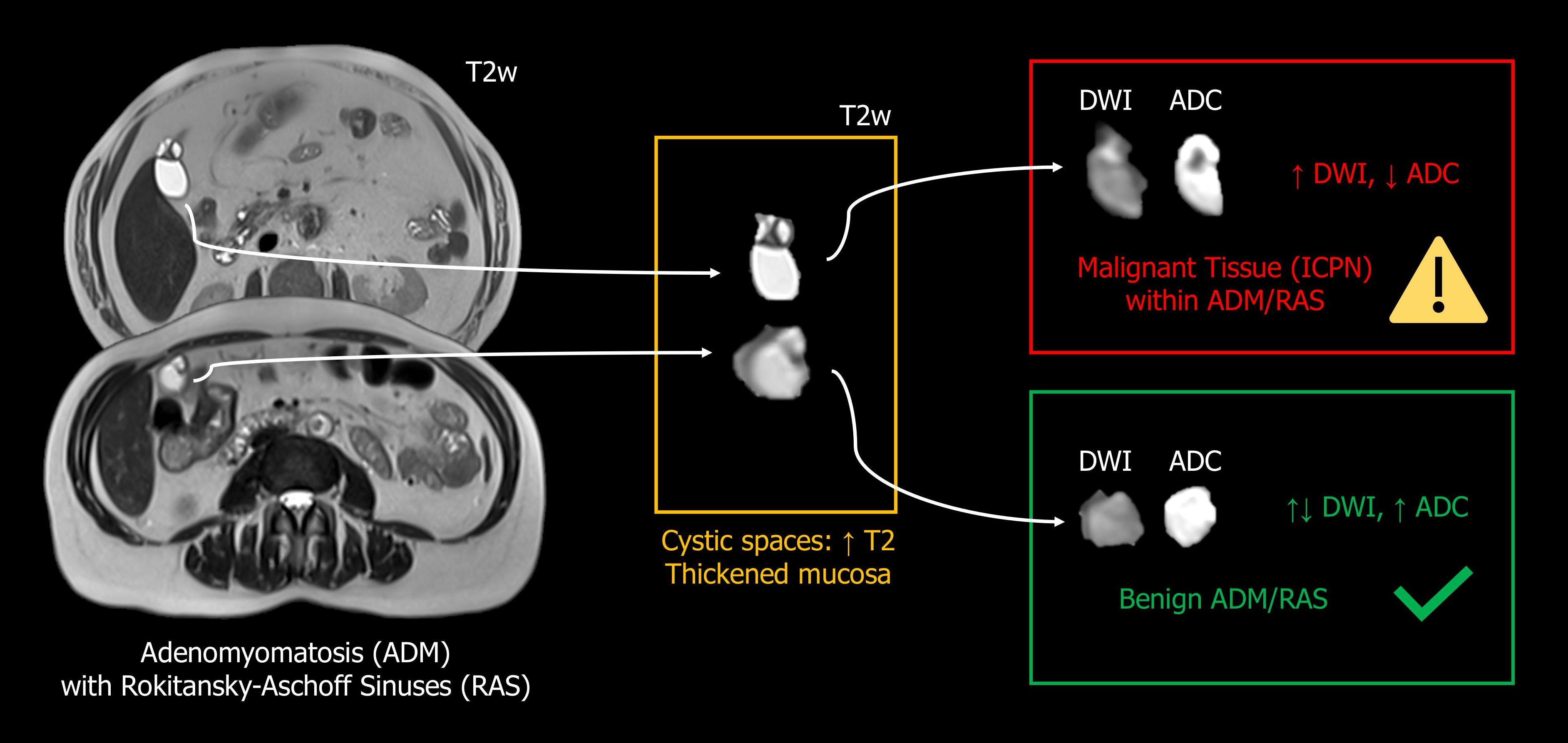
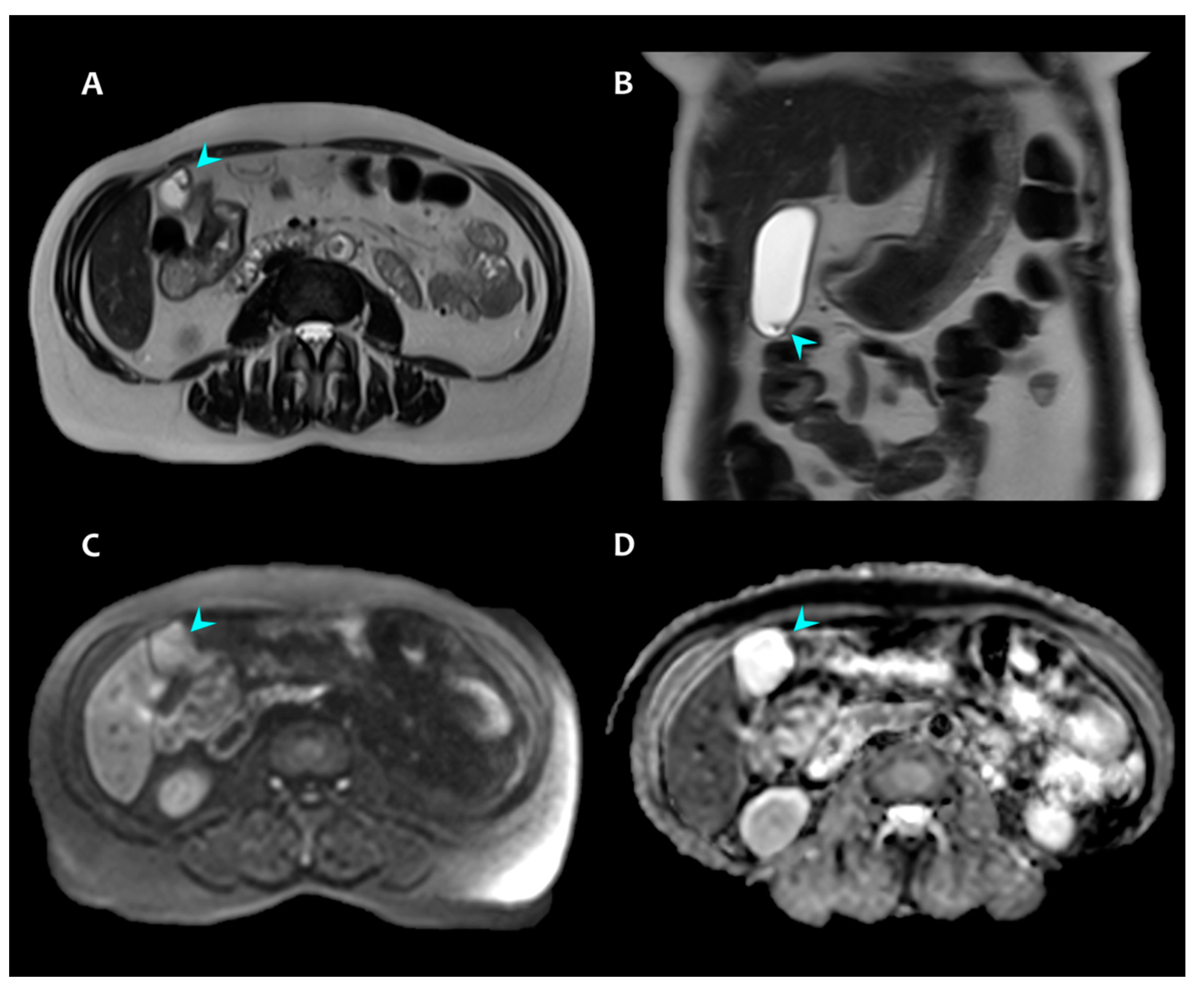
2. Presentation of Case #1

A 71-year-old man underwent abdominal contrast-enhanced computed tomography (CECT) for post-procedural assessment of an aorto-bi-iliac prosthesis positioning. In the absence of any systemic or abdominal symptoms, CECT showed gallbladder fundal cystic spaces as an incidental finding. Despite this finding being compatible with RAS, CECT also demonstrated an enhancing solid-tissue component within the cystic spaces, which could be interpreted as thickened ADM mucosa or thickened septa between cystic spaces (Figure 1A,B). As this solid tissue was atypical for simple RAS, the patient underwent an abdominal MRI scan to further characterize this alteration.
The MRI scan confirmed the presence of solid tissue within cystic areas. The solid tissue component appeared hypointense on T2-weighted sequence and spontaneously hyperintense on T1-weighted sequence, with early contrast enhancement in the arterial-phase and with no evidence of wash-out in portal- or late-phases (Figure 1C–H). In addition, a restriction of water molecule movements was documented as MR-DWI hyperintensity, with a corresponding hypointensity on the ADC map (Figure 1I,J). Quantitative ADC values, measured with an axial single-plane manual region of interest (ROI), were the following: mean 1.43 × 10⁻³ mm2/s, range 1.35–1.50 × 10⁻³ mm2/s. Since MR-DWI restriction supported the hypothesis of malignancy, a cholecystectomy was performed.
Histopathological macroscopic examination of the specimen found a fundal nodule of 15 mm with underlying cystic spaces. The microscopic examination of the nodule exhibited papillary epithelial high-grade dysplasia/intraepithelial neoplasia, according to World Health Organization 2019 classification [13], compatible with ICPN. Immunophenotype markers were as follows: cytokeratin 7 positive; p53, synaptophysin, chromogranin A negative. The microscopic examination of the cystic spaces found cystic spaces covered in markedly atypical epithelium, reflecting either a pre-existing ADM area with RAS colonized by ICPN cells or ICPN arising from pre-existing ADM area with RAS (Figure 2). In addition, the presence of necrotic areas and muscular invasion (extended to the peritoneal side of the peri-muscular connective tissue) by atypical glands supported a histopathological diagnosis of localized foci of adenocarcinoma pT2a.

3. Presentation of Case #2

A 59-year-old woman presented with dyspepsia and abdominal pain. Ultrasound (US) findings were compatible with cholecystitis, and the patient was scheduled for cholecystectomy. Abdominal MRI was performed 5 days later as a further pre-surgical evaluation in order to rule out choledocholithiasis.
Abdominal MRI (Figure 3A,B) showed a gallbladder fundal cystic space, compatible with RAS, and a linear endoluminal tissue component, representing the corresponding ADM mucosa. MR-DWI and an ADC map (Figure 3C,D) demonstrated no restriction of water molecule movements, advocating for the absence of malignant tissue. The measurement of ADC values was not appropriate in this case, as there were no hypointense regions in the ADC map that warranted the placement of an ROI. Given the benign features of the findings, gadolinium administration was considered not necessary.
Histopathological macroscopic examination of the specimen found a strawberry-like gallbladder mucosa; microscopic examination demonstrated signs of acute cholecystitis and chronic cholecystitis, cholesterolosis of the gallbladder wall, pyloric metaplasia of the epithelium, and RAS. No signs of epithelial dysplasia or neoplasia were found.

4. Discussion and Review of the Literature

In this article, we compare two cases in which Magnetic Resonance Diffusion-Weighted Imaging (MR-DWI) was able to detect and rule out, respectively, an underlying fundal intracholecystic papillary neoplasm (ICPN, a pre-malignant or malignant condition) involving an area of adenomyomatosis (ADM) with Rokitansky-Aschoff sinuses (RAS) (a benign condition). To our knowledge, this is the first article reporting on the role of MR-DWI in the specific case of ICPN involving RAS mucosa.
We performed a literature review in order to analyze other articles addressing the imaging features of small gallbladder malignant lesions involving Rokitansky-Aschoff sinuses. In addition, we reviewed studies assessing the role of MR-DWI in detecting gallbladder malignancy when conventional imaging techniques were unable to achieve a diagnosis and studies discussing the usefulness of MR-DWI for assessing gallbladder tumor invasiveness.

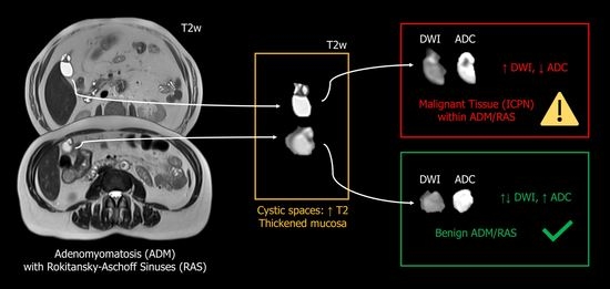
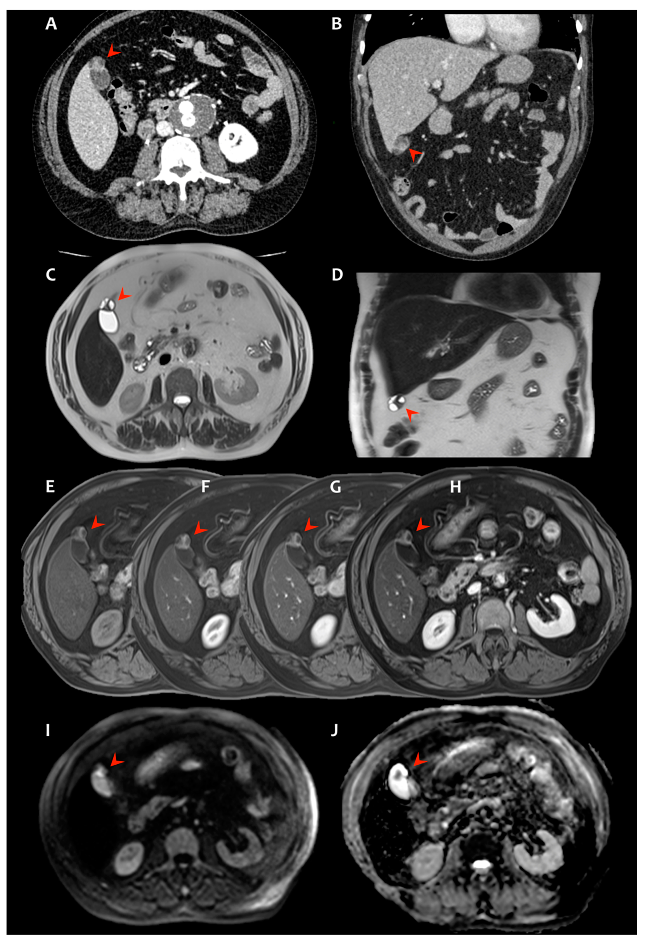
4.1. Intracholecystic Papillary Neoplasm (ICPN) and Rokitansky-Aschoff Sinuses (RAS)

We found no published case series of ICPN involving Rokitansky-Aschoff sinuses, but we found two case reports published, respectively, by Muranushi et al. [14] and Sato et al. [15], describing fundal ICPN arising inside Rokitansky-Aschoff sinuses, both appearing as lesions protruding outward from the gallbladder. In the first case report [14], Contrast-Enhanced Computed Tomography (CECT) and T2-weighted MRI shared similar features with case #1 of the present article, with a combination of solid enhancing components (representing the neoplastic tissue) and cystic components (the Rokitansky-Aschoff sinuses); however, MR-DWI characteristics were not reported in this article. In the second case report [15], CECT and MRI also depicted a cystic lesion, but the density of the cyst content was inhomogeneous (corresponding to mucin, as ICPN was associated with mucinous adenocarcinoma in this case); the authors stated that no solid nodules were clearly identifiable prior to cholecystectomy and that no MR-DWI restriction was visualized, but a preoperative diagnosis of gallbladder tumor was achieved nonetheless. An additional case report described ICPN arising from RAS, but the diagnosis was posed post mortem during organ harvesting for the purpose of donation; therefore, this case report did not feature any imaging data [16].
Overall, detecting malignant tissue within RAS is particularly important for two reasons. First, early detection of gallbladder malignancy results in a better prognosis. Second, the involvement of RAS by malignant tissue has been proven to represent a negative prognostic factor, resulting in shorter survival [17].

4.2. Diffusion MRI to Discriminate Malignant and Benign Gallbladder Lesions

Multiple articles advocate for the diagnostic value of MR-DWI in discriminating malignant and benign gallbladder lesions [18,19,20].
In a review focusing on the imaging features of uncommon intraluminal tumors of the gallbladder and the bile ducts, Chatterjee and colleagues report that MR-DWI restriction is a common finding in ICPN, and they advocate for a role of MR-DWI in distinguishing intraluminal tumors from stones and sludge [19].
As for adenomyomatous mucosa, differential diagnosis with malignant neoplasms is made challenging in some cases by the presence of inflamed thickened mucosa mimicking tumor tissue (and vice versa) [21]. In such cases, MR-DWI may play a role. For instance, in a study by Ogawa and colleagues, high MR-DWI signal was reported to be more likely in malignant lesions than in benign ones [22]. In this article, the authors proposed a ‘MR-DWI sign’ that should be considered ‘positive’ when some areas within the gallbladder show MR-DWI signal similar or higher than the spinal cord. With this method, however, they obtained a 30% and 22% rate of false positive MR-DWI sign (i.e., no histopathologic evidence of malignancy), respectively in patients with cholecystitis and and adenomyomatosis. Some potential factors contributing to these false positive results may be represented by the choice of the spinal cord as a reference and, more importantly, by readers not evaluating ADC maps qualitatively. Additionally, findings from other authors highlighted the possibility of a false positive MR-DWI in benign conditions, especially when chronic cholecystitis is present [23]. On the other hand, Sugita et al. reported a higher accuracy (AUC 0.94–0.98) of MR-DWI in detecting gallbladder adenocarcinoma and distinguishing it from benign wall thickening or nodules (including adenomyomatous thickening) [24]. These authors proposed to advocate for adenocarcinoma when a nodule/thickening with a spleen-like MR-DWI signal is found in the gallbladder wall.
More recently, So Yeon Cha and colleagues [18] conducted a retrospective study on MRI and CT images from 101 patients with both benign (chronic cholecystitis, focal adenomyomatosis) and malignant (gallbladder carcinoma) gallbladder conditions, which all presented as focal mild wall thickening. These authors reviewed these cases in order to identify imaging signs that would aid the differential diagnosis between benign and malignant conditions. In their cohort, MRI had a better diagnostic performance than CECT in distinguishing malignant from benign cholecyst wall thickening, showing higher sensitivity (98.6–100% of MRI vs. 79.2–84.7% of CECT), specificity (95.4–96.9% of MRI vs. 79.2–80.7% of CECT), and accuracy (97.0–97.5% of MRI vs. 80.2–81.2% of CECT). Notably, non-contrast MRI showed similar diagnostic performance indices as contrast-enhanced MRI (CE-MRI). These authors identified MR-DWI restriction, T2 moderate hyperintensity of the thickened wall, and papillary appearance as MRI markers of malignancy; on the other hand, T2 and T1 hyperintensity within the thickened wall, as well as the T2 necklace sign corresponding to Rokitansky-Aschoff sinuses, were associated with benign lesions.
In addition, the results from a recent meta-analysis [20] further supported the role of MR-DWI to discriminate malignant from benign gallbladder lesions. In this article, the reported pooled sensitivity/specificity values were 0.87/0.84 across studies. Notably, the diagnostic accuracy rate was higher for conventional-MRI and MR-DWI combined (89–94%) than for conventional-MRI alone (64–75%). These authors also attempted at proposing cutoff values of ADC that may help distinguishing malignant from benign gallbladder lesions. In fact, across the studies analyzed in this article, lesions usually exhibited a mean-ADC < 1.83 × 10⁻³ mm2/s when malignant and >1.62 × 10⁻³ mm2/s when benign. Similarly, the aforementioned study by Ogawa and colleagues had reported a remarkable difference between ADC values in malignant and benign lesions: (1.83 ± 0.69) × 10−3 mm2/s vs. (2.60 ± 0.54) × 10−3 mm2/s [22]. Our Case #1 (malignant ICPN) further confirms these data, showing mean-ADC 1.43 × 10⁻³ mm2/s, consistent with the values reported in the previous studies.
Overall, in the light of the current literature and of our presented cases, most cases of benign gallbladder lesions, including ADM/RAS, do not present with MR-DWI hyperintensity. When high MR-DWI signal is encountered, the evaluation of the ADC map and possibly the measurement of ADC values could aid the differential diagnosis with malignancies.

4.3. Diffusion MRI in ICPN/IPNB

Two studies [25,26] assessed the usefulness of MR-DWI and ADC measures specifically on ICPN/IPNB. However, these studies did not assess the diagnostic performance of this technique in distinguishing between the presence and the absence of these types of tumors, but rather demonstrated that MR-DWI can provide information regarding tumor invasiveness.
The first study, authored by Yoon et al. [25], discussed the usefulness of MR-DWI in a cohort of 23 patients with a specific diagnosis of intraductal papillary neoplasm of the bile duct (IPNB). In this study, they reported that adding MR-DWI to the abdominal CE-MRI protocol improved tumor conspicuity and aided the prediction of tumor invasiveness in a subset of patients (7 out of 23.30%), although it was not considered useful to assess tumor extension. Similarly, in the second study, performed on 52 patients with IPNB, Jin and colleagues [26] provided evidence that a histogram analysis of ADC maps may be useful to predict the invasiveness of these neoplasms. More in detail, these authors found that multiple histogram-derived metrics of ADC (mean; median; 10th, 25th, 75th, and 90th percentiles; skewness; kurtosis) differed between pathologically defined invasive and non-invasive IPNB, with lower ADC values in invasive tumors. However, after a multiple regression, only a higher ADC histogram skewness—as well as mural nodularity on conventional MRI—was found as an independent factor predicting invasive histopathological features.
Taken together, these two studies advocated for a role of MR-DWI in predicting the invasiveness and biologic features of IPNB (and consequently ICPN), with ADC representing a potential prognostic biomarker.

4.4. Clinical Relevance

In the presence of vast amounts of evidence supporting the role of MR-DWI in the evaluation of cholecystic neoplasm, we hereby report a peculiar case where MR-DWI enabled the detection of malignant neoplastic tissue (ICPN) within areas of adenomyomatosis (ADM with RAS). In the light of all the aforementioned studies, our case comparison suggests that the visualization of cystic spaces with a high T2 signal, corresponding to Rokitansky-Aschoff sinuses, does not suffice as a “benign marker”. In fact, this sign alone cannot be employed to rule out the presence of malignancy since neoplasms may involve adenomyomatous areas as well. On the contrary, MR-DWI restriction should be assessed in order to potentially detect or rule out the presence of malignant tissue (ICPN in this case) within Rokitansky-Aschoff sinuses. Future studies with a higher sample size may better evaluate the incidence of MR-DWI restriction within RAS and the degree of correlation between this radiological sign and histopathology-defined malignancies. In addition, future literature may further explore the differences in ADC measurements between benign and malignant gallbladder lesions in order to better validate the cutoff values of ADC proposed in the recent meta-analysis by Kuipers and colleagues [20].

5. Conclusions

The findings provided by our two cases, along with the evidence in the literature, advocate for the importance of evaluating Rokitansky-Aschoff sinuses (RAS) using Magnetic Resonance Diffusion-Weighted Imaging (MR-DWI) restriction in order to rule out malignant tissue (i.e., intracholecystic papillary neoplasm or adenocarcinoma) within the adenomyomatous area. The evaluation of MR-DWI should also include the corresponding ADC map in order to correctly assess an actual diffusion restriction when present. The visualization of T2-hyperintense cystic spaces representing RAS should not be used to rule out an underlying malignancy, as neoplasms and RAS can co-localize.

Author Contributions

Data collection, image interpretation, literature review, and manuscript drafting and editing, F.S.; data collection, image interpretation, article design, and manuscript editing, A.G.; surgical intervention, manuscript editing, L.C.; histopathological evaluation, manuscript editing, A.V.; literature review, manuscript editing, N.S.C.; supervision, article design, and manuscript editing, L.P. All authors have read and agreed to the published version of the manuscript.

Funding

N.S.C.’s work was supported by NIH-NIGMS Training Grant GM008042.

Institutional Review Board Statement

The study was conducted according to the guidelines of the Declaration of Helsinki. All diagnostic and therapeutic procedures were performed according to the current standard-of-care and after receiving the patients’ informed consent. No diagnostic or therapeutic acts were performed for the purpose of research. This is only a report of two cases treated according to the standard-of-care; a formal approval from the Ethic Committee is not required.

Informed Consent Statement

Informed consent was obtained from the patients prior to all diagnostic and therapeutic procedures.

Data Availability Statement

The data presented in this study are available on request from the corresponding author.

Conflicts of Interest

The authors declare no conflict of interest.

Abbreviations

ADCApparent Diffusion Coefficient
AUCarea under the curve
ADMadenomyomatosis
CECTcontrast-enhanced computed tomography
CE-MRIcontrast-enhanced MRI
ICPNintracholecystic papillary neoplasm
IPMNintraductal papillary-mucinous neoplasm
IPNBintraductal papillary neoplasm of the bile duct
MR-DWIMagnetic Resonance Diffusion-Weighted Imaging
MRIMagnetic Resonance Imaging
RASRokitansky-Aschoff sinuses
USUltrasound

References

  1. Golse, N.; Lewin, M.; Rode, A.; Sebagh, M.; Mabrut, J.-Y. Gallbladder adenomyomatosis: Diagnosis and management. J. Visc. Surg. 2017, 154, 345–353. [Google Scholar] [CrossRef]
  2. Bonatti, M.; Vezzali, N.; Lombardo, F.; Ferro, F.; Zamboni, G.; Tauber, M.; Bonatti, G. Gallbladder adenomyomatosis: Imaging findings, tricks and pitfalls. Insights Imaging 2017, 8, 243–253. [Google Scholar] [CrossRef] [Green Version]
  3. Adsay, V.; Jang, K.-T.; Roa, J.C.; Dursun, N.; Ohike, N.; Bagci, P.; Basturk, O.; Bandyopadhyay, S.; Cheng, J.D.; Sarmiento, J.M.; et al. Intracholecystic Papillary-Tubular Neoplasms (ICPN) of the Gallbladder (Neoplastic Polyps, Adenomas, and Papillary Neoplasms That Are ≥1.0 cm): Clinicopathologic and immunohistochemical analysis of 123 cases. Am. J. Surg. Pathol. 2012, 36, 1279–1301. [Google Scholar] [CrossRef]
  4. Albores-Saavedra, J.; Adsey, N.V. Carcinoma of gallbladder and extrahepatic bile ducts. In World Health Organization Classification of Tumours of the Digestive System, 4th ed.; Bosman, F.T., Carneiro, F., Hruban, R.H., Theise, N.D., Eds.; IARC: Lyon, France, 2010; pp. 266–272. [Google Scholar]
  5. Rocha, F.G.; Lee, H.; Katabi, N.; DeMatteo, R.P.; Fong, Y.; D’Angelica, M.I.; Allen, P.J.; Klimstra, D.S.; Jarnagin, W. Intraductal papillary neoplasm of the bile duct: A biliary equivalent to intraductal papillary mucinous neoplasm of the pancreas? Hepatology 2012, 56, 1352–1360. [Google Scholar] [CrossRef]
  6. Albores-Saavedra, J.; Shukla, D.; Carrick, K.; Henson, D.E. In Situ and Invasive Adenocarcinomas of the Gallbladder Extending Into or Arising From Rokitansky-Aschoff Sinuses: A Clinicopathologic Study of 49 Cases. Am. J. Surg. Pathol. 2004, 28, 621–628. [Google Scholar] [CrossRef]
  7. Rowan, D.J.; Pehlivanoglu, B.; Memis, B.; Bagci, P.; Erbarut, I.; Dursun, N.; Jang, K.-T.; Sarmiento, J.; Mucientes, F.; Cheng, J.D.; et al. Mural Intracholecystic Neoplasms Arising in Adenomyomatous Nodules of the Gallbladder: An Analysis of 19 Examples of a Clinicopathologically Distinct Entity. Am. J. Surg. Pathol. 2020, 44, 1649–1657. [Google Scholar] [CrossRef]
  8. Le Bihan, D. Looking into the functional architecture of the brain with diffusion MRI. Nat. Rev. Neurosci. 2003, 4, 469–480. [Google Scholar] [CrossRef]
  9. Chen, L.; Liu, M.; Bao, J.; Xia, Y.; Zhang, J.; Zhang, L.; Huang, X.; Wang, J. The Correlation between Apparent Diffusion Coefficient and Tumor Cellularity in Patients: A Meta-Analysis. PLoS ONE 2013, 8, e79008. [Google Scholar] [CrossRef] [Green Version]
  10. Barral, M.; Jemal-Turki, A.; Beuvon, F.; Soyer, P.; Camparo, P.; Cornud, F. Cellular density of low-grade transition zone prostate cancer: A limiting factor to correlate restricted diffusion with tumor aggressiveness. Eur. J. Radiol. 2020, 131. [Google Scholar] [CrossRef]
  11. Driessen, J.P.; Caldas-Magalhaes, J.; Janssen, L.M.; Pameijer, F.A.; Kooij, N.; Terhaard, C.H.J.; Grolman, W.; Philippens, M.E.P. Diffusion-weighted MR Imaging in Laryngeal and Hypopharyngeal Carcinoma: Association between Apparent Diffusion Coefficient and Histologic Findings. Radiology 2014, 272, 456–463. [Google Scholar] [CrossRef] [Green Version]
  12. Sanvito, F.; Castellano, A.; Falini, A. Advancements in Neuroimaging to Unravel Biological and Molecular Features of Brain Tumors. Cancers 2021, 13, 424. [Google Scholar] [CrossRef] [PubMed]
  13. Nagtegaal, I.D.; Odze, R.D.; Klimstra, D.; Paradis, V.; Rugge, M.; Schirmacher, P.; Washington, K.M.; Carneiro, F.; Cree, I.A.; The WHO Classification of Tumours Editorial Board. The 2019 WHO classification of tumours of the digestive system. Histopathology 2020, 76, 182–188. [Google Scholar] [CrossRef] [PubMed] [Green Version]
  14. Muranushi, R.; Saito, H.; Matsumoto, A.; Kato, T.; Tanaka, N.; Nakazato, K.; Morinaga, N.; Shitara, Y.; Ishizaki, M.; Yoshida, T.; et al. A case report of intracholecystic papillary neoplasm of the gallbladder resembling a submucosal tumor. Surg. Case Rep. 2018, 4, 124. [Google Scholar] [CrossRef] [PubMed] [Green Version]
  15. Sato, R.; Ando, T.; Tateno, H.; Rikiyama, T.; Furukawa, T.; Ebina, N. Intracystic papillary neoplasm with an associated mucinous adenocarcinoma arising in Rokitansky-Aschoff sinus of the gallbladder. Surg. Case Rep. 2016, 2, 62. [Google Scholar] [CrossRef] [PubMed] [Green Version]
  16. Nam, H.S.; Kang, D.H.; Choi, B.H.; Kim, S.Y.; Lee, J.H. Intracystic Papillary Neoplasm of the Gallbladder Arising from a Localized Adenomyomatous Hyperplasia. Korean J. Pancreas Biliary Tract 2018, 23, 182–189. [Google Scholar] [CrossRef] [Green Version]
  17. Roa, J.C.; Tapia, O.; Manterola, C.; Villaseca, M.; Guzman, P.; Araya, J.C.; Bagci, P.; Saka, B.; Adsay, V. Early gallbladder carcinoma has a favorable outcome but Rokitansky-Aschoff sinus involvement is an adverse prognostic factor. Virchows Arch. 2013, 463, 651–661. [Google Scholar] [CrossRef] [PubMed]
  18. Cha, S.Y.; Kim, Y.K.; Min, J.H.; Lee, J.; Cha, D.I.; Lee, S.J. Usefulness of noncontrast MRI in differentiation between gallbladder carcinoma and benign conditions manifesting as focal mild wall thickening. Clin. Imaging 2019, 54, 63–70. [Google Scholar] [CrossRef]
  19. Chatterjee, A.; Vendrami, C.L.; Nikolaidis, P.; Mittal, P.K.; Bandy, A.J.; Menias, C.O.; Hammond, N.A.; Yaghmai, V.; Yang, G.-Y.; Miller, F.H. Uncommon Intraluminal Tumors of the Gallbladder and Biliary Tract: Spectrum of Imaging Appearances. Radiographics 2019, 39, 388–412. [Google Scholar] [CrossRef] [Green Version]
  20. Kuipers, H.; Hoogwater, F.J.; Holtman, G.A.; van der Hoorn, A.; de Boer, M.T.; de Haas, R.J. Clinical value of diffusion-weighted MRI for differentiation between benign and malignant gallbladder disease: A systematic review and meta-analysis. Acta Radiol. 2021, 62, 987–996. [Google Scholar] [CrossRef]
  21. Yu, M.H.; Kim, Y.J.; Park, H.S.; Jung, S.I. Benign gallbladder diseases: Imaging techniques and tips for differentiating with malignant gallbladder diseases. World J. Gastroenterol. 2020, 26, 2967–2986. [Google Scholar] [CrossRef]
  22. Ogawa, T.; Horaguchi, J.; Fujita, N.; Noda, Y.; Kobayashi, G.; Ito, K.; Koshita, S.; Kanno, Y.; Masu, K.; Sugita, R. High b-value diffusion-weighted magnetic resonance imaging for gallbladder lesions: Differentiation between benignity and malignancy. J. Gastroenterol. 2012, 47, 1352–1360. [Google Scholar] [CrossRef] [PubMed]
  23. Tomizawa, M.; Shinozaki, F.; Fugo, K.; Sunaoshi, T.; Sugiyama, E.; Kano, D.; Shite, M.; Haga, R.; Fukamizu, Y.; Kagayama, S.; et al. Negative signals for adenomyomatosis of the gallbladder upon diffusion-weighted whole body imaging with background body signal suppression/T2-weighted image fusion analysis. Exp. Ther. Med. 2016, 11, 1777–1780. [Google Scholar] [CrossRef] [Green Version]
  24. Sugita, R.; Yamazaki, T.; Furuta, A.; Itoh, K.; Fujita, N.; Takahashi, S. High b-value diffusion-weighted MRI for detecting gallbladder carcinoma: Preliminary study and results. Eur. Radiol. 2009, 19, 1794–1798. [Google Scholar] [CrossRef]
  25. Yoon, H.J.; Kim, Y.K.; Jang, K.-T.; Lee, K.T.; Lee, J.K.; Choi, D.W.; Lim, J.H. Intraductal papillary neoplasm of the bile ducts: Description of MRI and added value of diffusion-weighted MRI. Abdom. Imaging 2013, 38, 1082–1090. [Google Scholar] [CrossRef] [PubMed]
  26. Jin, K.-P.; Rao, S.-X.; Sheng, R.-F.; Zeng, M.-S. Skewness of apparent diffusion coefficient (ADC) histogram helps predict the invasive potential of intraductal papillary neoplasms of the bile ducts (IPNBs). Abdom. Radiol. 2018, 44, 95–103. [Google Scholar] [CrossRef]
Figure 1. Imaging features of case #1, demonstrating fundal cystic spaces with a solid endoluminal tissue component (red arrowheads), representing a thickened cyst wall or a septum. The tissue shows: inhomogeneous enhancement in portal-phase CECT (A,B); T2-weighted MRI hypointensity (C,D); T1-weighted MRI spontaneous hyperintensity (E) with early contrast enhancement in arterial-phase (F) and with no wash-out in portal- (G) or late- (H) phases; MR-DWI restriction (I) with corresponding ADC hypointensity (J). ADC values = mean 1.43 × 10⁻³ mm2/s, range 1.35–1.50. Histopathology: intracholecystic papillary neoplasia (ICPN) involving areas of pre-existing adenomyomatosis (ADM) with Rokitansky-Aschoff sinuses (RAS).
Radiation 02 00004 g001
Figure 2. Histopathological images of case #1, demonstrating an intracholecystic papillary neoplasm (ICPN) involving Rokitansky-Aschoff sinuses (RAS). Images on the left (A,E) display the findings at lower magnification; images on the right (BD,FH) display the corresponding details. The images show a dysplastic/neoplastic epithelium (B) that involves the RAS mucosa (C) and exhibits a papillary growth pattern (F,G). Other RAS are spared by the tumor and show typical benign features (D,H).
Radiation 02 00004 g002
Figure 3. Imaging features of case #2, demonstrating a fundal cystic space with a linear endoluminal tissue component (teal arrowheads), representing a cyst wall or a septum. The tissue shows: T2-weighted MRI hypointensity (A,B); no MR-DWI restriction (C); no ADC hypointensity (D). Histopathology examination: Rokitansky-Aschoff sinuses (RAS) and cholecystitis.
Radiation 02 00004 g003
Publisher’s Note: MDPI stays neutral with regard to jurisdictional claims in published maps and institutional affiliations.

