
Preliminary Evaluation of Potential Impacts Associated with Small Cetacean Remote Biopsy Sampling by Controlled Testing on Stranded Common Bottlenose Dolphins (Tursiops truncatus)


Abstract
1. Introduction
2. Materials and Methods
2.1. Stranded Dolphin Carcasses
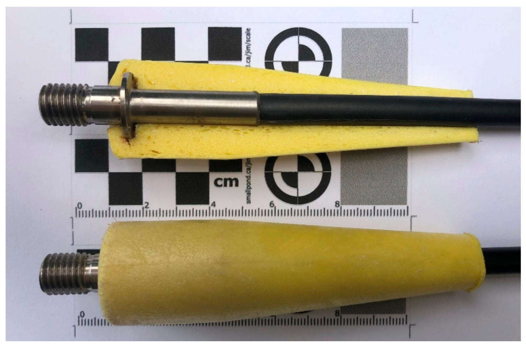
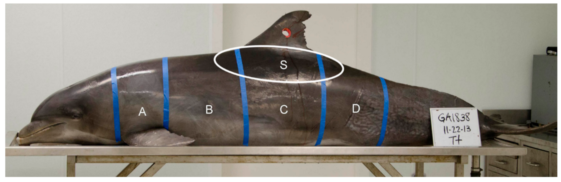
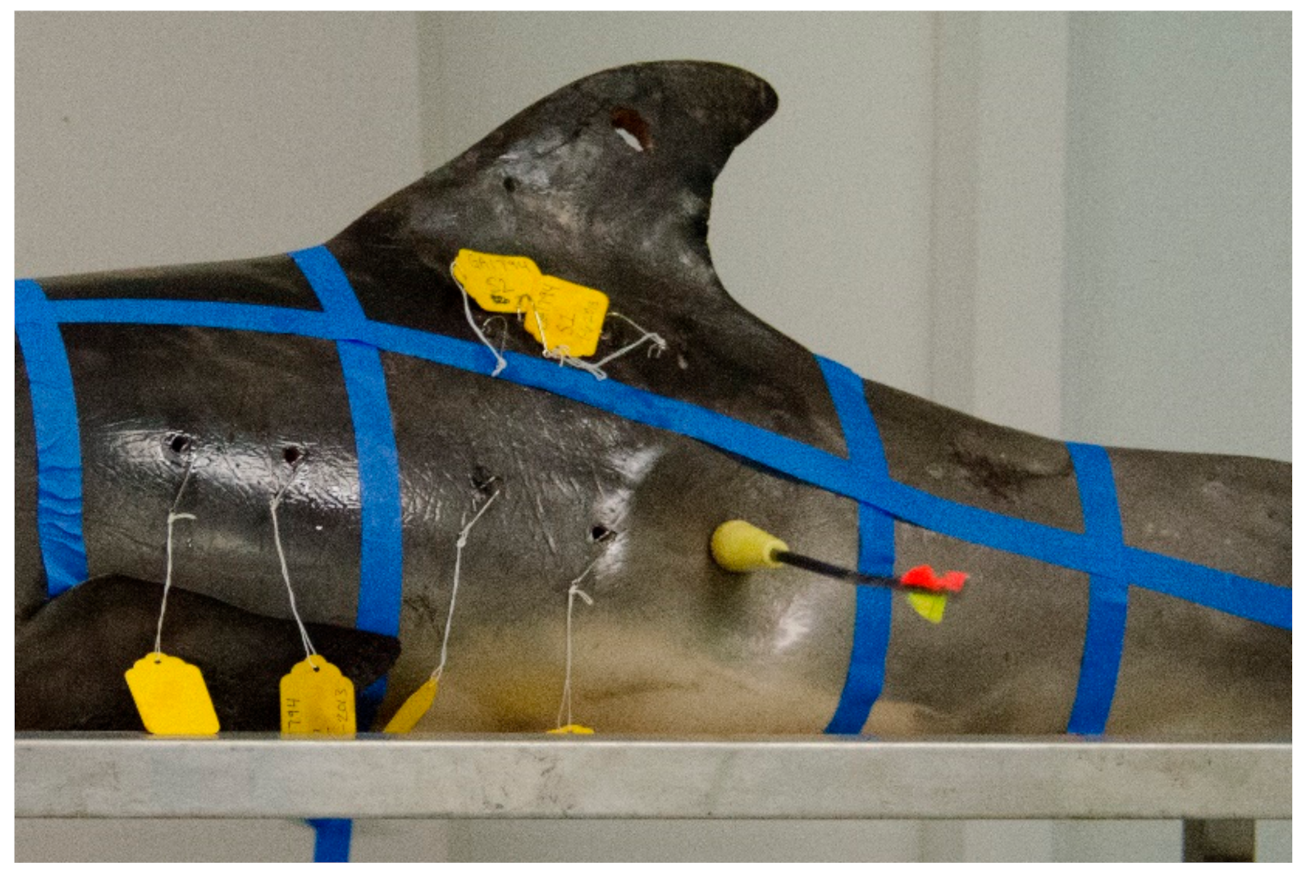
2.2. Carcass Sampling
2.3. Lesion Examination
3. Results
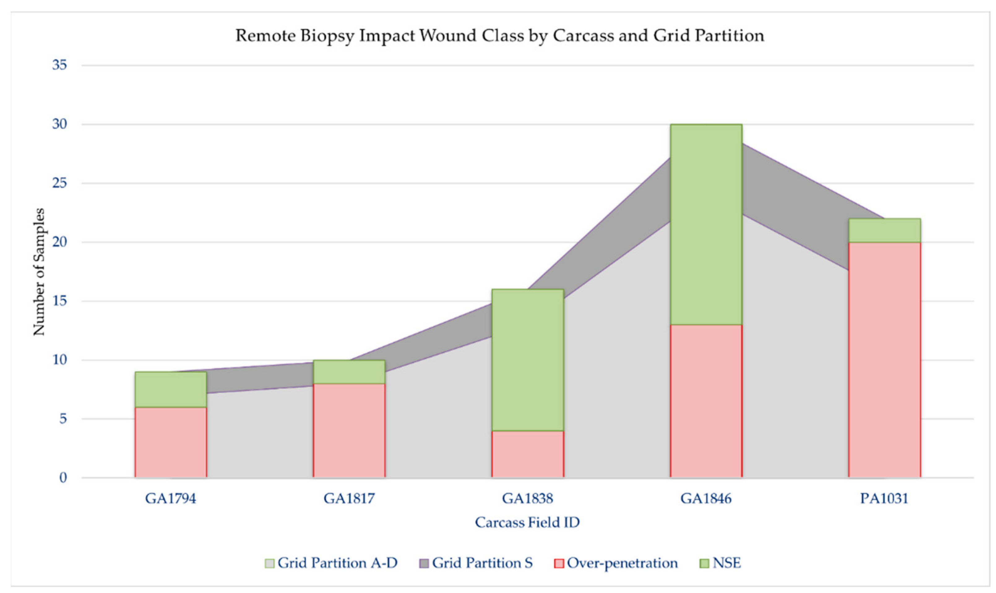
Carcass Sampling and Lesion Examination
4. Discussion
Author Contributions
Funding
Institutional Review Board Statement
Data Availability Statement
Acknowledgments
Conflicts of Interest
References
- Noren, D.P.; Mocklin, J.A. Review of cetacean biopsy techniques: Factors contributing to successful sample collection and physiological and behavioral impacts. Mar. Mammal Sci. 2012, 28, 154–199. [Google Scholar] [CrossRef]
- Hayes, S.A.; Josephson, E.; Maze-Foley, K.; Rosel, P.E. US Atlantic and Gulf of Mexico Marine Mammal Stock Assessments—2018, NOAA Technical Memorandum NMFS-NE-258; US Department of Commerce, National Oceanic and Atmospheric Administration, National Marine Fisheries Service, Northeast Fisheries Science Center: Woods Hole, MA, USA, 2019; 306p.
- Kiszka, J.J.; Méndez-Fernandez, P.; Heithaus, M.R.; Ridoux, V. The foraging ecology of coastal bottlenose dolphins based on stable isotope mixing models and behavioural sampling. Mar. Biol. 2014, 161, 953–961. [Google Scholar] [CrossRef]
- Galligan, T.M.; Balmer, B.C.; Schwacke, L.; Bolton, J.L.; Quigley, B.M.; Rosel, P.; Ylitalo, G.M.; Boggs-Russell, A.S. Examining the Relationships Between Blubber Steroid Hormone and Persistent Organic Pollutant Measurements in Common Bottlenose Dolphins. Environ. Pollut. 2019, 249, 982–991. [Google Scholar] [CrossRef] [PubMed]
- Righetti, B.P.H.; Mattos, J.J.; Siebert, M.N.; Daura-Jorge, F.G.; Bezamat, C.; Fruet, P.F.; Genoves, R.C.; Taniguchi, S.; da Silva, J.; Montone, R.C. Biochemical and molecular biomarkers in integument biopsies of free-ranging coastal bottlenose dolphins from southern Brazil. Chemosphere 2019, 225, 139–149. [Google Scholar] [CrossRef] [PubMed]
- Sinclair, C.; Barry, K.; Ronje, E.I.; Gorgone, A.; Martinez, A.; Speakman, T.; Mullin, K.D. Terrebonne Bay—Timbalier Bay, Lousiana Common Bottlenose Dolphin (Tursiops truncatus) Stock Photo-ID Capture-Recapture and Biopsy Field Summary; NOAA Technical Memorandum NMFS-SEFSC-717; NOAA: Washington, DC, USA, 2017; Volume 717, p. 21. [Google Scholar] [CrossRef]
- Parsons, K.; Durban, J.; Claridge, D. Comparing two alternative methods for sampling small cetaceans for molecular analysis. Mar. Mammal Sci. 2003, 19, 224–231. [Google Scholar] [CrossRef]
- Gorgone, A.M.; Haase, P.A.; Griffith, E.S.; Hohn, A.A. Modeling response of target and nontarget dolphins to biopsy darting. J. Wildl. Manag. 2008, 72, 926–932. [Google Scholar] [CrossRef]
- Wenzel, F.; Nicolas, J.; Larsen, F.; Pace, R.M., III. National Marine Fisheries Service, Northeast Fisheries Science Center Cetacean Biopsy Training Manual. (Northeast Fisheries Science Center Reference Document 10–11); National Oceanic and Atmospheric Administration: Woods Hole, MA, USA, 2010; 18p. Available online: https://repository.library.noaa.gov/view/noaa/3888 (accessed on 10 October 2021).
- Fruet, P.F.; Rosa, L.D.; Genoves, R.C.; Valiati, V.H.; de Freitas, T.R.; Möller, L.M. Biopsy darting of common bottlenose dolphins (Tursiops truncatus) in southern Brazil: Evaluating effectiveness, short-term responses and wound healing. Lat. Am. J. Aquat. Mamm. 2017, 11, 121–132. [Google Scholar] [CrossRef][Green Version]
- Pabst, D.A. Axial muscles and connective tissues of the bottlenose dolphin. In The Bottlenose Dolphin; Leatherwood, S., Reeves, R.R., Eds.; Academic Press: San Diego, CA, USA, 1990. [Google Scholar]
- Patenaude, N.; White, B. Skin biopsy sampling of beluga whale carcasses: Assessment of biopsy darting factors for minimal wounding and effective sample retrieval. Mar. Mammal Sci. 1995, 11, 163–171. [Google Scholar] [CrossRef]
- Weller, D.W.; Cockcroft, V.G.; Würsig, B.; Lynn, S.K.; Fertl, D. Behavioral responses of bottlenose dolphins to remote biopsy sampling and observations of surgical biopsy wound healing. Aquat. Mamm. 1997, 23, 49–58. [Google Scholar]
- Krutzen, M.; Barre, L.M.; Moller, L.M.; Heithaus, M.R.; Simms, C.; Sherwin, W.B. A biopsy system for small cetaceans: Darting success and wound healing in Tursiops spp. Mar. Mammal Sci. 2002, 18, 863–878. [Google Scholar] [CrossRef]
- Jefferson, T.A.; Hung, S.K. Effects of biopsy sampling on Indo-Pacific humpback dolphins (Sousa chinensis) in a polluted coastal environment. Aquat. Mamm. 2008, 34, 310. [Google Scholar] [CrossRef]
- Kiszka, J.; Simon-Bouhet, B.; Charlier, F.; Pusineri, C.; Ridoux, V. Individual and group behavioural reactions of small delphinids to remote biopsy sampling. Anim. Welf. 2010, 19, 411–417. [Google Scholar]
- Tezanos-Pinto, G.; Baker, C.S. Short-term reactions and long-term responses of bottlenose dolphins (Tursiops truncatus) to remote biopsy sampling. N. Z. J. Mar. Freshw. Res. 2012, 46, 13–29. [Google Scholar] [CrossRef]
- Kowarski, K.A.; Augusto, J.F.; Frasier, T.R.; Whitehead, H. Effects of Remote Biopsy Sampling on Long-Finned Pilot Whales (Globicephala melas) in Nova Scotia. Aquat. Mamm. 2014, 40, 117. [Google Scholar] [CrossRef]
- Reisinger, R.R.; Oosthuizen, W.C.; Peron, G.; Toussaint, D.C.; Andrews, R.D.; de Bruyn, P.J. Satellite tagging and biopsy sampling of killer whales at subantarctic Marion Island: Effectiveness, immediate reactions and long-term responses. PLoS ONE 2014, 9, e111835. [Google Scholar] [CrossRef]
- Gales, N.J.; Bowen, W.D.; Johnston, D.W.; Kovacs, K.M.; Littnan, C.L.; Perrin, W.F.; Reynolds, J.E.; Thompson, P.M. Guidelines for the treatment of marine mammals in field research. Mar. Mammal Sci. 2009, 25, 725–736. [Google Scholar] [CrossRef]
- Palsbøll, P.J.; Larsen, F.; Hansen, E.S. Sampling of skin biopsies from free-ranging large cetaceans in West Greenland: Development of new biopsy tips and bolt designs. Genetic ecology of whales and dolphins. Int. Whal. Comm. Spec. Issue 1991, 13, 71–79. [Google Scholar]
- Geraci, J.R.; Lounsbury, V.J. Marine Mammals Ashore: A Field Guide for Strandings, 2nd ed.; John Schmitz & Sons Inc.: Sparks, MD, USA, 2005. [Google Scholar]
- Read, A.J.; Wells, R.S.; Hohn, A.A.; Scott, M.D. Patterns of Growth in Wild Bottle-Nosed Dolphins, Tursiops-truncatus. J. Zool. 1993, 231, 107–123. [Google Scholar] [CrossRef]
- Mazzoil, M.S.; McCulloch, S.D.; Youngbluth, M.J.; Kilpatrick, D.S.; Murdoch, E.M.; Mase-Guthrie, B.; Odell, D.K.; Bossart, G.D. Radio-Tracking and Survivorship of Two Rehabilitated Bottlenose Dolphins (Tursiops truncatus) in the Indian River Lagoon, Florida. Aquat. Mamm. 2008, 34, 54–64. [Google Scholar] [CrossRef]
- Sinclair, C.; Sinclair, J.; Zolman, E.; Martinez, A.; Balmer, B.; Barry, K. Remote Biopsy Sampling Field Procedures for Cetaceans Used during the Natural Resource Damage Assessment of the MSC252 Deepwater Horizon Oil Spill (NOAA Technical Memorandum NMFS-SEFSC-670); National Oceanic and Atmospheric Administration, National Marine Fisheries Service, Southeast Fisheries Science Center: Pascagoula, MS, USA, 2015; 36p. [CrossRef]
- Kellar, N.M.; Trego, M.L.; Chivers, S.J.; Archer, F.I.; Minich, J.J.; Perryman, W.L. Are there biases in biopsy sampling? Potential drivers of sex ratio in projectile biopsy samples from two small delphinids. Mar. Mammal Sci. 2013, 29, E366–E389. [Google Scholar] [CrossRef]
- Ronje, E.I. Dart Speed and Energy for Potential Cetacean Remote Sampling Devices. Aquat. Mamm. 2020, 46, 454–460. [Google Scholar] [CrossRef]
- Bruce-Allen, L.J.; Geraci, J.R. Wound-healing in the bottlenose dolphin (Tursiops truncatus). Can. J. Fish. Aquat. Sci. 1985, 42, 216–228. [Google Scholar] [CrossRef]
- Corkeron, P.; Morris, R.; Bryden, M. A note on healing of large wounds in bottlenose dolphins, Tursiops truncatus. Aquat. Mamm. 1987, 13, 96–98. [Google Scholar]
- Bloom, P.; Jager, M. The injury and subsequent healing of a serious propeller strike to a wild bottlenose dolphin (Tursiops truncatus) resident in cold waters off the Northumberland coast of England. Aquat. Mamm. 1994, 20, 59–64. [Google Scholar]
- Orams, M.; Deakin, R. Report on the healing of a large wound in a bottlenose dolphin (Tursiops truncatus). In Marine Mammal Research in the Southern Hemisphere: Status, Ecology and Medicine; Hindell, M.A., Kempner, C.M., Eds.; Surrey Beatty & Sons: Chipping Norton, UK, 1997; Volume 1. [Google Scholar]
- Wells, R.S.; Allen, J.B.; Hofmann, S.; Bassos-Hull, K.; Fauquier, D.A.; Barros, N.B.; DeLynn, R.E.; Sutton, G.; Socha, V.; Scott, M.D. Consequences of injuries on survival and reproduction of common bottlenose dolphins (Tursiops truncatus) along the west coast of Florida. Mar. Mammal Sci. 2008, 24, 774–794. [Google Scholar] [CrossRef]
- Dwyer, S. Cookie cutter shark (Isistius sp.) bites on cetaceans, with particular reference to killer whales (orca) (Orcinus orca). Aquat. Mamm. 2011, 37, 111–138. [Google Scholar] [CrossRef]
- Best, P.B.; Photopoulou, T. Identifying the “demon whale-biter”: Patterns of scarring on large whales attributed to a cookie-cutter shark (Isistius sp.). PLoS ONE 2016, 11, e0152643. [Google Scholar] [CrossRef]
- Bearzi, G. First report of a common dolphin (Delphinus delphis) death following penetration of a biopsy dart. J. Cetacean Res. Manag. 2000, 2, 217–222. [Google Scholar]
- Barrett-Lennard, L.; Smith, T.G.; Ellis, G.M. A cetacean biopsy system using lightweight pneumatic darts, and its effect on the behavior of killer whales. Mar. Mammal Sci. 1996, 12, 14–27. [Google Scholar] [CrossRef]
- Fierro, M.F.; Ongley, J.P. Blunt force injuries. In Handbook of Forensic Pathology; Froede, R.C., Ed.; College of American Pathologists: Northfield, IL, USA, 1990. [Google Scholar]
- Mckenna, M.F.; Goldbogen, J.A.; Leger, J.S.; Hildebrand, J.A.; Cranford, T.W. Evaluation of postmortem changes in tissue structure in the bottlenose dolphin (Tursiops truncatus). Anat. Rec.-Adv. Integr. Anat. Evol. Biol. 2007, 290, 1023–1032. [Google Scholar] [CrossRef]
- Soldevilla, M.S.; McKenna, M.F.; Wiggins, S.M.; Shadwick, R.E.; Cranford, T.W.; Hildebrand, J.A. Cuvier’s beaked whale (Ziphius cavirostris) head tissues: Physical properties and CT imaging. J. Exp. Biol. 2005, 208, 2319–2332. [Google Scholar] [CrossRef] [PubMed]
- Fitzgerald, E.R.; Fitzgerald, J.W. Blubber and compliant coatings for drag reduction in water I. Viscoelastic properties of blubber and compliant coating materials. Mater. Sci. Eng. C 1995, 2, 209–214. [Google Scholar] [CrossRef]
- Marsh, B.B. Observations on rigor mortis in whale muscle. Biochim. Biophys. Acta 1952, 9, 127–132. [Google Scholar] [CrossRef]
- Van Ee, C.; Chasse, A.; Myers, B. Quantifying skeletal muscle properties in cadaveric test specimens: Effects of mechanical loading, postmortem time, and freezer storage. J. Biomech. Eng. 2000, 122, 9–14. [Google Scholar] [CrossRef] [PubMed]


| Field ID | Sex | Length (cm) | Weight (kg) | Code | Body Score | Stranding Reported | Placed in Freezer | Sampled |
|---|---|---|---|---|---|---|---|---|
| GA1794 | M | 156 | 50.7 | 3 (early) | Ideal | 4 February 2013 | 4 February 2013 | 6 September 2013 |
| GA1817 | M | 167 | 62.8 | 2 | Ideal | 13 March 2013 | 13 March 2013 | 6 September 2013 |
| GA1838 | M | 268 | 253.6 | 2 | Overweight | 22 November 2013 | Not frozen | 23 November 2013 |
| GA1846 | M | 272 | 244.0 | 2 | Ideal | 21 December 2013 | Not frozen | 22 December 2013 |
| PA1031 | M | 218 | 84.4 | 2 | Underweight | 7 March 2014 | 7 March 2014 | 25 June 2014 |
| Impact ID | Wound Class | Sample Blubber Thickness | Gross Description |
|---|---|---|---|
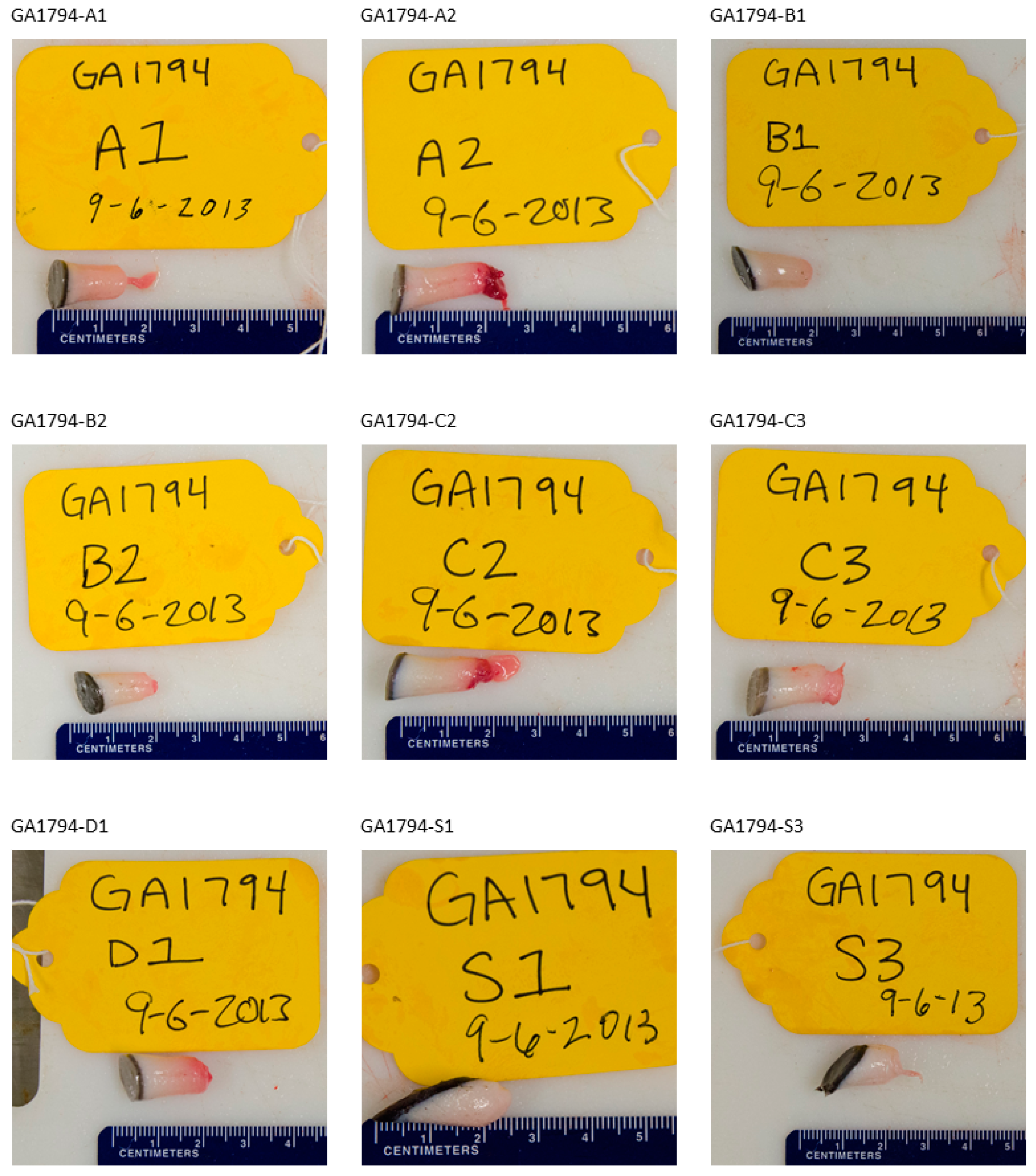
| GA1794-A1 | OP | 15 | 15 mm penetration of the supraspinatus muscle of scapula. Mid scapular impact. Impact created a linear dorsoventral split in the long axis of the scapula: Glenohumeral joint subchondral bone integrity compromised but cartilage layer intact. |
| GA1794-A2 | OP | 20 | 10 mm penetration. Crainial edge of scapula and 1/3 ventral from dorsal aspect. The edge of the scapula is splintered and irreglar with a fragment imbedded in the underlying muscle belly. |
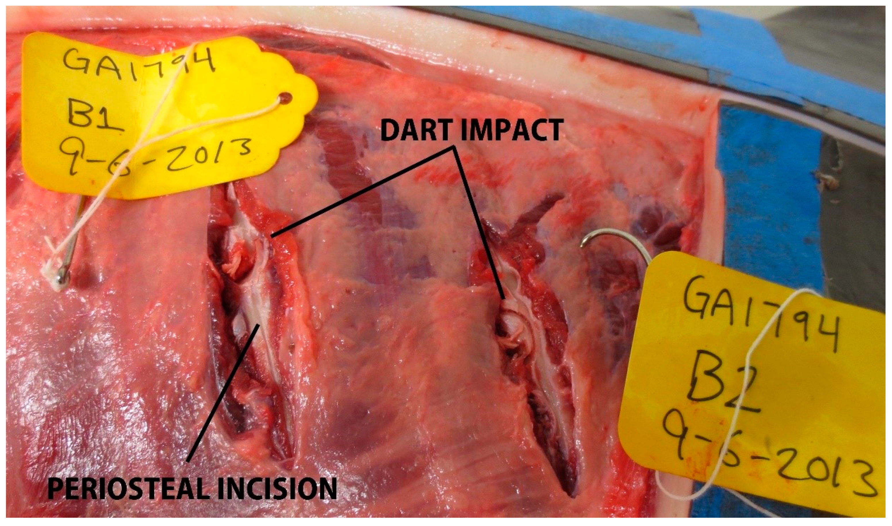
| GA1794-B1 | OP | 19 | Impact on caudal aspect of 6th rib causing a semicircular defect with complete rib fracture. Rib is unstable (i.e., possible flail chest type injury) |
| GA1794-B2 | OP | 17 | Impact on proximal 8th rib on cranial aspect of rib. A semicircular imprint has been left in the bone. Lesion does not penetrate pleural space. Rib integrity intact. |
| GA1794-C2 | OP | 20 | 15 mm penetration. Lesion is within the abdominal muscles approximately 5 cm below associated transverse process. |
| GA1794-C3 | OP | 17 | 20 mm penetration. Dart impact is approximately 20 mm ventral to associated transverse process. The biopsy hole is slightly oblique creating an oval shaped lesion. A small spiral pigmented epithelial tag has been left hanging within the biopsy site (possibly secondary to a direction change of dart within the blubber layer). Abdominal muscle penetration incomplete and peritoneum intact. |
| GA1794-D1 | NSE | 17 | Minimal subdermal penetration (<2 mm). Approximately 15 cm ventral to associated transverse process. The blubber layer appears thicker as the lesion is near the level of the anal slit. |
| GA1794-S1 | NSE | 10 | No visible penetration of the subdermal fascia |
| GA1794-S2 | NSE | 20 | No visible penetration of the subdermal fascia |
| GA1817-A1 | OP | 18 | 12 mm penetration. Suprascapular muscle penetration. Pleural space intact. Contusion of muscle but no evidence of scapula damage. |
| GA1817-A2 | OP | 20 | 15 mm penetration. At dorsal border of scapula where superficial muscles thinner. Scapula penetrated with fragmentation. |
| GA1817-B1 | OP | 20 | 13 mm penetration. Semicircular divot into the periosteum of the cranial edge of rib #6. Rib integrity appears intact. |
| GA1817-B2 | OP | 20 | Impact in center of the 9th rib, creating a shovel shaped divot that has been separated. Rib integrity appears intact. |
| GA1817-C1 | OP | 17 | 20 mm penetration. Caudal to last rib over abdomen. Palpation of this asymmetric lesion’s depth indicates no penetration of the peritoneum |
| GA1817-C2 | OP | 17 | 12 mm penetration. Ventral to transverse processes. |
| GA1817-D1 | OP | 14 | 15 mm penetration. Penetration is approximately 30 mm ventral to transverse processes of closest vertebra and appears to penetrate the ventral spinal muscles (iliopsoas or equivalent). Peritoneum is intact. |
| GA1817-D2 | OP | 15 | 40 mm penetration. Lesion slightly (12 mm) above the transverse process. Impact is within a tissue/muscle plane that with gentle probing extends deep to the pedicle of the proximate vertebrae. |
| GA1817-S1 | NSE | 18 | Incomplete penetration of blubber layer, however there is a peri-lesion tissue disruption (a divot) palpable from deeper layer. Impact located at crux of dorsal fin attachment to body wall. |
| GA1817-S2 | NSE | 26 | Minimal impact to subdermal fascia, muscle belly has palpable impact crater but no exposed muscle fibers |
| GA1838-A1 | NSE | 14 | Fascia traumatized showing redness and gelatinous appearance (carcass is relatively fresh). Location 10 mm dorsal to transverse process. |
| GA1838-A2 | NSE | 14 | Ventral to A1, sub-dermal fascia traumatized but minimal effect. |
| GA1838-A3 | NSE | 10 | Contusion to supraspinatus muscle—minimal effect. Impact of dart slightly askew (oblique) may have limited penetration depth. |
| GA1838-B1 | NSE | 22 | Minimal observable effect. |
| GA1838-B2 | OP | 20 | Oblique muscles over rib penetrated but ribs unaffected. |
| GA1838-B3 | OP | 20 | Muscular band over transverse process significant in this larger animal; Penetrated but minimal effect. |
| GA1838-C1 | NSE | 19 | Fascia discolored and cut from impact. |
| GA1838-C2 | NSE | 20 | Slight fascial evidence of impact. |
| GA1838-C3 | NSE | 20 | Slight fascial evidence of impact. |
| GA1838-C4 | OP | 21 | 12 mm penetration into lateral (transverse) thoracic muscle belly. |
| GA1838-D1 | OP | 20 | 4 mm penetration of dorsal epaxial muscle fascia (but not belly). Oblique impact secondary to body wall curvature at this level with blubber and fascia impacts slightly mis-aligned. |
| GA1838-D2 | NSE | 17 | 5 mm penetration into fatty fascia. Slightly oblique lesion. |
| GA1838-D3 | OP | 18–20 | Penetrated 4–5 mm into muscle belly |
| GA1838-S1 | NSE | 22 | Slight fascial evidence of impact, did not penetrate the fascial layer. |
| GA1838-S2 | NSE | 23 | Slight oblique impact, lesion is asymmetric due to angle of body wall, does not penetrate the subcutaneous fat into the muscle. |
| GA1838-S3 | NSE | 22 | Slight oblique impact, tip of probe stays within limits of blubber layer, backside does not appear discolored. |
| GA1846-A1 | NSE | 15–21 | Slight fascial evidence of impact. |
| GA1846-A2 | NSE | 22–23 | Dorsal caudal boarder of scapula. Fascia minimally effected. |
| GA1846-A3 | NSE | 23–24 | Slight fascial evidence of impact. |
| GA1846-A4 | OP | 18 | 20 mm penetration over scapula, penetrated into muscle but no trauma to scapula. |
| GA1846-A5 | OP | 22 | 4 mm penetration. Caudal scapula bone scored (periosteal) but no cortical bone fragments apparent. |
| GA1846-A6 | OP | 26 | Caudal dorsal scapula impact. Infraspinatus muscle biopsied but no trauma to scapula. |
| GA1846-B1 | NSE | 21–24 | Dart has penetrated the fibrous layer covering the muscles but does not penetrate through muscle. |
| GA1846-B2 | OP | 27–28 | 3–4 mm penetration through the fibrous layer between blubber and dorsal epaxial muscles. |
| GA1846-B3 | NSE | 22–23 | Penetrates blubber layer into thick fascia. No damage to latissimus dorsi muscle. |
| GA1846-B4 | OP | 22.5 | Intercostal muscle impacted and damaged but no rib impact. Pleural space intact. |
| GA1846-B5 | OP | 26 | Rib impacted with circular divot and bone splintering. |
| GA1846-B6 | OP | 22 | 12 mm penetration. Close to costo-chondral junction of rib and sternum. No perturbation of rib found. |
| GA1846-C1 | OP | 23 | 5 mm muscle penetration |
| GA1846-C2 | NSE | 21–22 | Dorsal epaxial muscle superficially penetrated but minimal effect. |
| GA1846-C3 | NSE | 19–21 | Pronounced fascia at this level, superficially penetrated subcutaneous layer. |
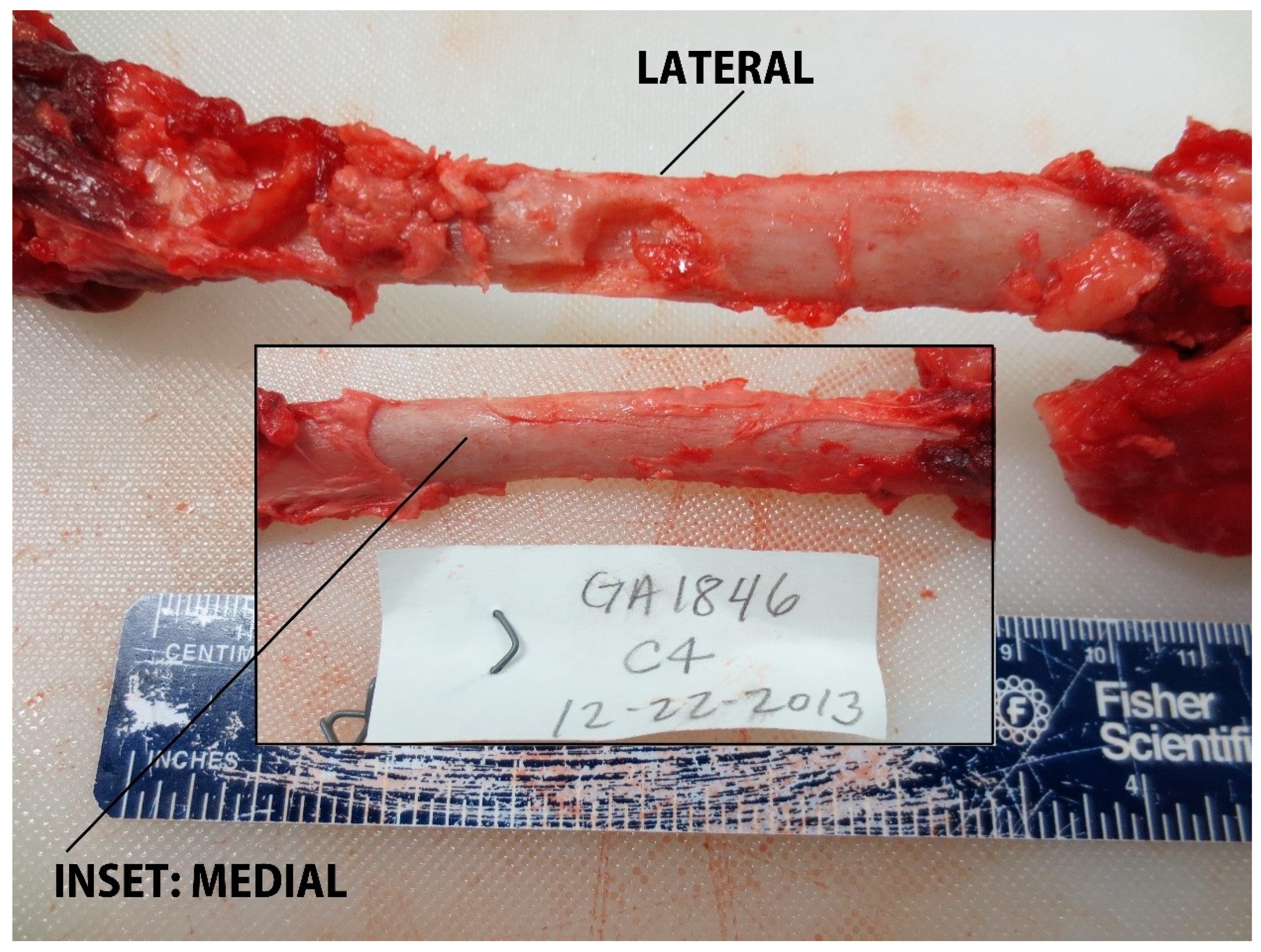
| GA1846-C4 | OP | 24 | Rib impact with fragmentation and linear scoring of cortex (impact slightly to caudal edge of rib). Possible greenstick fracture. |
| GA1846-C5 | NSE | 20 | Superficial muscle penetration. |
| GA1846-C6 | OP | 18 | 15 mm muscular penetration |
| GA1846-D1 | NSE | 21 | Slight fascial evidence of impact. |
| GA1846-D2 | OP | 19–20 | Muscle evident poking out from cut in fascia. |
| GA1846-D3 | NSE | 21–24 | Slight fascial evidence of impact. |
| GA1846-D4 | NSE | 20–21 | Slight fascial evidence of impact. |
| GA1846-D5 | OP | 17 | Penetrated fascia, muscle evident poking out from cut in fascia. |
| GA1846-D6 | OP | 19 | Very close to transverse process. Penetrated fascia and removed a muscle biopsy but no damage to transverse process. |
| GA1846-S1 | NSE | 15–24 | Subdermal fascia not penetrated |
| GA1846-S2 | NSE | 13–21 | Subdermal fascia not penetrated. |
| GA1846-S3 | NSE | 15–21 | Subdermal fascia not penetrated |
| GA1846-S4 | NSE | 18–22 | Incomplete penetration through the blubber layer to the subdermal fascia |
| GA1846-S5 | NSE | 13–22 | Incomplete penetration through the blubber layer to the subdermal fascia |
| GA1846-S6 | NSE | 15–23 | Incomplete penetration through the blubber layer to the subdermal fascia |
| PA1031-A1 | OP | 20 | Divot removed from rib #3, also taken cortical bone, fragmented-linear fragment of rib taken out in addition to divot |
| PA1031-A2 | OP | 22 | Perforated scapula, well circumscribed hole, little fragmenting of bone on lateral aspect of scapula, fragments on medial side of scapula |
| PA1031-B1 | OP | 27 | Significant impact to top of rib head, only 1 cm tissue over rib head, impacted rib #8 and left small divot in bone, trough periosteum and into cortical bone |
| PA1031-B2 | OP | 25 | Impacted caudal edge of rib #7. Semi-circular bone biopsy, no penetration into pleural space, large bone chip extends along rib on medial side, fragments of bone on medial axial component of rib. Decreased girth of rib substantially. |
| PA1031-B3 | OP | 24 | Impacted rib #9 and removed divot out of rib. |
| PA1031-B4 | OP | 20–27 | Hit but sample not retained, 20 mm penetration into dorsal epaxial muscle |
| PA1031-B5 | OP | 18–26 | 10 mm muscle penetration |
| PA1031-B6 | OP | 20 | 10 mm muscle penetration |
| PA1031-C1 | OP | 20 | 10 mm muscle penetration |
| PA1031-C2 | NSE | 19 | NSE, minimal muscle penetration |
| PA1031-C3 | OP | 18 | Significant damage to intercostal muscle between rib #3 and #4, ribs not impacted. |
| PA1031-C4 | OP | 19 | 10 mm muscular penetration, no impact to transverse process or penetration into abdominal cavity |
| PA1031-C5 | OP | 18 | Sample not retained. 5 mm muscle penetration under transverse process. |
| PA1031-D1 | OP | 19 | 20 mm penetration of epaxial muscle |
| PA1031-D2 | OP | 20 | 20 mm penetration of epaxial muscle |
| PA1031-D3 | OP | 17 | 20 mm penetration of epaxial muscle |
| PA1031-S1 | OP | 27 | 20 mm penetration of epaxial muscle |
| PA1031-S2 | OP | 20 | 20 mm penetration of epaxial muscle |
| PA1031-S3 | OP | 18–21 | 10 mm penetration of epaxial muscle |
| PA1031-S4 | OP | 18–28 | 20 mm penetration of epaxial muscle |
| PA1031-S5 | NSE | 20 | NSE, No penetration of the subdermal fascia into muscle |
| PA1031-S6 | OP | 20 | 15 mm muscle penetration directly over dorsal spinous process. No apparent trauma to the dorsal spinous process. |
Publisher’s Note: MDPI stays neutral with regard to jurisdictional claims in published maps and institutional affiliations. |
© 2021 by the authors. Licensee MDPI, Basel, Switzerland. This article is an open access article distributed under the terms and conditions of the Creative Commons Attribution (CC BY) license (https://creativecommons.org/licenses/by/4.0/).
Share and Cite
Ronje, E.I.; Brechtel, C. Preliminary Evaluation of Potential Impacts Associated with Small Cetacean Remote Biopsy Sampling by Controlled Testing on Stranded Common Bottlenose Dolphins (Tursiops truncatus). J. Zool. Bot. Gard. 2021, 2, 544-558. https://doi.org/10.3390/jzbg2040039
Ronje EI, Brechtel C. Preliminary Evaluation of Potential Impacts Associated with Small Cetacean Remote Biopsy Sampling by Controlled Testing on Stranded Common Bottlenose Dolphins (Tursiops truncatus). Journal of Zoological and Botanical Gardens. 2021; 2(4):544-558. https://doi.org/10.3390/jzbg2040039
Chicago/Turabian StyleRonje, Errol I., and Casey Brechtel. 2021. "Preliminary Evaluation of Potential Impacts Associated with Small Cetacean Remote Biopsy Sampling by Controlled Testing on Stranded Common Bottlenose Dolphins (Tursiops truncatus)" Journal of Zoological and Botanical Gardens 2, no. 4: 544-558. https://doi.org/10.3390/jzbg2040039
APA StyleRonje, E. I., & Brechtel, C. (2021). Preliminary Evaluation of Potential Impacts Associated with Small Cetacean Remote Biopsy Sampling by Controlled Testing on Stranded Common Bottlenose Dolphins (Tursiops truncatus). Journal of Zoological and Botanical Gardens, 2(4), 544-558. https://doi.org/10.3390/jzbg2040039