Article

IFN-τ Modulates PBMC Cytokine Profile and T Cell Phenotype to Improve Endometrial Immune Composition in the Implantation Window: A Combined In Vitro and In Vivo Study

1
Research Department, Nadezhda Women’s Health Hospital, 1373 Sofia, Bulgaria
2
Pathology Department, Nadezhda Women’s Health Hospital, 1373 Sofia, Bulgaria
3
Obstetrics & Gynecology Department, Nadezhda Women’s Health Hospital, 1373 Sofia, Bulgaria
4
Genetics Department, Nadezhda Women’s Health Hospital, 1373 Sofia, Bulgaria
5
Department of Medical Genetics, Faculty of Medicine, Medical University-Sofia, 1431 Sofia, Bulgaria
*
Author to whom correspondence should be addressed.
Immuno 2025, 5(4), 51; https://doi.org/10.3390/immuno5040051
Submission received: 2 October 2025 / Revised: 21 October 2025 / Accepted: 22 October 2025 / Published: 24 October 2025

Abstract

Embryo implantation requires a finely tuned immune balance at the maternal–fetal interface. Interferon tau (IFN-τ), a key immunomodulator in ruminant implantation, may have therapeutic potential in human reproduction. This study investigated its effects on peripheral blood mononuclear cells (PBMCs) in vitro and the subsequent impact on endometrial immune composition following intrauterine administration of these cells. The work was conducted in two stages. First, in vitro assays were performed with PBMCs from 20 patients with recurrent implantation failure (RIF) cultured with or without IFN-τ for 24 h. Cytokines (IL-10, IL-4, TNF-α, IL-6) were measured by ELISA, and T cell subsets (Th, cytT, Th1, Th2, Th9, Tfh, Th17, Treg) were analyzed by flow cytometry. IFN-τ increased IL-4 and reduced TNF-α and IL-6, indicating a Th2 profile shift. T-cell analysis revealed fewer cytT, Th1, Th9, and Th17 cells, more Th2 cells, and improved Th/Tk, Th1/Th2, and Th17/Treg ratios after IFN-τ. A second clinical study included 55 RIF patients who received intrauterine IFN-τ-modulated PBMCs. Post-treatment endometrial biopsies revealed more helper T cells and macrophages, with higher Th/total T, Th/cytT, and Th/macrophage ratios, suggesting a tolerogenic environment. Overall, IFN-τ modulates PBMCs in vitro and promotes a favorable endometrial immune profile in vivo, highlighting its potential as an immunotherapy in assisted reproduction.

1. Introduction

Successful embryo implantation is a complex and finely regulated process that requires a receptive endometrial environment and an appropriately modulated immune response [1,2]. At the maternal–fetal interface, immune tolerance must be established to permit embryo acceptance, while maintaining the capacity to respond to and defend against potential pathogens. Dysregulation of immune cell composition or cytokine signalling within the endometrium has been implicated in implantation failure and infertility among patients undergoing assisted reproductive technologies (ARTs) and particularly in those with repeated implantation failure (RIF) [3].
T helper (Th) cell subsets play a pivotal role in shaping the local immune environment, with a delicate balance between pro-inflammatory (e.g., Th1, Th17) and anti-inflammatory or regulatory (e.g., Th2, Treg) responses being critical for implantation success [4,5]. A Th2-dominant immune profile is generally associated with immune tolerance and favorable pregnancy outcomes, whereas an overrepresentation of Th1 or Th17 cells can contribute to implantation failure and early pregnancy loss [6,7].
Peripheral blood mononuclear cells (PBMCs) are a readily accessible source of immune cells and have been explored as intrauterine immunotherapy in reproductive medicine. They represent a heterogeneous population of circulating mononuclear cells, largely composed of T lymphocytes with a substantial proportion of Th cells, in addition to monocytes, dendritic cells, B cells and natural killer (NK) cells [8]. These cells are also present in the endometrium and play key roles in maternal tissue remodelling and embryo–maternal immune crosstalk during the implantation window [9]. Clinically, intrauterine administration of autologous PBMCs has emerged as an adjunctive treatment in infertility, with several clinical trials and meta-analyses reporting improved endometrial receptivity, clinical pregnancy rates, and live birth rates in ART patients [10,11,12,13,14]. PBMC-based therapies are typically prepared through short-term culture and are often activated in vitro with human chorionic gonadotropin (hCG)—one of the earliest human embryonic signals. Several in vitro invasion assays have demonstrated a synergistic effect of hCG and PBMCs on processes relevant to implantation [15,16]. However, other trophoblast-derived factors may exert more potent or specific immunomodulatory effects on PBMCs.
Interferon tau (IFN-τ) is a type I interferon uniquely expressed in ruminants, where it acts as the maternal pregnancy recognition signal. Beyond its endocrine role, IFN-τ exhibits potent immunomodulatory properties, contributing to the establishment of a tolerogenic uterine environment during implantation. While IFN-τ is not endogenously expressed in humans, it is structurally related to IFN-α and IFN-β, and exerts its effect through binding to the common type I interferon receptor (IFNAR), which is expressed in human immune cells. Activation of this receptor initiates downstream signaling via STAT1, STAT2, and Tyk2, leading to the induction of immunomodulatory cytokines such as IL-10, IL-6, and IL-4 [17]. Notably, IFN-τ exhibits significantly lower cytotoxicity compared to IFN-α and has demonstrated therapeutic potential in various in vitro and animal models of autoimmune and viral diseases [18,19,20]. Therefore, its immunological effects may be harnessed therapeutically to modulate immune responses in the human endometrium.
This study aimed to investigate the immunological effects of IFN-τ on PBMCs in vitro and assess the subsequent impact of intrauterine administration on endometrial immune composition in RIF patients undergoing assisted reproduction.

2. Materials and Methods

2.1. Study Population

Two prospective cohorts of RIF patients were recruited (Figure 1). The first included 20 reproductive-age women, who provided blood samples for the initial in vitro PBMC characterisation after immunomodulation with IFN-τ. The second pilot clinical arm consisted of 55 RIF patients undergoing assisted reproduction therapy at Nadezhda Women’s Health Hospital. They were recruited to assess the endometrial composition before and after intrauterine treatment with IFN-τ-munomodulated PBMC. Eligibility criteria included at least two failed in vitro fertilization (IVF) cycles with transfer of good-quality embryos and regular cycles. Exclusion criteria included uterine anomalies, chronic inflammatory, infectious or autoimmune disorders, oncological conditions, and immunological or hormonal treatments within three months of sampling.
The study was approved by the hospital’s ethics committee (protocol 7/28 February 2023), and all participants provided informed consent. The treatment arm was part of a registered clinical trial (NCT05775211) [21].

2.2. Autologous Immune Cell Isolation and Modulation

Autologous peripheral blood mononuclear cells (PBMCs) were isolated from 9 mL of peripheral venous blood using 1.077 g/mL Pancoll (P04-60100, Pan Biotech, Aidenbach, Germany) density gradient centrifugation at 400× g for 25 min. Isolated cells were washed with PBS and cultured in vitro for 24 h at 37 °C in RPMI-1640 medium (P04-22100, Pan Biotech) supplemented with 1 mg/mL human serum albumin (A1A029AA, Takeda, Zurich, Switzerland), antibiotics mix (penicillin/streptomycin/amphotericin B; P06-07300, Pan Biotech) with or without 500IU recombinant IFN-τ (CSB-YP350007SH, CusaBio, Houston, TX, USA). In the in vitro arm, cells were centrifuged to separate the supernatant for measurement of secreted cytokines and the cells were washed and prepared for flow cytometry quantification.
In the clinical arm, on day 7 post-luteinizing hormone peak (LH + 7), approximately 1 × 10⁷ washed modulated PBMCs were suspended in 0.5 mL sterile saline and administered intrauterinely using an IUI catheter by a trained assisted reproduction specialist. Patients were monitored for 2 h after the procedure and instructed to report any adverse events.

2.3. Cytokine Quantification

The PBMC medium was used to measure the quantities of tumor necrosis factor alpha (TNFα), interleukin-6 (IL6), interleukin-4 (IL4) and interleukin-10 (IL10) via sandwich enzyme-linked immunosorbent assay according to the manufacturer’s instructions (CSBE04740h, CSBE04638h, CSBE04633h and CSBE04593h, Cusabio, respectively). Media from IFN-τ-stimulated PBMCs were compared to those from unstimulated control cells after 24 h of culturing.

2.4. T Subpopulations Phenotyping

Washed PBMCs before and after 24 h culture with IFN-τ were incubated with a panel of fluorochrome-conjugated monoclonal antibodies specific to defined surface markers and analysed by multiparameter flow cytometry (BD FACS Lyric™, BD Biosciences, Eysins, Switzerland) to characterise and quantify distinct immune cell populations: total T cells (CD3+Lymphocytes), T helper cells (Th, CD3+CD4+CD8), cytotoxic T cells (cytT, CD3+CD4CD8+), and regulatory T cells (Treg, CD4+CD8CD25+CD127). Further subclassification of T helper cells was performed to identify T follicular helper cells (Tfh, CD3+CD4+CD8CD185+), as well as T helper functional subsets based on chemokine receptor expression: Th1 (CD4+CD183+CD196), Th2 (CD4+CD183CD194+CD196), Th9 (CD4+CD194CD196+) and Th17 (CD4+CD183CD194+CD196+). The following monoclonal antibodies were used for this immunophenotyping: CD4–APC-H7 (Cat# 560158, BD Biosciences, Eysins, Switzerland), CD8-APC-Cy7 (Cat# 344714, Biolegend, Amsterdam, Netherlands), CD25–BB515 (Cat# 564467, BD Biosciences), CD127–Alexa Fluor 647 (Cat# 558598, BD Biosciences), CD183 (CXCR3)–PE-Cy7 (Cat# 560831, BD Biosciences), CD185 (CXCR5)-BV480 (Cat# 566142, BD Biosciences), CD194 (CCR4)–BV421 (Cat# 562579, BD Biosciences) and CD196 (CCR6)–BB700 (Cat# 566477, BD Biosciences).

2.5. Endometrial Sample Collection and Processing

Endometrial biopsies were obtained using a Pipelle catheter (CooperSurgical, Trumbull, CT, USA) during the mid-luteal phase (LH + 7) in two consecutive cycles: one during a baseline cycle (pre-treatment) and a second one on the day after intrauterine PBMC administration. All samples were immediately fixed in 4% neutral-buffered formalin for 12–18 h, processed through Leica TP1020 Semi-enclosed Benchtop Tissue Processor (Leica Biosystems, Wetzlar, Hesse, Germany) and then embedded in paraffin.

2.6. Immunohistochemistry (IHC)

Serial tissue sections cut at 4 µm on a manual microtome (Leica Biosystems, Nussloch, Germany) were deparaffinized in xylene and rehydrated through a graded ethanol series. Antigen retrieval was performed using 0.01 M citrate buffer (pH 9.0) for 30 min at 95 °C. Endogenous peroxidase activity was blocked with 3% hydrogen peroxide in methanol for 5 min. Sections were incubated with 0.4% casein in phosphate-buffered saline (PBS) to block non-specific binding. Primary antibodies used included CD3 (1:50, rabbit polyclonal, BRB063, Zytomed Systems, Berlin, Germany) to identify T cells; CD4 (ready-to-use, mouse monoclonal, IS649, Dako, Santa Clara, CA, USA) as a marker of T helpers; CD8 (1:100, rabbit polyclonal, 1-CD040-02, Quartett, Berlin, Germany) for cytotoxic T cells; CD56 (1:200, mouse monoclonal, A00121-0007, ScyTek, Logan, UT, USA) to identify NK cells, and CD68 (ready-to-use, mouse monoclonal, IS613, Dako) to visualize macrophages. Sections were incubated with the respective primary antibody for 1 h at 37 °C. After washing, specimens were treated with a post-primary block composed of 10% (v/v) bovine serum in tris-buffered saline to facilitate penetration of the subsequently applied poly-HRP anti-rabbit/mouse IgG (NovoLink Polymer, Leica Biosystems, Nussloch, Germany). Visualization of the bound antibodies was performed via incubation with 3,3′-diaminobenzidine (DAB) chromogen, forming a brown precipitate. Ultimately, the tissue was counterstained with 0.02% hematoxylin. Negative controls for each sample were prepared using the same protocol, substituting PBS buffer for the primary antibody. Positive controls for antibody validation were prepared analogously to the experimental samples, using spleen tissue obtained from the pathology laboratory at Nadezhda Hospital.

2.7. Image Acquisition and Quantitative IHC Analysis

Stained sections from the functionalis layer of the endometrium were captured using an Olympus CKX41 inverted microscope and Olympus EP50 wireless digital camera (Olympus, Melville, NY, USA) at 40× magnification. For each marker, five non-overlapping fields per slide were analyzed using the Cell Quantification module on HALO™ image analysis software (IndicaLabs, Albuquerque, NM, USA, v. 3.4). The studied cells were quantified and expressed as a percentage of positively stained cells relative to stromal cells.

2.8. Statistical Analysis

Sample size estimation was performed using preliminary data from a pilot study involving immune profiling of PBMCs and endometrial assessment in RIF patients. Power analysis indicated that a minimum of 18 paired immune cell samples would be required to detect a 20% change in the Th1/Th2 or Th17/Treg ratio in PBMC, and a minimum of 50 paired samples to detect a 20% change in endometrial immune cell quantities a with 80% power at a 5% significance level (α = 0.05), assuming a standard deviation based on pilot data. To account for potential dropout or assay failure, we included 20 patients in the in vitro arm and 55 patients in the clinical study arm.
Data were first assessed for normality using the Shapiro–Wilk test. Parametric data are presented as mean ± standard deviation (SD), and non-parametric data as median with interquartile range [IQR]. Statistical comparisons were made using paired t-tests for normally distributed paired data, and the Wilcoxon signed-rank test for non-normally distributed paired data. Between-group comparisons were conducted using independent t-tests or chi-squared tests, depending on the variable type.
Outliers were identified as values lying beyond 1.5 times the IQR from the first or third quartiles. Outliers were retained to preserve the integrity of biological variation.
All statistical analyses were performed using SPSS version 27.0 (IBM Corp, Armonk, NY, USA). A p-value of <0.05 was considered statistically significant.

3. Results

3.1. Patient Characteristics

Baseline characteristics of the patients in the two cohorts are summarized in Table 1. Patients in the two cohorts were similar in terms of age and reproductive history.

3.2. In Vitro Experiments

3.2.1. IFN-τ Modulates Cytokine Secretion in PBMCs Toward a Th2 Profile

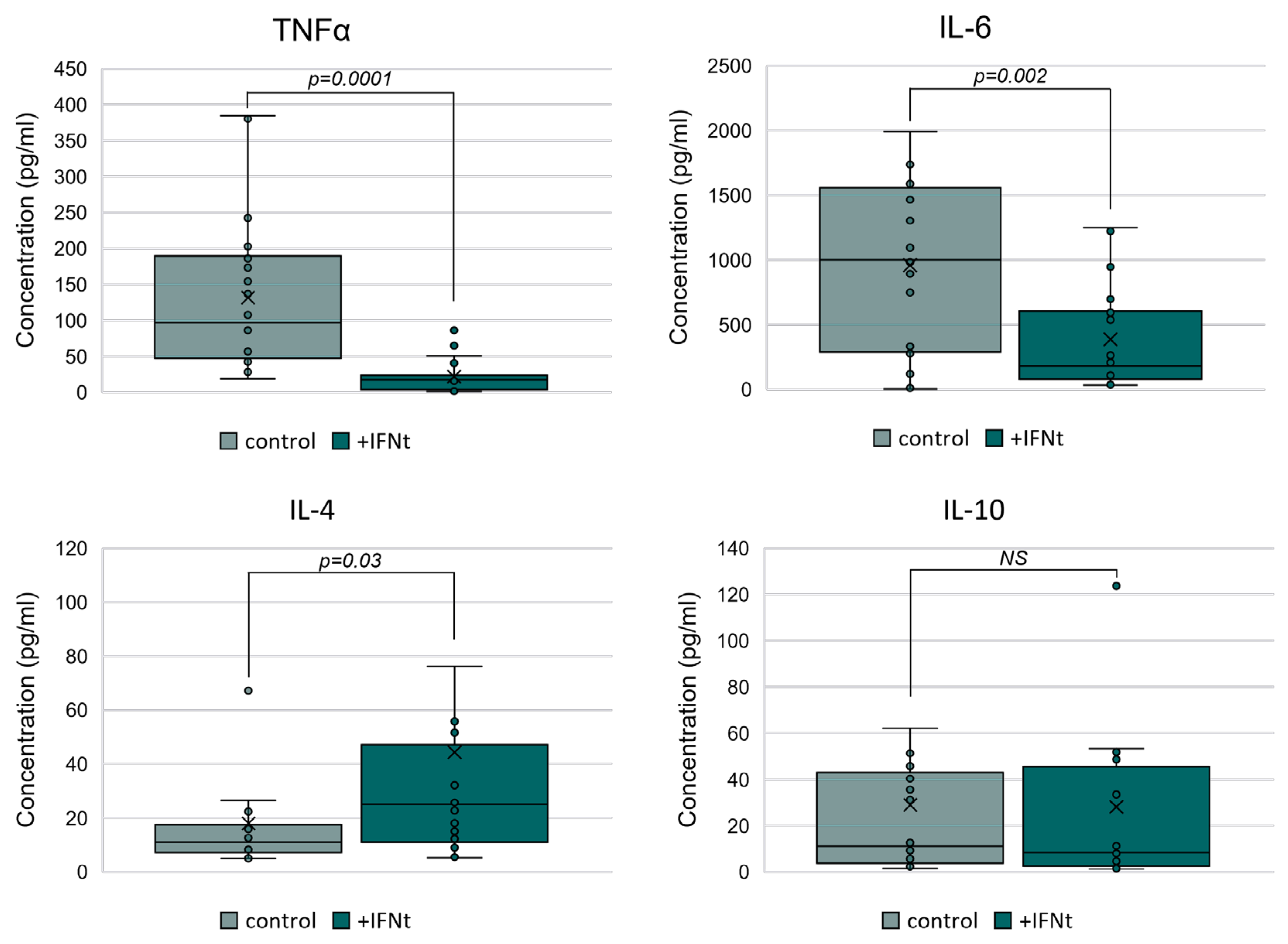
Incubation of PBMCs with IFN-τ for 24 h significantly altered their cytokine secretion profiles compared to unstimulated control cells. ELISA analysis demonstrated a significant increase in IL-4 secretion (median [IQR]: 25.1 pg/mL [36.3] vs. 13.5 pg/mL [18.2]; p = 0.03), while levels of the pro-inflammatory cytokines TNF-α and IL-6 were significantly decreased (median [IQR]: 17.7 pg/mL [19.9] vs. 96.8 pg/mL [142.6]; p = 0.0001 and 180.1 pg/mL [526.9] vs. 1001.8 pg/mL [1267.0]; p = 0.002, respectively) (Figure 2). IFN-τ did not induce significant changes in IL-10 secretion compared to 24 h control (p > 0.05). These findings suggest that IFN-τ promotes a shift from a pro-inflammatory (Th1/Th17) to an anti-inflammatory (Th2) cytokine environment in vitro.

3.2.2. IFN-τ Alters T Cell Subpopulation Profiles in PBMCs

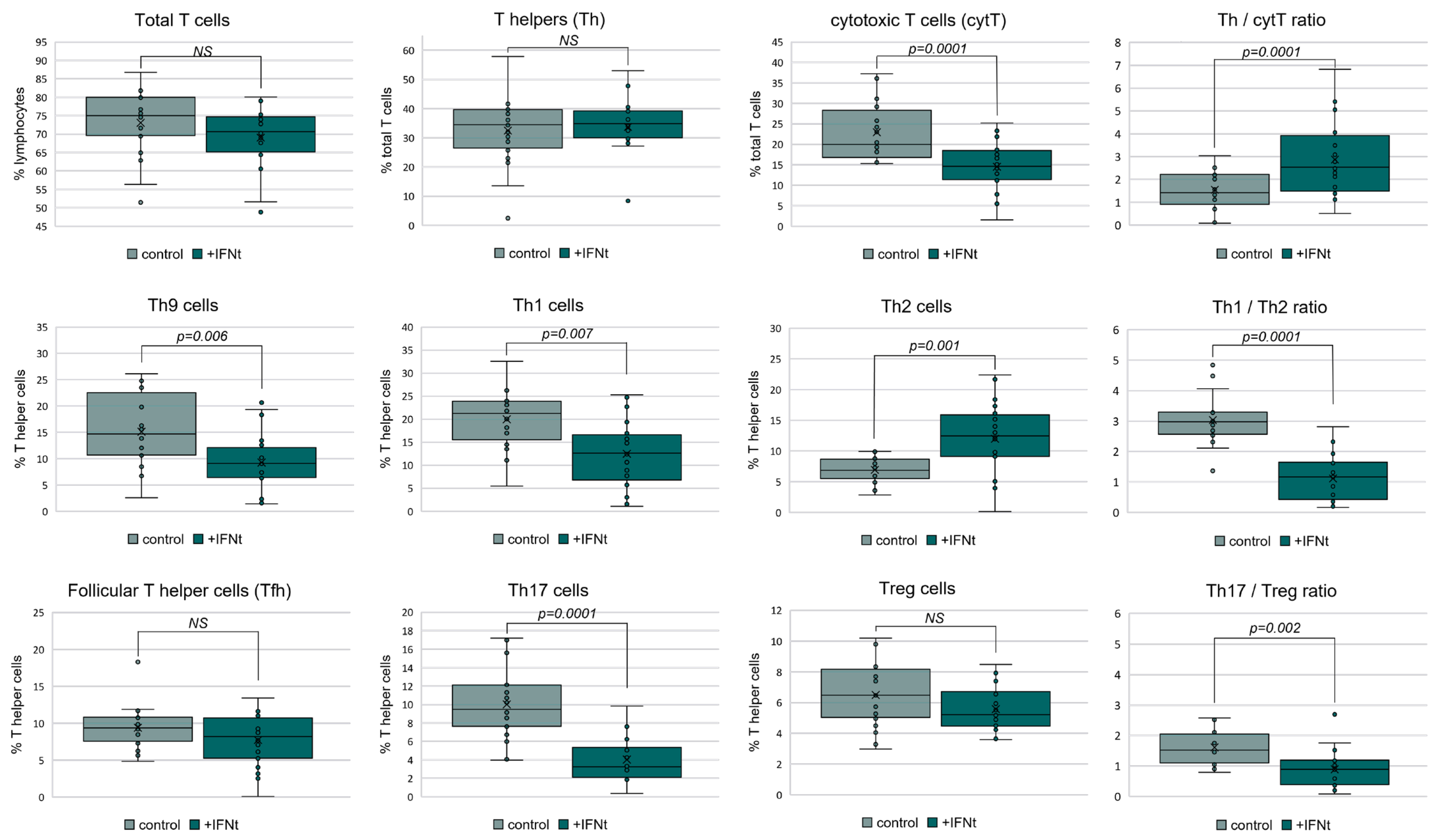
Flow cytometry analysis (Figure 3) revealed that IFN-τ stimulation led to a notable increase in Th2 cells (median [IQR]: 12.5% [6.9] vs. 6.8% [3.2]; p = 0.0001), alongside a significant reduction in cytotoxic T cells (median [IQR]: 14.6% [7.1] vs. 20.0% [11.5]; p = 0.0001), Th1 (12.6% [9.8] vs. 21.3% [8.4]; p = 0.007), Th9 (9.1% [5.7] vs. 14.7% [11.9]; p = 0.006), and Th17 cells (median [IQR]: 3.2% [3.2] vs. 9.5% [4.5]; p = 0.0001) in the PBMC compared to before immunomodulation. The Th/cytT ratio was significantly elevated following immunomodulation with IFN-τ (median [IQR]: 2.5 [2.4] vs. 1.4 [1.3]; p = 0.0001), indicating a shift toward T helper cell dominance. Importantly, the Th1/Th2 (1.2 [1.2] vs. 3.0 [0.7]; p = 0.0001) and Th17/Treg ratios (0.9 [0.8] vs. 1.5 [1.0]; p = 0.002) were significantly decreased, further supporting polarization toward a tolerogenic phenotype.

3.3. Intrauterine Administration of IFN-τ-Modulated PBMCs Enhances Endometrial Immune Composition

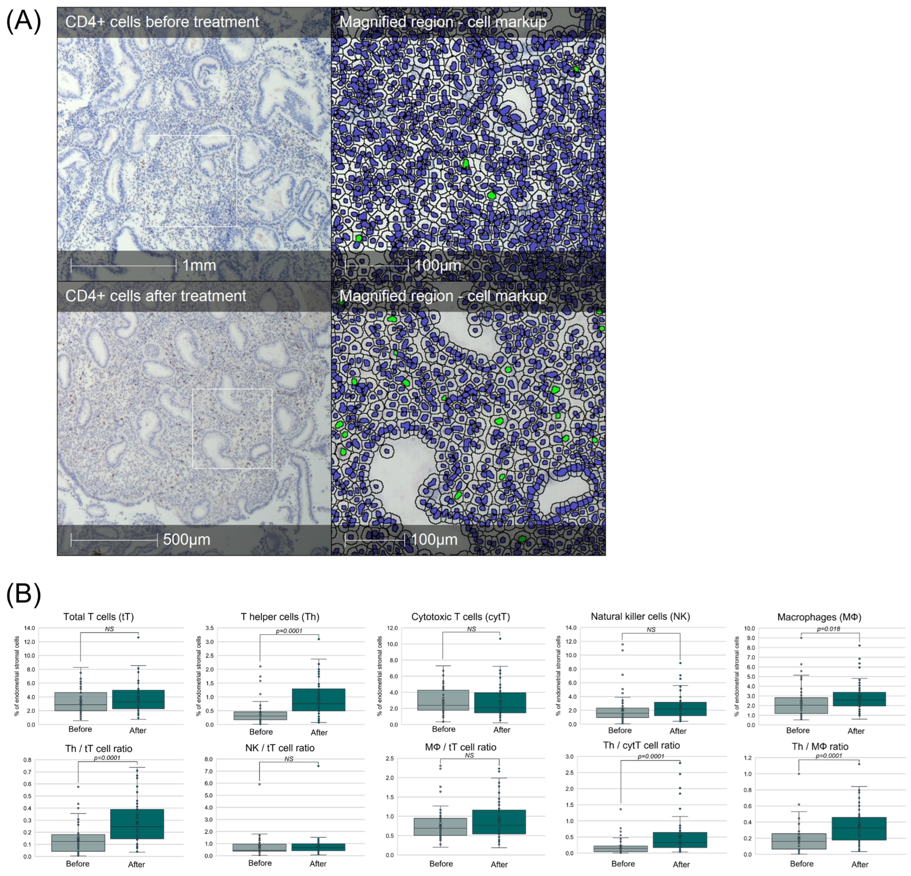
In our cohort of 55 RIF patients, endometrial biopsies obtained before and after intrauterine administration of IFN-τ-modulated PBMCs revealed marked changes in local immune composition (Figure 4). Immunohistochemical analysis showed a significant increase in CD4+ T helper cells (p < 0.05); increased CD68+ macrophages elevated Th/total T cell, Th/cytT, and Th/Macrophage ratios post-treatment. These shifts reflect a more tolerogenic endometrial immune environment, which is favourable for embryo implantation.

3.4. Safety and Tolerability

No patients in the clinical arm required medical intervention following treatment, nor reported procedure-related serious adverse events (fever, infection, pain). Transient mild cramping, which resolved on its own, was reported by 4 patients.

4. Discussion

Our study provides novel evidence that IFN-τ-modulated PBMCs may represent a promising therapeutic strategy for recurrent implantation failure (RIF) by promoting a favorable immunological environment at both the cellular and cytokine levels.
Through cytokine secretion assays, we demonstrated that IFN-τ stimulation shifts PBMC cytokine production toward an anti-inflammatory, implantation-permissive profile, with increased IL-4 and decreased TNF-α and IL-6 (Figure 2). This pattern is consistent with a Th2-biased response, which has been widely associated with successful implantation and maintenance of early pregnancy both in animal models and humans [5,22]. In contrast, elevated TNF-α and IL-6 have been implicated in endometrial inflammation and impaired receptivity [3]. Our findings suggest that IFN-τ counteracts pathogenic cytokine signalling, consistent with data from an animal model of implantation failure [23].
Flow cytometry analysis further confirmed that IFN-τ-modulated PBMCs adopt a less cytotoxic phenotype by shifting key cell subsets. Specifically, we observed a significant increase in Th2 cells, with concomitant decreases in cytotoxic T cells, Th1, Th9, and Th17 subsets, consistent with animal reports [24]. This resulted in improved Th/cytT, Th1/Th2, and Th17/Treg ratios, all of which are critical immunological checkpoints for implantation (Figure 3). These findings align with previous reports that dysregulated Th1/Th2 or Th17/Treg ratios contribute to implantation failure [25,26], and extend them by showing that IFN-τ can actively restore the balance. In addition to the altered cytokine secretion profile, such changes may be more relevant for the persistence of the treatment effect in vivo.
Translation of these findings into an in vivo setting demonstrated that intrauterine infusion of IFN-τ-modulated PBMCs led to increased infiltration of T helper cells within the endometrium, as detected by IHC (Figure 4). This increase is consistent with the in vitro polarization shift toward a Th2 phenotype and may reflect a more supportive local immune microenvironment for embryo implantation. Stemming from the increase in T helpers, we also observed an increase in their ratios, specifically Th/total T, Th/cyt T cells and Th/macrophages, in the endometrial stroma. Our previous work has found a link between an increase in these ratios and subsequent successful implantation in RIF patients [27]. There has been an increasing interest in biomarkers reflecting endometrial immune balance rather than absolute quantities of cells and cytokines when assessing the likelihood of implantation and guiding therapy in this population of patients [7,28,29,30].
We also observed a modest rise in endometrial macrophages, which, although limited, could contribute to tissue remodelling, angiogenesis and tolerance induction, particularly if they adopt a tolerogenic (“M2-like”) phenotype. Due to limitations in biopsy tissue quantity, we were unable to perform further phenotypic analysis (e.g., CD163 or CD206 expression for M2) in this study; however, follow-up studies using immunohistochemistry or flow cytometry to assess macrophage subsets are warranted. This is particularly relevant since there have been reports of dysregulated macrophage polarisation in the endometrium of RIF patients [31]. Evidence from a mouse model of obesity suggests oral IFN-τ reduces proinflammatory cytokines (IL-6, TNF-α) and M1 macrophages, while increasing anti-inflammatory M2-macrophages in adipose tissue [32]. Building on that in the reproductive context, the latest animal work by Feng et al. (2025) reported that IFN-τ exposure in vitro promotes M2 polarisation of endometrial macrophages by downregulating bta-miR-30b-5p and upregulating SOCS1, thereby restraining NF-κB signaling, and increasing the expression of CD206 and IL-10 [33]. The decrease in IL-6 and TNF-α we observed may also suggest suppression of signalling pathways like NF-κB, which are major drivers of inflammatory gene expression in both T cells and macrophages.
Recent animal studies further support the idea that PBMCs exert broad modulatory effects on the endometrium beyond simple cytokine changes. For example, a mouse model of implantation failure showed that intrauterine PBMC administration normalizes dysregulated gene expression in the pre-implantation endometrium, especially of estrogen-responsive genes, as well as increasing glucocorticoid receptor expression and suppressing inflammatory genes [34]. Mechanistic insight into IFN-τ itself has also advanced with recent studies in cows, where it has been reported to induce gene expression changes in both endometrial epithelial and stromal cells and in peripheral immune cells [35,36]. Such findings are consistent with our in vitro immune shifts and suggest that IFN-τ-modulated PBMCs may additionally act via modulation of steroid receptor and inflammation gene pathways.
IFN-τ is known to exert its immunomodulatory effects through binding to the type I interferon receptor complex (IFNAR1/2), initiating downstream signaling cascades. Upon ligand binding, IFNAR-associated kinases—Tyk2 and Jak1—become activated, leading to the phosphorylation of STAT1 and STAT2. These phosphorylated STATs form a complex with IRF9, known as ISGF3, which translocates into the nucleus to drive transcription of interferon-stimulated genes (ISGs) [17,18]. Although this study did not directly assess pathway activation at the protein or transcriptomic level, the observed changes in cytokine secretion and T cell polarization (Th1/Th2 and Th17/Treg ratios) are consistent with downstream IFNAR-mediated signaling effects [37,38,39]. Future work should include transcriptomic or phospho-STAT profiling to validate these mechanisms in human PBMCs.
Taken together, these results suggest that IFN-τ-modulated PBMCs promote a tolerogenic, Th2-dominant immune landscape that is more conducive to embryo implantation. Unlike other immunomodulatory strategies explored in RIF—such as corticosteroids, IVIG, or G-CSF—this approach leverages local action of a patient’s own immune cells, reducing systemic immunosuppression and potentially improving safety. The dual action of IFN-τ at the cytokine and cellular levels underscores its therapeutic potential, particularly given its ability to downregulate pro-inflammatory T cell subsets (Th1, Th9, Th17) while enhancing Th2-mediated immune support. The detailed characterization we performed helps to address several of the uncertainties that often surround cell therapies—specifically in terms of standardisation—dose and content of PBMCs, timing relative to menstrual cycle or embryo transfer, amount and duration of modulation. Moreover, the study of the local effect provides a measurable improvement in a surrogate that is related to the pathogenesis of the condition, which would strengthen future investigation of the effectiveness of this treatment in terms of clinical outcomes.
Nonetheless, several limitations warrant consideration. Our sample size, particularly in the in vivo arm, was relatively small, and we did not assess pregnancy outcomes following treatment, which limits conclusions about clinical efficacy. Moreover, while we demonstrated changes in broad immune subsets, further phenotyping, especially of macrophage polarization and Treg function, would help clarify the mechanistic basis of the observed effects. Future randomized trials with implantation and live birth rates as endpoints are essential to determine whether IFN-τ-modulated PBMCs can be translated into routine clinical practice for RIF patients.

5. Conclusions

In summary, our study demonstrates that IFN-τ modulation reprograms PBMCs toward an anti-inflammatory, Th2-favoring profile that translates into measurable changes in the endometrial immune landscape of RIF patients. These findings provide mechanistic evidence and support the concept of IFN-τ-modulated PBMCs as a novel immunotherapy to enhance endometrial receptivity.

Author Contributions

Conceptualization, M.R., G.S. and S.H.; methodology, M.R., D.P. and R.G.; software, M.R., D.P. and R.G.; formal analysis, M.R. and D.P.; investigation, M.R., M.H., J.S. and D.M.; resources, G.S. and S.H.; data curation, M.R., M.H. and J.S.; writing—original draft preparation, M.R.; writing—review and editing, M.R., D.P., R.G. and S.H.; visualization, M.R.; supervision, D.P. and G.S.; project administration, S.H.; funding acquisition, S.H. All authors have read and agreed to the published version of the manuscript.

Funding

This research was funded by the European Union-NextGenerationEU, through the National Recovery and Resilience Plan of the Republic of Bulgaria, project No. BG-RRP-2.004-0004-C01.

Institutional Review Board Statement

The study was conducted in accordance with the Declaration of Helsinki, and approved by the Institutional Ethics Committee of Nadezhda Women’s Health Hospital (protocol code 7/28 February 2023).

Informed Consent Statement

Informed consent was obtained from all subjects involved in the study.

Data Availability Statement

The data supporting the findings presented in this study are available at: https://doi.org/10.5281/zenodo.17250137.

Conflicts of Interest

The authors declare no conflicts of interest. The funders had no role in the design of the study; in the collection, analyses or interpretation of data; in the writing of the manuscript; or in the decision to publish the results.

Abbreviations

The following abbreviations are used in this manuscript:
PBMCPeripheral blood mononuclear cells
IFN-τInterferon τ
hCGhuman chorionic gonadotropin
RIFRecurrent implantation failure
ARTAssisted reproductive therapy
ThT helper lymphocyte
cytTCytotoxic T cell
TfhFollicular T helper
TregRegulatory T cell
IL-Interleukin-
TNFαTumor necrosis factor α

References

  1. Robertson, S.A.; Moldenhauer, L.M.; Green, E.S.; Care, A.S.; Hull, M.L. Immune determinants of endometrial receptivity: A biological perspective. Fertil. Steril. 2022, 117, 1107–1120. [Google Scholar] [CrossRef]
  2. Saito, S. Role of immune cells in the establishment of implantation and maintenance of pregnancy and immunomodulatory therapies for patients with repeated implantation failure and recurrent pregnancy loss. Reprod. Med. Biol. 2024, 23, e12600. [Google Scholar] [CrossRef]
  3. Guo, L.; Guo, A.; Yang, F.; Li, L.; Yan, J.; Deng, X.; Dai, C.; Li, Y. Alterations of Cytokine Profiles in Patients With Recurrent Implantation Failure. Front. Endocrinol. 2022, 13, 949123. [Google Scholar] [CrossRef]
  4. Saito, S.; Nakashima, A.; Shima, T.; Ito, M. Th1/Th2/Th17 and regulatory T-cell paradigm in pregnancy. Am. J. Reprod. Immunol. 2010, 63, 601–610. [Google Scholar] [CrossRef] [PubMed]
  5. Piccinni, M.P.; Raghupathy, R.; Saito, S.; Szekeres-Bartho, J. Cytokines, Hormones and Cellular Regulatory Mechanisms Favoring Successful Reproduction. Front. Immunol. 2021, 12, 717808. [Google Scholar] [CrossRef] [PubMed]
  6. Piccinni, M.P.; Lombardelli, L.; Logiodice, F.; Kullolli, O.; Romagnani, S.; Le Boulteiller, P. T helper cell mediated-tolerance towards fetal allograft in successful pregnancy. Clin. Mol. Allergy 2015, 13, 9. [Google Scholar] [CrossRef]
  7. Li, Y.; Chen, S.; Yang, J.; Zhang, K.; Feng, X.; Sun, C.; Feng, Q.; Li, Z. Associations of Th1, Th2, Th17, and Treg cell levels and imbalance with recurrent spontaneous abortion: A meta-analysis. J. Obstet. Gynaecol. Res. 2025, 51, e16207. [Google Scholar] [CrossRef] [PubMed]
  8. Kleiveland, C.R. Peripheral Blood Mononuclear Cells. In The Impact of Food Bioactives on Health: In Vitro and Ex Vivo Models; Verhoeckx, K., Cotter, P., López-Expósito, I., Kleiveland, C., Lea, T., Mackie, A., Requena, T., Swiatecka, D., Wichers, H., Eds.; Springer: Cham, Switzerland, 2015; Chapter 15. [Google Scholar] [CrossRef]
  9. Liu, S.; Diao, L.; Huang, C.; Li, Y.; Zeng, Y.; Kwak-Kim, J.Y.H. The role of decidual immune cells on human pregnancy. J. Reprod. Immunol. 2017, 124, 44–53. [Google Scholar] [CrossRef]
  10. Yu, N.; Zhang, B.; Xu, M.; Wang, S.; Liu, R.; Wu, J.; Yang, J.; Feng, L. Intrauterine administration of autologous peripheral blood mononuclear cells (PBMCs) activated by HCG improves the implantation and pregnancy rates in patients with repeated implantation failure: A prospective randomized study. Am. J. Reprod. Immunol. 2016, 76, 212–216. [Google Scholar] [CrossRef]
  11. Yakin, K.; Oktem, O.; Urman, B. Intrauterine administration of peripheral mononuclear cells in recurrent implantation failure: A systematic review and meta-analysis. Sci. Rep. 2019, 9, 3897. [Google Scholar] [CrossRef]
  12. Busnelli, A.; Somigliana, E.; Cirillo, F.; Baggiani, A.; Levi-Setti, P.E. Efficacy of therapies and interventions for repeated embryo implantation failure: A systematic review and meta-analysis. Sci. Rep. 2021, 11, 1747. [Google Scholar] [CrossRef]
  13. Liu, M.; Yuan, Y.; Qiao, Y.; Tang, Y.; Sui, X.; Yin, P.; Yang, D. The effectiveness of immunomodulatory therapies for patients with repeated implantation failure: A systematic review and network meta-analysis. Sci. Rep. 2022, 12, 18434. [Google Scholar] [CrossRef] [PubMed]
  14. Jiang, L.; Wen, L.; Lv, X.; Tang, N.; Yuan, Y. Comparative efficacy of intrauterine infusion treatments for recurrent implantation failure: A network meta-analysis of randomized controlled trials. J. Assist. Reprod. Genet. 2025, 42, 1177–1190. [Google Scholar] [CrossRef] [PubMed]
  15. Schumacher, A.; Zenclussen, A.C. Human Chorionic Gonadotropin-Mediated Immune Responses That Facilitate Embryo Implantation and Placentation. Front. Immunol. 2019, 10, 2896. [Google Scholar] [CrossRef] [PubMed]
  16. Yu, N.; Yan, W.; Yin, T.; Wang, Y.; Guo, Y.; Zhou, D.; Xu, M.; Ding, J.; Yang, J. HCG-Activated Human Peripheral Blood Mononuclear Cells (PBMC) Promote Trophoblast Cell Invasion. PLoS ONE 2015, 10, e0125589. [Google Scholar] [CrossRef]
  17. Murira, A.; Lamarre, A. Type-I Interferon Responses: From Friend to Foe in the Battle against Chronic Viral Infection. Front. Immunol. 2016, 7, 609. [Google Scholar] [CrossRef]
  18. Graber, J.; Dhib-Jalbut, S. Interferons. In Encyclopedia of the Neurological Sciences, 2nd ed.; Aminoff, M.J., Daroff, R.B., Eds.; Academic Press: San Diego, CA, USA, 2014; pp. 718–723. [Google Scholar] [CrossRef]
  19. Bazer, F.W.; Thatcher, W.W. Chronicling the discovery of interferon tau. Reproduction 2017, 154, F11–F20. [Google Scholar] [CrossRef]
  20. Li, S.; Gong, M.; Zhao, F.; Shao, J.; Xie, Y.; Zhang, Y.; Chang, H. Type I Interferons: Distinct Biological Activities and Current Applications for Viral Infection. Cell. Physiol. Biochem. 2018, 51, 2377–2396. [Google Scholar] [CrossRef]
  21. ClinicalTrials.gov. Effects of Intrauterine Administration of Autologous PBMC Modulated With IFNτ on Endometrial Cell Populations, NCT05775211. Available online: https://clinicaltrials.gov/ct2/show/NCT05775211 (accessed on 27 July 2025).
  22. Wang, W.; Sung, N.; Gilman-Sachs, A.; Kwak-Kim, J. T Helper (Th) Cell Profiles in Pregnancy and Recurrent Pregnancy Losses: Th1/Th2/Th9/Th17/Th22/Tfh Cells. Front. Immunol. 2020, 11, 2025. [Google Scholar] [CrossRef]
  23. Jiang, K.; Yang, J.; Chen, Y.; Guo, S.; Zhao, G.; Wu, H.; Deng, G. Protective Effects of Interferon-tau Against Lipopolysaccharide-Induced Embryo Implantation Failure in Pregnant Mice. J. Interferon Cytokine Res. 2018, 38, 226–234. [Google Scholar] [CrossRef]
  24. Soos, J.M.; Stüve, O.; Youssef, S.; Bravo, M.; Johnson, H.M.; Weiner, H.L.; Zamvil, S.S. Cutting edge: Oral type I IFN-tau promotes a Th2 bias and enhances suppression of autoimmune encephalomyelitis by oral glatiramer acetate. J. Immunol. 2002, 169, 2231–2235. [Google Scholar] [CrossRef]
  25. Kuroda, K.; Nakagawa, K.; Horikawa, T.; Moriyama, A.; Ojiro, Y.; Takamizawa, S.; Ochiai, A.; Matsumura, Y.; Ikemoto, Y.; Yamaguchi, K.; et al. Increasing number of implantation failures and pregnancy losses associated with elevated Th1/Th2 cell ratio. Am. J. Reprod. Immunol. 2021, 86, e13429. [Google Scholar] [CrossRef]
  26. Berdiaki, A.; Vergadi, E.; Makrygiannakis, F.; Vrekoussis, T.; Makrigiannakis, A. Repeated implantation failure is associated with increased Th17/Treg cell ratio, during the secretory phase of the human endometrium. J. Reprod. Immunol. 2024, 161, 104170. [Google Scholar] [CrossRef]
  27. Ganeva, R.; Parvanov, D.; Vidolova, N.; Ruseva, M.; Handzhiyska, M.; Arsov, K.; Decheva, I.; Metodiev, D.; Moskova-Doumanova, V.; Stamenov, G. Endometrial immune cell ratios and implantation success in patients with recurrent implantation failure. J. Reprod. Immunol. 2023, 156, 103816. [Google Scholar] [CrossRef]
  28. Lédée, N.; Petitbarat, M.; Chevrier, L.; Vitoux, D.; Vezmar, K.; Rahmati, M.; Dubanchet, S.; Gahéry, H.; Bensussan, A.; Chaouat, G. The uterine immune profile may help women with repeated unexplained embryo implantation failure after in vitro fertilization. Am. J. Reprod. Immunol. 2016, 75, 388–401. [Google Scholar] [CrossRef]
  29. Meng, S.; Ren, J.; Zhang, T.; Jia, Y.; Han, H.; Shen, H. Immunoregulatory therapy improves live birth in Th17/Treg-cell-elevated women with embryo transfer failure. Int. J. Gynaecol. Obstet. 2023, 161, 1101–1103. [Google Scholar] [CrossRef] [PubMed]
  30. Lédée, N.; Petitbarat, M.; Dray, G.; Chevrier, L.; Kazhalawi, A.; Rahmati, M.; Vicaut, E.; Diallo, A.; Cassuto, N.G.; Ruoso, L.; et al. Endometrial immune profiling and precision therapy increase live birth rate after embryo transfer: A randomised controlled trial. Front. Immunol. 2025, 16, 1523871. [Google Scholar] [CrossRef] [PubMed]
  31. Liu, Y.; Tan, X.; He, T. Macrophage polarization disorder in the endometrial immune microenvironment may contribute to recurrent implantation failure. Reprod. Dev. Med. 2024, 8, 162–168. [Google Scholar] [CrossRef]
  32. Zhao, X.; Song, M.; Sun, L.; Zou, H.; Yang, Y.; Wu, C.; Zhang, Y.; Li, Y. Interferon Tau Alleviates Obesity-Induced Adipose Tissue Inflammation and Insulin Resistance by Regulating Macrophage Polarization. PLoS ONE 2014, 9, e98835. [Google Scholar] [CrossRef]
  33. Feng, X.; Yang, C.; Wang, T.; Zhang, J.; Zhou, H.; Ma, B.; Xu, M.; Deng, G. IFN-τ Maintains Immune Tolerance by Promoting M2 Macrophage Polarization via Modulation of Bta-miR-30b-5p in Early Uterine Pregnancy in Dairy Cows. Cells 2025, 14, 87. [Google Scholar] [CrossRef]
  34. Kitawaki, Y.; Horie, A.; Ikeda, A.; Shitanaka, S.; Yanai, A.; Ohara, T.; Nakakita, B.; Sagae, Y.; Okunomiya, A.; Tani, H.; et al. Intrauterine administration of peripheral blood mononuclear cells helps manage recurrent implantation failure by normalizing dysregulated gene expression including estrogen-responsive genes in mice. Cell Commun. Signal. 2024, 22, 587. [Google Scholar] [CrossRef]
  35. Chaney, H.L.; Grose, L.F.; Charpigny, G.; Behura, S.K.; Sheldon, I.M.; Cronin, J.G.; Lonergan, P.; E Spencer, T.; Mathew, D.J. Conceptus-induced, interferon tau-dependent gene expression in bovine endometrial epithelial and stromal cells. Biol. Reprod. 2021, 104, 669–683. [Google Scholar] [CrossRef]
  36. Casano, A.B.; Barile, V.L.; Menchetti, L.; Guelfi, G.; Brecchia, G.; Agradi, S.; De Matteis, G.; Scatà, M.C.; Grandoni, F.; Barbato, O. Interferon Tau (IFNt) and Interferon-Stimulated Genes (ISGs) Expression in Peripheral Blood Leukocytes and Correlation with Circulating Pregnancy-Associated Glycoproteins (PAGs) during Peri-Implantation and Early Pregnancy in Buffalo Cows. Animals 2022, 12, 3068. [Google Scholar] [CrossRef] [PubMed]
  37. Villarino, A.V.; Gallo, E.; Abbas, A.K. STAT1-activating cytokines limit Th17 responses through both T-bet-dependent and -independent mechanisms. J. Immunol. 2010, 185, 6461–6471. [Google Scholar] [CrossRef] [PubMed]
  38. Guo, B.; Chang, E.Y.; Cheng, G. The type I IFN induction pathway constrains Th17-mediated autoimmune inflammation in mice. J. Clin. Invest. 2008, 118, 1680–1690. [Google Scholar] [CrossRef] [PubMed]
  39. Sadreev, I.I.; Chen, M.Z.Q.; Umezawa, Y.; Kajimoto, T.; Li, X.; Okuda, K.; Ishihara, T.; Okamura, H.; O’Shea, J.J.; Koga, T.; et al. The Competitive Nature of Signal Transducer and Activator of Transcription Complex Formation Drives Phenotype Switching of T Cells. Immunology 2018, 153, 488–501. [Google Scholar] [CrossRef]
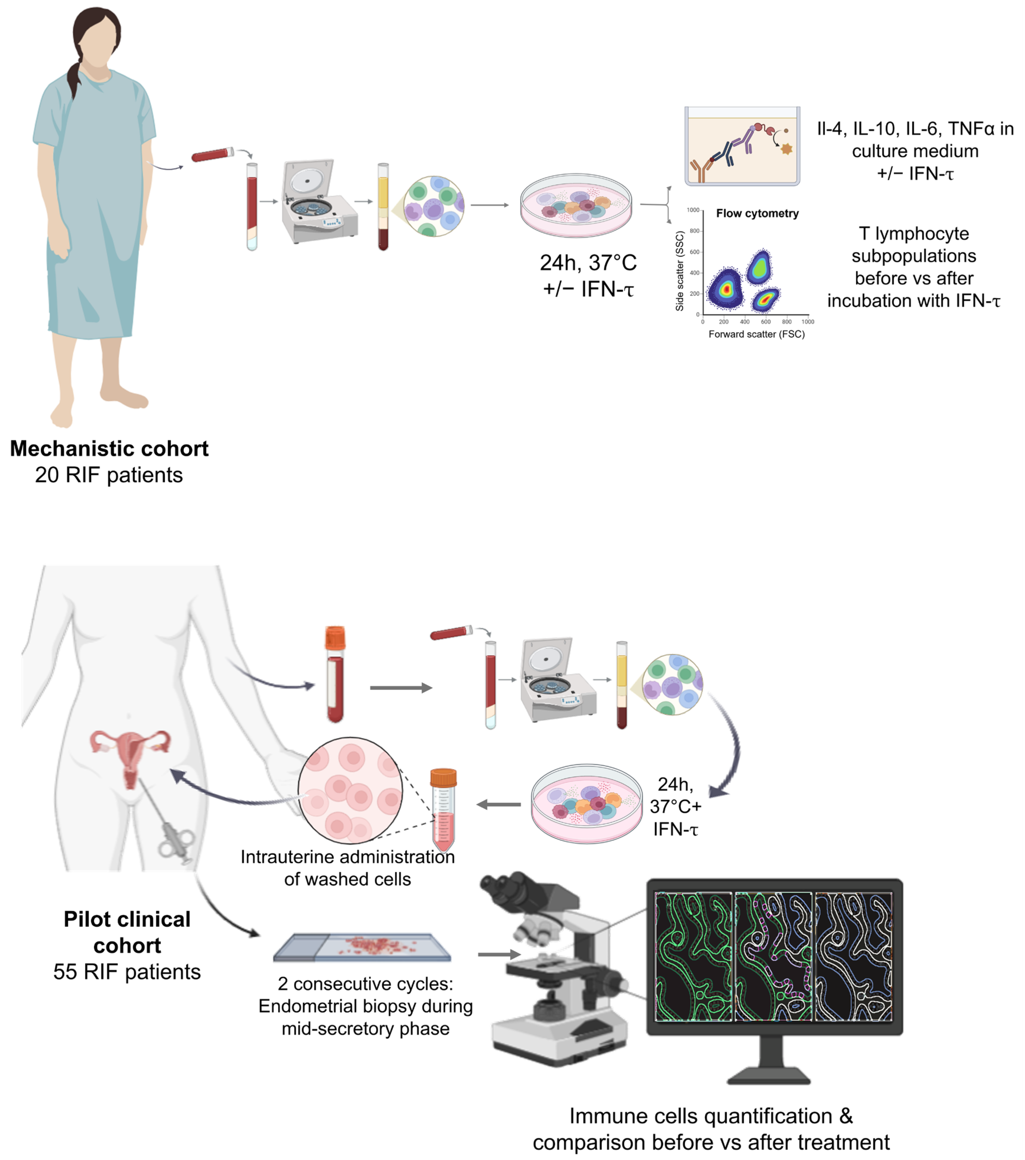
Figure 1. Schematic representation of the experimental design. Two separate cohorts of RIF patients were recruited—one for mechanistic investigation of the modulatory effects of IFN-τ on PBMC (n = 20), and a second for analysis of the in vivo effect of intrauterine treatment with IFN-τ-modulated PBMC on endometrial immune composition (n = 55). Created with BioRender.com.
Immuno 05 00051 g001
Figure 2. IFN-τ alters the cytokine secretion profile of human PBMCs. Boxplots presenting levels of TNF-α, IL-6, IL-4, and IL-10 secreted by peripheral blood mononuclear cells (PBMCs) after 24 h in vitro stimulation with interferon tau (IFN-τ) (n = 20 patients). Each patient’s PBMCs were divided into two conditions: IFN-τ-treated and unstimulated (control). Cytokine concentrations were measured in culture supernatants using ELISA. Compared to unstimulated controls (n = 20 matched samples), IFN-τ stimulation significantly reduced secretion of pro-inflammatory cytokines TNF-α (p = 0.0001) and IL-6 (p = 0.002), and significantly increased the Th2-associated cytokine IL-4 (p = 0.03). No significant change was observed in IL-10 secretion. Statistical comparisons were made using the Wilcoxon signed-rank test for related samples. Boxplots represent the distribution of values with the mean indicated by an “X”. NS—not significant.
Immuno 05 00051 g002
Figure 3. IFN-τ induces T cell polarization in PBMCs. Flow cytometry was used to assess the frequencies of T cell subsets in peripheral blood mononuclear cells (PBMCs) isolated from 20 patients (n = 20), before and after 24 h in vitro stimulation with interferon tau (IFN-τ). Each sample was analyzed in paired conditions (unstimulated vs. IFN-τ-stimulated). T cell subsets were defined by intracellular or surface marker expression and are expressed as a percentage of total lymphocytes, total T cells, or CD4+ T helper cells, as appropriate. Following IFN-τ treatment, there was a significant increase in Th2 cell frequency (p = 0.001), along with significant decreases in Th1, Th9, Th17, and cytotoxic T cells (cytT) (all p < 0.05). These changes were accompanied by modulation of key immunological ratios, including an increased Th/cytT ratio and decreased Th1/Th2 and Th17/Treg ratios, consistent with a shift toward an anti-inflammatory profile. Statistical analysis was performed using the Wilcoxon signed-rank test for related samples. Boxplots display the distribution of values, with the mean indicated by an “X”. NS—not significant.
Immuno 05 00051 g003
Figure 4. Endometrial immune remodelling after intrauterine treatment with IFN-τ-modulated PBMC. (A) Representative IHC images from paired endometrial biopsies collected before (top panels) and after (bottom panels) treatment illustrate an increase in CD4+ T helper cell infiltration following intrauterine infusion of IFN-τ-treated autologous PBMCs. (B) Quantitative analysis of immune cell populations was performed on paired biopsies from 55 recurrent implantation failure (RIF) patients (n = 55). Post-treatment samples showed significant increases in the percentages of CD4+ T helper cells and macrophages, as well as increases in immunological ratios including Th/total T cells, Th/cytotoxic T cells (cytT), and Th/macrophage ratios (all p < 0.05). Data are presented as boxplots, with each pair representing a patient’s pre- and post-treatment biopsy. The mean value is indicated by an “X”. Immunostaining was performed on formalin-fixed, paraffin-embedded tissue sections, with quantification performed on HALO™ Image analysis of positively stained cells per total stromal cells. Statistical comparisons were conducted using the related samples Wilcoxon signed-rank test. NS—not significant.
Immuno 05 00051 g004
Table 1. Baseline characteristics of study cohorts.
CharacteristicsIn Vitro Cohort
(n = 20)
Clinical Cohort
(n = 55)
p Value
Age (years)39.8 ± 5.338.4 ± 6.2NS
Type of infertility NS
    Primary (n, %)11 (55%)32 (58%)
    Secondary (n, %)9 (45%)23 (42%)
Previous IVF attempts (n)3.0 ± 0.93.1 ± 1.0NS
Data are presented as mean ± SD or counts (percentage), as appropriate. Comparisons were performed via an independent t-test for continuous data and a chi-squared test for categorical data. NS—non-significant.
Disclaimer/Publisher’s Note: The statements, opinions and data contained in all publications are solely those of the individual author(s) and contributor(s) and not of MDPI and/or the editor(s). MDPI and/or the editor(s) disclaim responsibility for any injury to people or property resulting from any ideas, methods, instructions or products referred to in the content.