Perspective

The “6B” Strategy: Build Back a Better Blood–Brain Barrier

by
Laurent Calvier
1,2,*,
Anna E. Alexander
1,2 and
Joachim Herz
1,2,3,4
1
Department of Molecular Genetics, University of Texas (UT) Southwestern Medical Center, Dallas, TX 75390, USA
2
Center for Translational Neurodegeneration Research, University of Texas (UT) Southwestern Medical Center, Dallas, TX 75390, USA
3
Department of Neuroscience, University of Texas (UT) Southwestern Medical Center, Dallas, TX 75390, USA
4
Department of Neurology and Neurotherapeutics, University of Texas (UT) Southwestern Medical Center, Dallas, TX 75390, USA
*
Author to whom correspondence should be addressed.
Immuno 2022, 2(3), 506-511; https://doi.org/10.3390/immuno2030032
Submission received: 5 July 2022 / Revised: 26 August 2022 / Accepted: 28 August 2022 / Published: 2 September 2022
(This article belongs to the Section Autoimmunity and Immunoregulation)

Abstract

:
Under pathological conditions such as multiple sclerosis (MS), leukocytes infiltrate the central nervous system where they, in concert with activated microglia, promote inflammatory demyelination resulting in a broad spectrum of symptoms, including paralysis. Therefore, all current therapeutic approaches to MS target the immune system, blocking inflammation and paralysis progression, but may compromise the immune system. In this focused review, we present an underestimated compartment, the blood–brain barrier, which is compromised during MS and becomes permeable to leukocytes infiltrating the central nervous system. This barrier has the potential to offer new therapeutic strategies and is easily accessible for drugs. We highlight this paradigm using the example of the therapeutic anti-Reelin strategy we have developed. Reelin is a plasma protein that regulates the expression of adhesion markers on the endothelial surface, thus promoting the infiltration of inflammatory cells and propagating inflammation. Building Back a Better Blood–Brain Barrier (the “6B” strategy) may have advantages compared to actual immunosuppressive drugs because it restores a physiological function rather than suppressing the immune system.

1. Introduction

Chronic inflammation and excessive recruitment of circulating leukocytes is an early event and a key mechanism in several pathologies, including autoimmune diseases. Multiple sclerosis (MS) is a typical example of this disorder when unbalanced recruitment and activation of circulating immune cells lead them into the central nervous system (CNS) where they attack the myelin sheath that insulates nerve fibers, eventually causing paralysis [1,2,3,4,5,6]. Approximately 85% of patients affected by MS present a relapsing–remitting disease form, characterized by alternating episodes of neurological disability (relapses or attacks) followed by complete or partial recovery of symptoms [7]. Approximately two-thirds of these patients will eventually make a transition to secondary progressive MS within two decades after initial onset [8]. On the other hand, approximately 10% of MS patients present a primary progressive MS form with gradual worsening of the neurological disability [7]. Although substantial progress has been made in the development of effective treatments over the past two decades for relapsing–remitting MS [9], no convincingly effective therapies exist for progressive forms of MS (primary and secondary) [10,11].
With an immune system going out of control and attacking healthy tissue, logically, all the available MS therapies directly target this immune response (Figure 1) to dampen neuroinflammation and associated adverse effects [5]. As expected, these immune-suppressing therapies have yielded tremendous progress for relapsing–remitting MS, slowing down the relapses, but it appears that they are reaching a glass ceiling. Despite the approval of almost twenty drugs targeting the cellular immune system, three challenges remain: (i) paralysis can hardly be reversed, (ii) effective therapies are lacking for progressive forms of MS, and (iii) immunotherapeutic interventions may compromise the immune system [12,13]. For example, Natalizumab is an antibody widely used in clinics that binds to all leukocytes via α4-integrin and prevents their interaction with receptors on the endothelium such as vascular cell adhesion molecule 1 (VCAM-1) [14,15]. Natalizumab appears to indiscriminately abolish the ability of leukocytes to adhere and access the CNS. While this drug has striking results in decreasing the number of relapses, it also blocks physiological diapedesis and is associated with a high risk for progressive multifocal leukoencephalopathy caused by reactivation of the John Cunningham (JC) virus, resulting in a fatal viral infection of the brain [16]. Therefore, identifying optimal, non-immunosuppressive therapies for all forms of MS continues to constitute a major unmet need for patients [17].

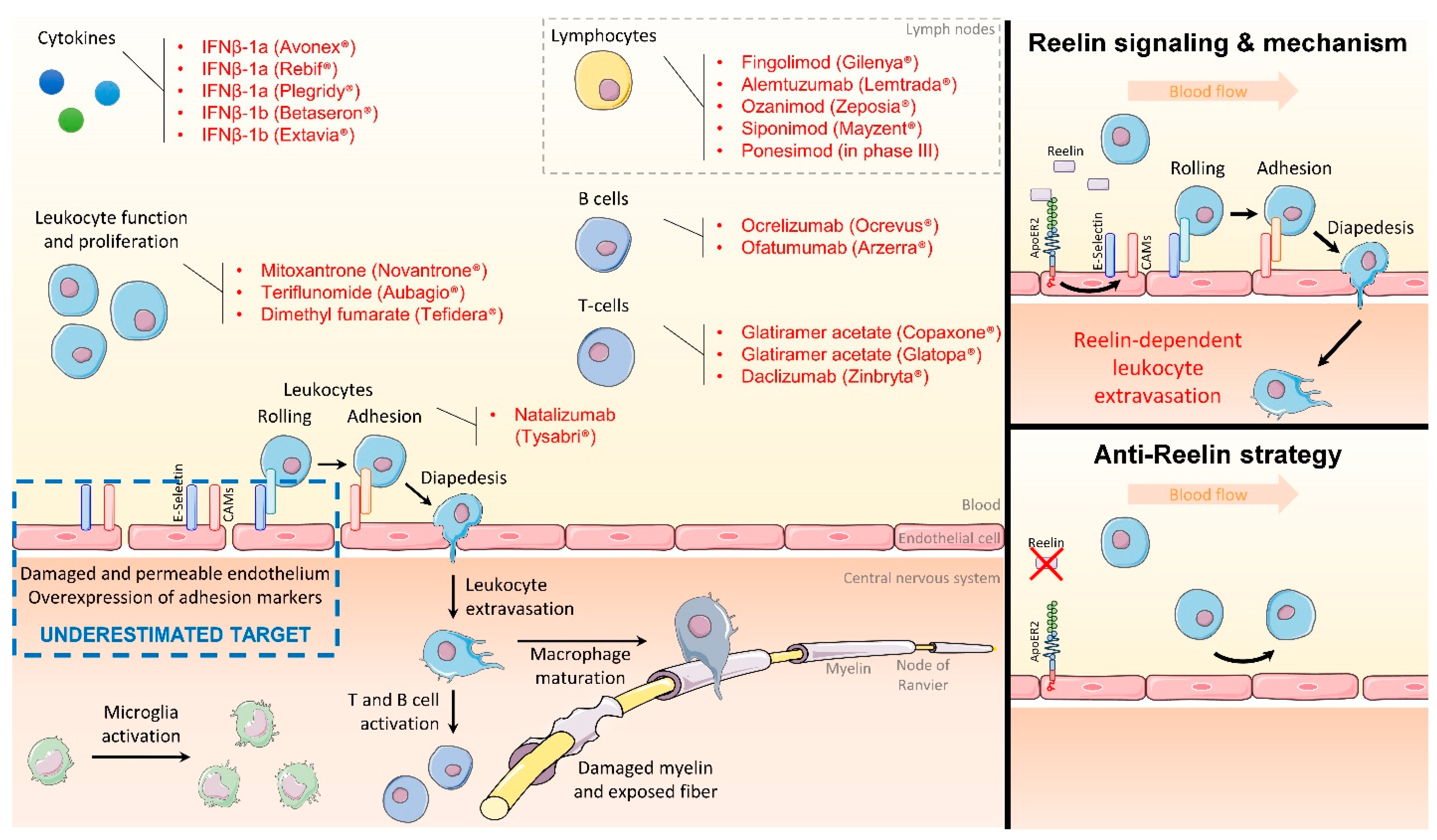
2. Targeting the Endothelial Function to Block Inflammation

To identify new targets, it may appear counterintuitive, but innovative, to step back from immunotherapies and explore other MS components. While the CNS is difficult to access for drugs due to the blood–brain barrier (BBB), the vascular barrier itself is the last key compartment for neuroinflammation. Under normal conditions, this endothelial wall effectively regulates the passage of immune cells into the CNS. However, under inflammatory conditions such as MS, this barrier is compromised, becoming more adhesive and permeable, which dramatically increases leukocyte rolling, adhesion, and infiltration [1,18]. This BBB breakdown hypothesis is supported by a substantial body of radiological and pathological clinical evidence. BBB leakage has been observed in MS patients by Gadolinium-enhancing lesions on MRI as well as by fibrinogen (a plasma protein) deposition in developing lesions [19,20]. On the cellular level, tight-junction abnormalities are also seen in endothelial cells around active lesions, which is a hallmark of a permeable endothelium [20]. This results in increased infiltration of leukocytes into the CNS as demonstrated in the postmortem MS brain [20]. Based on this new paradigm, restoring this BBB would prevent leukocyte infiltration into the CNS without targeting and dampening the immune system.
During inflammation, endothelial cells overexpress on their surface “rolling” proteins such as E-selectin and adhesion proteins such as intercellular adhesion molecule 1 (ICAM-1) or VCAM-1, massively increasing the number of leukocytes patrolling the vessel wall and infiltrating the surrounding tissues [21,22,23]. Natalizumab has proven that total obliteration of only one adhesion protein on leukocytes is very effective, but it also disrupts basic diapedesis function [5,12,14,15]. Learning from this flaw to identify the next game changer and to preserve this physiological function, dampening the expression of a large set of adhesion molecules back to a physiologically normal baseline level may be a superior strategy. In this respect, Reelin may be a potential target that fulfills these criteria. Initially recognized only for its role in guiding neurons during brain development and as a synaptic homeostatic regulator [24,25,26,27,28], it has recently been recognized for a previously unknown non-neuronal function as an NF-κB activator in the vasculature.

3. Reelin Inhibition Protects the Endothelial Function and Presents Clinical Potential

In human aortic endothelial cells, adhesion and permeability are largely regulated via NF-κB pathway activation [29]. NF-κB target genes include adhesion molecules such as E-selectin, ICAM-1, or VCAM-1, but also cytokines and chemokines. Interestingly, it has been demonstrated in human aortic endothelial cells that Reelin activates NF-κB and thereby controls the expression of its target genes, especially adhesion molecules [30,31,32], via its membrane receptor apolipoprotein E receptor 2 (ApoER2) [30], a member of the LDLR family [33]. Accordingly, it has been shown that Reelin promotes leukocyte adhesion to endothelial cells and that Reelin-blocking antibodies can prevent this adhesion, in vitro using an adhesion assay with human aortic endothelial cells and human monocytes U937 and in vivo by intravital microscopy on mesenteric vessels [30,31,32]. Using autoimmune encephalomyelitis (EAE) as a murine MS model, both genetic deletion of either Reelin or its receptor ApoER2, as well as pharmacological (antibody-mediated) Reelin depletion substantially reduce paralysis progression [32,34]. In all these models, Reelin or ApoER2 depletion normalizes the expression of vascular adhesion markers in the CNS, consequently decreasing leukocyte infiltration, demyelination, and paralysis.
In a proof-of-concept study, we have demonstrated that administration of a Reelin-neutralizing antibody given at the onset of EAE symptoms results in diminished neuroinflammation, paralysis, and improved recovery [32]. Importantly, the antibody depletes Reelin from circulation, but not from the CNS due to the blood–brain barrier, thus preserving the neurophysiological functions of Reelin in the brain. In addition, there is considerable genetic and biochemical evidence that supports the relevance of anti-Reelin therapy in humans. In two independent genomewide association studies [35,36], Reelin single-nucleotide polymorphisms were found to be associated with MS severity score. Finally, in serum from MS patients, Reelin concentration was increased during relapse but returned to baseline levels during remission phases [32].
The discrimination of Reelin’s functions in the CNS versus circulation is a relatively new finding and some uncertainties remain, requiring additional studies. For example, the source of plasma Reelin expression is not well-understood. Reelin is expressed in the CNS by the neurons; however, it is unlikely that it leaks from there (or the cerebrospinal fluid) into circulation in significant amounts. To confirm this, we have previously shown that we can deplete Reelin from plasma by injecting Reelinfl/fl mice through the tail vein with an adenovirus expressing Cre recombinase (Ad-Cre) [30]. Immunoblot analysis demonstrated efficient and specific ablation of Reelin from plasma but not from the brain. In the literature, the liver and especially the hepatic stellate cells are believed to be the main source of peripheral Reelin [30,37,38,39,40]; we challenge this paradigm as we have not observed an increased expression in hepatic Reelin mRNA expression matching plasma Reelin accumulation during EAE (data not shown). In addition, Reelin expression has been reported in lymphatic vessels [41] but remains unknown in the specific glymphatic system. Therefore, plasma Reelin secretory cells during neuroinflammation remain to be determined.

4. Conclusions and Perspectives

The “6B” strategy—an acronym for Building Back a Better Blood–Brain Barrier—that we are proposing here stands out from previous approaches by restoring the endothelial barrier and targeting a broad range of adhesion proteins. As illustrated in Figure 1, targeting plasma Reelin allows the regulation of vascular permeability to devise an alternative endothelial-specific tunable approach for the treatment of MS. In addition, studying the function of Reelin in the plasma unexpectedly revealed a novel mechanism and illustrated how the organism reuses a neuronal guidance molecule (Reelin) crucial for normal brain development to “guide” leukocytes into the perivascular space during inflammatory processes by modulating extravasation. These recent findings establish a new paradigm for the role of guidance molecules across different functions and cell types, with far-reaching implications for several chronic inflammatory diseases aside from MS, such as Alzheimer’s disease, arthritis, atherosclerosis [30,31], or potentially Crohn’s disease. Besides Reelin, other targets can be used to modulate the BBB properties, either from the same LDLR ligand/receptor family [33] such as the LDL receptor related protein 1 (LRP1) [42,43], or outside of this family such as the peroxisome proliferator activated receptor γ (PPARγ) [44,45,46,47,48,49].

Author Contributions

L.C. wrote the manuscript. A.E.A. and J.H. have revised it for interpretation and content. All authors contributed to the article and approved the submitted version. All authors have read and agreed to the published version of the manuscript.

Funding

J.H. was supported by grants from the NHLBI (R37 HL063762), NIA (RF AG053391), then NINDS and NIA (R01 NS093382), BrightFocus A2016396S, the Bluefield Project to Cure FTD and a Harrington Scholar Innovator Award (2019).

Institutional Review Board Statement

Not applicable.

Informed Consent Statement

Not applicable.

Data Availability Statement

Not applicable.

Conflicts of Interest

L.C. and J.H. are co-shareholders of Reelin Therapeutics Inc. and coinventors of a patent related to anti-Reelin strategies (Application Number: 15/763,047 and Publication Number: 20180273637).

References

  1. Ajami, B.; Bennett, J.L.; Krieger, C.; McNagny, K.M.; Rossi, F.M.V. Infiltrating monocytes trigger EAE progression, but do not contribute to the resident microglia pool. Nat. Neurosci. 2011, 14, 1142–1149. [Google Scholar] [CrossRef] [PubMed]
  2. Benveniste, E.N. Role of macrophages/microglia in multiple sclerosis and experimental allergic encephalomyelitis. J. Mol. Med. 1997, 75, 165–173. [Google Scholar] [CrossRef] [PubMed]
  3. Bauer, J.; Sminia, T.; Wouterlood, F.G.; Dijkstra, C.D. Phagocytic activity of macrophages and microglial cells during the course of acute and chronic relapsing experimental autoimmune encephalomyelitis. J. Neurosci. Res. 1994, 38, 365–375. [Google Scholar] [CrossRef] [PubMed]
  4. Dendrou, C.A.; Fugger, L.; Friese, M.A. Immunopathology of multiple sclerosis. Nat. Rev. Immunol. 2015, 15, 545–558. [Google Scholar] [CrossRef] [PubMed]
  5. Dobson, R.; Giovannoni, G. Multiple sclerosis—A review. Eur. J. Neurol. 2019, 26, 27–40. [Google Scholar] [CrossRef] [PubMed]
  6. Manouchehri, N.; Hussain, R.Z.; Cravens, P.D.; Esaulova, E.; Artyomov, M.N.; Edelson, B.T.; Wu, G.F.; Cross, A.H.; Doelger, R.; Loof, N.; et al. CD11c+CD88+CD317+ myeloid cells are critical mediators of persistent CNS autoimmunity. Proc. Natl. Acad. Sci. USA 2021, 118, e2014492118. [Google Scholar] [CrossRef]
  7. Ransohoff, R.M.; Hafler, D.A.; Lucchinetti, C.F. Multiple sclerosis-a quiet revolution. Nat. Rev. Neurol. 2015, 11, 134–142. [Google Scholar] [CrossRef]
  8. Compston, A.; Coles, A. Multiple sclerosis. Lancet 2008, 372, 1502–1517. [Google Scholar] [CrossRef]
  9. Atarashi, K.; Hirata, T.; Matsumoto, M.; Kanemitsu, N.; Miyasaka, M. Rolling of Th1 cells via P-selectin glycoprotein ligand-1 stimulates LFA-1-mediated cell binding to ICAM-1. J. Immunol. 2005, 174, 1424–1432. [Google Scholar] [CrossRef]
  10. Feinstein, A.; Freeman, J.; Lo, A.C. Treatment of progressive multiple sclerosis: What works, what does not, and what is needed. Lancet Neurol. 2015, 14, 194–207. [Google Scholar] [CrossRef]
  11. Ontaneda, D.; Fox, R.J. Progressive multiple sclerosis. Curr. Opin. Neurol. 2015, 28, 237–243. [Google Scholar] [CrossRef] [PubMed]
  12. Rommer, P.S.; Milo, R.; Han, M.H.; Satyanarayan, S.; Sellner, J.; Hauer, L.; Illes, Z.; Warnke, C.; Laurent, S.; Weber, M.S.; et al. Immunological Aspects of Approved MS Therapeutics. Front. Immunol. 2019, 10, 1564. [Google Scholar] [CrossRef] [PubMed]
  13. Callegari, I.; Derfuss, T.; Galli, E. Update on treatment in multiple sclerosis. La Presse Médicale 2021, 50, 104068. [Google Scholar] [CrossRef] [PubMed]
  14. Stüve, O.; Marra, C.M.; Jerome, K.R.; Cook, L.; Cravens, P.D.; Cepok, S.; Frohman, E.M.; Theodore Phillips, J.; Arendt, G.; Hemmer, B.; et al. Immune surveillance in multiple sclerosis patients treated with natalizumab. Ann. Neurol. 2006, 59, 743–747. [Google Scholar] [CrossRef]
  15. Stüve, O.; Marra, C.M.; Bar-Or, A.; Niino, M.; Cravens, P.D.; Cepok, S.; Frohman, E.M.; Theodore Phillips, J.; Arendt, G.; Jerome, K.R.; et al. Altered CD4+/CD8+ T-cell ratios in cerebrospinal fluid of natalizumab-treated patients with multiple sclerosis. Arch. Neurol. 2006, 63, 1383–1387. [Google Scholar] [CrossRef]
  16. Cutter, G.R.; Stüve, O. Does risk stratification decrease the risk of natalizumab-associated PML? Where is the evidence? Mult. Scler. 2014, 20, 1304–1305. [Google Scholar] [CrossRef]
  17. Thompson, A.J. A much-needed focus on progression in multiple sclerosis. Lancet Neurol. 2015, 14, 133–135. [Google Scholar] [CrossRef]
  18. Lucchinetti, C.; Brück, W.; Parisi, J.; Scheithauer, B.; Rodriguez, M.; Lassmann, H. Heterogeneity of multiple sclerosis lesions: Implications for the pathogenesis of demyelination. Ann. Neurol. 2000, 47, 707–717. [Google Scholar] [CrossRef]
  19. McGinley, M.P.; Goldschmidt, C.H.; Rae-Grant, A.D. Diagnosis and Treatment of Multiple Sclerosis: A Review. JAMA 2021, 325, 765–779. [Google Scholar] [CrossRef]
  20. Spencer, J.I.; Bell, J.S.; DeLuca, G.C. Vascular pathology in multiple sclerosis: Reframing pathogenesis around the blood-brain barrier. J. Neurol. Neurosurg. Psychiatry 2018, 89, 42–52. [Google Scholar] [CrossRef]
  21. Schnoor, M.; Alcaide, P.; Voisin, M.-B.; van Buul, J.D. Crossing the Vascular Wall: Common and Unique Mechanisms Exploited by Different Leukocyte Subsets during Extravasation. Mediat. Inflamm. 2015, 2015, 946509. [Google Scholar] [CrossRef] [PubMed]
  22. Moore, K.J.; Tabas, I. Macrophages in the pathogenesis of atherosclerosis. Cell 2011, 145, 341–355. [Google Scholar] [CrossRef] [PubMed]
  23. Yuan, S.Y.; Shen, Q.; Rigor, R.R.; Wu, M.H. Neutrophil transmigration, focal adhesion kinase and endothelial barrier function. Microvasc. Res. 2012, 83, 82–88. [Google Scholar] [CrossRef] [PubMed]
  24. D’Arcangelo, G.; Nakajima, K.; Miyata, T.; Ogawa, M.; Mikoshiba, K.; Curran, T. Reelin is a secreted glycoprotein recognized by the CR-50 monoclonal antibody. J. Neurosci. 1997, 17, 23–31. [Google Scholar] [CrossRef]
  25. Herz, J.; Chen, Y. Reelin, lipoprotein receptors and synaptic plasticity. Nat. Rev. Neurosci. 2006, 7, 850–859. [Google Scholar] [CrossRef]
  26. Tissir, F.; Goffinet, A.M. Reelin and brain development. Nat. Rev. Neurosci. 2003, 4, 496–505. [Google Scholar] [CrossRef]
  27. Trommsdorff, M.; Gotthardt, M.; Hiesberger, T.; Shelton, J.; Stockinger, W.; Nimpf, J.; Hammer, R.E.; Richardson, J.A.; Herz, J. Reeler/Disabled-like disruption of neuronal migration in knockout mice lacking the VLDL receptor and ApoE receptor 2. Cell 1999, 97, 689–701. [Google Scholar] [CrossRef]
  28. Marckx, A.T.; Fritschle, K.E.; Calvier, L.; Herz, J. Reelin changes hippocampal learning in aging and Alzheimer’s disease. Behav. Brain Res. 2021, 414, 113482. [Google Scholar] [CrossRef]
  29. Madonna, R.; De Caterina, R. Relevance of new drug discovery to reduce NF-κB activation in cardiovascular disease. Vascul. Pharmacol. 2012, 57, 41–47. [Google Scholar] [CrossRef]
  30. Ding, Y.; Huang, L.; Xian, X.; Yuhanna, I.S.; Wasser, C.R.; Frotscher, M.; Mineo, C.; Shaul, P.W.; Herz, J. Loss of Reelin protects against atherosclerosis by reducing leukocyte-endothelial cell adhesion and lesion macrophage accumulation. Sci. Signal. 2016, 9, ra29. [Google Scholar] [CrossRef] [Green Version]
  31. Calvier, L.; Xian, X.; Lee, R.G.; Sacharidou, A.; Mineo, C.; Shaul, P.W.; Kounnas, M.Z.; Tsai, S.; Herz, J. Reelin Depletion Protects Against Atherosclerosis by Decreasing Vascular Adhesion of Leukocytes. Arter. Thromb. Vasc. Biol. 2021, 41, 1309–1318. [Google Scholar] [CrossRef]
  32. Calvier, L.; Demuth, G.; Manouchehri, N.; Wong, C.; Sacharidou, A.; Mineo, C.; Shaul, P.W.; Monson, N.L.; Kounnas, M.Z.; Stüve, O.; et al. Reelin depletion protects against autoimmune encephalomyelitis by decreasing vascular adhesion of leukocytes. Sci. Transl. Med. 2020, 12, eaay7675. [Google Scholar] [CrossRef] [PubMed]
  33. Calvier, L.; Herz, J.; Hansmann, G. Interplay of Low-Density Lipoprotein Receptors, LRPs, and Lipoproteins in Pulmonary Hypertension. JACC Basic Transl. Sci. 2022, 7, 164–180. [Google Scholar] [CrossRef] [PubMed]
  34. Calvier, L.; Manouchehri, N.; Sacharidou, A.; Mineo, C.; Shaul, P.W.; Hui, D.Y.; Kounnas, M.Z.; Stüve, O.; Herz, J. Apolipoprotein E receptor 2 deficiency decreases endothelial adhesion of monocytes and protects against autoimmune encephalomyelitis. Sci. Immunol. 2021, 6, eabd0931. [Google Scholar] [CrossRef] [PubMed]
  35. International Multiple Sclerosis Genetics Consortium. Genome-wide association study of severity in multiple sclerosis. Genes Immun. 2011, 12, 615–625. [Google Scholar] [CrossRef]
  36. Baranzini, S.E.; Wang, J.; Gibson, R.A.; Galwey, N.; Naegelin, Y.; Barkhof, F.; Radue, E.-W.; Lindberg, R.L.; Uitdehaag, B.M.; Johnson, M.R.; et al. Genome-wide association analysis of susceptibility and clinical phenotype in multiple sclerosis. Hum. Mol. Genet. 2009, 18, 767–778. [Google Scholar] [CrossRef]
  37. Carotti, S.; Perrone, G.; Amato, M.; Gentilucci, U.V.; Righi, D.; Francesconi, M.; Pellegrini, C.; Zalfa, F.; Zingariello, M.; Picardi, A.; et al. Reelin expression in human liver of patients with chronic hepatitis C infection. Eur. J. Histochem. 2017, 61, 2745. [Google Scholar] [CrossRef]
  38. Smalheiser, N.R.; Costa, E.; Guidotti, A.; Impagnatiello, F.; Auta, J.; Lacor, P.; Kriho, V.; Pappas, G.D. Expression of reelin in adult mammalian blood, liver, pituitary pars intermedia, and adrenal chromaffin cells. Proc. Natl. Acad. Sci. USA 2000, 97, 1281–1286. [Google Scholar] [CrossRef]
  39. Samama, B.; Boehm, N. Reelin immunoreactivity in lymphatics and liver during development and adult life. Anat. Rec. A Discov. Mol. Cell. Evol. Biol. 2005, 285, 595–599. [Google Scholar] [CrossRef]
  40. Botella-López, A.; De-Madaria, E.; Jover, R.; Bataller, R.; Sancho-Bru, P.; Candela, A.; Compañ, A.; Pérez-Mateo, M.; Martinez, S.; Sáez-Valero, J. Reelin is overexpressed in the liver and plasma of bile duct ligated rats and its levels and glycosylation are altered in plasma of humans with cirrhosis. Int. J. Biochem. Cell Biol. 2008, 40, 766–775. [Google Scholar] [CrossRef]
  41. Lutter, S.; Xie, S.; Tatin, F.; Makinen, T. Smooth muscle-endothelial cell communication activates Reelin signaling and regulates lymphatic vessel formation. J. Cell Biol. 2012, 197, 837–849. [Google Scholar] [CrossRef] [PubMed]
  42. Calvier, L.; Boucher, P.; Herz, J.; Hansmann, G. LRP1 Deficiency in Vascular SMC Leads to Pulmonary Arterial Hypertension That Is Reversed by PPARγ Activation. Circ. Res. 2019, 124, 1778–1785. [Google Scholar] [CrossRef] [PubMed]
  43. Storck, S.E.; Kurtyka, M.; Pietrzik, C.U. Brain endothelial LRP1 maintains blood–brain barrier integrity. Fluids Barriers CNS 2021, 18, 1–7. [Google Scholar] [CrossRef] [PubMed]
  44. Sagheddu, C.; Melis, M.; Muntoni, A.L.; Pistis, M. Repurposing Peroxisome Proliferator-Activated Receptor Agonists in Neurological and Psychiatric Disorders. Pharmaceuticals 2021, 14, 1025. [Google Scholar] [CrossRef]
  45. Calvier, L.; Chouvarine, P.; Legchenko, E.; Hansmann, G. Transforming Growth Factor β1– and Bone Morphogenetic Protein 2/PPARγ–regulated MicroRNAs in Pulmonary Arterial Hypertension. Am. J. Respir. Crit. Care Med. 2017, 196, 1227–1228. [Google Scholar] [CrossRef]
  46. Kökény, G.; Calvier, L.; Legchenko, E.; Chouvarine, P.; Mózes, M.M.; Hansmann, G. PPARγ is a gatekeeper for extracellular matrix and vascular cell homeostasis. Curr. Opin. Nephrol. Hypertens. 2020, 29, 171–179. [Google Scholar] [CrossRef]
  47. Hansmann, G.; Calvier, L.; Risbano, M.G.; Chan, S.Y. Activation of the Metabolic Master Regulator PPARγ: A Potential PIOneering Therapy for Pulmonary Arterial Hypertension. Am. J. Respir. Cell Mol. Biol. 2020, 62, 143–156. [Google Scholar] [CrossRef]
  48. Németh, Á.; Mózes, M.M.; Calvier, L.; Hansmann, G.; Kökény, G. The PPARγ agonist pioglitazone prevents TGF-β induced renal fibrosis by repressing EGR-1 and STAT3. BMC Nephrol. 2019, 20, 1–9. [Google Scholar] [CrossRef]
  49. Calvier, L.; Chouvarine, P.; Legchenko, E.; Hoffmann, N.; Geldner, J.; Borchert, P.; Jonigk, D.; Mozes, M.M.; Hansmann, G. PPARγ Links BMP2 and TGFβ1 Pathways in Vascular Smooth Muscle Cells, Regulating Cell Proliferation and Glucose Metabolism. Cell Metab. 2017, 25, 1118–1134.e7. [Google Scholar] [CrossRef] [Green Version]
Figure 1. MS therapeutic targets and therapeutic opportunities. The left panel lists the presently used MS drugs that target the immune system while therapeutic opportunities of targeting the blood–brain barrier appear undervalued. The right panels illustrate how plasma Reelin controls the expression of endothelial adhesion proteins via activation of its receptor ApoER2, which induces NF-κB signaling.
Immuno 02 00032 g001
Publisher’s Note: MDPI stays neutral with regard to jurisdictional claims in published maps and institutional affiliations.

