Case Report

Poliosis Is Associated with Response to Checkpoint-Inhibitor Therapy: A Case Report of Two Patients with Multifocal Metastatic Melanoma

Department of Dermatology, University Medical Center of the Johannes-Gutenberg University Mainz, 55131 Mainz, Germany
*
Author to whom correspondence should be addressed.
Immuno 2022, 2(2), 307-316; https://doi.org/10.3390/immuno2020020
Submission received: 1 March 2022 / Revised: 27 March 2022 / Accepted: 29 March 2022 / Published: 31 March 2022
(This article belongs to the Section Cancer Immunology and Immunotherapy)

Abstract

:
The advent of immune-checkpoint inhibitors (ICIs) led to significant improvements in the treatment of patients with advanced melanoma and resulted in durable tumor responses in a considerable number of advanced melanoma patients. Next to the immune-mediated anti-neoplastic effects, ICIs may cause various immune-related adverse events (irAEs), often requiring early discontinuation of therapy. By contrast, cutaneous irAE rarely enforce treatment discontinuation but may represent simple and robust predictive markers for treatment response. The relevance of irAEs as clinical markers for an improved response to immunotherapy is still debated. We report here on two patients with multifocal metastatic melanoma who developed the rare event of generalized poliosis during combined immunotherapy with ipilimumab plus nivolumab, followed by a near-complete and durable response. Our observations suggest that poliosis may be a useful and simple clinical indicator of anti-tumor immunity, clinical response and favorable survival outcome in advanced melanoma patients treated with ICI.

1. Introduction

Targeting of immunological checkpoints, including programmed death protein (PD)-1 and cytotoxic T-lymphocyte antigen (CTLA)-4, significantly improved the treatment of metastatic melanoma. Both monotherapy with monoclonal antibodies targeting PD-1, as well as combined PD-1 and CTLA-4 checkpoint blockade, are approved for the treatment of metastatic melanoma, and response rates of approximately 40% for anti-PD1 monotherapy and 60% for combined ICI therapy have been documented in clinical trials [1]. The strong immune stimulation induced by checkpoint-inhibitors may, however, result in a wide range of immune-related adverse events (irAEs) [2].
Combined checkpoint blockade in particular is associated with severe treatment-related adverse events in 50% to 60% of cases, while lower side effect rates are seen in patients receiving monotherapy with anti-CTLA-4 (10–40%) or anti-PD-1 (12–20%) [3,4,5]. IrAEs typically arise between 3 and 14 weeks from initial application, but may occur at any point during treatment or even after treatment discontinuation [5]. Most commonly, irAEs include the occurrence of gastrointestinal, hepatic, endocrinological or cutaneous symptoms. A number of clinical trials have assessed the relationship of treatment outcome with the occurrence of irAEs in malignant melanoma, but could not consistently demonstrate a relationship between the development of irAEs and clinical outcome [6,7,8,9]: Notably, in these analyses, time-delay bias is a significant obstacle for the assessment of the relationship of irAEs with clinical outcome, because patients with toxicities have to remain on treatment long enough to observe the toxicities [10]. While gastrointestinal, hepatic and endocrinological irAEs often enforce an early discontinuation of ICI therapy, cutaneous irAEs require discontinuation of treatment only in about 3% of cases [11]. Notably, an association of any grade irAEs with treatment outcomes, has been found strongest for skin toxicities [10].
Cutaneous irAEs in ICI-treated melanoma patients are heterogeneous and may present as inflammatory eruptions, cutaneous sarcoid or vitiligo-like depigmentation, or as specific immune-mediated skin diseases such as psoriasis, lichen planus, bullous autoimmune disease, and others [5,12]. In particular, vitiligo-like depigmentations in patients with melanoma have been associated with more favorable clinical outcomes in some, but not in other publications [13]. More specifically, the most common class of vitiligo-like depigmentations, that is vitiligo, has been observed in approximately 10% of melanoma patients treated with ICI and has early been reported to be a predictive marker for response to ICI-based immunotherapy [5]. Vitiligo in general is considered a type of autoimmunity that results from the depigmentation of the skin and most commonly presents as depigmented, well-demarcated macules that may merge into patches that predominantly occur on the upper torso, upper limbs, and the head-neck area [5,14]. By contrast, the development of poliosis is a rare event during ICI therapy [15]. Poliosis also belongs to the class of vitiligo-like depigmentations, but as opposed to vitiligo, is defined as depigmentation that is confined to the hair, and the eyebrows and eyelashes in particular [14]. Vitiligo-like depigmentations, such as vitiligo and poliosis, result from the same pathophysiological process, that is the destruction of melanocytes by cytotoxic T lymphocytes which show cross-reactivity towards antigens being expressed on melanocytes and melanoma cells, such as gp100, MART-1 and the tyrosinase-associated proteins [16]. However, due to the immune privilege of hair follicles, poliosis is generally considered a rarer event as compared to vitiligo. We here report on two patient cases with multifocal metastatic melanoma who developed concomitant poliosis during initial combined checkpoint inhibitor therapy, which did not result in treatment discontinuation, but was associated with a strong and durable response to treatment.

2. Case Description

Case 1: A 53-year-old male was initially diagnosed with stage IB melanoma of the upper back in February 2016. After surgical excision with adequate safety margins and no pathological findings from sentinel node biopsy, the patient had been observed in clinical melanoma follow-up. In May 2020 the patient presented with a painless subcutaneous swelling of the abdominal region, which had rapidly increased in size over the last weeks. MRI and CT-scans revealed an extensive, multifocal metastases of malignant melanoma including lymphatic, pulmonal, hepatic, cerebral, osseous, subcutaneous, cardial, adrenal gland and peritoneal metastases. Serum LDH (275 U/L) and S100B (0.20 µg/L) levels were elevated at initial diagnosis. An incisional biopsy of a subcutaneous lesion confirmed the diagnosis of metastatic melanoma (see Figure 1). Molecular analysis of the tumor tissue biopsy revealed a mutation of the NRAS gene (p.Q61R); BRAF, and KIT genes were wildtype.
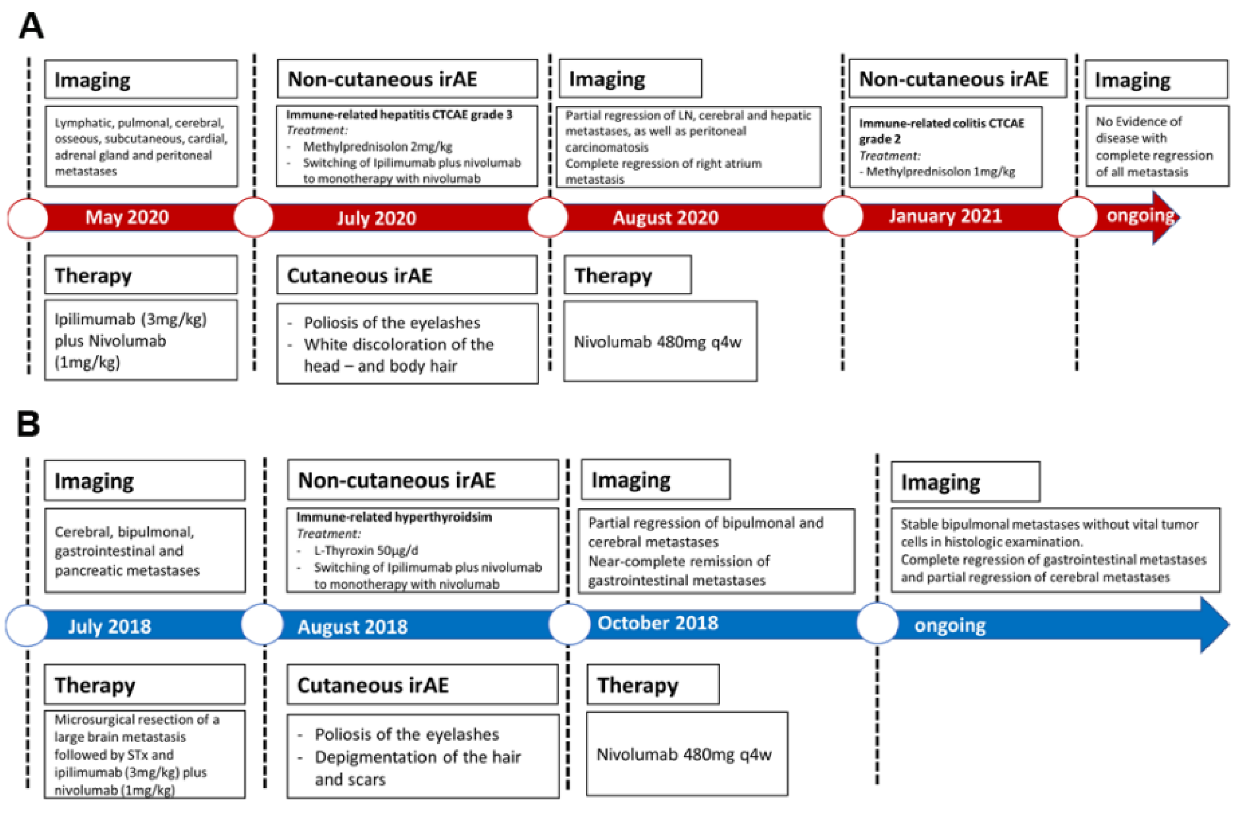
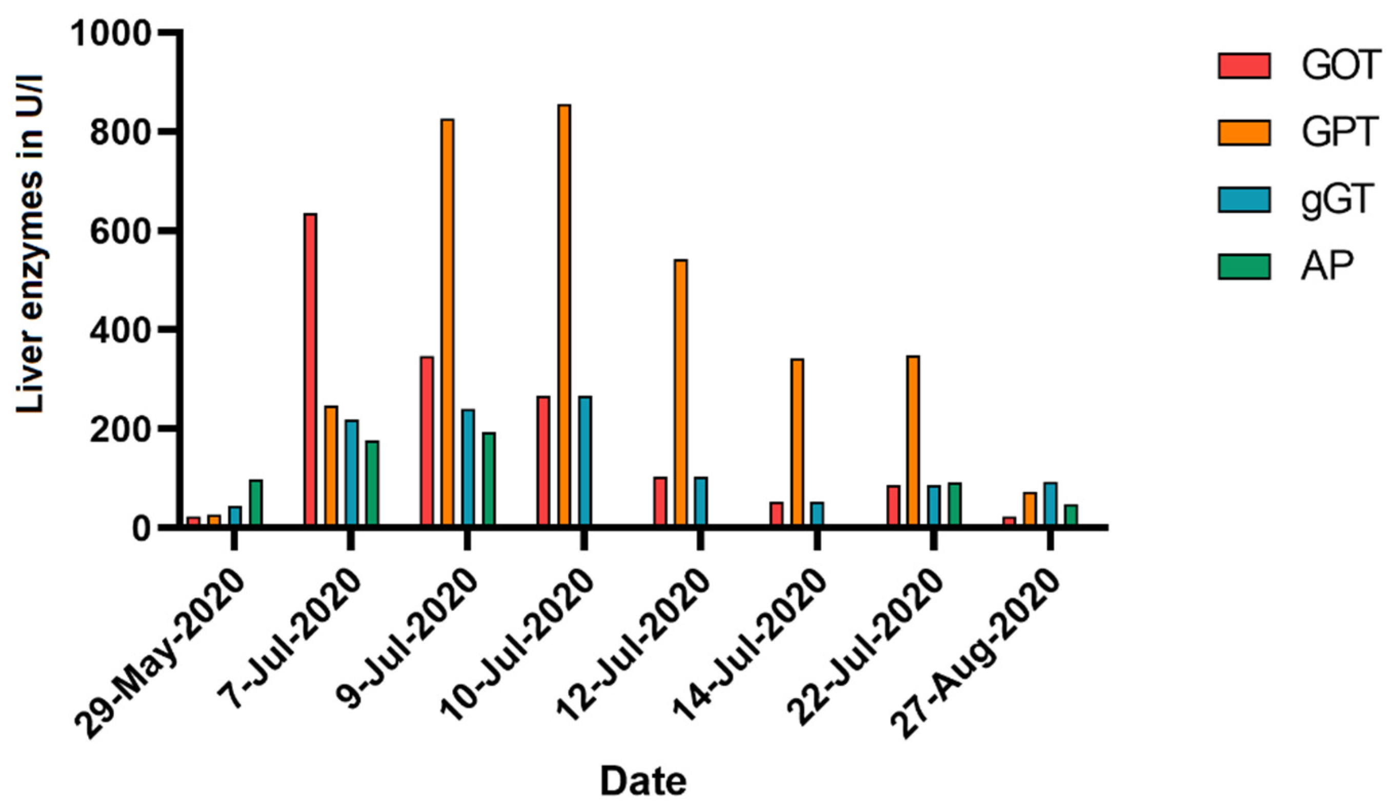
After interdisciplinary tumor board discussion, combined immunotherapy with ipilimumab (3 mg/kg) plus nivolumab (1 mg/kg) q3w was initiated in May 2020 (for timelines see Figure 2A). After three administrations of combined immunotherapy the patient showed a sudden increase in liver enzymes, namely, serum levels of alanine transaminase (ALT) and aspartate transaminase (AST), deterioration of his general condition and nausea in July 2020 (see Figure 3). An immune-related hepatitis was diagnosed (CTCAE grade 3), treatment with combined immunotherapy discontinued and treatment with methylprednisolone (2 mg/kg) initiated. After all laboratory parameters returned to normal values and immune-related hepatitis resolved, slow tapering of methylprednisolone was initiated, and immunotherapy was switched to anti-PD1 monotherapy with nivolumab 480 mg q4w in September 2020.
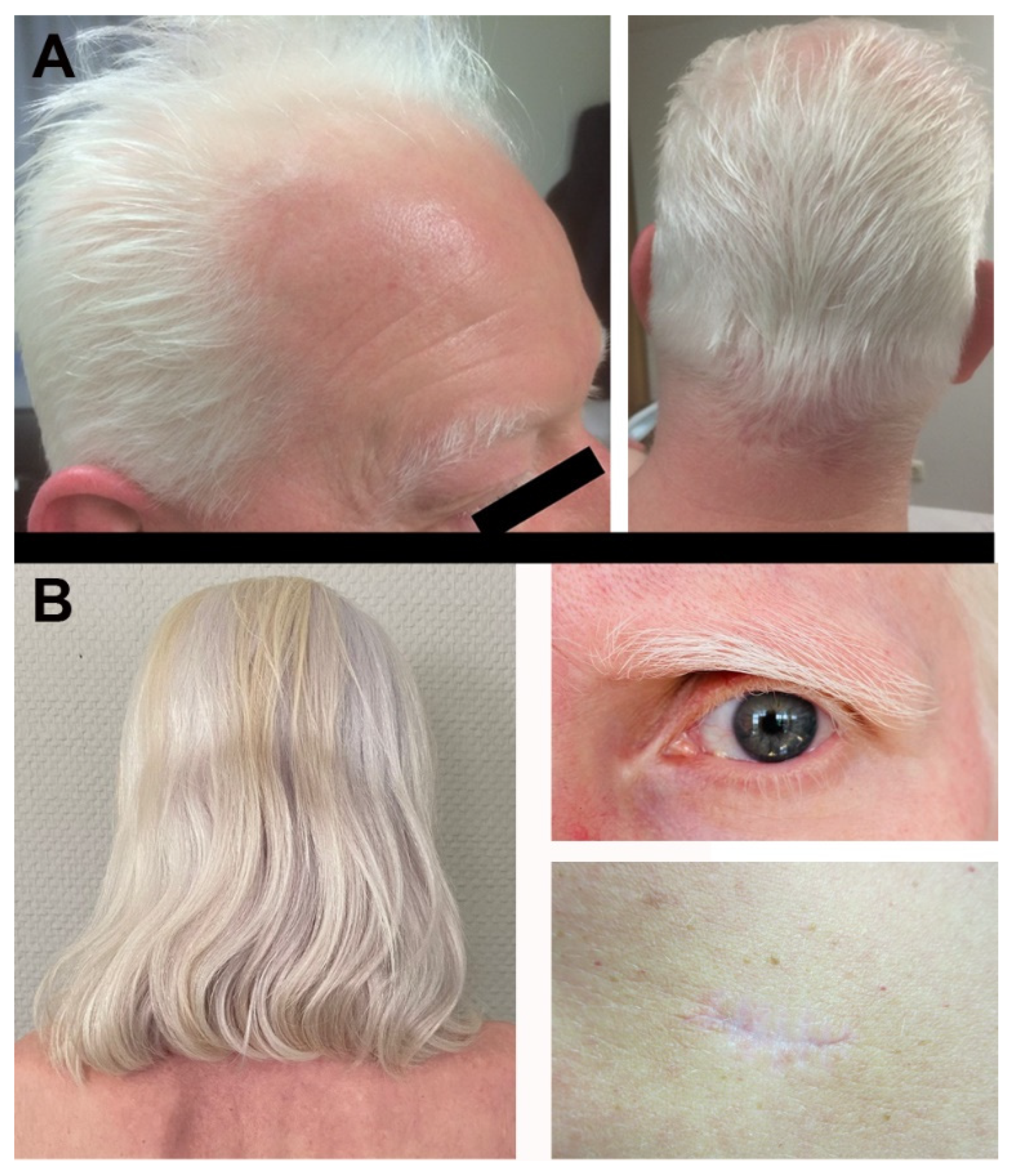
Concomitantly, in July 2020, the patient reported for the first time an increasing white discoloration of the scalp hair, eyelashes and body hair (see Figure 4A). In addition, an ophthalmologic examination revealed uveitis with subjective visual impairment. With subjective mild hearing loss and dizziness, an ENT examination was performed without pathological findings.
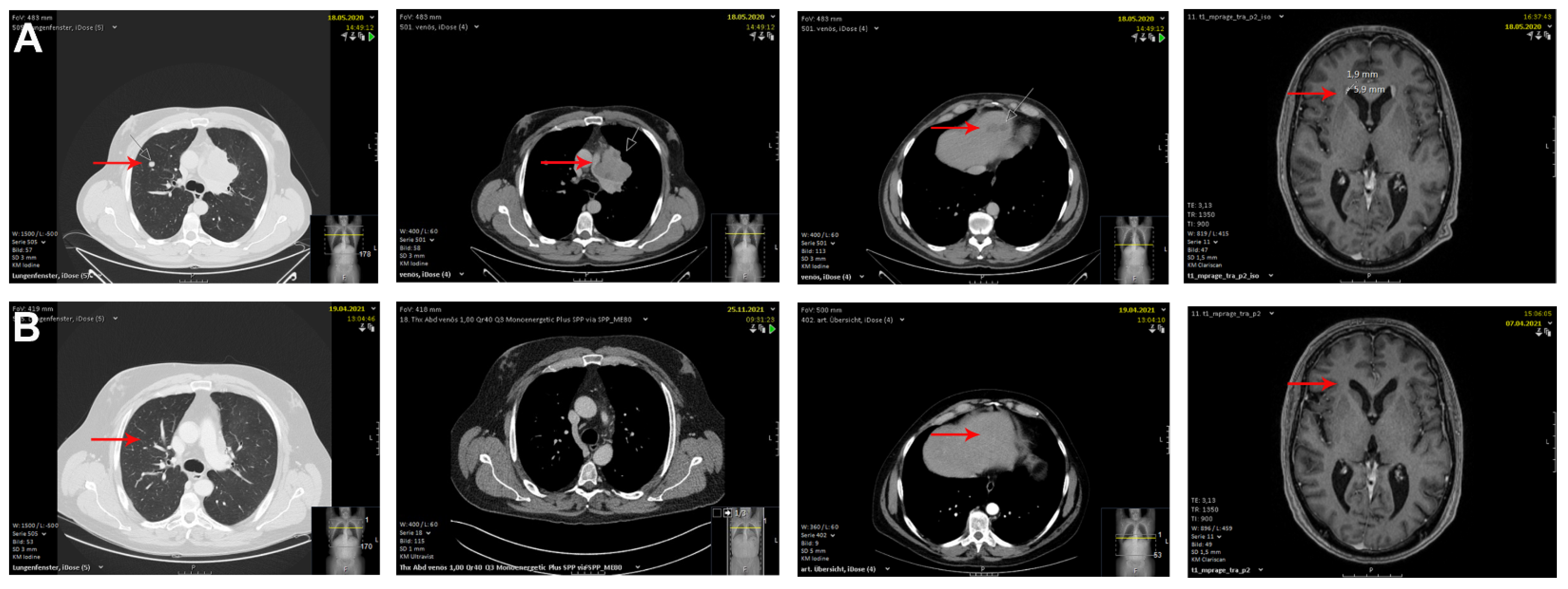
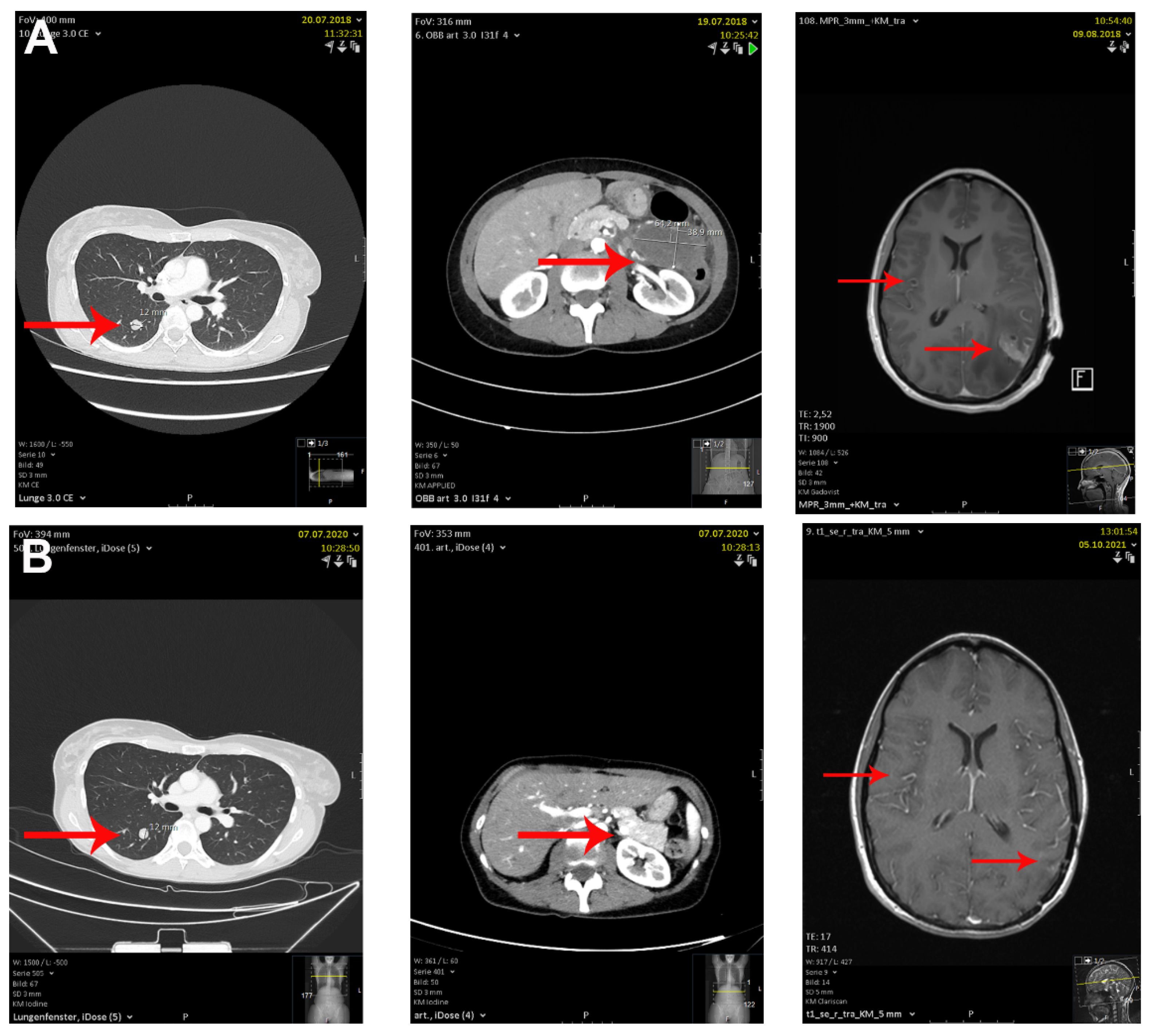
Three months after the first administration of combined immunotherapy, CT and MRI scans documented a substantial response to combined immune checkpoint blockade (cICB) with significant decreases in the size and number of lymph node metastases, cerebral and hepatic metastases, peritoneal carcinomatosis, and complete regression of the metastasis in the right atrium (see Figure 5). Therefore, the patient was classified as a partial responder. In January 2021 the patient presented with a sudden onset of severe diarrhea with >5 stools/day and weight loss of 1 kg. An immune-related colitis (CTCAE grade 2) was diagnosed, which resolved after treatment with methylprednisolone 1 mg/kg. After all laboratory parameters had returned to normal values, immune-related colitis had resolved, and the patient did not need further treatment of irAEs, treatment with nivolumab 480 mg q4w was continued.
Twelve months after the first administration of nivolumab the latest MRI and CT scans showed a complete regression of cerebral, pulmonal, osseous and cutaneous metastasis and only small residuals of the former hepatic and adrenal gland metastasis, and the patient was re-classified as complete responder by September 2021. In conjunction with normal LDH and S100B levels in the peripheral blood, this finding documented the durable and complete response to immunotherapy in this patient. Meanwhile, all therapy-related side effects, except for the depigmentation of the hair and eyelids, corresponding to poliosis, subjective visual impairment and photophobia, had completely resolved, supporting the patient’s overall satisfaction with the therapeutic intervention.
Case 2: We observed a similar association of clinical response to cICB with concomitant development of poliosis in a 48-year-old female patient who first presented in July 2018 with abdominal pain and vomiting. MRI scans revealed a tumor lesion of 6.5 × 3.9 cm in the gastric corpus. A CT-guided puncture of the gastric metastases and histologic examination yielded the diagnosis of metastatic melanoma. Subsequent MRI and CT scans detected cerebral, bipulmonal, gastrointestinal and pancreatic metastasis (see Figure 6). The primary tumor could not be detected even after a thorough physical examination so that the diagnosis of a melanoma of unknown primary was made. While serum LDH levels were within the normal range (216 U/L), S100B levels were elevated (0.18 µg/L) at initial diagnosis. Molecular analysis of the tumor tissue biopsy revealed a BRAFV600E mutation, while KIT gene was wildtype.
After interdisciplinary tumor board discussion and surgical resection of a large cerebral metastasis followed by stereotactic radiation therapy with 30 Gy, cICB with ipilimumab (3 mg/kg) plus nivolumab (1 mg/kg) q3w was initiated in August 2018 (for timelines see Figure 2B). After the second administration of cICB the patient developed a mild uveitis and gradual depigmentation of the hair, eyelids, consistent with poliosis, as well as vitiligo-like depigmentation of the skin and scars (see Figure 4B). Further, the patient developed manifest hyperthyroidism (TSH: 0.03 mU/L; fT3: 3.5 ng/L) with negative TSH receptor antibodies. The patient was started on thyroistatic therapy leading to euthyroidism after 8 months of treatment. By January 2018 the patient showed a latent hypothyroidism in laboratory tests which was subsequently controlled with L-thyroxine therapy. Despite these unanticipated side effects, the treatment in general was well tolerated by the patients and concomitant tumor response benefited therapy adherence.
Three months after initial administration of cICB, CT and MRI scans showed a partial regression of the bipulmonal and a near-complete regression of gastrointestinal metastasis. Therefore, the patient was classified as a partial responder and immunotherapy continued with nivolumab 480 mg q4w. A total of 18 Months after initiation of nivolumab, the latest CT and MRI scans in July 2021 again showed a significant regression of gastrointestinal metastasis, whereas the cerebral and pulmonal lesions remained unchanged, unable to distinguish between residual tumor and scar tissue (see Figure 6). Subsequently, the intestinal mass was surgically removed, revealing a completely necrotic tissue with no residual viable tumor upon histology. In the absence of signs for tumor progression and with consistently normal tumor markers (LDH-A and S100B), the case presented here also demonstrates a sustained therapeutic response to combined immunotherapy with concomitant development of poliosis.

3. Discussion

Checkpoint-inhibitors such as PD-1 and CTLA-4 antibodies have revolutionized the treatment of metastatic melanoma. However, ICI therapy and combined immunotherapy in particular, can also cause a wide range of irAEs, which result in significant morbidity for patients and can limit the use of these beneficial drugs. The randomized phase III CheckMate 067 trial has demonstrated that colitis (13%), hepatitis (33%), and endocrinopathies (34%) are common side effects of cICB, that may enforce treatment discontinuation in case of severe adverse events [17]. However, discontinuation of ICI treatment due to irAEs may not necessarily be associated with a worse treatment outcome as shown by previous studies [2]. Indeed, there is evidence of a possible association of any type of irAEs and favorable response to ICI. In this regard, a retrospective analysis of 576 patients treated with nivolumab demonstrated that the response rates for patients who experienced any-grade irAEs was significantly higher as compared to patients without irAEs. By contrast, a more recent study of more than 1500 patients with metastatic melanoma demonstrated no association of irAE with progression-free survival or overall survival [8]. Since non-cutaneous irAEs more often result in treatment discontinuation, time-delay bias remains a significant challenge in the analysis of the relationship between irAEs and clinical outcome [10]. Hence, the value of these irAEs as a predictive marker for better patient survival is still controversial.
On the other hand, cutaneous irAEs, occurring in up to 30% of patients treated with cICB, may be more robust predictors, as suggested by Hosoya and coworkers [18]. Cutaneous irAEs most commonly include vitiligo-like depigmentations, skin rash and pruritus.
While the incidence of vitiligo in melanoma patients treated with immune-checkpoint inhibitors was estimated to be between 3.4% and 10%, vitiligo has also been observed in approx. 3.4% of patients who did not receive ICI treatment, which is still 10-fold higher as compared to the incidence of vitiligo found in the general population [19,20] and highlights the strongly immunogenic character of melanoma. Notably, the prevalence of vitiligo was reported to be higher in high- and intermediate-risk groups of melanoma patients as compared to low-risk patients [20]. Furthermore, a reverse analysis of data yielded a 180-fold higher prevalence of melanoma in patients first diagnosed with vitiligo which indicates that spontaneous regression of primary melanoma lesions may frequently occur in these patients and may account for a relevant number of melanoma patients with unknown primary [21,22,23].
As the development of vitiligo in patients with melanoma has been attributed to the strong immune response induced by immunogenic melanoma antigens, such as Melanom-A, MART-1 and gp100 [22], culminating evidence suggests a strong association of vitiligo-like depigmentation with a favorable clinical outcome [5,13]. As such, in a recent retrospective analysis of patients with nivolumab showing irAEs, Freeman-Keller and coworkers reported an overall survival benefit in patients presenting with a rash and vitiligo, whereas no significant survival benefit was seen with other irAEs, such as colitis or endocrinopathies [24]. Another study by Quach et al. suggested that cutaneous irAEs might be associated with a favorable response in patients treated with anti-PD1 therapies. Notably, superior outcomes regarding response rates were seen in patients presenting with vitiligo and rash as compared to patients with pruritus [25]. Previous studies conducted by Hua et al. and Nakamura et al. support these findings, as they confirmed a higher incidence of vitiligo in patients who developed a complete or partial response to cICB [26,27].
Unlike vitiligo-like depigmentation, the development of poliosis is a rare event in patients with metastatic melanoma who were treated with ICI. Mechanistically, it has been postulated that depigmentation of the hair follicles results from a strong anti-melanoma immunity, so that CD8 positive T-lymphocytes target melanocytes as a result of shared expression of melanocyte differentiation antigens on previously immune privileged areas on the hair follicles [12]. Due to the rareness of poliosis, the relationship between this depigmentation event and the outcome of ICI therapy in melanoma patients is still largely unknown.
The development of poliosis during melanoma immune-checkpoint inhibitor therapy, which has been associated with a strong treatment response, has only rarely been described in a couple of case reports (see Table 1): Dalle and coworkers described a case of unilateral poliosis of the right eyebrow together with a halo of depigmentation around skin metastases during treatment with nivolumab [28]. Another case describes the development of eyelash poliosis within 2 months of cICB initiation that persisted for 3 years of follow-up [15]. Also, Thomas and coworkers previously described a case in which anti-PD1 monotherapy with Pembrolizumab was associated with complete response upon concomitant development of poliosis and non-segmental vitiligo [29]. It has thus been suggested that this rare irAE correlates with treatment efficacy [5]. In this case series, both patients developed eyelash poliosis and a complete depigmentation of the hair, nevi and scars during cICB but also showed other severe irAEs, and a subsequent durable partial response to cICB at three months after therapy onset, which has been followed by a near complete response at the end of the follow up period. Importantly, the co-occurrence of cutaneous and non-cutaneous irAE in both patients is in accordance with previous analysis from Freman-Keller and coworkers who demonstrated that the number of irAEs might further contribute to the predictive value for response to immunotherapy [24].
Furthermore, our male patient also developed bilateral uveitis and subjective dysacusis, which triggered further diagnostic steps to exclude the suspected diagnosis of Vogt–Koyanagi–Harada syndrome (VKHS). VKHS is a rare condition presenting with the development of poliosis, alongside uveitis, otitis, hypomelanosis and alopecia. A total of 12 melanoma patients who experienced ICI-induced VKHD-like symptoms have been reported so far. Almost all patients showed a significant anti-tumor response during or after the onset of VKHD [31]. Notably, VKHD-like symptoms have also been reported in patients treated with BRAF/MEK inhibitors, which might be explained both by the immunomodulatory effects of BRAF/MEK inhibitors and the massive apoptosis of melanoma cells induced by BRAF-inhibition that results in increased recognition of melanoma-specific antigens and subsequent autoimmunity against melanocyte-rich tissues [32].
In conclusion, the development of poliosis during ICI therapy is a rare cutaneous irAE. Here, we report that poliosis may be a useful and simple clinical indicator of anti-tumor immunity, clinical response and favorable survival outcome, since both patients have shown an impressive response to cICB even in a setting of multifocal metastases. Due to the methodological limitation of our findings as a case report, the value of cutaneous irAEs as a predictive marker for better patient survival and response to treatment with ICI in general, and poliosis in particular, requires further multicentric validation by clinical studies on larger patient cohorts. Also, it seems conceivable that the overall number of irAEs may be strongly indicative of a response to checkpoint-inhibitor therapy, as both patients in this case report series additionally developed non-cutaneous irAEs.

4. Conclusions

In this case report series, we report on two patients with multifocal metastatic melanoma who developed the rare event of poliosis during combined checkpoint-inhibitor therapy with ipilimumab plus nivolumab. Unlike vitiligo-like depigmentation, the development of poliosis is a rare event in patients treated with checkpoint inhibitors, which has not been found to be associated with response to immunotherapy so far.
Given the strong association of clinical response and the concomitant development of eyelash poliosis, we suggest that poliosis may be a useful and simple clinical indicator of anti-tumor immunity, clinical response and favorable survival outcome in advanced melanoma patients treated with ICI.

Author Contributions

All authors (M.H., H.S., V.M., M.H.B., S.G. and C.L.) contributed to the data collection, documentation, critical revision of the paper and final approval of the manuscript. M.H. conceived the work and wrote the manuscript. All authors have read and agreed to the published version of the manuscript.

Funding

M.H. is funded by the Clinician Scientist Fellowship “TransMed Jumpstart Program: 2019_A72” supported by the Else Kröner Fresenius Foundation and by an intramural research funding of the UMC Mainz. H.S. is supported by intramural research funding of the UMC Mainz.

Institutional Review Board Statement

The study was conducted in accordance with the Declaration of Helsinki. Ethical review and approval were waived due to the anonymized analysis of retrospective patient data, which have been routinely collected during the clinical treatment at the Skin Cancer Center of the Department of Dermatology (University Hospital Mainz). This procedure is regulated in Rhineland-Palatinate by the State Hospital Act (§ 36 and § 37). Also, written informed consent for publication of the individual cases was obtained from the patients concerned.

Informed Consent Statement

Written informed consent was obtained from the individuals for the publication of any potentially identifiable images or data included in this article.

Data Availability Statement

The original contributions presented in the study are included in the article. Further inquiries can be directed to the corresponding author.

Acknowledgments

The authors would like to thank the team of the skin cancer center Mainz for comprehensive documentation and helpful comments on the manuscript. Also, we are grateful for the valuable comments and participation of both patients in this case report.

Conflicts of Interest

M.H., H.S., V.M. and M.H.B. declare that the research was conducted in the absence of any commercial or financial relationships that could be construed as a potential conflict of interest. S.G. declares honoraria for advisory boards, oral presentations and travel expenses from Roche, Novartis, MSD, and BMS outside the submitted work. C.L. declares speakers, advisory board honoraria and travel support from Bristol Myers Squibb, Merck Sharp and Dohme, Merck Serono, Novartis, Roche, Pierre Fabre, Sun Pharma, Kiowa Kirin, Sanofi, Biontech, Almirall Hermal outside the submitted work.

References

  1. Hodi, F.S.; Chiarion-Sileni, V.; Gonzalez, R.; Grob, J.J.; Rutkowski, P.; Cowey, C.L.; Lao, C.D.; Schadendorf, D.; Wagstaff, J.; Dummer, R.; et al. Nivolumab plus ipilimumab or nivolumab alone versus ipilimumab alone in advanced melanoma (CheckMate 067): 4-year outcomes of a multicentre, randomised, phase 3 trial. Lancet Oncol. 2018, 19, 1480–1492. [Google Scholar] [CrossRef]
  2. Matull, J.; Livingstone, E.; Wetter, A.; Zimmer, L.; Zaremba, A.; Lahner, H.; Schadendorf, D.; Ugurel, S. Durable Complete Response in a Melanoma Patient with Unknown Primary, Associated with Sequential and Severe Multi-Organ Toxicity After a Single Dose of CTLA-4 Plus PD-1 Blockade: A Case Report. Front. Oncol. 2020, 10, 592609. [Google Scholar] [CrossRef] [PubMed]
  3. Martins, F.; Sofiya, L.; Sykiotis, G.P.; Lamine, F.; Maillard, M.; Fraga, M.; Shabafrouz, K.; Ribi, C.; Cairoli, A.; Guex-Crosier, Y.; et al. Adverse effects of immune-checkpoint inhibitors: Epidemiology, management and surveillance. Nat. Rev. Clin. Oncol. 2019, 16, 563–580. [Google Scholar] [CrossRef] [PubMed]
  4. Palmieri, D.J.; Carlino, M.S. Immune Checkpoint Inhibitor Toxicity. Curr. Oncol. Rep. 2018, 20, 72. [Google Scholar] [CrossRef]
  5. Gault, A.; Anderson, A.E.; Plummer, R.; Stewart, C.; Pratt, A.G.; Rajan, N. Cutaneous immune-related adverse events in patients with melanoma treated with checkpoint inhibitors. Br. J. Dermatol. 2021, 185, 263–271. [Google Scholar] [CrossRef]
  6. Mandalà, M.; Larkin, J.M.G.; Ascierto, P.A.; Del Vecchio, M.; Gogas, H.; Cowey, C.L.; Arance Fernandez, A.N.A.M.; Dalle, S.; Schenker, M.; Grob, J.J.; et al. An analysis of nivolumab-mediated adverse events and association with clinical efficacy in resected stage III or IV melanoma (CheckMate 238). J. Clin. Oncol. 2019, 37, 9584. [Google Scholar] [CrossRef]
  7. Weber, J.S.; Hodi, F.S.; Wolchok, J.D.; Topalian, S.L.; Schadendorf, D.; Larkin, J.; Sznol, M.; Long, G.V.; Li, H.; Waxman, I.M.; et al. Safety Profile of Nivolumab Monotherapy: A Pooled Analysis of Patients with Advanced Melanoma. J. Clin. Oncol. 2017, 35, 785–792. [Google Scholar] [CrossRef]
  8. Robert, C.; Hwu, W.J.; Hamid, O.; Ribas, A.; Weber, J.S.; Daud, A.I.; Hodi, F.S.; Wolchok, J.D.; Mitchell, T.C.; Hersey, P.; et al. Long-term safety of pembrolizumab monotherapy and relationship with clinical outcome: A landmark analysis in patients with advanced melanoma. Eur. J. Cancer 2021, 144, 182–191. [Google Scholar] [CrossRef]
  9. Eggermont, A.M.M.; Kicinski, M.; Blank, C.U.; Mandala, M.; Long, G.V.; Atkinson, V.; Dalle, S.; Haydon, A.; Khattak, A.; Carlino, M.S.; et al. Association Between Immune-Related Adverse Events and Recurrence-Free Survival among Patients with Stage III Melanoma Randomized to Receive Pembrolizumab or Placebo: A Secondary Analysis of a Randomized Clinical Trial. JAMA Oncol. 2020, 6, 519–527. [Google Scholar] [CrossRef] [Green Version]
  10. Sullivan, R.J.; Weber, J.S. Immune-related toxicities of checkpoint inhibitors: Mechanisms and mitigation strategies. Nat. Rev. Drug Discov. 2021. [Google Scholar] [CrossRef]
  11. Hofmann, L.; Forschner, A.; Loquai, C.; Goldinger, S.M.; Zimmer, L.; Ugurel, S.; Schmidgen, M.I.; Gutzmer, R.; Utikal, J.S.; Goppner, D.; et al. Cutaneous, gastrointestinal, hepatic, endocrine, and renal side-effects of anti-PD-1 therapy. Eur. J. Cancer 2016, 60, 190–209. [Google Scholar] [CrossRef] [PubMed]
  12. Burzi, L.; Alessandrini, A.M.; Quaglino, P.; Piraccini, B.M.; Dika, E.; Ribero, S. Cutaneous Events Associated with Immunotherapy of Melanoma: A Review. J. Clin. Med. 2021, 10, 3047. [Google Scholar] [CrossRef] [PubMed]
  13. Teulings, H.E.; Limpens, J.; Jansen, S.N.; Zwinderman, A.H.; Reitsma, J.B.; Spuls, P.I.; Luiten, R.M. Vitiligo-like depigmentation in patients with stage III-IV melanoma receiving immunotherapy and its association with survival: A systematic review and meta-analysis. J. Clin. Oncol. 2015, 33, 773–781. [Google Scholar] [CrossRef] [PubMed]
  14. Alsuhaibani, A.H.; Alhumayed, M. Primary orbital melanoma with poliosis and a palpable mass. Arch. Ophthalmol. 2011, 129, 1382–1383. [Google Scholar] [CrossRef] [PubMed] [Green Version]
  15. Ediriwickrema, L.S.; Liu, C.Y.; Kikkawa, D.O.; Korn, B.S. Development of Poliosis Following Checkpoint Inhibitor Treatment for Cutaneous Melanoma. Ophthalmic Plast Reconstr. Surg. 2019, 35, e121–e122. [Google Scholar] [CrossRef]
  16. Failla, C.M.; Carbone, M.L.; Fortes, C.; Pagnanelli, G.; D’Atri, S. Melanoma and Vitiligo: In Good Company. Int. J. Mol. Sci. 2019, 20, 5731. [Google Scholar] [CrossRef] [Green Version]
  17. Larkin, J.; Chiarion-Sileni, V.; Gonzalez, R.; Grob, J.J.; Rutkowski, P.; Lao, C.D.; Cowey, C.L.; Schadendorf, D.; Wagstaff, J.; Dummer, R.; et al. Five-Year Survival with Combined Nivolumab and Ipilimumab in Advanced Melanoma. N. Engl. J. Med. 2019, 381, 1535–1546. [Google Scholar] [CrossRef] [Green Version]
  18. Hosoya, K.; Fujimoto, D.; Morimoto, T.; Kumagai, T.; Tamiya, A.; Taniguchi, Y.; Yokoyama, T.; Ishida, T.; Hirano, K.; Matsumoto, H.; et al. Association Between Early Immune-related Adverse Events and Clinical Outcomes in Patients with Non-Small Cell Lung Cancer Treated with Immune Checkpoint Inhibitors. Clin. Lung Cancer 2020, 21, e315–e328. [Google Scholar] [CrossRef]
  19. Wolner, Z.J.; Marghoob, A.A.; Pulitzer, M.P.; Postow, M.A.; Marchetti, M.A. A case report of disappearing pigmented skin lesions associated with pembrolizumab treatment for metastatic melanoma. Br. J. Dermatol. 2018, 178, 265–269. [Google Scholar] [CrossRef]
  20. Schallreuter, K.U.; Levenig, C.; Berger, J. Vitiligo and cutaneous melanoma. A case study. Dermatologica 1991, 183, 239–245. [Google Scholar] [CrossRef]
  21. Blessing, K.; McLaren, K.M. Histological regression in primary cutaneous melanoma: Recognition, prevalence and significance. Histopathology 1992, 20, 315–322. [Google Scholar] [CrossRef] [PubMed]
  22. Aung, P.P.; Nagarajan, P.; Prieto, V.G. Regression in primary cutaneous melanoma: Etiopathogenesis and clinical significance. Lab. Investig. 2017, 97, 657–668. [Google Scholar] [CrossRef] [PubMed]
  23. Del Fiore, P.; Rastrelli, M.; Dall’Olmo, L.; Cavallin, F.; Cappellesso, R.; Vecchiato, A.; Buja, A.; Spina, R.; Parisi, A.; Mazzarotto, R.; et al. Melanoma of Unknown Primary: Evaluation of the Characteristics, Treatment Strategies, Prognostic Factors in a Monocentric Retrospective Study. Front. Oncol. 2021, 11, 627527. [Google Scholar] [CrossRef] [PubMed]
  24. Freeman-Keller, M.; Kim, Y.; Cronin, H.; Richards, A.; Gibney, G.; Weber, J.S. Nivolumab in Resected and Unresectable Metastatic Melanoma: Characteristics of Immune-Related Adverse Events and Association with Outcomes. Clin. Cancer Res. 2016, 22, 886–894. [Google Scholar] [CrossRef] [Green Version]
  25. Quach, H.T.; Dewan, A.K.; Davis, E.J.; Ancell, K.K.; Fan, R.; Ye, F.; Johnson, D.B. Association of Anti-Programmed Cell Death 1 Cutaneous Toxic Effects with Outcomes in Patients with Advanced Melanoma. JAMA Oncol. 2019, 5, 906–908. [Google Scholar] [CrossRef]
  26. Nakamura, Y.; Tanaka, R.; Asami, Y.; Teramoto, Y.; Imamura, T.; Sato, S.; Maruyama, H.; Fujisawa, Y.; Matsuya, T.; Fujimoto, M.; et al. Correlation between vitiligo occurrence and clinical benefit in advanced melanoma patients treated with nivolumab: A multi-institutional retrospective study. J. Dermatol. 2017, 44, 117–122. [Google Scholar] [CrossRef]
  27. Hua, C.; Boussemart, L.; Mateus, C.; Routier, E.; Boutros, C.; Cazenave, H.; Viollet, R.; Thomas, M.; Roy, S.; Benannoune, N.; et al. Association of Vitiligo with Tumor Response in Patients with Metastatic Melanoma Treated with Pembrolizumab. JAMA Dermatol. 2016, 152, 45–51. [Google Scholar] [CrossRef] [Green Version]
  28. Amini Adle, M.; Chastagner, M.; Mansard, S.; Dalle, S. Image Gallery: Unilateral eyebrow depigmentation. Br. J. Dermatol. 2019, 180, e107. [Google Scholar] [CrossRef]
  29. Thomas, S.; Laino, A.; Sturm, R.; Nufer, K.; Lambie, D.; Shepherd, B.; Atkinson, V.; Adams, L.; Soyer, H.P.; Schaider, H. Focal regression of a primary melanoma, fading lentigines and poliosis in metastatic melanoma treated with anti-PD-1. J. Eur. Acad. Dermatol. Venereol. 2018, 32, e176–e177. [Google Scholar] [CrossRef]
  30. Zarbo, A.; Belum, V.R.; Sibaud, V.; Oudard, S.; Postow, M.A.; Hsieh, J.J.; Motzer, R.J.; Busam, K.J.; Lacouture, M.E. Immune-related alopecia (areata and universalis) in cancer patients receiving immune checkpoint inhibitors. Br. J. Dermatol. 2017, 176, 1649–1652. [Google Scholar] [CrossRef]
  31. Gambichler, T.; Seifert, C.; Lehmann, M.; Lukas, C.; Scheel, C.; Susok, L. Concurrent Vogt-Koyanagi-Harada disease and impressive response to immune checkpoint blockade in metastatic melanoma. Immunotherapy 2020, 12, 439–444. [Google Scholar] [CrossRef] [PubMed]
  32. Fusumae, T.; Kamiya, K.; Maekawa, T.; Komine, M.; Murata, S.; Inoda, S.; Takahashi, R.; Kawashima, H.; Ohtsuki, M. Vogt-Koyanagi-Harada disease-like uveitis induced by vemurafenib for metastatic cutaneous malignant melanoma. J. Dermatol. 2018, 45, e159–e160. [Google Scholar] [CrossRef] [PubMed]
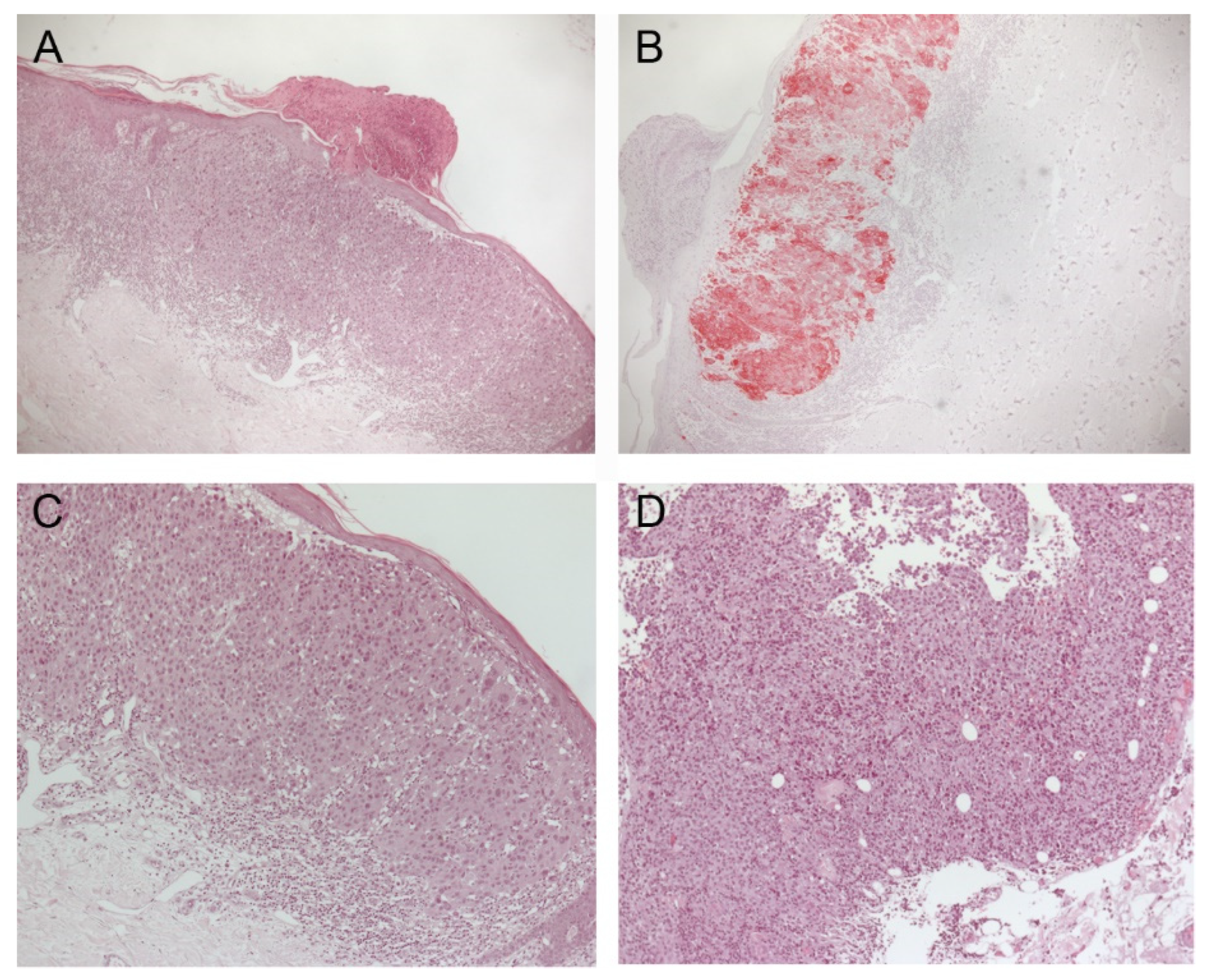
Figure 1. Histopathological samples from primary melanoma lesions (AC) and subcutaneous metastasis prior to initiation of combined checkpoint inhibitor therapy in patient #1. Initial diagnosis of melanoma was confirmed in February 2016 using H&E (5× in (A) and 10× in (C)) and IHC stains for Melan-A (B). Upon diagnosis of advanced stage melanoma, a biopsy from a subcutaneous lesion in the abdominal skin region confirmed advanced stage melanoma (10× in (D)). Both histopathological samples showed a strong infiltration by lymphocytes in the tumor-surrounding stroma.
Immuno 02 00020 g001
Figure 2. Schematic timelines of therapy, imaging and the occurrence of cutaneous and non-cutaneous adverse events in the course of the disease of patient #1 (A) and #2 (B).
Immuno 02 00020 g002
Figure 3. Laboratory results in the course of immune-related hepatitis of patient #1, which has been diagnosed in July 2020 and resolved in the course of the same month upon treatment with methylprednisolone 2 mg/kg body weight. Abbreviations: GOT = aspartate aminotransferase (AST); GPT = alanine aminotransferase (ALT); gGT = gamma-glutamyl-transferase; AP = alkaline phosphatase.
Immuno 02 00020 g003
Figure 4. Clinical presentation of poliosis after complete response to combined immunotherapy in a male patient (A) with initial melanoma metastatic to the skin, lung, liver, brain, peritoneum, right heart atrium, lymph nodes, adrenal gland and bones and a female patient (B) with clinical presentation of poliosis to the hair, eyelids and scars after complete response to initially multifocal metastatic melanoma showing cerebral, bipulmonal, gastric, pancreatic, renal and intestinal metastasis.
Immuno 02 00020 g004
Figure 5. Results from MRI and CT scans of the 53-year-old male patient prior to the administration of combined checkpoint blockade in May 2020 and results from the latest scans from July 2021. Compared to initial staging results showing melanoma pulmonary, cardiac, hepatic and cerebral metastasis (A), the staging results from July 2021 show a complete remission of all previously reported metastasis with no evidence of disease at this stage of follow up (B).
Immuno 02 00020 g005
Figure 6. Results from MRI and CT scans of the 48-year-old female prior to the administration of combined checkpoint blockade in August 2018 (A) and results from the latest scans from July 2021 (B). Whereas pulmonary and cerebral metastasis are largely unchanged, a complete regression of gastrointestinal metastases was found over the course of immunotherapy.
Immuno 02 00020 g006
Table 1. Summary of all reports describing the event of poliosis during checkpoint-inhibitor therapy for patients with advanced melanoma and response to immunotherapy.
AuthorPrimary MelanomaAgeTumor StageICI AgentFirst Signs of PoliosisTreatment ResponseOther irAERef.
Korn et al.right eyelid52 yearsIIICIpilimumab + Nivolumab2 months upon initiationcomplete remissionnone[15]
Gault et al.unknownunknownIVIpilimumab5 yearscomplete remissionnone[5]
Wolner et al.right lower back60 yearsIVPembrolizumab4 months upon treatment initiationpartial responsenone[19]
Zarbo et al.unknown65 yearsIVIpilimumab + Nivolumab3 months upon initiationpartial responsealopecia, hepatitis, hypothyroidism[30]
Thomas et al.right shoulderunknownIVPembrolizumab5 months upon initiationcomplete responsenon-segmental vitiligo[29]
Haist et al.unknown and upper back48 years and 53 yearsIVIpilimumab + NivolumabAfter 2 and 3 cycles of cICBcomplete responsehypothyroidism, vitiligo, hepatitis and colitis
Publisher’s Note: MDPI stays neutral with regard to jurisdictional claims in published maps and institutional affiliations.