Article

Effect of Wild Blueberry Metabolites on Biomarkers of Gastrointestinal and Immune Health In Vitro

by
Thirumurugan Rathinasabapathy
1,2,
Jade Lomax
1,2,
Kavin Srikanth
1,3,
Debora Esposito
1,4,
Colin D. Kay
1,2 and
Slavko Komarnytsky
1,2,*
1
Plants for Human Health Institute, NC State University, 600 Laureate Way, Kannapolis, NC 28081, USA
2
Department of Food, Bioprocessing, and Nutrition Sciences, North Carolina State University, 400 Dan Allen Drive, Raleigh, NC 27695, USA
3
Department of Biology, University of North Carolina Asheville, 1 University Heights Drive, Asheville, NC 28804, USA
4
Department of Animal Science, NC State University, 120 Broughton Drive, Raleigh, NC 27695, USA
*
Author to whom correspondence should be addressed.
Immuno 2022, 2(2), 293-306; https://doi.org/10.3390/immuno2020019
Submission received: 20 February 2022 / Revised: 18 March 2022 / Accepted: 21 March 2022 / Published: 25 March 2022

Abstract

:
Wild blueberries (Vaccinium angustifolium Aiton.) are a rich source of dietary fiber and (poly)phenols with gastrointestinal and immune health-promoting properties, however, their mechanisms of action on the intestinal epithelial cells and transient tissue macrophages remain to be elucidated. In this study, we evaluated the individual effects of anthocyanins, short-chain fatty acids (metabolites derived from fiber), and a series of hydroxycinnamic and hydroxybenzoic acid metabolites common to anthocyanins and other polyphenols on epithelial gut homeostasis in human colon epithelial CCD-18 cells and murine RAW 264.7 macrophages. Gastrointestinal cell migration was enhanced in response to anthocyanin glucosides with the maximum effect observed for malvidin-3-glucoside, and a structural subset of hydroxybenzoic acids, especially 2-hydroxybenzoic acid. Enhanced staining for ZO-1 protein in the junctional complexes was observed in CCD-18 cells treated with malvidin and butyrate, as well as several phenolic metabolites, including hydroxybenzoic and hydroxycinnamic acids. Nitric oxide production and pro-inflammatory gene expression profiles in the LPS-stimulated macrophages were mostly affected by treatments with 3-caffeoylquinic (chlorogenic) and 3,4-dihydroxycinnamic (caffeic) acids, as well as 2-hydroxybenzoic acid. This study lays the foundation for future investigations evaluating the effects of dietary interventions on managing gastrointestinal and inflammatory pathophysiological outcomes.

1. Introduction

Wild lowbush blueberries (Vaccinium angustifolium Aiton.) have a complex and dense phytonutrient profile enriched with dietary fiber (3–4 g/100 g fresh weight) [1] and bioactive polyphenols, including anthocyanins, proanthocyanidins, and hydroxycinnamic acids (300–600 mg/100 g FW) [2]. Their anthocyanidin profile is dominated by delphinidin and malvidin glycosides [3] and is in contrast to many other anthocyanin-rich fruits and berries that predominantly accumulate cyanidins [4]. The blueberry anthocyanin glycosides can be further acylated with organic acids [5], and this modification generally improves the stability of anthocyanins at wider pH ranges [6] and digestive conditions [7] but hinders their bioavailability [8]. Following absorption in the gut, low amounts of precursor polyphenolic compounds can be found in the systemic circulation in the form of intact or phase II-derived (glucuronide, methyl, or sulfate) conjugates, while significantly larger amounts of the polyphenolic compounds undergo colonic fermentation by the gut microbiome and enter the human body as low molecular weight phenolic microbial metabolites or catabolites [9]. Disruption of the intact microbiome with antibiotic treatment in rodents results in reduced absorption of microbial metabolites and predominant excretion of polyphenols in their unmodified form [10]. While different levels of phenolic intake affect the magnitude of biological effects [11], not all plasma phenolic metabolites show a linear response in an intake-dependent manner [12,13]. The lack of a linear relationship can be explained in part by the different affinities of individual phenolic acids towards microbial species, transporters, carrier proteins, and metabolic enzymes that are responsible for their phase-II metabolism.
In recent years, consumption of blueberries was increasingly associated with positive metabolic and immune health outcomes, in part by observing improvements in insulin sensitivity [14], vascular and endothelial function [15], inflammation [16], lipid status [17], and brain perfusion [18]. Blueberries, like any other phytochemical or nutrient-rich food, can influence the human state of health by modifying the integrity of the gastrointestinal system, conditioning the composition of gut microbiota, or directly shifting host metabolism and energy balance in the body.
The gastrointestinal wall, in the form of a single or double mucus layer, a single layer of the gut epithelial cells, and the vascular endothelium serves as the primary impervious barrier to all dietary components that are not hydrophobic or do not have a specific system of active transport [19]. This is achieved by the formation of tight junctions at the apical membrane, and adherence junctions at the basal membrane of the gut epithelial cells to control the trafficking of macromolecules, food tolerance, and immune responses in the gut [20]. Loss of barrier function secondary to release of zonulin, a prehaptoglobin-2 protein that disassembles tight junctions through EGF activation [21], generally leads to altered intestinal permeability and cytokine-mediated dysfunction that affects the intestinal mucosa and other tissues in the context of sustained chronic inflammation [22]. Zonulin expression is regulated in part by IL-6 responsive elements found in its promoter region [23] and correlates with the circulating IL-6 in subjects with obesity-associated insulin resistance [24]. At the same time, care should be taken to extrapolate zonulin findings directly [25]. While the beneficial effects of blueberry polyphenols and fiber on microbiota have been shown in clinical studies [26,27,28], little is known about the physiological effects and molecular targets of precursor polyphenol molecules and their metabolites on the activity of gastrointestinal epithelial cells and tissue-resident macrophages that directly sample luminal content [29].
In this study, we examined the ability of major metabolites of wild blueberry anthocyanins (hydroxycinnamic and hydroxybenzoic acids), hydroxycinnamic acid esters (chlorogenic acid), triterpenoid ursolic acid, and fiber (acetate, propionate, and butyrate short-chain fatty acids) to modulate biomarkers of the intestinal barrier and inflammation in vitro, in order to access their potential to support gastrointestinal and immune health outcomes.

2. Materials and Methods

2.1. Chemicals

The anthocyanin 3-O-β-glucosides cyanidin (C3G), delphinidin (D3G), and malvidin (M3G) were purchased from Polyphenols Laboratories AS (Sandnes, Norway). Sodium acetate, butyrate, propionate, ursolic acid, benzoic acid (BA), 3-hydroxyphenylpropionic acid (3HPPA), and 3-methoxy-4-hydroxycinnamic acid (ferulic acid, 3M4HCA) were from Sigma (St. Louis, MO, USA). 3-caffeoylquinic acid (chlorogenic acid, 3CQA), 3,4-dihydroxybenzoic acid (protocatechuic acid, 3,4DHBA), 3-methoxy-4-hydroxybenzoic acid (vanillic acid, 3M4HBA), 3-methoxy-4-hydroxyphenylacetic acid (homovanillic acid, 3M4HPAA), 4-hydroxyphenylpropionic acid (dihydrocoumaric acid, 4HPPA), and 4-methoxy-3-hydroxycinnamic acid (isoferulic acid, 4M3HCA) were from Alfa Aesar (Haverhill, MA, USA). 2-hydroxybenzoic acid (salicylic acid, 2HBA), 3-methoxy-4-hydroxyphenylpropionic acid (dihydroferulic acid, 3M4HPPA), and 3,4-dihydroxycinnamic acid (caffeic acid, 3,4DHCA) were from TCI (Tokyo, Japan). 3,4-dihydroxyphenylacetic acid (3,4DHPAA) was from Acros Organics (Fair Lawn, NJ, USA). The nomenclature of phenolic acid metabolites used in this study is listed in Table 1. All other chemical reagents were purchased from Sigma unless specified otherwise.

2.2. Cell Culture

The normal colonic epithelial CCD-18 Co cells (ATCC CRL-1459, obtained from American Type Culture Collection; Livingstone, MT, USA) and the mouse macrophage cell line RAW 264.7 (ATCC TIB-71) were maintained in Dulbecco’s modified Eagle’s medium (DMEM, Life Technologies, Carlsbad, CA, USA), supplemented with 10% fetal bovine serum (Life Technologies), 100 IU/mL of penicillin, and 100 µg/mL of streptomycin (Fisher Scientific, Pittsburg, PA, USA) at a density not exceeding 5 × 10⁵ cells/mL. All cell lines were grown in 57-cm2 Nunc cell culture dishes (Nalge Nunc International, Rochester, NY, USA) maintained at 37 °C in a humidified 5% CO2 Thermo Forma Series II incubator (Fisher Scientific), and routinely passaged every 3–4 days. Cell viability and dose range determination studies did not show any detrimental effects at all levels up to the maximum concentration tested as described earlier [30,31,32], with the exception of ursolic acid which was cytotoxic at 15 μM and therefore excluded from the subsequent experiments (data not shown).

2.3. Cell Migration and Formation of Junctional Complexes

CCD-18 cells were seeded in the 96-well Oris Cell Migration plates (Platypus Technologies, Madison, WI, USA) with a stopper positioned in the center of a well to create a cell-free detection zone after 24 h of incubation. Following the removal of the stoppers, cells were allowed to migrate (with and without treatments as indicated, using 0.1% DMSO as a vehicle control) into the clear field for 48 h, stained with NucBlue Hoechst 33342 Live ReadyProbes Reagent (Fisher Scientific, Waltham, MA, USA), and central fluorescence was measured at a 360 nm excitation/460 nm emission using a BioTek SynergyH1 microplate reader (Agilent, Santa Clara, CA, USA), both pre- and post-migration. Elevated levels of growth factors found in 20% FBS were used as a positive control.
In a separate set of experiments, CCD-18 cells were allowed to form monolayers for 5 days (with and without treatments as indicated, using 0.1% DMSO as a vehicle control), fixed with cold 4% formaldehyde for 15 min, permeabilized with 0.3% Triton X-100 for 5 min, and blocked with 5% BSA for 30 min at 37 °C. Zonulin accumulation in junction complexes was visualized using ZO-1/TJP1 antibody Alexa Fluor 488 (Fisher) (1:200 in 0.1% BSA for 3 h) and quantified using mean fluorescent intensity at a 490 nm excitation/525 nm emission.

2.4. Nitric Oxide Production and Gene Expression in Macrophages

RAW 264.7 cells were seeded in 96-well plates in triplicate at the concentration of 5 × 10⁴ cells/well in a 200 μL culture medium and allowed to adhere for 24 h. The cells were then pre-treated with the indicated lower dose ranges of wild blueberry metabolites established in the gastrointestinal cell assays and elicited with 1 µg/mL of LPS for an additional 6 h. Nitric oxide released from the stimulated macrophages was indirectly quantified by measuring nitrite accumulation in the medium using the Greiss reagent system (Promega, Madison, WI) and a SynergyH1 microplate reader (BioTek) at 530 nm. For gene expression studies, the cells were seeded in 24-well plates at the concentration of 5 × 10⁵ cells/well in a 1 mL culture medium and treated as indicated.

2.5. RNA Extraction, Purification, and cDNA Synthesis

The total RNA was isolated from RAW macrophages using TRIzol reagent (Life Technologies) following the manufacturer’s instructions. RNA was quantified using the SynergyH1/Take 3 plate (BioTek). The cDNAs were synthesized using 2 µg of RNA for each sample using a high-capacity cDNA Reverse Transcription kit following the manufacturer’s protocol on an ABI GeneAMP 9700 (Life Technologies).

2.6. Quantitative PCR Analysis

The resulting cDNA was amplified by real-time quantitative PCR using SYBR green PCR master mix (Life Technologies). To avoid interference due to genomic DNA contamination, only intron-overlapping primers were selected using Primer Express software, version 2.0 (Applied Biosystems, Foster City, CA) as follows: β-actin, forward primer: 5′-AAC CGT GAA AAG ATG ACC CAG AT-3′, reverse primer: 5′-CAC AGC CTG GAT GGC TAC GT-3′; COX-2, forward primer: 5′-TGG TGC CTG GTC TGA TGA TG-3′, reverse primer: 5′-GTG GTA ACC GCT CAG GTG TTG-3′; iNOS, forward primer: 5′-CCC TCC TGA TCT TGT GTT GGA-3′, reverse primer: 5߰-TCA ACC CGA GCT CCT GGA A-3′; IL-6, forward primer: 5′-TAG TCC TTC CTA CCC CAA TTT CC-3′, reverse primer: 5′-TTG GTC CTT AGC CAC TCC TTC-3′; and IL-1β, forward primer: 5′-CAA CCA ACA AGT GAT ATT CTC CAT G-3′, reverse primer: 5′-GAT CCA CAC TCT CCA GCT GCA-3′. Quantitative PCR (qPCR) amplifications were performed on an ABI 7500 Fast real-time PCR (Life Technologies) using 1 cycle at 50 °C for 2 min and 1 cycle at 95 °C for 10 min, followed by 40 cycles of 15 s at 95 °C and 1 min at 60 °C. The dissociation curve was completed with 1 cycle of 1 min at 95 °C, 30 s at 55 °C, and 30 s at 95 °C. mRNA expression was analyzed using the ΔΔCT method and normalized with respect to the expression of the β-actin housekeeping gene using 7500 Fast System SDS software, v1.3.0 (Life Technologies). Amplification of specific transcripts was confirmed by obtaining melting curve profiles.

2.7. Statistical Analysis

Statistical analyses were performed using Prism 8.0 (GraphPad Software, San Diego, CA, USA). Data were analyzed by one-way ANOVA with treatment as a factor. Post hoc analyses of differences between individual experimental groups were made using Dunnett’s multiple comparison tests. The significance was set at p < 0.05. Values are reported as means ± SEM.

3. Results

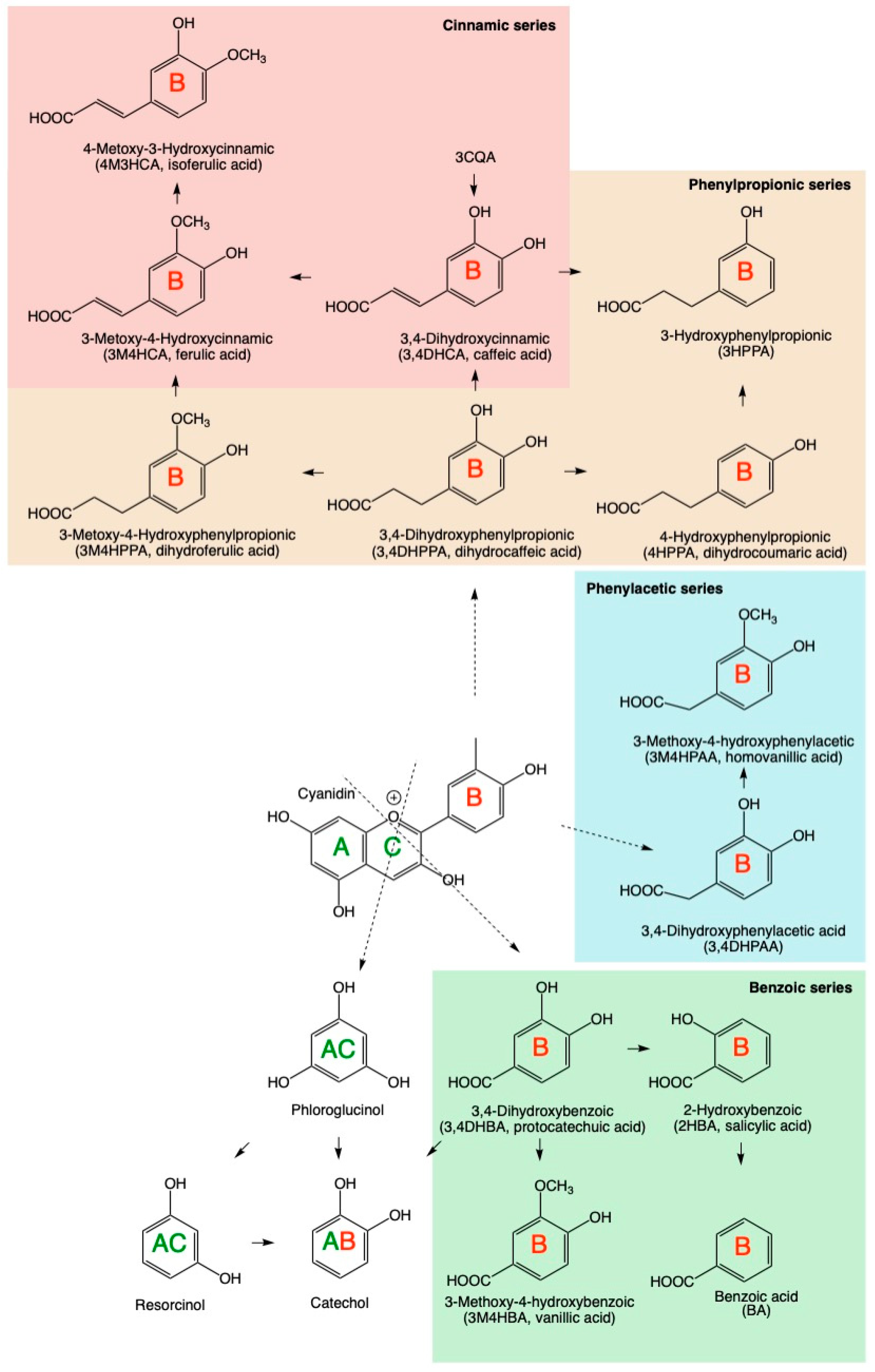
3.1. Physiological Dose Ranges of Gastrointestinal Metabolites

Certain dietary factors, such as glutamine [33], histidine [34], phenolic acids [35], and short-chain fatty acids derived from carbohydrate metabolism by microbiota [36] have the potential to directly affect intestinal mucosa and immunity. Gastrointestinal cells are typically exposed to higher levels of these metabolites than those found in the systemic circulation, as both intestinal tissues and fecal samples of healthy individuals were reported to contain around 50–150 mM of short-chain fatty acids [37] and 2–16 μmol of individual phenolic compounds [38]. These ranges were used to establish physiologically relevant concentrations of the gastrointestinal metabolites in the subsequent cell assays (up to 200 μM for short-chain fatty acids, and up to 60 μM for phenolic acid metabolites, respectively). The nomenclature of phenolic acid metabolites is listed in Table 1.

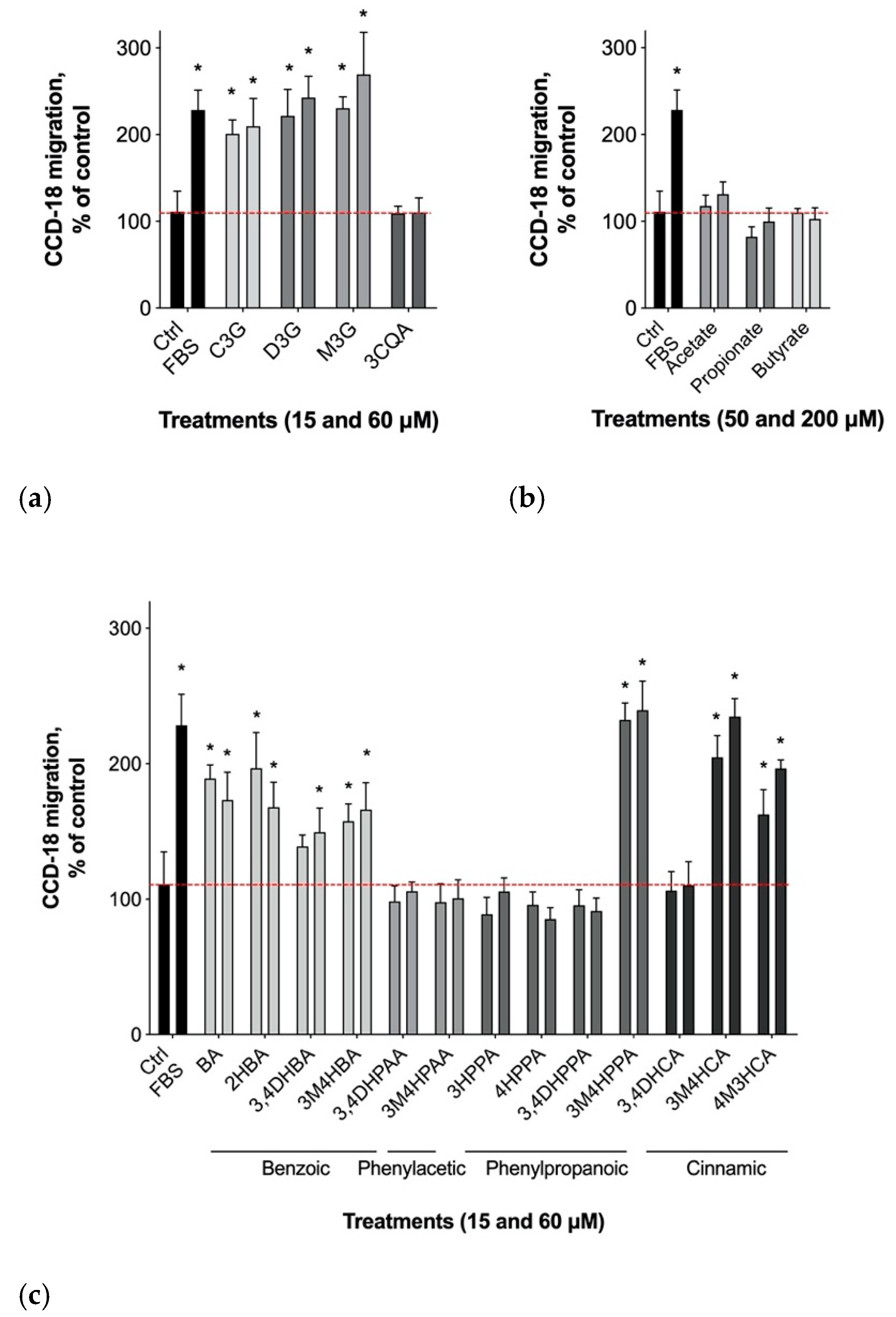
3.2. Gastrointestinal Epithelial Cell Migration

Active gastrointestinal cell migration is critical for steady-state epithelial turnover and healing to adequately maintain a selective permeability barrier separating luminal content from the underlying gastrointestinal tissues [39]. The effects of polyphenols, namely anthocyanins (cyanidin-, delphinidin-, and malvidin-3-glucosides), their major phenolic metabolites (Figure 1), and short-chain fatty acids derived from fiber (acetate, propionate, and butyrate) that naturally present in wild blueberry digests, were screened for their ability to promote CCD-18 gut epithelial cell migration. Cell migration was enhanced relative to the vehicle control in response to all three anthocyanin glucosides tested, with the maximum effect observed for malvidin-3-glucoside (169.5 ± 48.4%, p < 0.05) and no effects for either chlorogenic acid (Figure 2a) or short-chain fatty acid metabolites (Figure 2b).
Among the hydroxybenzoic acid metabolites, CCD-18 migration was enhanced by the application of all four hydroxybenzoic acids used in this study, including 3,4-dihydroxybenzoic (protocatechuic acid; 3,4DHBA), 4-hydroxy-3-methoxybenozic (vanillic acid; 4H3MBA), 2-hydroxybenzoic (salicylic acid; 2HBA), and benzoic acid (BA). The maximum effect was observed for 2-hydroxybenzoic and benzoic acids at 196.7 ± 61.1% and 189.1 ± 31.2% (p < 0.05), respectively. This effect was absent for all phenylacetic and phenylpropionic acids tested, with the exception of 4-hydroxy-3-methoxyphenylpropionic acid (dihydroferulic acid 4H3MCA; 239.5 ± 71.4%, p < 0.05). Two methylated cinnamic metabolites, 4-hydroxy-3-methoxycinnamic (ferulic acid; 4H3MCA) and 3-hydroxy-4-methoxycinnamic (isoferulic acid; 3H4MCA) were also equally effective at the maximum concentrations tested to enhance epithelial cell migration at 234.7 ± 51.8% and 196.5 ± 23.1%, respectively (Figure 2c).

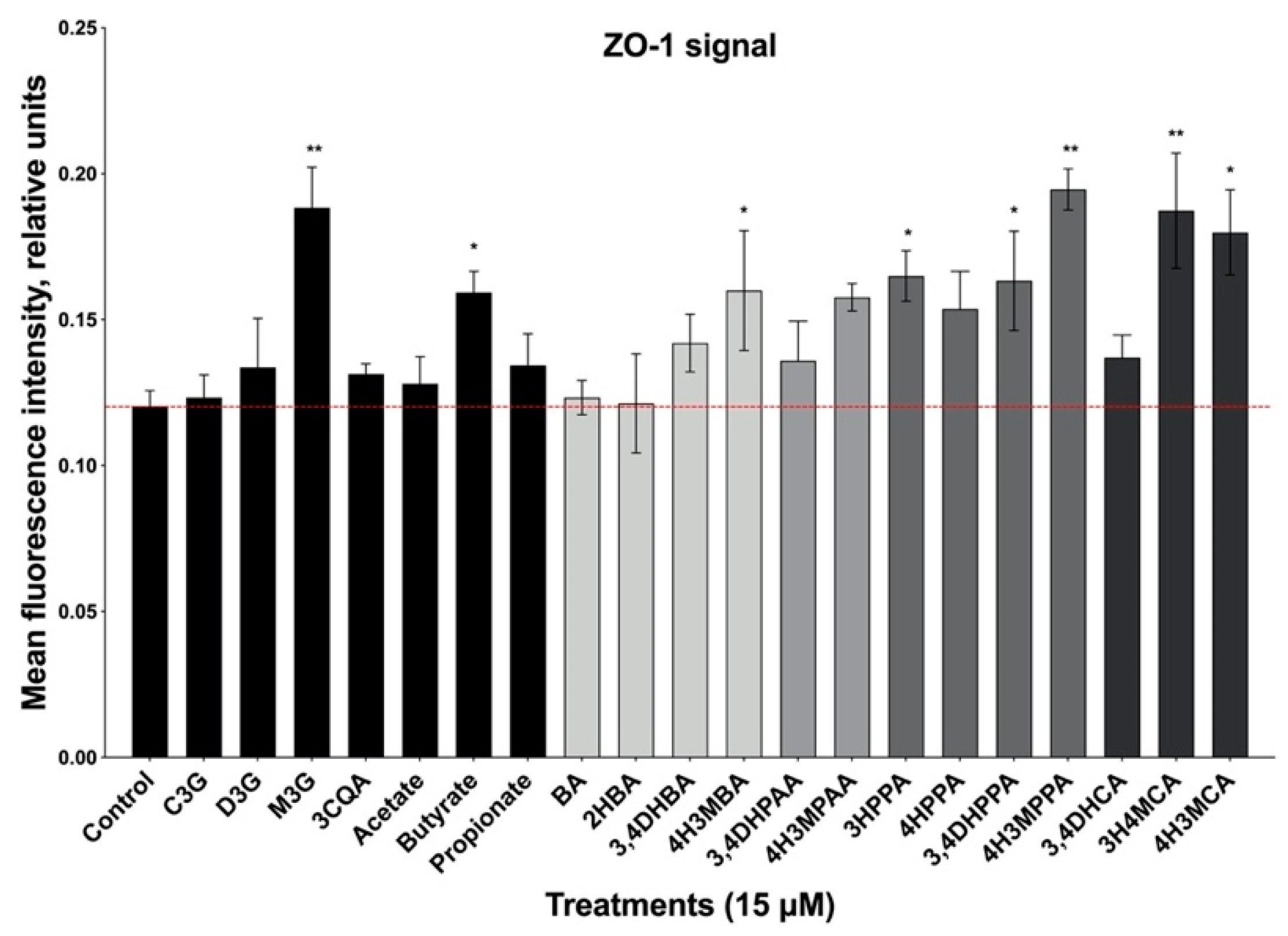
3.3. Formation of Epithelial Cell Monolayers

After establishing cell contacts, gastrointestinal epithelial CCD-18 cells developed junction complexes and established a monolayer resembling that of mature intestinal enterocytes [39]. The junction complexes are primarily composed of the membrane-anchored occludin and cytoplasmic zonula occludens-1 (ZO-1) proteins that regulate paracellular permeability [40]. Enhanced staining for ZO-1 protein was observed in CCD-18 cells treated with malvidin and butyrate (Figure 3). Additionally, among the wild blueberry anthocyanin metabolites tested at the lower dose effective in promoting gastrointestinal cell migration, only methylated derivatives of hydroxybenzoic (vanillic acid 4H3MBA) and 3 hydroxycinnamic acid (3HCA) enhanced accumulation of the ZO-1 protein. The maximum effect was similar for ferulic, isoferulic, and caffeic acids in the range of 50–58% increase in the ZO-1 mean fluorescence signal (p < 0.05). Phenylacetic metabolites, such as 3,4-dihydroxyphenylacetic and 3-methoxy-4-hydroxyphenylacetic (homovanillic) acids had no biological activity in this assay (Figure 3).

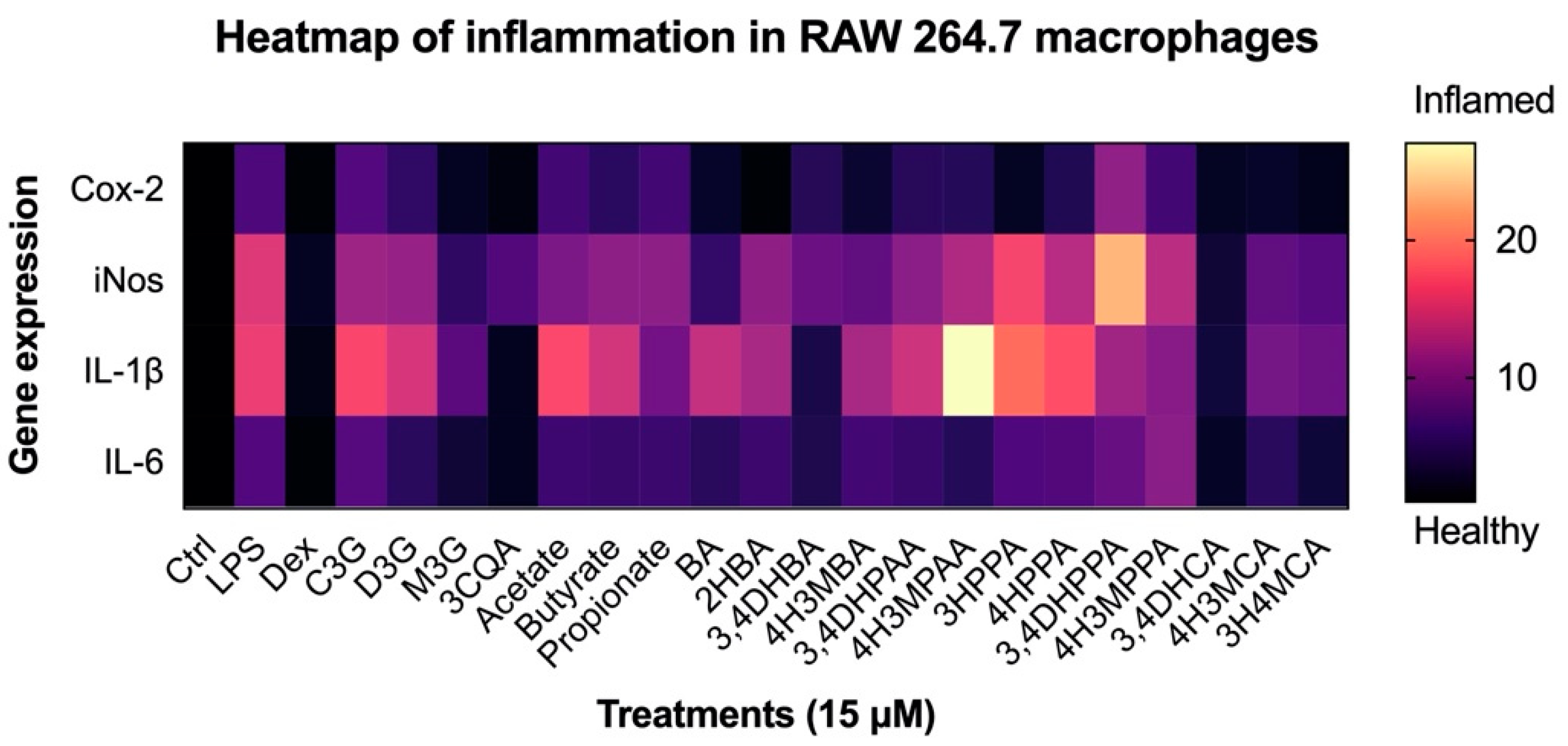
3.4. Reduction in Inflammatory Response in Macrophages

Next, we investigated the ability of wild blueberry metabolites to modulate nitric acid production and gene expression profiles characteristic of acute and chronic inflammation in the LPS-stimulated RAW 264.7 macrophages. Nitric oxide production was not significantly affected by the parent anthocyanin glucosides when tested at a lower concentration (Figure 4). On the contrary, treatments with both 3-caffeoylquinic acid (chlorogenic acid) and its major metabolite 3,4-dihydroxycinnamic acid (caffeic acid) led to strong reductions of nitric oxide release in the culture medium by 49.2% and 34.7%, respectively.
The remaining methylated hydroxycinnamic acid metabolites used in this study were also effective at reducing nitric oxide production to a similar degree of potency in the range of 22–31%. Finally, there was a trend to decrease nitric oxide release when macrophages were exposed to hydroxybenzoic acid metabolites, with benzoic acid being the most potent (a reduction of 54.6%, p < 0.05). Short-chain fatty acid, phenylacetic, and phenylpropanoic acid metabolites were largely ineffective in this assay.
All groups of wild blueberry metabolites showed varying levels of modulation of gene expression profiles associated with acute and chronic biomarkers of inflammation (Cox-2, iNOS, IL-1β, and IL-6). Cox-2 expression was most strongly affected by 3-caffeoylquinic acid −1.72x (−72%) and 2-hydroxybenzoic acid −1.76x (−76%), followed by metabolites from the hydroxybenzoic and hydroxycinnamic acids in the range of 22–56% and 38–61%, respectively, while short-chain fatty acids, phenylacetic, as well as phenylpropionic acids used in this study, showed little to no effects. A wider array of metabolites affected the expression of iNOS, albeit the overall magnitude of the effect was smaller than that of Cox-2. Many of the metabolites of both fiber and phenolic origin affected the expression levels of the early inflammatory IL-1β gene with no clear specificity towards any subclass used in this study. The strongest inhibition of IL-1β expression was achieved by treatment with 3-caffeoylquinic acid and its major metabolite 3,4-dihydroxycinnamic acid (−1.73x and −1.81x, respectively), as well as 2-hydroxybenzoic acid (−1.65x). The mRNA levels of IL-6 were least affected by bioactive wild blueberry metabolites, as significant IL-6 mRNA decreases were observed only when cells were exposed to parent anthocyanin molecules (22–59%) and their hydroxycinnamic acid metabolites were in the range of 24–55%, as observed for the 3,4-dihydroxycinnamic, 4-hydroxy-3-methoxycinnamic, and 3-hydroxy-4-methoxycinnamic acids (Figure 5).

4. Discussion

Although early humans relied on mixed diets of leafy greens, storage tubers, fruits (seeds, nuts), and opportunistic meats, diets based on modern crops show significant decreases in many health-promoting metabolites as we selected for new crop varieties with improved taste profiles and shelf life [40]. Wild blueberries are ideally positioned to start to close this gap, as they generally contain higher amounts of fibers and anthocyanins than their cultivated counterparts. Blueberries are considered a good source of fiber and health-promoting polyphenols, specifically anthocyanins [41]. Low bioavailability of these high molecular weight polyphenols and dietary fibers, at least partially resistant to digestion, has been a matter of research and debate for several decades. We now know that chemical degradation and bacterial catabolism of these molecules into the smaller and more bioavailable catabolites, such as phenolic acids [9] and short-chain fatty acids [42] form a critical connection between xenobiotic metabolism, bioactivation, and host-gut biochemical interactions with these structures. Microbiome-derived fermentation products from foods naturally rich in polyphenols and fibers may also play an important role in the gastrointestinal mucoprotection, feeding of the intestinal epithelial cells, maintaining the intestinal barrier, and regulating the immune outcomes in the gut [43].
This study described the differential effects of short-chain fatty acids and phenolic metabolites on migration and formation of tight junctions by the gastrointestinal epithelial cells. A group of hydroxybenzoic acid metabolites significantly enhanced CCD-18 cell migration. This outcome was the net of cell proliferation and migration into the target area. Benzoic acids were common in the microbial degradation metabolite pools originating from dietary aromatic compounds naturally present in berries [44] and fermented dairy products [45]. In recent studies, benzoic acid supplementation increased the digestibility of total nitrogen, therefore improving the utilization of nutrients [46] and improving the mucosal-epithelial integrity in pigs [47]. A generation of benzoic acid metabolites via the intestinal microbial metabolism of dietary aromatic compounds modified urinary hippuric acid profiles and correlated with positive health outcomes in patients with gastrointestinal disorders, such as Crohn’s disease [48]. Reduced synthesis of benzoic acid metabolites by the intestinal microbiota may therefore be implicated in the gut epithelial pathogenesis, loss of barrier function, and warrants further investigation. Benzoic acid metabolites also showed a partial tendency towards downregulating the COX-2/PGE2 signaling pathway at the level of gene expression (Figure 5).
Several hydroxycinnamic acids, including 3,4-dihydroxycinnamic (caffeic acid), 4-hydroxy-3-methoxycinnamic (ferulic acid), and 3-hydroxy-4-methoxycinnamic (isoferulic acid) also improved gut epithelial cell migration. This data was in agreement with the earlier reported effects of ferulic acid on the LPS-induced epithelial barrier dysfunction [49]. At the same time, short-chain fatty acids derived from dietary fibers had little effect on the migration of the gastrointestinal cells, suggesting that this process does not directly depend on additional colonic energy supplies and trophic factors provided by these metabolites [50]. However, one of the short-chain fatty acids, butyrate, showed a significant effect on the formation of junctional complexes in CCD-18 cell cultures. This effect can be partially explained by the ability of butyrate, and to a lesser extent, propionate, to modulate gastrointestinal gene transcription via histone acetylation and induce permeability changes in tight junctions [51]. Among phenolic metabolites tested, an increase in hydrophobicity because of methylation was generally associated with stronger changes in junctional complexes, as measured by the accumulation of ZO-1 protein. It is possible that more hydrophobic phenolic metabolites had an increased capacity to improve the gastrointestinal barrier function and therefore impede the effects of luminal bacteria on the circulation and mesenteric lymph content [52]. On the other hand, we would expect more polar hydroxybenzoic acid metabolites to have stronger effects on the gastrointestinal tissues that face luminal content as previously reported in the turkey [53] and pig [54] models.
The integrity of intestinal mucosa is not only the major critical factor for nutrient digestion and absorption, but also plays a key role in the recognition and activation of the immune system in response to physiological stimuli and pathogen infection. Lipopolysaccharide induces an increase in intestinal permeability and subsequently activates residential tissue macrophages that promote differentiation and development of the pro-inflammatory response [55]. These effects are mediated by changes in the expression levels of cytokine biomarkers that drive activation of the classical pro-inflammatory M1 pathway, including the inducible nitric oxide synthase (iNOS), interleukin-1β (IL-1β), and interleukin-6 (IL-6) [56]. Cyclooxygenase-2 (Cox-2)/PGE2 production in activated macrophages further drives distinct intracellular pathways that modulate inflammation and host immune responses [57].
In this study, caffeoylquinic, hydroxybenzoic, and hydroxycinnamic acids reduced nitric oxide release and associated gene expression levels of iNOS in macrophages, and this effect was largely absent when cells were exposed to hydroxyphenylacetic and hydroxyphenylpropionic acids (Figure 4 and Figure 5). Among those, 3,4-dihydroxycinnamic (caffeic acid) and chlorogenic acid (that carries the caffeic acid moiety as a part of its structure) showed the strongest inhibition of pro-inflammatory gene expression in macrophages at the concentration tested. This was an interesting parallel to a previous study that showed the importance of the caffeic acid pharmacophore to metabolic health [31]. Methylated derivatives of caffeic acid showed stronger effects on COX-2 and IL-6 expression as compared to iNOS and IL-1β genes, suggesting different efficacies at various pathways of the inflammatory regulatory networks.
While all subclasses of phenolic metabolites are typically observed in human plasma or serum metabolome following the consumption of plant foods, only some metabolites achieve maximum observed concentrations of 10–25 µM, as reported for 3-phenylpropanoic, 2-hydroxybenzoic, and hippuric acids, while the individual concentrations of the remaining phenolic metabolites typically range between 10 nM and 1 µM [58]. The mean cumulative concentrations of these series of phenolic metabolites, however, often reach 30–40 µM in the systemic circulation and may be responsible for additive biological effects as previously shown for hydroxybenzoic acid metabolites in vascular smooth muscle cells [59]. Ranges of concentrations for major phenolic acid metabolites identified in plasma, urine, and fecal water support the notion that these can be achieved by dietary supplementation with various foods, including blueberries [41]. It, therefore, seems plausible that body tissues located in immediate proximity to the gastrointestinal luminal content (gut epithelial and smooth muscle cells) and blood vessels (endothelial and smooth muscle cells) are exposed to higher levels of polar microbial catabolites derived from dietary fiber and polyphenols, while methylated phenolic metabolites are better positioned to achieve biologically relevant outcomes in the more distant metabolically active tissues (adipose, liver, and tissue residential immune cells). The yet unexplored structure-activity relationships between different subclasses of phenolic metabolites with respect to their methylation and hydrophobicity, which facilitates passive membrane permeation and interaction with molecular and intercellular targets, may partially explain the various findings reported in adipocyte [32], liver [31,60], smooth muscle [59], and endothelial [61] cell culture models.

5. Conclusions

The observed activity of wild blueberry metabolites on key biomarker measures of gastrointestinal and immune cells was specific to the particular series and structures derived from digestion and fermentation of dietary fiber and polyphenolic compounds, and varied depending on the tissue analyzed. Further work is required to elucidate the underlying mechanisms and targets involved in the recognition of these signals. This work also raised an intriguing possibility that hydroxybenzoic acid metabolites play an important role in the modulation of the physiological outcomes in the gastrointestinal epithelial cells, similar to the short-chain fatty acids.
Overall, findings also pointed to the hydroxybenzoic and hydroxycinnamic phenolic acid metabolites as being more potent in regulating gene expression profiles that are central to sustaining acute and chronic inflammation in the gastrointestinal and immune cells. Thus, our results suggested that diets that incorporate wild blueberries hold a high potential to supplement humans with small molecular weight metabolites that may improve the management and outcomes of immune and metabolic risk factors linked to sustained inflammation.

Author Contributions

C.D.K. and S.K. conceived the study and outlined the scope of work; J.L. and D.E. established cell line assays; T.R. managed metabolite stocks and treatments; K.S. performed RNA extractions and qPCR analysis; S.K. performed assay quantifications and overall data analysis; T.R. and S.K. wrote the manuscript; C.D.K. and D.E. reviewed the manuscript. All authors have read and agreed to the published version of the manuscript.

Funding

This work was supported in part by the Wild Blueberry Association of North America (C.D.K., S.K.) and the USDA National Institute of Food and Agriculture Hatch projects #1023927 (S.K.), #1011757 (C.D.K.), and #1016019 (D.E.).

Institutional Review Board Statement

Not applicable.

Informed Consent Statement

Not applicable.

Data Availability Statement

Data may be obtained from a corresponding author upon a reasonable request.

Acknowledgments

We wish to express sincere gratitude and appreciation to UNC’s Chapel Hill interns, Ashley Buige and Mikayla Bowen, for establishing the zonulin accumulation assay in the lab.

Conflicts of Interest

The authors declare no conflict of interest.

References

  1. Nile, S.H.; Park, S.W. Edible Berries: Bioactive Components and Their Effect on Human Health. Nutrition 2014, 30, 134–144. [Google Scholar] [CrossRef] [PubMed]
  2. Yousef, G.G.; Brown, A.F.; Funakoshi, Y.; Mbeunkui, F.; Grace, M.H.; Ballington, J.R.; Loraine, A.; Lila, M.A. Efficient Quantification of the Health-Relevant Anthocyanin and Phenolic Acid Profiles in Commercial Cultivars and Breeding Selections of Blueberries (Vaccinium Spp.). J. Agric. Food Chem. 2013, 61, 4806–4815. [Google Scholar] [CrossRef] [PubMed]
  3. Rodriguez-Mateos, A.; Cifuentes-Gomez, T.; Tabatabaee, S.; Lecras, C.; Spencer, J.P.E. Procyanidin, Anthocyanin, and Chlorogenic Acid Contents of Highbush and Lowbush Blueberries. J. Agric. Food Chem. 2012, 60, 5772–5778. [Google Scholar] [CrossRef] [PubMed]
  4. Fang, J. Classification of Fruits Based on Anthocyanin Types and Relevance to Their Health Effects. Nutrition 2015, 31, 1301–1306. [Google Scholar] [CrossRef]
  5. Wang, Y.; Fong, S.K.; Singh, A.P.; Vorsa, N.; Johnson-Cicalese, J. Variation of Anthocyanins, Proanthocyanidins, Flavonols, and Organic Acids in Cultivated and Wild Diploid Blueberry Species. HortScience 2019, 54, 576–585. [Google Scholar] [CrossRef] [Green Version]
  6. Zhao, C.-L.; Yu, Y.-Q.; Chen, Z.-J.; Wen, G.-S.; Wei, F.-G.; Zheng, Q.; Wang, C.-D.; Xiao, X.-L. Stability-Increasing Effects of Anthocyanin Glycosyl Acylation. Food Chem. 2017, 214, 119–128. [Google Scholar] [CrossRef]
  7. Correa-Betanzo, J.; Allen-Vercoe, E.; McDonald, J.; Schroeter, K.; Corredig, M.; Paliyath, G. Stability and Biological Activity of Wild Blueberry (Vaccinium angustifolium) Polyphenols during Simulated in Vitro Gastrointestinal Digestion. Food Chem. 2014, 165, 522–531. [Google Scholar] [CrossRef]
  8. Kurilich, A.C.; Clevidence, B.A.; Britz, S.J.; Simon, P.W.; Novotny, J.A. Plasma and Urine Responses Are Lower for Acylated vs Nonacylated Anthocyanins from Raw and Cooked Purple Carrots. J. Agric. Food Chem. 2005, 53, 6537–6542. [Google Scholar] [CrossRef]
  9. Kay, C.D.; Pereira-Caro, G.; Ludwig, I.A.; Clifford, M.N.; Crozier, A. Anthocyanins and Flavanones Are More Bioavailable than Previously Perceived: A Review of Recent Evidence. Annu. Rev. Food Sci. Technol. 2017, 8, 155–180. [Google Scholar] [CrossRef]
  10. Esposito, D.; Damsud, T.; Wilson, M.; Grace, M.H.; Strauch, R.; Li, X.; Lila, M.A.; Komarnytsky, S. Black Currant Anthocyanins Attenuate Weight Gain and Improve Glucose Metabolism in Diet-Induced Obese Mice with Intact, but Not Disrupted, Gut Microbiome. J. Agric. Food Chem. 2015, 63, 6172–6180. [Google Scholar] [CrossRef]
  11. Basu, A.; Du, M.; Leyva, M.J.; Sanchez, K.; Betts, N.M.; Wu, M.; Aston, C.E.; Lyons, T.J. Blueberries Decrease Cardiovascular Risk Factors in Obese Men and Women with Metabolic Syndrome. J. Nutr. 2010, 140, 1582–1587. [Google Scholar] [CrossRef] [PubMed] [Green Version]
  12. De Ferrars, R.M.; Czank, C.; Zhang, Q.; Botting, N.P.; Kroon, P.A.; Cassidy, A.; Kay, C.D. The Pharmacokinetics of Anthocyanins and Their Metabolites in Humans. Br. J. Pharmacol. 2014, 171, 3268–3282. [Google Scholar] [CrossRef] [PubMed] [Green Version]
  13. Rodriguez-Mateos, A.; Feliciano, R.P.; Cifuentes-Gomez, T.; Spencer, J.P.E. Bioavailability of Wild Blueberry (Poly)Phenols at Different Levels of Intake. J. Berry Res. 2016, 6, 137–148. [Google Scholar] [CrossRef] [Green Version]
  14. Stull, A.J.; Cash, K.C.; Johnson, W.D.; Champagne, C.M.; Cefalu, W.T. Bioactives in Blueberries Improve Insulin Sensitivity in Obese, Insulin-Resistant Men and Women. J. Nutr. 2010, 140, 1764–1768. [Google Scholar] [CrossRef]
  15. Rodriguez-Mateos, A.; Rendeiro, C.; Bergillos-Meca, T.; Tabatabaee, S.; George, T.; Heiss, C.; Spencer, J. Intake and Time Dependence of Blueberry Flavonoid-Induced Improvements in Vascular Function: A Randomized, Controlled, Double-Blind, Crossover Intervention Study with Mechanistic Insights into Biological Activity. Am. J. Clin. Nutr. 2013, 98, 1179–1191. [Google Scholar] [CrossRef] [Green Version]
  16. Johnson, S.A.; Feresin, R.G.; Navaei, N.; Figueroa, A.; Elam, M.L.; Akhavan, N.S.; Hooshmand, S.; Pourafshar, S.; Payton, M.E.; Arjmandi, B.H. Effects of Daily Blueberry Consumption on Circulating Biomarkers of Oxidative Stress, Inflammation, and Antioxidant Defense in Postmenopausal Women with Pre- and Stage 1-Hypertension: A Randomized Controlled Trial. Food Funct. 2017, 8, 372–380. [Google Scholar] [CrossRef]
  17. Curtis, P.J.; van der Velpen, V.; Berends, L.; Jennings, A.; Feelisch, M.; Umpleby, A.M.; Evans, M.; Fernandez, B.O.; Meiss, M.S.; Minnion, M.; et al. Blueberries Improve Biomarkers of Cardiometabolic Function in Participants with Metabolic Syndrome-Results from a 6-Month, Double-Blind, Randomized Controlled Trial. Am. J. Clin. Nutr. 2019, 109, 1535–1545. [Google Scholar] [CrossRef] [Green Version]
  18. Bowtell, J.L.; Aboo-Bakkar, Z.; Conway, M.E.; Adlam, A.-L.R.; Fulford, J. Enhanced Task-Related Brain Activation and Resting Perfusion in Healthy Older Adults after Chronic Blueberry Supplementation. Appl. Physiol. Nutr. Metab. 2017, 42, 773–779. [Google Scholar] [CrossRef]
  19. Vancamelbeke, M.; Vermeire, S. The Intestinal Barrier: A Fundamental Role in Health and Disease. Expert Rev. Gastroenterol. Hepatol. 2017, 11, 821–834. [Google Scholar] [CrossRef]
  20. Turner, J.R. Intestinal Mucosal Barrier Function in Health and Disease. Nat. Rev. Immunol. 2009, 9, 799–809. [Google Scholar] [CrossRef]
  21. Tripathi, A.; Lammers, K.M.; Goldblum, S.; Shea-Donohue, T.; Netzel-Arnett, S.; Buzza, M.S.; Antalis, T.M.; Vogel, S.N.; Zhao, A.; Yang, S.; et al. Identification of Human Zonulin, a Physiological Modulator of Tight Junctions, as Prehaptoglobin-2. Proc. Natl. Acad. Sci. USA 2009, 106, 16799–16804. [Google Scholar] [CrossRef] [PubMed] [Green Version]
  22. Zak-Gołąb, A.; Kocełak, P.; Aptekorz, M.; Zientara, M.; Juszczyk, L.; Martirosian, G.; Chudek, J.; Olszanecka-Glinianowicz, M. Gut Microbiota, Microinflammation, Metabolic Profile, and Zonulin Concentration in Obese and Normal Weight Subjects. Int. J. Endocrinol. 2013, 2013, 674106. [Google Scholar] [CrossRef] [PubMed] [Green Version]
  23. Oliviero, S.; Cortese, R. The Human Haptoglobin Gene Promoter: Interleukin-6-Responsive Elements Interact with a DNA-Binding Protein Induced by Interleukin-6. EMBO J. 1989, 8, 1145–1151. [Google Scholar] [CrossRef]
  24. Moreno-Navarrete, J.M.; Sabater, M.; Ortega, F.; Ricart, W.; Fernández-Real, J.M. Circulating Zonulin, a Marker of Intestinal Permeability, is Increased in Association with Obesity-Associated Insulin Resistance. PLoS ONE 2012, 7, e37160. [Google Scholar] [CrossRef] [PubMed] [Green Version]
  25. Massier, L.; Chakaroun, R.; Kovacs, P.; Heiker, J.T. Blurring the Picture in Leaky Gut Research: How Shortcomings of Zonulin as a Biomarker Mislead the Field of Intestinal Permeability. Gut 2021, 70, 1801–1802. [Google Scholar] [CrossRef]
  26. Vendrame, S.; Guglielmetti, S.; Riso, P.; Arioli, S.; Klimis-Zacas, D.; Porrini, M. Six-Week Consumption of a Wild Blueberry Powder Drink Increases Bifidobacteria in the Human Gut. J. Agric. Food Chem. 2011, 59, 12815–12820. [Google Scholar] [CrossRef]
  27. Guglielmetti, S.; Fracassetti, D.; Taverniti, V.; Del Bo’, C.; Vendrame, S.; Klimis-Zacas, D.; Arioli, S.; Riso, P.; Porrini, M. Differential Modulation of Human Intestinal Bifidobacterium Populations after Consumption of a Wild Blueberry (Vaccinium angustifolium) Drink. J. Agric. Food Chem. 2013, 61, 8134–8140. [Google Scholar] [CrossRef]
  28. Hester, S.N.; Mastaloudis, A.; Gray, R.; Antony, J.M.; Evans, M.; Wood, S.M. Efficacy of an Anthocyanin and Prebiotic Blend on Intestinal Environment in Obese Male and Female Subjects. J. Nutr. Metab. 2018, 2018, 7497260. [Google Scholar] [CrossRef]
  29. Allaire, J.M.; Crowley, S.M.; Law, H.T.; Chang, S.-Y.; Ko, H.-J.; Vallance, B.A. The Intestinal Epithelium: Central Coordinator of Mucosal Immunity. Trends Immunol. 2018, 39, 677–696. [Google Scholar] [CrossRef]
  30. Esposito, D.; Chen, A.; Grace, M.H.; Komarnytsky, S.; Lila, M.A. Inhibitory Effects of Wild Blueberry Anthocyanins and Other Flavonoids on Biomarkers of Acute and Chronic Inflammation in Vitro. J. Agric. Food Chem. 2014, 62, 7022–7028. [Google Scholar] [CrossRef]
  31. Jackson, K.M.P.; Rathinasabapathy, T.; Esposito, D.; Komarnytsky, S. Structural Constraints and Importance of Caffeic Acid Moiety for Anti-Hyperglycemic Effects of Caffeoylquinic Acids from Chicory. Mol. Nutr. Food Res. 2017, 61, 1601118. [Google Scholar] [CrossRef] [PubMed]
  32. Skates, E.; Overall, J.; DeZego, K.; Wilson, M.; Esposito, D.; Lila, M.A.; Komarnytsky, S. Berries Containing Anthocyanins with Enhanced Methylation Profiles Are More Effective at Ameliorating High Fat Diet-Induced Metabolic Damage. Food Chem. Toxicol. 2018, 111, 445–453. [Google Scholar] [CrossRef] [PubMed]
  33. Swaid, F.; Sukhotnik, I.; Matter, I.; Berkowitz, D.; Hadjittofi, C.; Pollak, Y.; Lavy, A. Dietary Glutamine Supplementation Prevents Mucosal Injury and Modulates Intestinal Epithelial Restitution Following Acetic Acid Induced Intestinal Injury in Rats. Nutr. Metab. 2013, 10, 53. [Google Scholar] [CrossRef] [PubMed] [Green Version]
  34. Andou, A.; Hisamatsu, T.; Okamoto, S.; Chinen, H.; Kamada, N.; Kobayashi, T.; Hashimoto, M.; Okutsu, T.; Shimbo, K.; Takeda, T.; et al. Dietary Histidine Ameliorates Murine Colitis by Inhibition of Proinflammatory Cytokine Production from Macrophages. Gastroenterology 2009, 136, 564–574.e2. [Google Scholar] [CrossRef] [PubMed]
  35. Russell, W.; Duthie, G. Plant Secondary Metabolites and Gut Health: The Case for Phenolic Acids. Proc. Nutr. Soc. 2011, 70, 389–396. [Google Scholar] [CrossRef] [Green Version]
  36. Louis, P.; Flint, H.J. Formation of Propionate and Butyrate by the Human Colonic Microbiota. Environ. Microbiol. 2017, 19, 29–41. [Google Scholar] [CrossRef] [Green Version]
  37. Venegas, D.P.; De la Fuente, M.K.; Landskron, G.; González, M.J.; Quera, R.; Dijkstra, G.; Harmsen, H.J.M.; Faber, K.N.; Hermoso, M.A. Short Chain Fatty Acids (SCFAs)-Mediated Gut Epithelial and Immune Regulation and Its Relevance for Inflammatory Bowel Diseases. Front. Immunol. 2019, 10, 277. [Google Scholar] [CrossRef] [Green Version]
  38. Kahle, K.; Kraus, M.; Scheppach, W.; Ackermann, M.; Ridder, F.; Richling, E. Studies on Apple and Blueberry Fruit Constituents: Do the Polyphenols Reach the Colon after Ingestion? Mol. Nutr. Food Res. 2006, 50, 418–423. [Google Scholar] [CrossRef]
  39. Krndija, D.; Marjou, F.E.; Guirao, B.; Richon, S.; Leroy, O.; Bellaiche, Y.; Hannezo, E.; Vignjevic, D.M. Active Cell Migration is Critical for Steady-State Epithelial Turnover in the Gut. Science 2019, 365, 705–710. [Google Scholar] [CrossRef]
  40. Komarnytsky, S.; Retchin, S.; Vong, C.I.; Lila, M.A. Gains and Losses of Agricultural Food Production: Implications for the Twenty-First Century. Annu. Rev. Food Sci. Technol. 2021, 13. [Google Scholar] [CrossRef]
  41. Vong, C.I.; Rathinasabapathy, T.; Moncada, M.; Komarnytsky, S. All Polyphenols Are Not Created Equal: Exploring the Diversity of Phenolic Metabolites. J. Agric. Food Chem. 2022, 70, 2077–2091. [Google Scholar] [CrossRef] [PubMed]
  42. Canfora, E.E.; Jocken, J.W.; Blaak, E.E. Short-Chain Fatty Acids in Control of Body Weight and Insulin Sensitivity. Nat. Rev. Endocrinol. 2015, 11, 577–591. [Google Scholar] [CrossRef] [PubMed]
  43. Cummings, J.H.; Macfarlane, G.T. The Control and Consequences of Bacterial Fermentation in the Human Colon. J. Appl. Bacteriol. 1991, 70, 443–459. [Google Scholar] [CrossRef] [PubMed]
  44. Walsh, M.C.; Brennan, L.; Pujos-Guillot, E.; Sébédio, J.-L.; Scalbert, A.; Fagan, A.; Higgins, D.G.; Gibney, M.J. Influence of Acute Phytochemical Intake on Human Urinary Metabolomic Profiles. Am. J. Clin. Nutr. 2007, 86, 1687–1693. [Google Scholar] [CrossRef] [PubMed]
  45. Sieber, R.; Bütikofer, U.; Bosset, J.O. Benzoic Acid as a Natural Compound in Cultured Dairy Products and Cheese. Int. Dairy J. 1995, 5, 227–246. [Google Scholar] [CrossRef]
  46. Halas, D.; Hansen, C.F.; Hampson, D.J.; Mullan, B.P.; Kim, J.C.; Wilson, R.H.; Pluske, J.R. Dietary Supplementation with Benzoic Acid Improves Apparent Ileal Digestibility of Total Nitrogen and Increases Villous Height and Caecal Microbial Diversity in Weaner Pigs. Anim. Feed Sci. Technol. 2010, 160, 137–147. [Google Scholar] [CrossRef]
  47. Halas, D.; Hansen, C.F.; Hampson, D.J.; Mullan, B.P.; Wilson, R.H.; Pluske, J.R. Effect of Dietary Supplementation with Inulin and/or Benzoic Acid on the Incidence and Severity of Post-Weaning Diarrhoea in Weaner Pigs after Experimental Challenge with Enterotoxigenic Escherichia Coli. Arch. Anim. Nutr. 2009, 63, 267–280. [Google Scholar] [CrossRef]
  48. Williams, H.R.; Cox, I.J.; Walker, D.G.; Cobbold, J.F.; Taylor-Robinson, S.D.; Marshall, S.E.; Orchard, T.R. Differences in Gut Microbial Metabolism Are Responsible for Reduced Hippurate Synthesis in Crohn’s Disease. BMC Gastroenterol. 2010, 10, 108. [Google Scholar] [CrossRef] [Green Version]
  49. He, S.; Guo, Y.; Zhao, J.; Xu, X.; Wang, N.; Liu, Q. Ferulic Acid Ameliorates Lipopolysaccharide-Induced Barrier Dysfunction via MicroRNA-200c-3p-Mediated Activation of PI3K/AKT Pathway in Caco-2 Cells. Front. Pharmacol. 2020, 11, 376. [Google Scholar] [CrossRef]
  50. Pascale, A.; Marchesi, N.; Marelli, C.; Coppola, A.; Luzi, L.; Govoni, S.; Giustina, A.; Gazzaruso, C. Microbiota and Metabolic Diseases. Endocrine 2018, 61, 357–371. [Google Scholar] [CrossRef]
  51. Ohata, A.; Usami, M.; Miyoshi, M. Short-Chain Fatty Acids Alter Tight Junction Permeability in Intestinal Monolayer Cells via Lipoxygenase Activation. Nutrition 2005, 21, 838–847. [Google Scholar] [CrossRef] [PubMed] [Green Version]
  52. Zhu, R.; Ma, X. Role of Metabolic Changes of Mucosal Layer in the Intestinal Barrier Dysfunction Following Trauma/Hemorrhagic Shock. Pathol. Res. Pract. 2018, 214, 1879–1884. [Google Scholar] [CrossRef] [PubMed]
  53. Giannenas, I.; Papaneophytou, C.P.; Tsalie, E.; Pappas, I.; Triantafillou, E.; Tontis, D.; Kontopidis, G.A. Dietary Supplementation of Benzoic Acid and Essential Oil Compounds Affects Buffering Capacity of the Feeds, Performance of Turkey Poults and Their Antioxidant Status, PH in the Digestive Tract, Intestinal Microbiota and Morphology. Asian Australas. J. Anim. Sci. 2014, 27, 225–236. [Google Scholar] [CrossRef] [PubMed] [Green Version]
  54. Pu, J.; Chen, D.; Tian, G.; He, J.; Zheng, P.; Mao, X.; Yu, J.; Huang, Z.; Zhu, L.; Luo, J.; et al. Protective Effects of Benzoic Acid, Bacillus Coagulans, and Oregano Oil on Intestinal Injury Caused by Enterotoxigenic Escherichia Coli in Weaned Piglets. BioMed Res. Int. 2018, 2018, 1829632. [Google Scholar] [CrossRef] [PubMed] [Green Version]
  55. De Sousa, J.R.; Da Costa Vasconcelos, P.F.; Quaresma, J.A.S. Functional Aspects, Phenotypic Heterogeneity, and Tissue Immune Response of Macrophages in Infectious Diseases. Infect. Drug Resist. 2019, 12, 2589–2611. [Google Scholar] [CrossRef] [Green Version]
  56. Novoselov, V.V.; Sazonova, M.A.; Ivanova, E.A.; Orekhov, A.N. Study of the Activated Macrophage Transcriptome. Exp. Mol. Pathol. 2015, 99, 575–580. [Google Scholar] [CrossRef]
  57. Cabral, G.A. Lipids as Bioeffectors in the Immune System. Life Sci. 2005, 77, 1699–1710. [Google Scholar] [CrossRef]
  58. Chandra, P.; Rathore, A.S.; Kay, K.L.; Everhart, J.L.; Curtis, P.; Burton-Freeman, B.; Cassidy, A.; Kay, C.D. Contribution of Berry Polyphenols to the Human Metabolome. Molecules 2019, 24, 4220. [Google Scholar] [CrossRef] [Green Version]
  59. Warner, E.F.; Rodriguez-Ramiro, I.; O’Connell, M.A.; Kay, C.D. Cardiovascular Mechanisms of Action of Anthocyanins May Be Associated with the Impact of Microbial Metabolites on Heme Oxygenase-1 in Vascular Smooth Muscle Cells. Molecules 2018, 23, 898. [Google Scholar] [CrossRef] [Green Version]
  60. Murray, A.F.; Palatini, K.; Komarnytsky, S.; Gianfagna, T.J.; Munafo, J.P. Phenylpropanoid Glycerol Glucosides Attenuate Glucose Production in Hepatocytes. ACS Omega 2019, 4, 10670–10676. [Google Scholar] [CrossRef]
  61. Paixão, J.; Dinis, T.C.P.; Almeida, L.M. Malvidin-3-Glucoside Protects Endothelial Cells up-Regulating Endothelial NO Synthase and Inhibiting Peroxynitrite-Induced NF-KB Activation. Chem. Biol. Interact. 2012, 199, 192–200. [Google Scholar] [CrossRef] [PubMed]
Figure 1. Schematic representation of a parental anthocyanin molecule and the major phenolic metabolites that are produced during the subsequent degradation and microbial fermentation of the parental anthocyanins. The nomenclature of phenolic acid metabolites is also listed in Table 1.
Immuno 02 00019 g001
Figure 2. Effects of wild blueberry (a) anthocyanin glucosides, (b) short-chain fatty acid metabolites from dietary fiber, and (c) different subclasses of phenolic acid metabolites on CCD-18 gastrointestinal epithelial cell migration. Cells were seeded in Oris migration plates and their migration into exclusion zones was followed for 48 h with/without the indicated treatments and compared against the vehicle (0.1% DMSO) and positive control (20% FBS). Pre- and post-migration central fluorescence was quantified by NucBlue (Hoechst 33342) staining at 360 nm excitation/460 nm emission and reported as mean ± SEM (* p < 0.05). Anthocyanin and phenolic acids were tested at 15 μM (lower dose) and 60 μM (higher dose); short-chain fatty acids were tested at 50 μM (lower dose) and 200 μM (higher dose), as indicated on each panel.
Immuno 02 00019 g002
Figure 3. Effects of wild blueberry anthocyanins, short-chain fatty acids, and phenolic acid metabolites on the formation of junctional complexes in the CCD-18 gastrointestinal cells monolayers. Cells were allowed to form monolayers for 5 days with/without the indicated treatments and compared against vehicle control (0.1% DMSO). Zonulin accumulation in the junction complexes was visualized using ZO-1/TJP1 antibody Alexa Fluor 488, mean fluorescent intensity at 490 nm excitation/525 nm emission and reported as mean ± SEM (* p < 0.05, ** p < 0.01).
Immuno 02 00019 g003
Figure 4. Effects of wild blueberry anthocyanins, short-chain fatty acids, and phenolic acid metabolites on nitric oxide production in activated macrophages. Cells were pre-treated with target metabolites and inflammatory response was induced with 1 µg/mL LPS for 6 h. Changes in nitrite concentration as an indirect measure of nitric oxide production were reported as mean ± SEM relative to the LPS controls (* p < 0.05, ** p < 0.01, *** p < 0.001, **** p < 0.0001).
Immuno 02 00019 g004
Figure 5. Heatmap of anti-inflammatory effects of wild blueberry anthocyanins, short-chain fatty acids, and phenolic acid metabolites based on qPCR gene expression profiles of key biomarkers of acute and chronic inflammation, including cyclooxygenase-2 (Cox-2), inducible nitric oxide synthase (iNOS), interleukin-1β (IL-1β), and interleukin-6 (IL-6). Macrophages were pre-treated with individual compounds as the specified and inflammatory response was induced with 1 µg/mL LPS for 6 h. Dexamethasone (Dex) at 10 µM was used as a positive control. Total RNAs were isolated from duplicate treatments and pooled for qPCR analysis. Fold changes in gene expression are reported as means relative to healthy controls.
Immuno 02 00019 g005
Table 1. Phenolic acids used in this study.
SeriesCommon NameChemical NameAbbr.
BenzoicBenzoicBenzoic BA
Salicylic 2-Hydroxybenzoic2HBA
Protocatechuic3,4-Dihydroxybenzoic3,4DHBA
Vanillic4-Hydroxy-3-methoxybenzoic 4H3MBA
PhenylaceticDOPAC3,4-Dihydroxyphenylacetic3,4DHPAA
Homovanillic3-Hydroxy-4-methoxyphenylacetic4H3MPAA
Phenylpropionic m-Dihydrocoumaric3-Hydroxyphenylpropionic3HPPA
p-Dihydrocoumaric4-Hydroxyphenylpropionic4HPPA
Dihydrocaffeic3,4-Dihydroxyphenylpropionic3,4DHPPA
Dihydroferulic4-Hydroxy-3-methoxyphenylpropionic4H3MPPA
CinnamicCaffeic3,4-Dihydroxycinnamic3,4DHCA
Isoferulic3-Hydroxy-4-methoxycinnamic3H4MCA
Ferulic4-Hydroxy-3-methoxycinnamic4H3MCA
Publisher’s Note: MDPI stays neutral with regard to jurisdictional claims in published maps and institutional affiliations.

