Review

Busting the Myths of DLco for Pulmonary Trainees: Isolated Reductions in DLco and the Relationship with VA

1
Department of Thoracic Medicine and Surgery, Temple University Hospital, Philadelphia, PA 19140, USA
2
Department of Thoracic Medicine and Surgery, Lewis Katz School of Medicine, Temple University, Philadelphia, PA 19140, USA
*
Author to whom correspondence should be addressed.
J. Respir. 2025, 5(3), 8; https://doi.org/10.3390/jor5030008
Submission received: 6 March 2025 / Revised: 12 June 2025 / Accepted: 18 June 2025 / Published: 24 June 2025

Abstract

Background: DLco remains one of the most commonly performed tests in the pulmonary lab. An isolated reduction in DLco is a unique abnormality with specific differentials when evaluating a patient with dyspnea. There remains a significant misunderstanding amongst young pulmonologists and pulmonary trainees regarding DLco and its relationship with alveolar volume and kco. Objective: This review aims to provide a physiological basis for the DLco test and bust the myth of “DLco corrected for lung volume.” Method: A systematic review of the available literature regarding alveolar gas-exchange physiology, measurement methods of DLco, the interplay of different variables associated with it, and the causes of its reduction was performed. Focused physiological data were used to put together a comprehensive review of isolated reductions in DLco. The second part of this review addresses the critical and interdependent relationship between DLco and alveolar volume (VA). Results: DLco has a unique relationship with lung volume that needs to be considered while interpreting its value. Diffusion capacity per unit volume (kco) is an independent factor that, when combined with DLco and VA, helps accurately interpret the test and narrow down differentials. Conclusion: DLco is an extremely valuable test and an important prognostic tool in many patients with dyspnea. An isolated reduction in DLco is increasingly recognized these days as an early marker of detection for various pulmonary parenchymal and vascular diseases. A detailed physiopathological explanation, followed by the proposed algorithm, should help pulmonary physicians and trainees understand and implement DLco’s relationships in their daily patient care.

1. Introduction

The diffusion capacity of the lung for carbon monoxide (DLco, also known as transfer factor for carbon monoxide, or TLco) is one of the most vital tests routinely performed for patients during the evaluation of dyspnea. It is a measure of the conductance of gas transfer from inspired gas to the red blood cells. The diffusing capacity of the lungs for carbon monoxide (DLco) is designed to reflect the properties of the alveolar–capillary membrane, precisely the ease with which oxygen moves from inhaled air to the red blood cells in the pulmonary capillaries. The DLco can be affected by factors that change the membrane properties and also by changes in hemoglobin and capillary blood volume.

Methods of DLco Measurement

Methodology involving DLco measurement can be divided into two phases: preparation for the test and the actual test.
  • Preparation for the test:
    No cigarette smoking on the day of the test.
    No supplemental oxygen for at least 15 min prior to the test. Supplemental oxygen can decrease DLco by approximately 0.35 percent per mmHg change in arterial oxygen tension (PaO2). The DLco test cannot be accurately performed in patients who are unable to discontinue supplemental oxygen for at least 15 min.
    It can be performed after the bronchodilator test.
  • DLco maneuver and techniques:
    DLco is measured using the following techniques [1].
    • Single-breath method;
    • Intra-breath method;
    • Rebreathing technique.
The single-breath DLco maneuver begins with a full exhalation to residual volume (RV). The mouthpiece is connected to test gas (0.3 percent carbon monoxide [CO], tracer gas [e.g., 10 percent helium or 0.3 percent methane], oxygen, and nitrogen), and the subject inhales rapidly to total lung capacity in <4 s [2,3].
Following a 10 ± 2 s breath-hold, the subject then exhales quickly and completely to RV. An alveolar sample of the exhaled gas is collected immediately following dead space washout and analyzed to calculate the dilution of the tracer gas and the uptake of CO.
The intra-breath method involves measurement during exhalation only. The gas that exits during the initial phase of exhalation has less time to diffuse from alveoli to capillaries and will have a higher concentration of CO compared to the gas during later stages of exhalation. The difference between various exhaled gas samples can be used to calculate DLco. Another study conducted by Suzuki et al. showed that the intra-breath method is a reliable alternative to the single-breath method to measure diffusion capacity [4].
The rebreathing technique is less frequently used and is only deployed during exercise studies. A study by Liu et al. showed that the rebreathing technique is more consistent with respiratory system physiology and could be a better test for measuring diffusing capacity than the intra-breath method [5].
This article focuses on DLco measured through a single-breath maneuver and addresses two key areas pertinent to DLco, i.e., isolated reduced DLco and the relationship between alveolar volume with kco and DLco.

2. Search Strategy

We performed a comprehensive (but nonsystematic) literature search for articles related to DLco. References included in this narrative review were identified by searching PubMed (https://pubmed.ncbi.nlm.nih.gov, accessed on 1 August 2024), an online search engine, for articles published up to December 2023, using the terms “Diffusion capacity of carbon monoxide,” “DLco,” “DLco and lung volume,” “lung mechanics” “Physiology of gas exchange,” “Krough factor or kco,” “Diffusion coefficient of carbon monoxide,” “Isolated reduction in DLco,” “DLco calculation and methodology,” “Physiology behind DLco,” AND “Causes of DLco reduction.” Relevant references cited in these articles were also screened. We limited our search to articles published in English and reviewed them manually. Articles in other languages with abstracts in English were also reviewed if sufficient detail was present in the abstract. The final reference list was generated based on the relevance to the topic covered in this review article.

3. Isolated Reduced DLco

An “isolated reduction in DLco” is defined as reduced carbon monoxide diffusion capacity in the presence of otherwise normal spirometry. The differential diagnosis of isolated reduced DLco is broad and needs an investigative approach to pinpoint the underlying cause of it. In patients with unexplained dyspnea, this may be an early marker of an underlying lung disease and often provides valuable information [6]. The following are common causes of this abnormality.

3.1. Pulmonary Vascular Disease

In about 75% of patients with idiopathic pulmonary arterial hypertension (IPAH), the diffusing capacity of the lung for carbon monoxide (DLco) is reduced [7,8]. A reduction in DLco in pulmonary arterial hypertension (PAH) may result from vascular remodeling and is related to proportionate reductions in alveolar–capillary membrane diffusing capacity and total pulmonary capillary blood volume available for gas exchange [9]. The reduction in DLco is moderate (Z score 2.5–4) in the majority of patients, and the presence of a severely reduced DLco (Z score <4.1) during the diagnostic workup should raise suspicion of secondary causes of pulmonary hypertension, such as connective tissue disease [10].
Lower DLco is associated with worse functional status and is a poor prognostic indicator in patients with pulmonary hypertension (PHTN) [11]. Further, a reduction in DLco is an independent predictor of mortality in patients with WHO class I, II, and III PHTN [12,13,14]. It can be the only abnormality in the early stages of PHTN [15,16].

3.2. Smoking

In never, former, and current smokers, the prevalence of reduced DLco was 6.7%, 14.4%, and 26.7%, respectively [17].
A reduced DLco may predict a future decline in forced expiratory volume in 1 s (FEV1) [17]. In smokers with normal spirometry, a reduced DLco increases the risk of developing COPD [18]. A DLco predicted below 85% is a significant predictor of all-cause mortality [19].

3.3. Anemia with Sickle Cell Disease

Although restrictive physiology is the most common PFT abnormality in patients with sickle cell disease (SS-D), about 13% of subjects have isolated reduced DLco [20,21]. Anemia is considered a key determinant of DLco. Low hemoglobin (Hgb) typically leads to underestimation of DLco; therefore, DLco is typically reported after correction for Hgb. Interestingly, DLco in sickle cell patients remains lower than normal despite adjusting for Hgb, reflecting that low Hgb alone does not account for reduced DLco in sickle cell patients. The persistence of decreased DLco after adjusting for low Hb points to a disruption of the alveolar–capillary membrane as a predominant mechanism of this finding.

3.4. Heart Failure

Reduction in DLco is well documented in chronic heart failure (CHF). Although the functional significance of this reduction remains controversial, DLco is an independent predictor of peak exercise oxygen uptake in heart failure [22]. Often, a reduction in DLco is the only abnormality in patients who present for the evaluation of dyspnea and have underlying congestive heart failure. While the exact mechanism behind this remains unclear, it is mainly attributed to CHF-related increase in resistance to gas transfer across the alveolar–capillary membrane. In addition, the volume of air available for gas exchange (VA) is reduced in patients with CHF.

3.5. Interstitial Lung Disease

Reduction in DLco may be the only abnormality seen on single-breath testing in patients with early interstitial lung disease (ILD) [23]. In patients with systemic sclerosis (SSC), isolated reduced DLco is commonly reported to be the only abnormality that frequently precedes the radiographic changes in systemic sclerosis-related ILD (SSC-ILD) [23]. In this patient population, moderately reduced DLco (<55%) has also been proposed to represent a certain subset of patients at risk of developing pulmonary hypertension [24]. Another study performed on adult-onset dermatomyositis patients showed isolated DLco reduction in 25% of patients without CT evidence of ILD [25].
Several mechanisms may account for DLco reduction in ILD patients depending on the specific ILD type. The predominant mechanism is the disruption of the alveolar membrane and the lung vasculature.

3.6. Combined Pulmonary Fibrosis and Emphysema

The coexistence of opposing disease processes could be present with normal spirometry, lung volumes, and isolated reduction in DLco. A study looked at isolated DLco reduction and correlated that with echocardiogram and CT chest scans, showing that most patients had evidence of emphysema with pulmonary fibrosis [26].

3.7. HIV Infection

Reduction in DLco is the most common abnormality on pulmonary function testing (PFTs) in patients with HIV [27]. An isolated reduction in DLco has a prevalence of 16–56% in patients with HIV, compared to 0.5–12% in the general population [28]. This isolated abnormality not only indicates chronic respiratory symptom burden in the HIV patient population, but it is also associated with increased mortality [29,30]. The exact pathologic mechanism by which HIV may contribute independently to a decrease in DLco is uncertain. Possible mechanisms include increased inflammation, upregulation of metalloproteinase in alveoli, and lymphocytic alveolitis [30].

3.8. Occupational Exposures

Isolated reduction in DLco is also a frequent abnormality encountered in populations with occupational exposures. It is present in about 19% of coal miners at the time of screening. This demonstrates that spirometry alone is an inadequate screening test for such a population [31]. About 70% of such patients with isolated abnormality had an abnormal A-a gradient. Similar results were seen in patients with asbestosis in another study [31].
The possible mechanism in this patient population is likely the subclinical active inflammation in the lungs related to occupational dust exposure.

4. Algorithmic Approach

We propose the following algorithmic approach for the evaluation of patients with an isolated reduction in DLco (as shown in Figure 1).

4.1. DLco and Va Relationship

To better understand this, let us first examine the relationship between DLco and Va using the following equation:
kco × Va = DLco
Here, kco is efficiency per lung unit, Va is the number of contributing units, and DLco is the gas-exchange capacity.
A common misconception is that DLco/Va is “DLco corrected for lung volume”. Changes in DLco and kco with alveolar volume are relevant for accurately interpreting diffusion in patients with low lung volumes. Adjusting predicted DLco and kco for alveolar volume provides a better assessment of lung function [32].

4.2. Components of DLco

The DLco is made of two conductances in series: the membrane conductance (Dm), which represents the diffusion component, and the reactive conductance (ϴ. Qc), where ϴ (q factor) is the rate of reaction of CO with hemoglobin and Qc is the blood volume in the pulmonary capillaries. The Dm factor is reduced when the alveolar–capillary membrane surface is reduced, or its thickness is increased. The q factor (ϴ) varies with the hemoglobin concentration: the lower value of q factor accounts for the lower value of DLco measured in anemia [32]. Since these conductances are in series, these properties are, therefore, related, as shown below in what is called the Roughton–Forster equation [33]:
1/DLco = 1/Dm + 1/ϴ. Qc
A number of physiological changes can affect ϴ. Qc to influence DLco. As the lung volume increases (largely due to increasing alveolar surface area), effects on Qc are variable due to differential stretching and flattening of alveolar and extra-alveolar capillaries. The net effect of these changes is that DLco tends to increase as the lung inflates, but the change in DLco is proportionally less than the change in Va. Exercise, supine position, and Müller maneuvers (inspiratory efforts against a closed glottis) can all recruit and dilate alveolar capillaries, thereby increasing Qc and DLco [32].
After pneumonectomy, alveolar–capillary recruitment occurs in the remaining lung tissue since the cardiac output now flows through a smaller capillary network. This causes a less-than-expected loss of Qc for the amount of lung tissue removed. In contrast, the Valsalva maneuver (expiratory efforts against a closed glottis) can reduce Qc and thereby reduce DLco.
The measurement of carbon monoxide uptake is also affected by the ventilation distribution with respect to Dm or ϴ. Qc (i.e., carbon monoxide uptake can only be measured in lung units where carbon monoxide was inspired and expired). This is particularly important in diseases such as emphysema, where the inhaled carbon monoxide may preferentially go to the better-ventilated regions of the lung, and the subsequently measured carbon monoxide uptake will be determined primarily by the uptake properties of these regions. Under these conditions, the tracer gas dilution used to calculate Va will also reflect primarily regional dilution and underestimate the alveolar volume as a whole. The resulting calculated DLco value should thus be considered as primarily reflecting the gas-exchange properties of the better-ventilated regions of the lung [34].
In other words, DLco is not the measurement of an actual physical variable. Instead, it is a calculation of what would be the flux of CO from the alveoli to the blood in the hypothetical condition of the subject’s lungs being filled with 100% CO. Hence, the introduction of alveolar volume, which represents the volume of distribution of CO, and of barometric pressure which corresponds to the driving pressure for diffusion, is required for its accurate interpretation [35].

4.3. What Is Alveolar Volume (Va)?

The alveolar volume (Va) is an “accessible” volume involved in gas exchange. It is derived from the single-breath helium dilution volume after subtracting an “estimated” anatomic dead space (Vdanat) from the inspired volume (Vi). This is a measurement made simultaneously at the time of DLco testing. Since the test involves taking maximum inspiration breath (∼TLC), Va is within 10% of TLC in normal subjects.
The following Table 1 shows a few important clinical causes of low “accessible” alveolar volume (Va).

4.4. What Is kco?

No clarifying definition has emerged from the American Thoracic Society/European Respiratory Society (ATS/ERS) Task Force on Standardization of Lung Function Testing, which still refers to kco or DLco/Va, like most authors, as “diffusing capacity per unit alveolar volume” [35]. It has been named differently in various studies where it is often referred to as a permeability coefficient, transfer coefficient, or Krog factor [35].
kco is the rate constant for alveolar CO uptake, generally mentioned as s−1 or min−1. During breath-holding in the single-breath DLco, CO is removed from alveolar gas at an exponential rate. kco is the fractional change in CO concentration, which can also be defined as “the rate constant of carbon monoxide uptake.” Physiologically, and for the purpose of discussion, kco (s−1 or min−1), kco/Pb* (min−1 ⋅ mm Hg−1), and DLco/Va (= kco) (mL min−1 mm Hg−1 L−1 BTPS) are physiologically equivalent, except in their units, to the rate of removal of CO from alveolar gas. Factors affecting kco in normal subjects are listed in the Table 2.

5. Relationship Between Lung Volume (Va) and kco

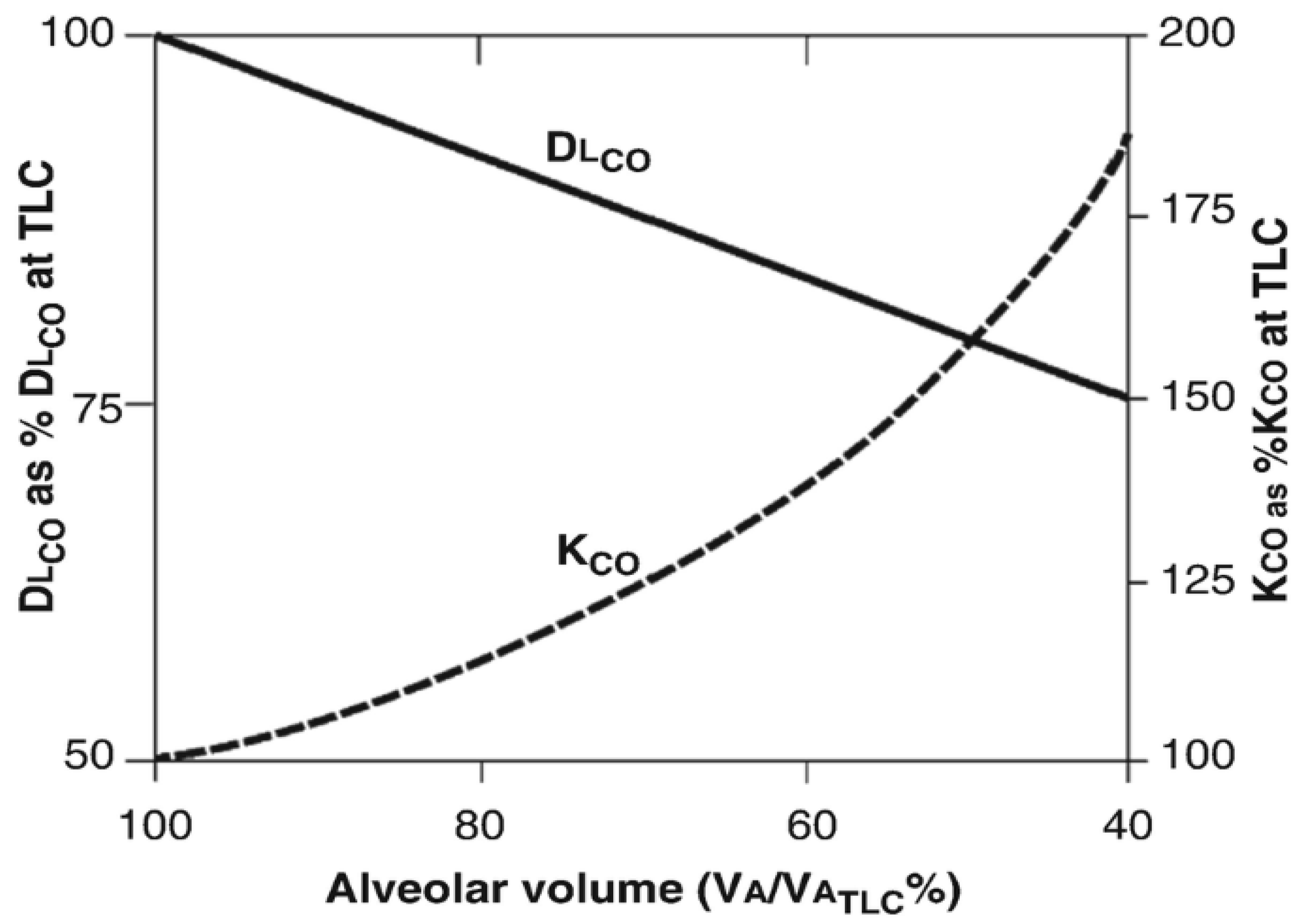
As the lung volume decreases from TLC to FRC, the DLco falls and kco rises ([36,37], see Figure 2). Vamax is a percentage of the predicted TLC. DLco at 50% Vamax is 79%, and kco is 158% [35]. This increased efficiency of alveolar uptake of carbon monoxide (kco) at resting breathing volumes protects DLco against undue volume dependence; that is, DLco is 80% of its TLC value at 50% Vamax rather than the expected 50%.
The physiological reason for the increase in kco with decreasing alveolar expansion is that with a decrease in alveolar expansion, the ratio Va/Dm remains almost constant. Dm here is the membrane diffusion capacity [38,39]. Pulmonary blood volume (Vc) stays stable with the decrease in lung volume so that the fall in VA/DLco (=rise in kco) is caused by the decrease in Va/Vc (=rise in Vc/Va).

5.1. Alveolar Po2

Another important factor that influences kco is alveolar Po2, which has important implications for accurately measuring DLco. If the patient is an active smoker and has smoked before the test is performed, it causes an increase in the partial pressure of carbon monoxide (Pco) in the blood. This, in turn, it generates what is called “back pressure” and results in lower DLco measurement. The same thing happens if we repeat testing multiple times. The test gas includes 21–25% oxygen, so kco is usually measured at a normal alveolar PaO2.
The following Table 3 summarizes the important factors affecting kco.

5.2. Clearing the Misconception of “Volume-Adjusted DLco (DLco/Va)”

Now, with the baseline knowledge of all three components of our base equation, let us talk about the misconception we alluded to earlier. kco, expressed as DLco/Va, is the carbon monoxide diffusing capacity per unit alveolar volume at the alveolar volume (Va) at which the measurement is made. It remains, in essence, a pressure-adjusted rate constant for alveolar carbon monoxide uptake.
Confusion arises in how PFT laboratories, by convention, report DLco and the related measurements Va and DLco/Va. This has had the unintended consequence of many clinicians considering DLco/Va to be the DLco “corrected” for the Va, when it is actually kco —a rate constant for CO uptake in the lung. kco is not the lung CO diffusing capacity.
The term DLco/Va is best avoided because Kco (the preferred term) is not derived from measurement of either DLco or Va. Furthermore, kco is not a surrogate measurement for DLco. Whenever DLco is reduced, the predominant reason for this reduction (e.g., whether it is predominantly a reduced Va, or reduced kco, or both) has critical diagnostic and pathophysiologic implications. The key questions that should be asked include the following: is the reduction in DLco due to a reduction in Va, kco, or both [39,40]?

5.3. Integrated Approach Toward Assessment of Lung Diffusion Capacity

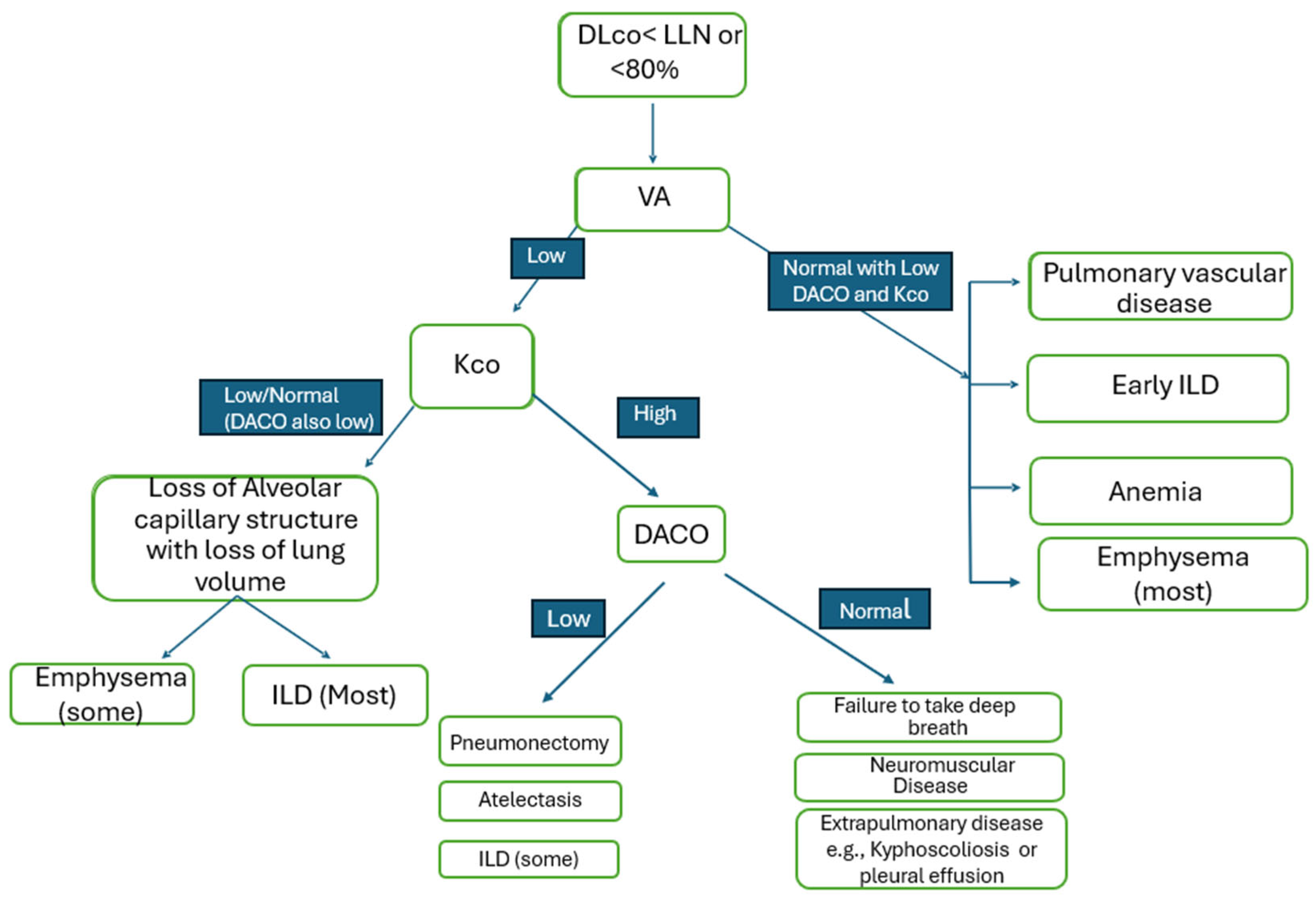
DLco interpretation requires integrating its two major components, kco and Va. Isolated interpretation of either component leads to misinformation or consequently can lead to the wrong diagnosis. We identified the following algorithm (Figure 3) to better interpret reduced DLco and to understand its relationship with Va [41].
As shown in the above algorithm, looking at DLco alone is insufficient for interpretation. All of the above diseases cause low DLco values on pulmonary function testing, but they can be differentiated when we integrate values of kco and Va.
Another approach, which is physiologically sounder, is to incorporate DACO (predicted DLco for patient’s lung volume). Dr Presti et al. described this relationship, emphasizing how interpreting DLco without DACO can lead to misinterpretation [42]. They described that while acknowledging that kco increases at lower lung volumes, the standard does not incorporate the predictable relationship of kco and DLco to lung volume, that patients with interstitial lung disease (ILD) may have low, normal, or elevated kco, or that patients with low lung volume due to incomplete lung expansion would have normal DLco adjusted for lung volume. Predicted DACO, the predicted DLco for the patient’s lung volume, should also be reported.
A fine example of the above-mentioned integrated approach is an early interstitial lung disease preceding the overt fibrotic stage. In such cases, the kco may be within the “normal” range (say 80–100%), but in the presence of a low Va, this could be interpreted as “abnormal” because the expected compensation via the “loss of units” model is lacking.

6. Conclusions

Isolated reduction in DLco has a distinct set of differentials in patients with dyspnea. This finding should urge physicians to initiate the appropriate workup to identify the underlying etiology. Interpreting DLco is not as straightforward as it may seem. Its complex relationship with alveolar volume and kco is critical to understand in order to accurately interpret the result of this commonly performed test.

7. Summary

The diffusion capacity of carbon monoxide (DLco) is a valuable test in evaluating patients with various lung diseases. It assists in diagnosing patients, following them over time, and has prognostic value.
Accurate interpretation of DLco requires understanding its relationship with lung volume (Va) and the diffusion coefficient (kco).
It is a common misunderstanding in everyday clinical practice to interpret DLco/Va as diffusion capacity “corrected” for lung volume. This notion is physiologically incorrect and has clinical implications.
Knowing the physiological basis of this test should help the readers understand the necessity of incorporating all the variables required for its correct interpretation.
Further, a readily available algorithm incorporating the above factors can provide a quick reference guide for trainees and pulmonary physicians to interpret lung diffusion capacity results.
Isolated reduced DLco is a unique abnormality that is often overlooked and not given due importance. It often points to early/developing lung disease and/or pulmonary vascular abnormality.
The presence of isolated DLco on full PFTs should alarm the physicians so that further workups can be initiated to find the cause of this abnormality.

Funding

This research received no external funding.

Acknowledgments

Several references were made to the outstanding work performed in the field by colleagues and experts in the field.

Conflicts of Interest

The authors declare no conflicts of interest.

References

  1. Enright, P. Office-based DLCO tests help pulmonologists to make important clinical decisions. Respir. Investig. 2016, 54, 305–311. [Google Scholar] [CrossRef]
  2. Macintyre, N.; Crapo, R.O.; Viegi, G.; Johnson, D.C.; Van Der Grinten, C.P.; Brusasco, V.; Burgos, F.; Casaburi, R.; Coates, A.; Enright, P.; et al. Standardisation of the single-breath determination of carbon monoxide uptake in the lung. Eur. Respir. J. 2005, 26, 720–735. [Google Scholar] [CrossRef] [PubMed]
  3. Jensen, R.L.; Crapo, R.O. Diffusing capacity: How to get it right. Respir. Care 2003, 48, 777–782. [Google Scholar] [PubMed]
  4. Suzuki, T.; Yoshimi, K.; Ueki, J.; Fukuchi, Y. Measurement of diffusing capacity by the intrabreath method. Nihon Kokyuki Gakkai Zasshi J. Jpn. Respir. Soc. 2005, 43, 347–353. [Google Scholar]
  5. Liu, Q.X.; Zheng, J.P.; Xie, Y.Q.; Guan, W.J.; Jiang, C.Y.; An, J.Y.; Yu, X.X.; Liu, W.T.; Gao, Y. Single-breath and rebreathing methods for measurement of pulmonary diffusing function: A comparative study. Zhonghua Jie He He Hu Xi Za Zhi Chin. J. Tuberc. Respir. Dis. 2013, 36, 510–515. [Google Scholar]
  6. Abou Daya, I.H.; Anwer, M.U.; Diaz-Fuentes, G.; Blum, S.; Menon, L. Isolated abnormalities of diffusion capacity (DLCO) in pulmonary function tests among inner city patients. Chest 2008, 134, 49S. [Google Scholar] [CrossRef]
  7. Randhawa, E.; Sherman, M. Causes of Isolated Diffusing Capacity of the Lungs for Carbon Monoxide (Dlco) from Outpatient Pulmonary Function Testing. Chest 2018, 154, 966A. [Google Scholar] [CrossRef]
  8. Diamanti, E.; Karava, V.; Yerly, P.; Aubert, J.D. Carbon monoxide diffusion capacity as a severity marker in pulmonary hypertension. J. Clin. Med. 2021, 11, 132. [Google Scholar] [CrossRef]
  9. Farha, S.; Asosingh, K.; Xu, W.; Sharp, J.; George, D.; Comhair, S.; Park, M.; Tang, W.W.; Loyd, J.E.; Theil, K.; et al. Hypoxia-inducible factors in human pulmonary arterial hypertension: A link to the intrinsic myeloid abnormalities. Blood J. Am. Soc. Hematol. 2011, 117, 3485–3493. [Google Scholar] [CrossRef]
  10. Overbeek, M.J.; Groepenhoff, H.; Voskuyl, A.E.; Smit, E.F.; Peeters, J.W.; Vonk-Noordegraaf, A.; Spreeuwenberg, M.D.; Dijkmans, B.C.; Boonstra, A. Membrane diffusion-and capillary blood volume measurements are not useful as screening tools for pulmonary arterial hypertension in systemic sclerosis: A case control study. Respir. Res. 2008, 9, 68. [Google Scholar] [CrossRef]
  11. Szturmowicz, M.; Kacprzak, A.; Franczuk, M.; Burakowska, B.; Kurzyna, M.; Fijałkowska, A.; Skoczylas, A.; Wesołowski, S.; Kuś, J.; Torbicki, A. Low DLCO in idiopathic pulmonary arterial hypertension—Clinical correlates and prognostic significance. Adv. Respir. Med. 2016, 84, 87–94. [Google Scholar] [CrossRef] [PubMed]
  12. Chandra, S.; Shah, S.J.; Thenappan, T.; Archer, S.L.; Rich, S.; Gomberg-Maitland, M. Carbon monoxide diffusing capacity and mortality in pulmonary arterial hypertension. J. Heart Lung Transplant. 2010, 29, 181–187. [Google Scholar] [CrossRef] [PubMed]
  13. Hoeper, M.M.; Meyer, K.; Rademacher, J.; Fuge, J.; Welte, T.; Olsson, K.M. Diffusion capacity and mortality in patients with pulmonary hypertension due to heart failure with preserved ejection fraction. JACC Heart Fail. 2016, 4, 441–449. [Google Scholar] [CrossRef]
  14. Rose, L.; Prins, K.W.; Archer, S.L.; Pritzker, M.; Weir, E.K.; Misialek, J.R.; Thenappan, T. Survival in pulmonary hypertension due to chronic lung disease: Influence of low diffusion capacity of the lungs for carbon monoxide. J. Heart Lung Transplant. 2019, 38, 145–155. [Google Scholar] [CrossRef]
  15. Garcia-Rio, F.; Miravitlles, M.; Soriano, J.B.; Cosío, B.G.; Soler-Cataluña, J.J.; Casanova, C.; de Lucas, P.; Alfageme, I.; González-Moro, J.M.; Herrero, M.G.; et al. Prevalence of reduced lung diffusing capacity and CT scan findings in smokers without airflow limitation: A population-based study. BMJ Open Respir. Res. 2023, 10, e001468. [Google Scholar] [CrossRef]
  16. Sun, X.G.; Hansen, J.E.; Oudiz, R.J.; Wasserman, K. Pulmonary function in primary pulmonary hypertension. J. Am. Coll. Cardiol. 2003, 41, 1028–1035. [Google Scholar] [CrossRef]
  17. Knudson, R.J.; Kaltenborn, W.T.; Burrows, B. The effects of cigarette smoking and smoking cessation on the carbon monoxide diffusing capacity of the lung in asymptomatic subjects. Am. J. Respir. Crit. Care Med. 1989, 140, 645–651. [Google Scholar] [CrossRef] [PubMed]
  18. Harvey, B.G.; Strulovici-Barel, Y.; Kaner, R.J.; Sanders, A.; Vincent, T.L.; Mezey, J.G.; Crystal, R.G. Risk of COPD with obstruction in active smokers with normal spirometry and reduced diffusion capacity. Eur. Respir. J. 2015, 46, 1589–1597. [Google Scholar] [CrossRef]
  19. Neas, L.M.; Schwartz, J. Pulmonary function levels as predictors of mortality in a national sample of US adults. Am. J. Epidemiol. 1998, 147, 1011–1018. [Google Scholar] [CrossRef]
  20. Klings, E.S.; Wyszynski, D.F.; Nolan, V.G.; Steinberg, M.H. Abnormal pulmonary function in adults with sickle cell anemia. Am. J. Respir. Crit. Care Med. 2006, 173, 1264–1269. [Google Scholar] [CrossRef]
  21. Stinson, J.M.; McPherson, G.L. Lung volumes and diffusion capacity in sickle cell trait. J. Natl. Med. Assoc. 1986, 78, 505. [Google Scholar] [PubMed]
  22. Siegel, J.L.; Millen, A.; Brown, L.K.; DeLuca, A.; Teirstein, A.S. Pulmonary diffusing capacity in left ventricular dysfunction. Chest 1990, 98, 550–553. [Google Scholar] [CrossRef] [PubMed]
  23. Kraemer, M.D.; Kubo, S.H.; Rector, T.S.; Brunsvold, N.; Bank, A.J. Pulmonary and peripheral vascular factors are important determinants of peak exercise oxygen uptake in patients with heart failure. J. Am. Coll. Cardiol. 1993, 21, 641–648. [Google Scholar] [CrossRef] [PubMed]
  24. Tasnim, S.N.; Khan, R. Retrospective study of isolated reduction of diffusion capacity in pulmonary function test. Chest 2024, 166, A5682. [Google Scholar] [CrossRef]
  25. Colaci, M.; Giuggioli, D.; Sebastiani, M.; Manfredi, A.; Lumetti, F.; Luppi, F.; Cerri, S.; Ferri, C. Predictive value of isolated DLCO reduction in systemic sclerosis patients without cardio-pulmonary involvement at baseline. Reumatismo 2015, 67, 149–155. [Google Scholar] [CrossRef]
  26. Morganroth, P.A.; Kreider, M.E.; Okawa, J.; Taylor, L.; Werth, V.P. Interstitial lung disease in classic and skin-predominant dermatomyositis: A retrospective study with screening recommendations. Arch. Dermatol. 2010, 146, 729–738. [Google Scholar] [CrossRef]
  27. Aduen, J.F.; Zisman, D.A.; Mobin, S.I.; Venegas, C.; Alvarez, F.; Biewend, M.; Jolles, H.I.; Keller, C.A. Retrospective study of pulmonary function tests in patients presenting with isolated reduction in single-breath diffusion capacity: Implications for the diagnosis of combined obstructive and restrictive lung disease. In Mayo Clinic Proceedings; Elsevier: Amsterdam, The Netherlands, 2007; Volume 82, pp. 48–54. [Google Scholar]
  28. Crothers, K.; McGinnis, K.; Kleerup, E.; Wongtrakool, C.; Hoo, G.S.; Kim, J.; Sharafkhaneh, A.; Huang, L.; Luo, Z.; Thompson, B.; et al. HIV infection is associated with reduced pulmonary diffusing capacity. JAIDS J. Acquir. Immune Defic. Syndr. 2013, 64, 271–278. [Google Scholar] [CrossRef]
  29. Jan, A.K.; Moore, J.V.; Wang, R.J.; Mcging, M.; Farr, C.K.; Moisi, D.; Hartman-Filson, M.; Kerruish, R.; Jeon, D.; Lewis, E.; et al. Markers of inflammation and immune activation are associated with lung function in a multi-center cohort of persons with HIV. Aids 2021, 35, 1031–1040. [Google Scholar] [CrossRef]
  30. Gingo, M.R.; Nouraie, M.; Kessinger, C.J.; Greenblatt, R.M.; Huang, L.; Kleerup, E.C.; Kingsley, L.; McMahon, D.K.; Morris, A. Decreased lung function and all-cause mortality in HIV-infected individuals. Ann. Am. Thorac. Soc. 2018, 15, 192–199. [Google Scholar] [CrossRef]
  31. Byanova, K.L.; Fitzpatrick, J.; Jan, A.K.; McGing, M.; Hartman-Filson, M.; Farr, C.K.; Zhang, M.; Gardner, K.; Branchini, J.; Kerruish, R.; et al. Isolated abnormal diffusing capacity for carbon monoxide (iso↓ DLco) is associated with increased respiratory symptom burden in people with HIV infection. PLoS ONE 2023, 18, e0288803. [Google Scholar] [CrossRef]
  32. Gandhi, S.; Cohen, R.A.; Rasmussen, D.L.; Almberg, K.S.; Go, L.H. Impact of Diffusion Capacity Measurement in the Evaluation of Former Coal Miners for Coal Mine Dust Lung Disease. In C15. Occupational Exposures of the 21st Century; American Thoracic Society: New York, NY, USA, 2019; p. A4260. [Google Scholar]
  33. Johnson, D.C. Importance of adjusting carbon monoxide diffusing capacity (DLCO) and carbon monoxide transfer coefficient (KCO) for alveolar volume. Respir. Med. 2000, 94, 28–37. [Google Scholar] [CrossRef] [PubMed]
  34. Roughton, F.J.; Forster, R.E. Relative importance of diffusion and chemical reaction rates in determining rate of exchange of gases in the human lung, with special reference to true diffusing capacity of pulmonary membrane and volume of blood in the lung capillaries. J. Appl. Physiol. 1957, 11, 290–302. [Google Scholar] [CrossRef] [PubMed]
  35. Graham, B.L.; Brusasco, V.; Burgos, F.; Cooper, B.G.; Jensen, R.; Kendrick, A.; MacIntyre, N.R.; Thompson, B.R.; Wanger, J. 2017 ERS/ATS standards for single-breath carbon monoxide uptake in the lung. Eur. Respir. J. 2017, 49, 1600016. [Google Scholar] [CrossRef]
  36. Cotes, J.E. Carbon monoxide transfer coefficient KCO (TL/VA): A flawed index. Eur. Respir. J. 2001, 18, 893–898. [Google Scholar] [CrossRef]
  37. McGrath, M.W.; Thomson, M.L. The effect of age, body size and lung volume change on alveolar-capillary permeability and diffusing capacity in man. J. Physiol. 1959, 146, 572. [Google Scholar] [CrossRef]
  38. Nguyen, L.P.; Harper, R.W.; Louie, S. Using and interpreting carbon monoxide diffusing capacity (DLCO) correctly. Consultant 2016, 56, 440–445. [Google Scholar]
  39. Hughes, J.M.; Pride, N.B. Examination of the carbon monoxide diffusing capacity (DLCO) in relation to its KCO and VA components. Am. J. Respir. Crit. Care Med. 2012, 186, 132–139. [Google Scholar] [CrossRef]
  40. Stam, H.; Kreuzer, F.J.; Versprille, A. Effect of lung volume and positional changes on pulmonary diffusing capacity and its components. J. Appl. Physiol. 1991, 71, 1477–1488. [Google Scholar] [CrossRef]
  41. Stanojevic, S.; Kaminsky, D.A.; Miller, M.R.; Thompson, B.; Aliverti, A.; Barjaktarevic, I.; Cooper, B.G.; Culver, B.; Derom, E.; Hall, G.L.; et al. ERS/ATS technical standard on interpretive strategies for routine lung function tests. Eur. Respir. J. 2022, 60, 2101499. [Google Scholar] [CrossRef]
  42. Presti, T.P.; Johnson, D.C. Improving pulmonary function test interpretation. Eur. Respir. J. 2023, 61, 2201858. [Google Scholar] [CrossRef]
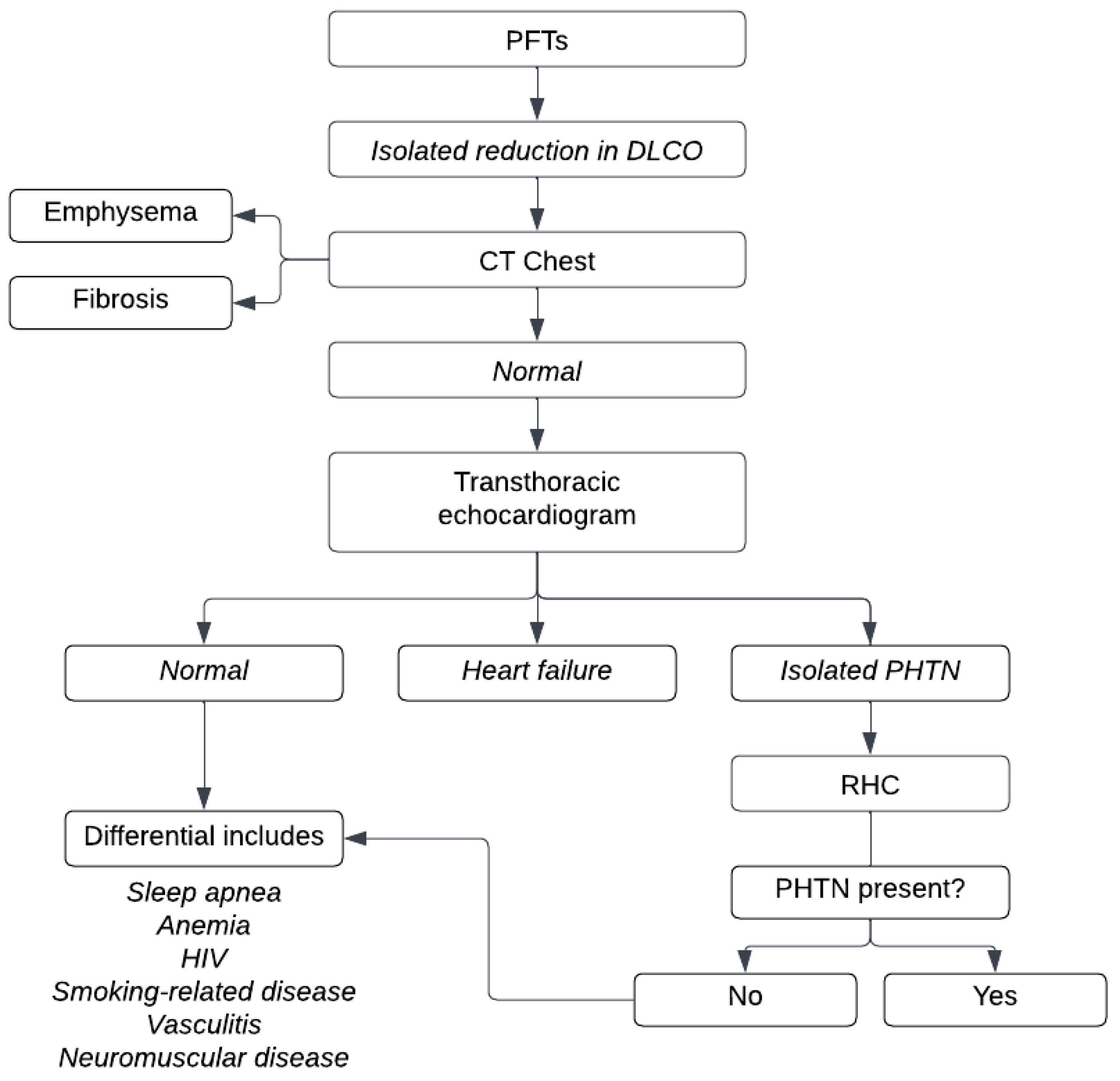
Figure 1. Proposed diagnostic algorithm for isolated DLco reduction.
Jor 05 00008 g001
Figure 2. The effect of voluntary changes in lung volume on DLco and kco plotted as the percentage of value at full inflation (approximately TLC) against alveolar expansion and expressed as the alveolar volume at the percent maximum. Definitions of abbreviations: DLco: CO diffusing capacity; kco: rate constant for CO uptake per unit barometric pressure (kco/Pb* z~ DLco/Va; Va; alveolar volume [32]).
Jor 05 00008 g002
Figure 3. An algorithm approach to reduced DLco. Abbreviations: PHTN (pulmonary hypertension); PE (pulmonary embolism); ILD (interstitial lung disease).
Jor 05 00008 g003
Table 1. Causes of low “accessible” alveolar volume.
Incomplete Alveolar ExpansionLoss of Lung Units
(Diffuse or Localized)
Poor Mixing with Normal or Loss of Function (Diffuse or Localized)
Inadequate inspirationPneumonectomyAsthma
Respiratory muscle weaknessFibrosisCOPD
Chest wall or pleural restriction Bronchiolitis obliterans
Table 2. Factors affecting Kco in Normal Subjects.
Factors Affecting kco in Normal Subjects
Lung volume
Exercise
Anemia
Alveolar Po2 (Pao2)
Age and height
Body position
Table 3. Factors affecting kco.
Pathophysiological MechanismClinical Example
Causes of increase in kco
Increased pulmonary blood flowAsthma, left to right shunt, alveolar hemorrhage
Incomplete alveolar expansion to TLCChest wall/pleural abnormalities
Microvascular dilatationObesity
Causes of decrease in kco
Pulmonary microvascular diseasesPulmonary HTN, pulmonary vasculitis
Alveolar destructionEmphysema, ILD
Microvascular destructionBronchiolitis obliterans, chronic severe heart failure
Disclaimer/Publisher’s Note: The statements, opinions and data contained in all publications are solely those of the individual author(s) and contributor(s) and not of MDPI and/or the editor(s). MDPI and/or the editor(s) disclaim responsibility for any injury to people or property resulting from any ideas, methods, instructions or products referred to in the content.