Abstract
Introduction: Computed tomography-guided biopsies (CTGB) are essential in diagnosing various conditions, particularly in respiratory medicine, with lung cancer being a primary focus. A significant complication associated with CTGB is pneumothorax, which can occur in up to 26% of cases. At Northumbria Healthcare NHS Foundation Trust, a large interventional service collaborates closely with radiologists and respiratory physicians. This study aims to evaluate the incidence of pneumothorax following CTGB. Methods: A retrospective service review was conducted on all lung parenchymal CTGBs performed between April 2011 and July 2023, with approval from the local information governance. Demographic data and clinical outcomes were analyzed using descriptive statistics. Continuous variables are presented as medians with interquartile ranges (IQR), while categorical variables are reported as frequencies and percentages. Results: A total of 1492 CT-guided lung biopsies were analyzed. The median age of patients was 72 years (IQR 10.5), and 50.9% were male. Pneumothorax occurred in 23.8% (n = 355) of cases. Of these, 159 (44.8%) were detected on post-biopsy CT scans. The average number of pleural passes was 1.8 (range 1–4). Among those with pneumothorax, 53.6% had radiologically evident emphysema. The median forced expiratory volume in 1 s (FEV1) was 1.97 L (IQR 1.04). Sixty-seven percent (n = 234) of patients had no pleural contact, and the median lesion size was 26 mm (IQR 24). Seventy-two percent (n = 255) of lesions with pneumothoraces were less than 3 cm deep. Forty-four percent of biopsies were performed using 18 French gauge tru-cut needles. Of the 355 pneumothoraces, 89% (n = 315) were managed conservatively, with 42 requiring pleural intervention (41 small-bore 12 Fr intercostal chest drains and one pleural vent). Symptoms were initially present in 40 cases, and two cases developed symptoms up to 7 days post-procedure. Conclusions: The incidence of pneumothorax is consistent with expected rates, with more occurrences observed in biopsies of smaller lesions lacking pleural contact, lesions with surrounding emphysema, and cases requiring multiple pleural passes. FEV1 does not appear to influence the risk of pneumothorax. Conservative management is generally effective, without significant complications.
1. Introduction
There are several different conditions that can affect the respiratory system—these include airway diseases such as chronic obstructive pulmonary disease, emphysema, or asthma; occupational lung diseases such as asbestosis; autoimmune conditions such as vasculitis; interstitial lung diseases; infections involving bacteria, viruses, or fungi; and benign or malignant conditions. While several diagnoses can be reached through histories and clinical examinations, complemented by tests such as lung function tests or computed tomogram (CT) scans, occasionally a biopsy of a lesion is required. This can be achieved either via bronchoscopy or thoracoscopy, or with the use of image guidance (on both pleural-based and parenchymal masses).
Our local population is predominantly Caucasian, with a high smoking rate, and we have very high lung cancer prevalence [1]. We serve a catchment area with a population of approximately 600,000 at Northumbria Healthcare NHS Foundation Trust. Our respiratory service thus has a huge burden of lung cancer and our services are geared to providing the required services. Lung cancer is the leading cause of cancer deaths worldwide and most prevalent cancer worldwide in men [2]. Incidence is expected to increase with the advent of lung cancer screening programmes [2,3]. For individualized approaches to care and treatment planning, whether this is curative or palliative, pathological confirmation of the type of lung cancer via a biopsy is often required. CT-guided biopsies (CTGB) for parenchymal masses have high sensitivity and specificity, and are thus common practice in the lung cancer diagnostic pathway [3]. However, as with any procedure, complications occur, with pneumothorax being the commonest, with quoted rates of up to 26% [4]. Previous meta-analyses have suggested that pneumothorax rates increase with the use of needles greater than 18 Gauge [G] in diameter, when needle passes through a fissure or through a bulla, when the biopsy is performed with more than one puncture and in non-coaxial fashion, when emphysema is present around the lesion, and when biopsying smaller lesions with no pleural contact at greater intrathoracic depths [4]. It makes sense to not perform biopsies of such lesions, but often a diagnosis is required and so a biopsy must be performed. Nakamura et al. describe the ways in which any complications can be reduced, specifically looking at pneumothorax and the use of sealing agents such as blood patches, collagen, fibrin, and aliquots of sodium chloride [5]. The studies looking at those techniques included a small number of patients in single centres, and while some of them included randomized groups, the use of sealants was not widespread. Other techniques, such as rapid roll-over, pleural patching, positioning biopsy-side down, and needle removal on expiration, have all been tried, sometimes together, with variable success; additionally, the studies were limited as they were from single centres, had non-randomized groups, and showed a widespread variability of techniques between centres [6]. Moad et al. also found that the majority of pneumothoraces can be conservatively managed [7].
Locally, in Northumbria Healthcare NHS Foundation Trust, all acute care is based at a flagship centre, the Northumbria Specialist Emergency Care Hospital, and all CTGBs are performed there by the radiologists, with post-biopsy management undertaken by the respiratory physicians [8]. Any anticoagulation is stopped as required and, if required, clotting and platelets counts are checked and any deficiencies corrected. On the day of the biopsy, the patients are admitted to a day care unit and, in the CT suite, are either in the prone or supine position, depending on where the lesion is. The needle size is dependent on the radiologist performing the biopsy. Local anesthetic is applied to the identified biopsy site. Typically, a chest radiograph is performed 4 h after the CTGB to assess for pneumothorax (or earlier if there is clinical concern), and if a pneumothorax is present, the respiratory physicians make the decision to intervene, admit, or discharge with adequate safety nets. Fluoroscopy is performed. Sealants and rapid roll-over techniques are not used locally, and, more recently, the manual aspiration of air is occasionally performed if a pneumothorax is present. Biopsies are performed either supine or prone, depending on the radiologist’s preference. Images 1 and 2 show a typical CTGB of a lung mass being performed.
Practice regarding CTGB and subsequent pneumothorax management is very varied and resource-dependent [9]. The current British Thoracic Society guidelines for radiologically guided procedures and pleural disease highlight the lack of evidence to guide the appropriate management of pneumothorax after CTGB [10,11]. It has been acknowledged that there is a need to conduct good-quality research in this area to inform future treatment guidelines for standardized, evidence-based patient care. Thus, we sought to perform a retrospective case-note review of local CTGB-induced pneumothorax, with a view to understanding local practice.
2. Methods
2.1. Project Registration
The project was registered as a service evaluation and was granted information governance clearance (C4453/4560) with no formal ethical approval required and waived informed consent.
2.2. Timeline and Inclusion Cirteria
A list of all lung CTGBs for parenchymal masses from April 2011 to July 2023 was obtained from the radiology department. These were then analyzed for pneumothoraces and those without were excluded from the final analysis.
2.3. Analysis
Simple demographics, radiographic and spirometric findings, pneumothorax rates, management strategies, and outcomes were collected. All results were analyzed descriptively and, where possible, continuous variables were presented as the median, with interquartile range (IQR) and categorical variables expressed as frequencies (n) and percentages (%). All analyses were performed in Microsoft® Excel, 2024 Edition.
3. Results
A total of 1492 CTGBs were performed during that period. The median age was 72 years (IQR 10.5), and 760 (50.9%) were male patients. There were 355 pneumothoraces (23.8%) overall, with 159 (44.8%) of those being visible on the post-biopsy CT images, as shown in Figure 1. The mean number of pleural passes was 1.8 (range 1–4). Of the patients with pneumothoraces, 190 (53.6%) had radiological emphysema and median forced expiratory volume in 1 s (FEV1) was 1.97 litres (L) (IQR 1.04). Table 1 represents these results pictorially.
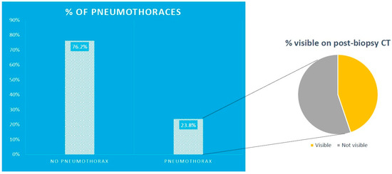
Figure 1.
Percentage of patients with pneumothoraces those visible on post-biopsy CT.

Table 1.
Pictorial representation of results of patients with pneumothoraces.
3.1. Pneumothoraces Associated with CTGB
Of those with pneumothoraces, 234 (67%) of the lesions biopsied had no pleural contact, and 255 (72%) of those lesions were less than 3 cm deep. The median size of biopsied lesions was 26 mm (IQR 24). In this same group, most biopsies were performed with an 18 French (Fr) gauge (44%) and 17Fr (32.1%) tru-cut needles, as shown in Figure 2.
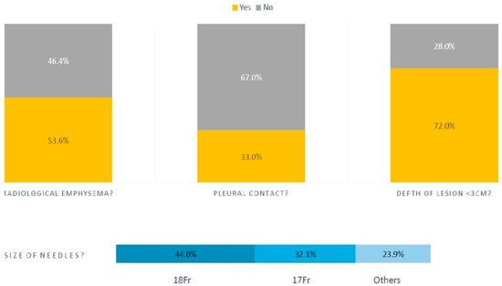
Figure 2.
Percentage of pneumothoraces with radiological emphysema and of patients with pleural contact according to lesion depth and needle size.
Of the 355 pneumothoraces, 315 (89%) pneumothoraces were managed conservatively. Of the 159 (44.8%) visible on the post-biopsy CT images, 101 (63.5%) were asymptomatic. A total of 143 (90%) patients had a chest radiograph between 2 and 4 h later. A total of 17 (12%) of those had a slight increase in the size of the pneumothorax, but no increase in symptoms. We did not measure the size of the pneumothoraces locally; instead, a symptom-based approach was used, which we will discuss later. A total of 333 (94%) of the patients had appropriate oxygenation levels for their respective ranges (locally 88–92% for those at risk of type 2 respiratory failure or with lung disease, and 90–95% for those without pre-existing lung disease, which is a slight adaptation from 94–98%) [12]. The rest had transient hypoxemias (lowest value 85%) lasting less than 24 h, which all resolved and were attributed to hypoventilation due to pain, which was in turn related to the biopsy procedure. Pain was treated with either paracetamol, codeine, or liquid morphine.
3.2. Pleural Interventions in Patients with Pneumothorax
A total of 42 out of 355 patients (12%) had a pleural intervention (41 small bore, 12Fr intercostal chest drains were inserted, and 1 one Rocket® Pleural Vent), as shown in Figure 3. Forty had documented symptoms of increased breathlessness at the onset of pneumothorax, either in the CT suite or developed thereafter. Thirty-five pneumothoraces resolved with small bore drainage, seven required large bore drain insertion (20Fr in two and 24Fr in five), and two required surgical cardiothoracic intervention.
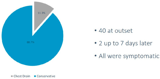
Figure 3.
Percentage of patients undergoing chest drain insertion and conservative management, depicting the total number of patients with symptoms.
Of note, there was a single case of mortality following CTGB within the dataset. A male who had no pneumothorax visible on post-biopsy CT images or on his 4 h post-biopsy chest radiograph developed progressive breathlessness a few hours later, and subsequently collapsed and had a cardiac arrest.
Despite paramedic-assisted resuscitation, he unfortunately died of a tension pneumothorax, which was proven on post-mortem—the death was attributed to the delayed presentation of a CTGB-induced pneumothorax. His pre-biopsy FEV1 was 2.1 L (110% predicted), the lesion biopsied was 2 cm deep, and two passes were performed with an 18Fr needle. No fissures were crossed and there was only minor radiological emphysema around the lesion.
4. Discussion
Our retrospective analysis shows that pneumothoraces due to CTGB are common and can be expected in 25% of all patients undergoing CTGB. Pneumothoraces are more common when biopsying smaller lesions with no pleural contact and with surrounding radiological emphysema. Most of the biopsies associated with pneumothorax were on lesions less than 3 cm deep and most were performed with lower-calibre needles, perhaps suggesting that the surrounding radiological emphysema is more contributory. FEV1 does not seem to influence the risk of pneumothorax for CTGB.
The main message to convey is that the vast majority of pneumothoraces can be observed and conservatively managed. We previously presented data showing even large pneumothoraces secondary to CTGB can be safely observed [13]. This concept was strengthened by large trials in primary pneumothoraces and extrapolated to the CTGB population [14,15,16]. Self-sealing of the lung from a CTGB insult is thought to be due to the recoil properties and collapsibility of the lung, which allows for the quick closing of any alveolar–pleural connection. We experimented with ambulatory devices, as emergent data has suggested that they are safe in CTGB pneumothoraces, but the risk of large air leaks in patients with underlying lung disease led us to abandon this practice [14,15,16].
Our study has some significant limitations—we do not have a control group of patients without pneumothorax for comparison, so, as a single-arm retrospective study, the generalizability of the results is questionable, and we did not attempt any statistical inferences. We also did not look at other complications of CTGB, overall diagnostic sensitivity, or co-axial or core needle techniques. The single case of mortality within the dataset was not felt to have been preventable. We also did not look at the type of lesion being biopsied (ground glass or solid or subsolid) to see if that had any bearing on pneumothorax occurrence.
Practice around the United Kingdom (UK) is very varied [9]. Tavare et al. surveyed UK practice in 2017. A total of 30.1% (72/239) of survey respondents did not require pre-biopsy lung function testing. A total of 55.9% of radiologists made one or two passes, and 40.8% made three or four passes. A total of 64% used the so-called chest drain prevention techniques, with 43.9% using needle aspiration. Other methods include the roll-over technique, saline injection into the biopsy track, and the use of hydrogel plugs or blood patches. We did not look at these techniques within this study. The timing of post-biopsy chest radiographs also varied, with 23% being performed at 1 h, 24.7% at 2 h, and 22.6% at 4 h. There is also a lack of standardization amongst centres, and thoracic surgery is often required for those lesions where CTGB is non-diagnostic, although exact figures are lacking. We also did not look for previous smoking habits (as this was very poorly documented) or other respiratory co-morbidities; this is another limitation of our study. Delayed pneumothorax has also been described in the literature as being at a population-based estimate of less than 1% [17], which is what we saw in our single case of mortality in this dataset.
We are also currently participating in the Pneumothorax after Lung Biopsy: Understanding the Management Basis (PLUMB) study, which is being run in the United Kingdom and is collecting all of the data we have not collected (Ethics Reference 24/NI/0111 and Integrated Research Application System Project Identification 331451) [18].
Another point should be made regarding our oxygen parameters. We agree that target saturations should be 88–92% in all patients with COPD and other conditions at heightened risk of oxygen toxicity. In patients receiving oxygen in the study by Echevarria et al., the lowest mortality was observed in those with sats 88–92%, including the cohort with normocapnia [10]. There was an adverse dose response of oxygen therapy at higher saturations, and the relationship was stronger, not weaker, after adjusting for baseline risk. Local expert opinion settled on a range of 90–95%, as the upper threshold lies between 94% and 96%, which are the two upper values suggested.
As formal randomized controlled trials on how to prevent complications such as pneumothorax are not available, the evidence stems from meta-analyses and retrospective reviews. As described above, Nakamura et al. suggested a host of factors to reduce pneumothorax rates, such as using small gauge needles, trying not to biopsy central or deep lesions with surrounding radiological emphysema, and not performing multiple pleural passes [5]. Heerink et al. similarly analyzed 32 articles and 8133 procedures, finding that larger needles caused more complications and were frequently used in parenchyma and when biopsying smaller lesion sizes [19]. Moad et al. reported similar findings but also noted that the FEV1 did not impact pneumothorax occurrence [7].
5. Conclusions
Until there are adequately powered randomized controlled trials looking at the various aspects of CTGB and, ultimately, pneumothorax management with patient-centred outcomes at its core, regular analyses of large volume centres are important. Whilst the data is often incomplete and laden with limitations, it can inform local practice, as we have attempted here. The widespread variation in practice also calls for a standardized approach, which can only succeed with radiology and respiratory societies working together collaboratively and at an international level.
Author Contributions
J.S., G.M., D.M., W.H.O. and A.A. all wrote the article, and revised the manuscript for content. All the authors performed data collection; A.A. performed the data analysis. All authors have read and agreed to the published version of the manuscript.
Funding
There was no funding for this study.
Institutional Review Board Statement
(C4453/4560) Northumbria Healthcare Foundation Trust.
Informed Consent Statement
As this was a retrospective review, there was no required for informed consent from the participants.
Data Availability Statement
Some of the data will be available with reasonable requests.
Conflicts of Interest
Avinash Aujayeb is part of the Editorial team for Journal of Respiration, but was not involved in peer-reviewer selection. None of the other authors have anything to declare.
References
- Rate of Newly Diagnosed Cases of Lung Cancer per 100,000 Population in England in 2020, by Region and Gender. 2024. Available online: https://www.statista.com/statistics/312896/lung-cancer-cases-rate-england-region-gender/ (accessed on 10 March 2025).
- Lung Cancer. 2023. Available online: https://www.who.int/news-room/fact-sheets/detail/lung-cancer#:~:text=GLOBOCAN%202020%20estimates%20of%20cancer,deaths%20(18%25)%20in%202020 (accessed on 11 November 2024).
- Hardavella, G.; Chorostowska-Wynimko, J.; Blum, T.G. Lung cancer: An update on the multidisciplinary approach from screening to palliative care. Breathe 2024, 20, 240117. [Google Scholar] [CrossRef]
- Huo, Y.R.; Chan, M.V.; Habib, A.R.; Lui, I.; Ridley, L. Pneumothorax rates in CT-Guided lung biopsies: A comprehensive systematic review and meta-analysis of risk factors. Br. J. Radiol. 2020, 93, 20190866. [Google Scholar] [CrossRef] [PubMed]
- Nakamura, K.; Matsumoto, K.; Inoue, C.; Matsusue, E.; Fujii, S. Computed Tomography-guided Lung Biopsy: A Review of Techniques for Reducing the Incidence of Complications. Interv. Radiol. 2021, 6, 83–92. [Google Scholar] [CrossRef]
- Najafi, A.; Al Ahmar, M.; Bonnet, B.; Delpla, A.; Kobe, A.; Madani, K.; Roux, C.; Deschamps, F.; de Baère, T.; Tselikas, L. The PEARL Approach for CT-guided Lung Biopsy: Assessment of Complication Rate. Radiology 2022, 302, 473–480. [Google Scholar] [CrossRef] [PubMed]
- Moad, M.; Narkhede, P.; Jackson, K.; Aujayeb, A. A note on pneumothorax post-CT-guided biopsy. Br. J. Radiol. 2021, 94, 20201010. [Google Scholar] [CrossRef]
- Northumbria Specialist Emergency Care Hospital. 2024. Available online: https://en.wikipedia.org/w/index.php?title=Northumbria_Specialist_Emergency_Care_Hospital&action=history (accessed on 11 November 2024).
- Tavare, A.N.; Hare, S.S.; Miller, F.; Hammond, C.; Edey, A.; Devaraj, A. A survey of UK percutaneous lung biopsy practice: Current practices in the era of early detection, oncogenetic profiling, and targeted treatments. Clin. Radiol. 2018, 73, 800–809. [Google Scholar] [CrossRef] [PubMed]
- Manhire, A.; Charig, M.; Clelland, C.; Gleeson, F.; Miller, R.; Moss, H.; Pointon, K.; Richardson, C.; Sawicka, E. Guidelines for radiologically guided lung biopsy. Thorax 2003, 58, 920–936. [Google Scholar] [CrossRef]
- Roberts, M.E.; Rahman, N.M.; Maskell, N.A.; Bibby, A.C.; Blyth, K.G.; Corcoran, J.P.; Edey, A.; Evison, M.; de Fonseka, D.; Hallifax, R. British Thoracic Society Guideline for pleural disease. Thorax 2023, 78, s1–s42. [Google Scholar] [CrossRef] [PubMed]
- Echevarria, C.; Steer, J.; Wason, J.; Bourke, S. Oxygen therapy and inpatient mortality in COPD exacerbation. Emerg. Med. J. 2021, 38, 170–177. [Google Scholar] [CrossRef] [PubMed]
- Aujayeb, A.; Narkhede, P. Pneumothorax rates after CT guided biopsy: Experience from a high volume cancer centre. Eur. Respir. J. 2021, 58 (Suppl. 65), PA3784. [Google Scholar] [CrossRef]
- Chopra, A.; Judson, M.A.; Rahman, N.M.; Doelken, P. The lung is not a balloon: The self-sealing property of the lung. Lancet Respir. Med. 2024, 12, 190–192. [Google Scholar] [CrossRef] [PubMed]
- Ball, M.; Babu, S.; Wallis, A.; Asciak, R. Promising role for pleural vent in pneumothorax following CT-guided biopsy of lung lesions. Br. J. Radiol. 2022, 95, 20210965. [Google Scholar] [CrossRef] [PubMed]
- Walker, S.P.; Keenan, E.; Bintcliffe, O.; Stanton, A.E.; Roberts, M.; Pepperell, J.; Fairbairn, I.; McKeown, E.; Goldring, J.; Maddekar, N. Ambulatory management of secondary spontaneous pneumothorax: A randomised controlled trial. Eur. Respir. J. 2021, 57, 2003375. [Google Scholar] [CrossRef] [PubMed]
- Pua, B.; Tang, E.; Bhat, A.; Zabih, R.; Winokur, R.; Madoff, D. Delayed pneumothorax after percutaneous lung biopsy in the state of California. J. Vasc. Interv. Radiol. 2015, 27, S90. [Google Scholar] [CrossRef]
- PLUMB (Pneumothorax after Lung Biopsy: Understanding the Management Basis). Available online: https://www.inspirerespiratory.co.uk/ (accessed on 12 March 2025).
- Heerink, W.J.; de Bock, G.H.; de Jonge, G.J.; Groen, H.J.M.; Vliegenthart, R.; Oudkerk, M. Complication rates of CT-guided transthoracic lung biopsy: Meta-analysis. Eur. Radiol. 2017, 27, 138–148. [Google Scholar] [CrossRef] [PubMed]
Disclaimer/Publisher’s Note: The statements, opinions and data contained in all publications are solely those of the individual author(s) and contributor(s) and not of MDPI and/or the editor(s). MDPI and/or the editor(s) disclaim responsibility for any injury to people or property resulting from any ideas, methods, instructions or products referred to in the content. |
© 2025 by the authors. Licensee MDPI, Basel, Switzerland. This article is an open access article distributed under the terms and conditions of the Creative Commons Attribution (CC BY) license (https://creativecommons.org/licenses/by/4.0/).