
Airway Stents in Interventional Pulmonology


Abstract
1. Introduction
2. Indications for Stent Placement
3. General Principles in Patient Selection
4. General Principles in Stent Selection
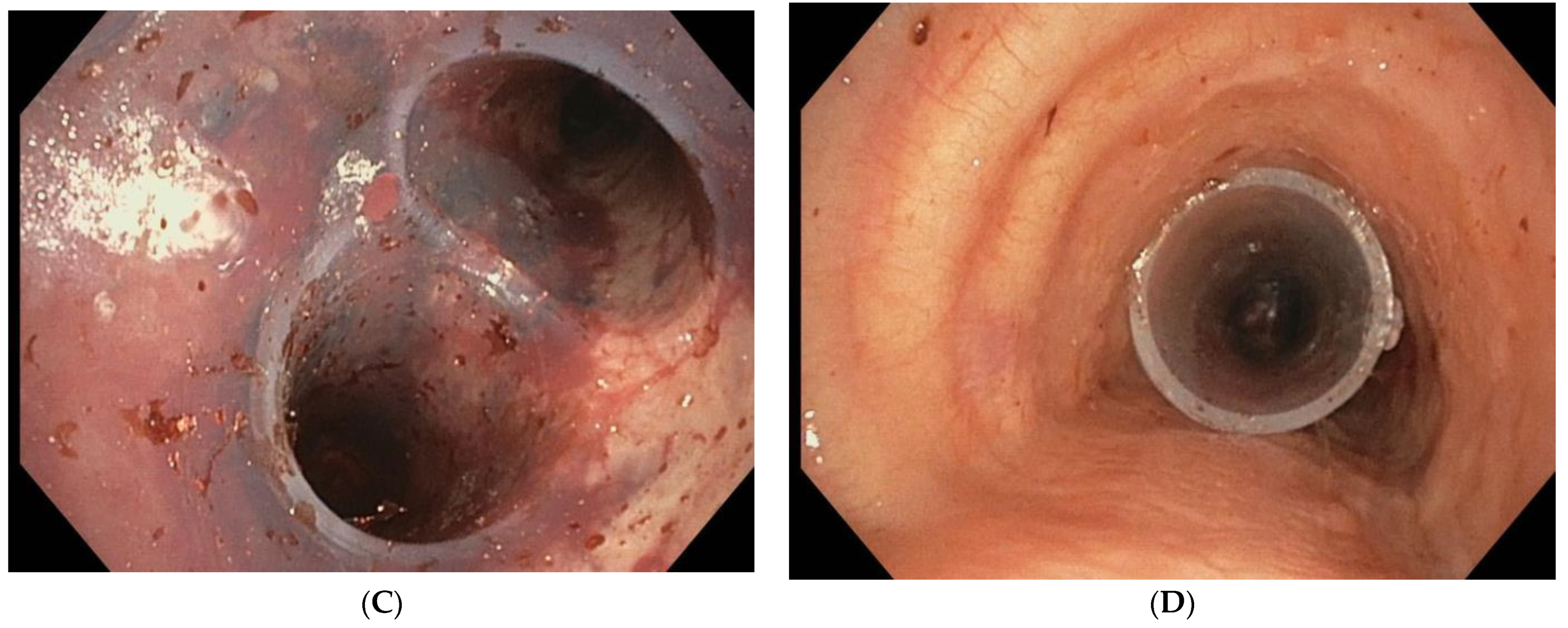
4.1. Metallic Stents
4.2. Silicone Stents
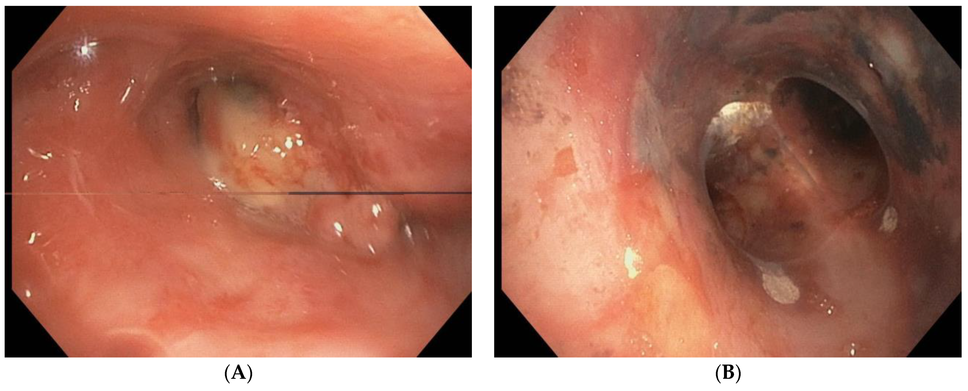
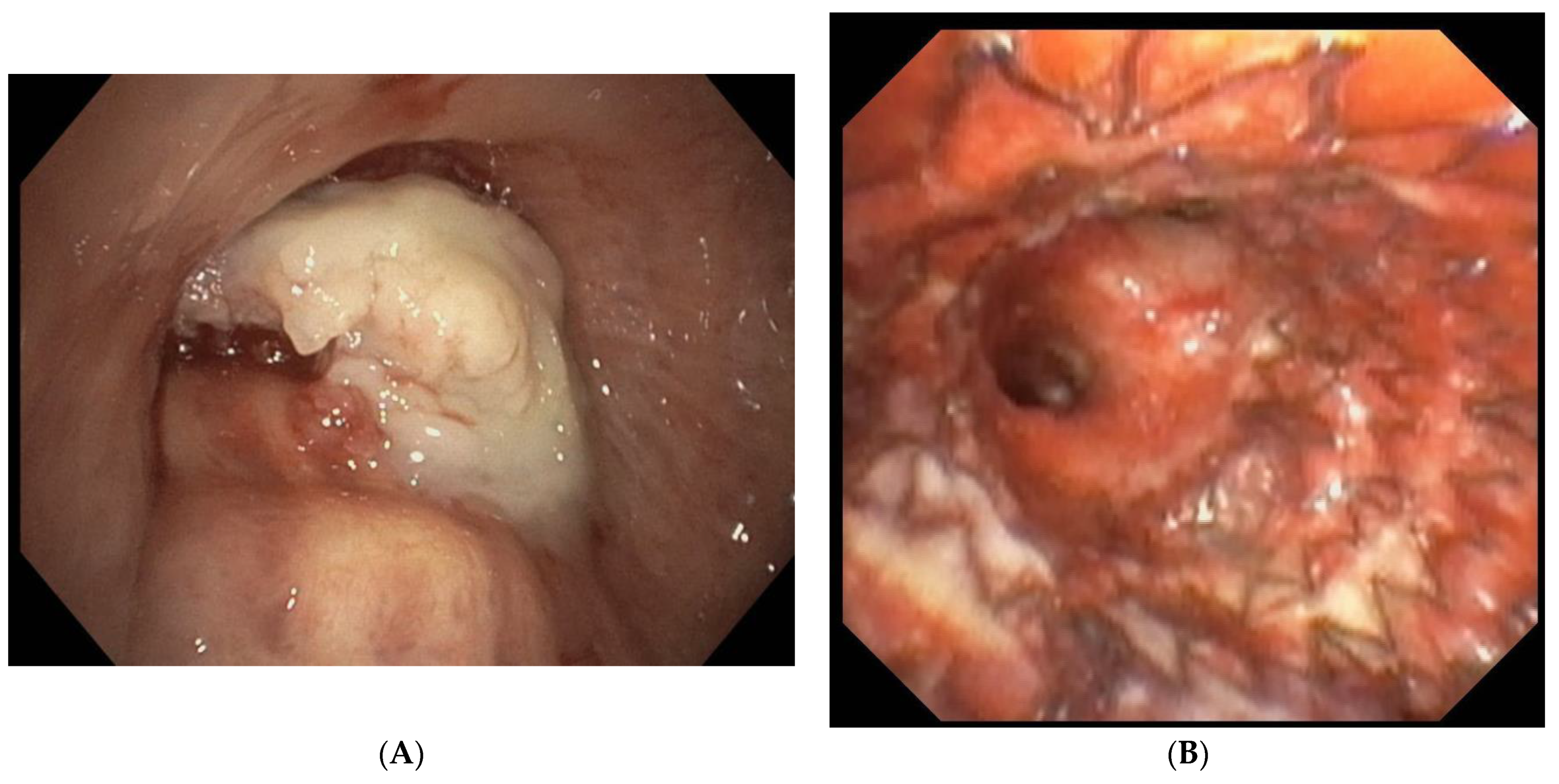
5. Complications
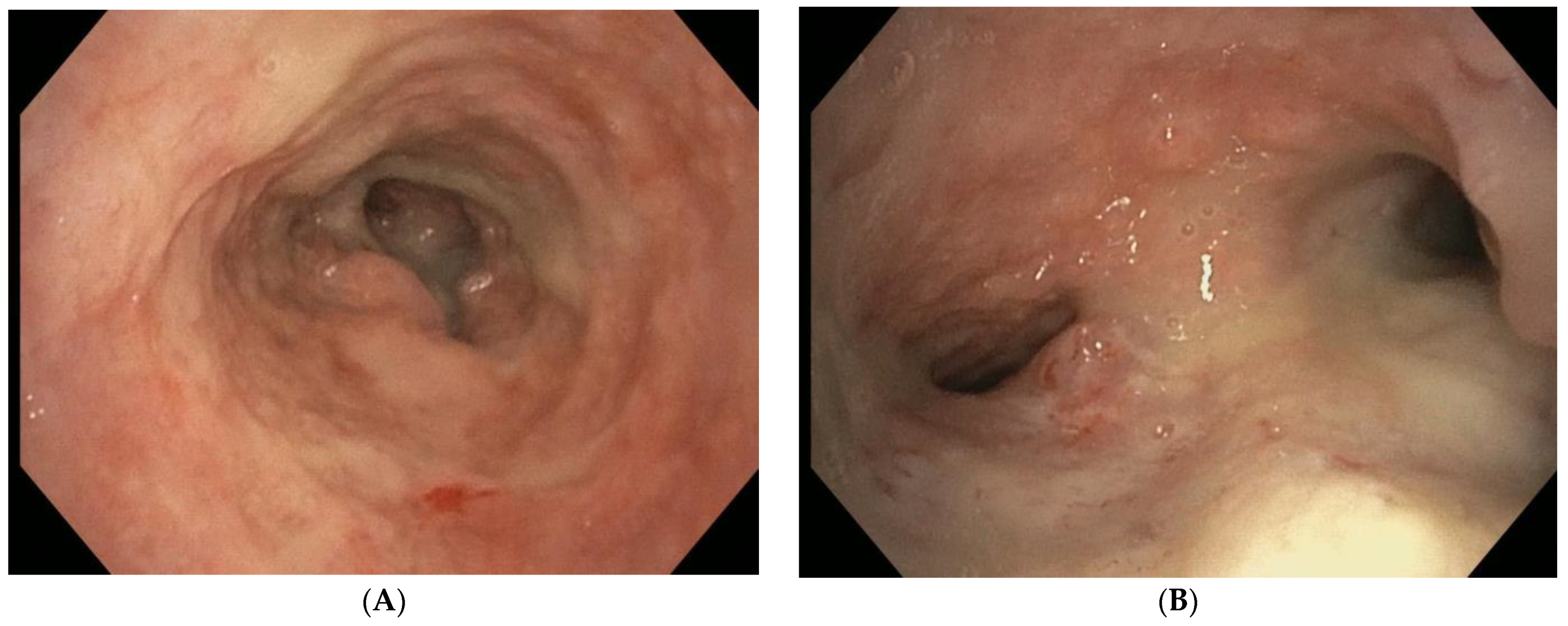
6. Stent Management
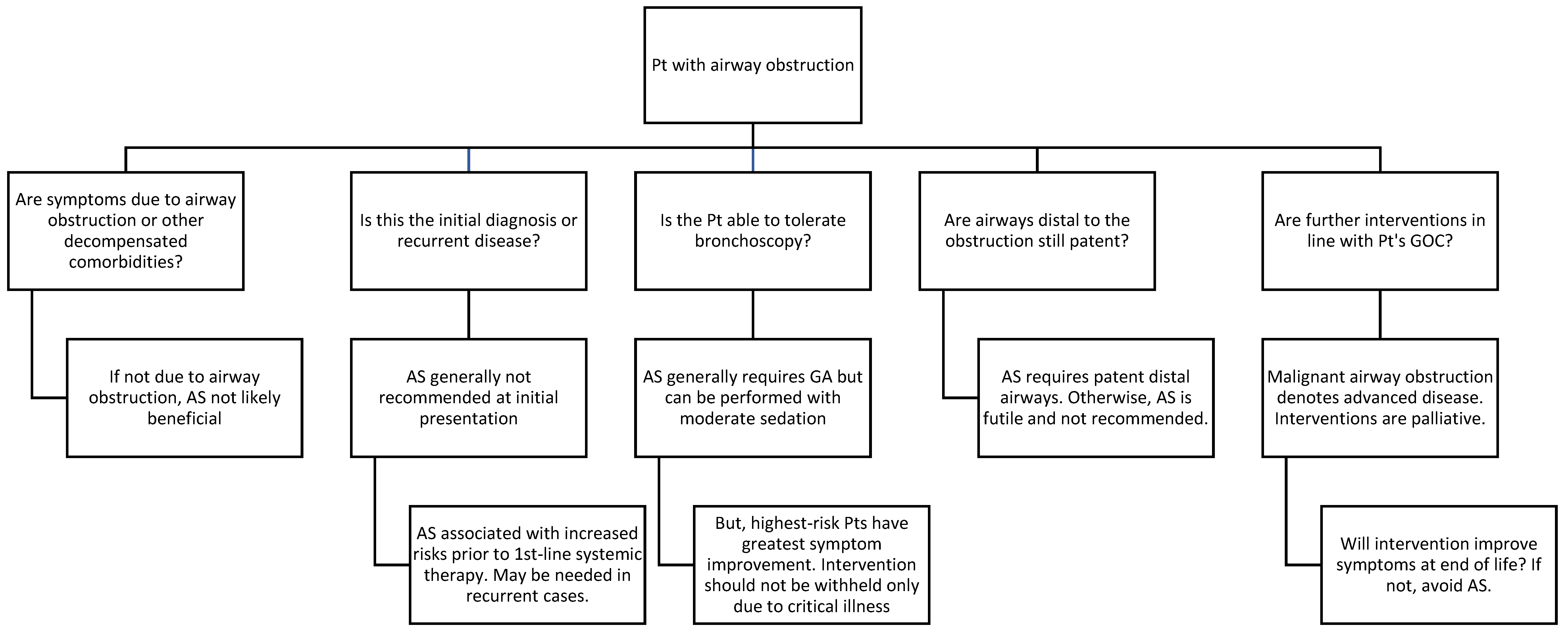
7. When Airway Stenting Is Not the Optimal Initial Strategy
8. Future Applications
9. Conclusions
Author Contributions
Funding
Institutional Review Board Statement
Informed Consent Statement
Conflicts of Interest
References
- Ernst, A.; Feller-Kopman, D.; Becker, H.D.; Mehta, A.C. Central airway obstruction. Am. J. Respir. Crit. Care Med. 2004, 169, 1278–1297. [Google Scholar] [CrossRef]
- Folch, E.; Keyes, C. Airway stents. Ann. Cardiothorac. Surg. 2018, 7, 273–283. [Google Scholar] [CrossRef]
- Aravena, C.; Gildea, T.R. Patient-specific airway stent using three-dimensional printing: A review. Ann. Transl. Med. 2023, 11, 360. [Google Scholar] [CrossRef]
- Ost, D.E.; Ernst, A.; Grosu, H.B.; Lei, X.; Diaz-Mendoza, J.; Slade, M.; Gildea, T.R.; Machuzak, M.; Jimenez, C.A.; Toth, J.; et al. Complications Following Therapeutic Bronchoscopy for Malignant Central Airway Obstruction: Results of the AQuIRE Registry. Chest 2015, 148, 450–471. [Google Scholar] [CrossRef]
- Bashour, S.I.; Lazarus, D.R. Therapeutic bronchoscopy for malignant central airway obstruction: Impact on quality of life and risk-benefit analysis. Curr. Opin. Pulm. Med. 2022, 28, 288–293. [Google Scholar] [CrossRef]
- Sabath, B.F.; Casal, R.F. Airway stenting for central airway obstruction: A review. Mediastinum 2023, 7, 18. [Google Scholar] [CrossRef] [PubMed]
- Murgu, S.D.; Egressy, K.; Laxmanan, B.; Doblare, G.; Ortiz-Comino, R.; Hogarth, D.K. Central Airway Obstruction: Benign Strictures, Tracheobronchomalacia, and Malignancy-related Obstruction. Chest 2016, 150, 426–441. [Google Scholar] [CrossRef] [PubMed]
- Ost, D.E.; Ernst, A.; Grosu, H.B.; Lei, X.; Diaz-Mendoza, J.; Slade, M.; Gildea, T.R.; Machuzak, M.S.; Jimenez, C.A.; Toth, J.; et al. Therapeutic bronchoscopy for malignant central airway obstruction: Success rates and impact on dyspnea and quality of life. Chest 2015, 147, 1282–1298. [Google Scholar] [CrossRef] [PubMed]
- Sabath, B.F.; Ost, D.E. Update on airway stents. Curr. Opin. Pulm. Med. 2018, 24, 343–349. [Google Scholar] [CrossRef] [PubMed]
- Kanchustambham, V.; Saladi, S.; Mehta, K.; Mwangi, J.; Jamkhana, Z.; Patolia, S. Vascular Air Embolism During Bronchoscopy Procedures- Incidence, Pathophysiology, Diagnosis, Management and Outcomes. Cureus 2017, 9, e1087. [Google Scholar] [CrossRef] [PubMed]
- Kizilgoz, D.; Aktas, Z.; Yilmaz, A.; Ozturk, A.; Segmen, F. Comparison of two new techniques for the management of malignant central airway obstruction: Argon plasma coagulation with mechanical tumor resection versus cryorecanalization. Surg. Endosc. 2018, 32, 1879–1884. [Google Scholar] [CrossRef]
- Mahajan, A.K.; Ibrahim, O.; Perez, R.; Oberg, C.L.; Majid, A.; Folch, E. Electrosurgical and Laser Therapy Tools for the Treatment of Malignant Central Airway Obstructions. Chest 2020, 157, 446–453. [Google Scholar] [CrossRef]
- Bashour, S.I.; Ost, D.E. An update on bronchopleural fistulae following cancer-related surgery. Curr. Opin. Pulm. Med. 2023, 29, 223–231. [Google Scholar] [CrossRef] [PubMed]
- Zhou, C.; Hu, Y.; Xiao, Y.; Yin, W. Current treatment of tracheoesophageal fistula. Ther. Adv. Respir. Dis. 2017, 11, 173–180. [Google Scholar] [CrossRef] [PubMed]
- Parikh, M.; Wilson, J.; Majid, A.; Gangadharan, S. Airway stenting in excessive central airway collapse. J. Vis. Surg. 2017, 3, 172. [Google Scholar] [CrossRef] [PubMed]
- Martinod, E.; Dutau, H.; Guibert, N. Management of airway complications after lung transplantation: Is there an ideal stent? J. Thorac. Dis. 2022, 14, 3111–3115. [Google Scholar] [CrossRef] [PubMed]
- Anton-Pacheco, J.L.; Luna, C.; Garcia, E.; Lopez, M.; Morante, R.; Tordable, C.; Palacios, A.; de Miguel, M.; Benavent, I.; Gomez, A. Initial experience with a new biodegradable airway stent in children: Is this the stent we were waiting for? Pediatr. Pulmonol. 2016, 51, 607–612. [Google Scholar] [CrossRef] [PubMed]
- Li, L.; Zhang, X.; Shi, J.; Chen, Y.; Wan, H.; Herth, F.J.; Luo, F. Airway Stents from Now to the Future: A Narrative Review. Respiration 2023, 102, 439–448. [Google Scholar] [CrossRef] [PubMed]
- Serio, P.; Fainardi, V.; Leone, R.; Baggi, R.; Grisotto, L.; Biggeri, A.; Mirabile, L. Tracheobronchial obstruction: Follow-up study of 100 children treated with airway stenting. Eur. J. Cardiothorac. Surg. 2014, 45, e100–e109. [Google Scholar] [CrossRef]
- Majid, A.; Ospina-Delgado, D.; Ayala, A.; Gangadharan, S.P.; Alape, D.; Buitrago, D.; Parikh, M.S.; Wilson, J.L.; Chee, A.C.; Fernandez-Bussy, S.; et al. Stent Evaluation for Expiratory Central Airway Collapse: Does the Type of Stent Really Matter? J. Bronchol. Interv. Pulmonol. 2023, 30, 37–46. [Google Scholar] [CrossRef]
- Plojoux, J.; Laroumagne, S.; Vandemoortele, T.; Astoul, P.J.; Thomas, P.A.; Dutau, H. Management of benign dynamic "A-shape" tracheal stenosis: A retrospective study of 60 patients. Ann. Thorac. Surg. 2015, 99, 447–453. [Google Scholar] [CrossRef]
- Sivaramakrishnan, P.; Mishra, M.; Sindhwani, G.; Sharma, P. Novel use of metallic stent to control post-debulking bleeding in a patient with central airway obstruction. BMJ Case Rep. 2022, 15, e252848. [Google Scholar] [CrossRef]
- Dutau, H.; Di Palma, F.; Thibout, Y.; Febvre, M.; Cellerin, L.; Naudin, F.; Hermant, C.; Vallerand, H.; Lachkar, S.; Fournier, C.; et al. Impact of Silicone Stent Placement in Symptomatic Airway Obstruction due to Non-Small Cell Lung Cancer—A French Multicenter Randomized Controlled Study: The SPOC Trial. Respiration 2020, 99, 344–352. [Google Scholar] [CrossRef] [PubMed]
- Grosu, H.B.; Eapen, G.A.; Morice, R.C.; Jimenez, C.A.; Casal, R.F.; Almeida, F.A.; Sarkiss, M.G.; Ost, D.E. Stents are associated with increased risk of respiratory infections in patients undergoing airway interventions for malignant airways disease. Chest 2013, 144, 441–449. [Google Scholar] [CrossRef] [PubMed]
- Barnwell, N.; Lenihan, M. Anaesthesia for airway stenting. BJA Educ. 2022, 22, 160–166. [Google Scholar] [CrossRef] [PubMed]
- Benn, B.S. Therapeutic bronchoscopy facilitates liberation from mechanical ventilation and improves quality of life for critically ill patients with central airway obstruction. J. Thorac. Dis. 2021, 13, 5135–5138. [Google Scholar] [CrossRef] [PubMed]
- Pandit, A.; Gupta, N.; Kumar, V.; Bharati, S.J.; Garg, R.; Madan, K.; Mishra, S.; Bhatnagar, S. Effect of Palliative Bronchoscopic Interventions on Symptom Burden in Patients with Central Airway Narrowing: A Retrospective Review. Indian. J. Palliat. Care 2019, 25, 250–253. [Google Scholar] [CrossRef] [PubMed]
- Marchese, R.; Poidomani, G.; Palumbo, V.D.; Lo Nigro, C.; Caterino, U.; Lo Monte, A.I.; Cajozzo, M. Secondary Carina and Lobar Bronchi Stenting in Patients with Advanced Lung Cancer: Is It Worth the Effort? A Clinical Experience. Ann. Thorac. Cardiovasc. Surg. 2020, 26, 320–326. [Google Scholar] [CrossRef] [PubMed]
- Tjahjono, R.; Chin, R.Y.; Flynn, P. Tracheobronchial stents in palliative care: A case series and literature review. BMJ Support. Palliat. Care 2018, 8, 335–339. [Google Scholar] [CrossRef] [PubMed]
- Vonk-Noordegraaf, A.; Postmus, P.E.; Sutedja, T.G. Tracheobronchial stenting in the terminal care of cancer patients with central airways obstruction. Chest 2001, 120, 1811–1814. [Google Scholar] [CrossRef]
- Catarata, M.J.P.; Saleiro, S.; Araujo, V.S. Outcomes of Airway Stents in the Palliative Care of Patients with Cancer. Am. J. Hosp. Palliat. Care 2021, 38, 19–24. [Google Scholar] [CrossRef]
- Mallow, C.; Hayes, M.; Semaan, R.; Smith, T.; Hales, R.; Brower, R.; Yarmus, L. Minimally invasive palliative interventions in advanced lung cancer. Expert. Rev. Respir. Med. 2018, 12, 605–614. [Google Scholar] [CrossRef]
- Guibert, N.; Saka, H.; Dutau, H. Airway stenting: Technological advancements and its role in interventional pulmonology. Respirology 2020, 25, 953–962. [Google Scholar] [CrossRef]
- Salguero, B.D.; Agrawal, A.; Lo Cascio, C.M.; So, M.; Chaddha, U. How risky is it to remove an airway stent? Respir. Med. 2023, 216, 107320. [Google Scholar] [CrossRef]
- Lund, M.E.; Force, S. Airway stenting for patients with benign airway disease and the Food and Drug Administration advisory: A call for restraint. Chest 2007, 132, 1107–1108. [Google Scholar] [CrossRef]
- Stoeckel, D.; Pelton, A.; Duerig, T. Self-expanding nitinol stents: Material and design considerations. Eur. Radiol. 2004, 14, 292–301. [Google Scholar] [CrossRef] [PubMed]
- Shayesteh Moghaddam, N.; Saedi, S.; Amerinatanzi, A.; Hinojos, A.; Ramazani, A.; Kundin, J.; Mills, M.J.; Karaca, H.; Elahinia, M. Achieving superelasticity in additively manufactured NiTi in compression without post-process heat treatment. Sci. Rep. 2019, 9, 41. [Google Scholar] [CrossRef]
- Patoir, A.; Luchez, A.; Tiffet, O.; Vercherin, P.; Grima, R.; Tronc, F.; Philit, F.; Mornex, J.F.; Vergnon, J.M.; Maury, J.M. Airway complications after lung transplantation: Benefit of a conservative bronchoscopy strategy. J. Thorac. Dis. 2020, 12, 2625–2634. [Google Scholar] [CrossRef]
- Holden, V.K.; Ospina-Delgado, D.; Chee, A.; Parikh, M.S.; Carreiro, M.M.; Alape Moya, D.; Fernandez-Bussy, S.; Herth, F.J.F.; Majid, A. Safety and Efficacy of the Tracheobronchial Bonastent: A Single-Center Case Series. Respiration 2020, 99, 353–359. [Google Scholar] [CrossRef] [PubMed]
- Ishida, A.; Oki, M.; Saka, H. Fully covered self-expandable metallic stents for malignant airway disorders. Respir. Investig. 2019, 57, 49–53. [Google Scholar] [CrossRef] [PubMed]
- Sinha, T.; Ho, T.A.; van der Rijst, N.; Lashari, B.; Weir, M. Safety of hybrid bronchial stents in transplant airway complications: A single center experience. J. Thorac. Dis. 2022, 14, 2071–2078. [Google Scholar] [CrossRef]
- Chung, F.T.; Chen, H.C.; Chou, C.L.; Yu, C.T.; Kuo, C.H.; Kuo, H.P.; Lin, S.M. An outcome analysis of self-expandable metallic stents in central airway obstruction: A cohort study. J. Cardiothorac. Surg. 2011, 6, 46. [Google Scholar] [CrossRef] [PubMed]
- Dumon, J.F. A dedicated tracheobronchial stent. Chest 1990, 97, 328–332. [Google Scholar] [CrossRef] [PubMed]
- Sabath, B.; Casal, R.F. The (Hour)glass Half-Full: Modified Silicone Hourglass Stents for the Treatment of Central Airway Obstruction. Cureus 2021, 13, e15501. [Google Scholar] [CrossRef] [PubMed]
- Schwalk, A.J.; Marcoux, M.; Swisher, S.G.; Casal, R.F. Development of Miniature Y Stent for Treatment of Postoperative Bronchial Stenosis. Ann. Thorac. Surg. 2020, 110, e99–e101. [Google Scholar] [CrossRef] [PubMed]
- Dutau, H.; Toutblanc, B.; Lamb, C.; Seijo, L. Use of the Dumon Y-stent in the management of malignant disease involving the carina: A retrospective review of 86 patients. Chest 2004, 126, 951–958. [Google Scholar] [CrossRef] [PubMed]
- Dumon, J.-F.; Cavaliere, S.; Diaz-Jimenez, J.P.; Vergnon, J.-M.; Venuta, F.; Dumon, M.-C.; Kovitz, K.L. Seven-Year Experience with the Dumon Prosthesis. J. Bronchol. Interv. Pulmonol. 1996, 3, 6–10. [Google Scholar] [CrossRef]
- Chen, D.F.; Chen, Y.; Zhong, C.H.; Chen, X.B.; Li, S.Y. Long-term efficacy and safety of the Dumon stent for benign tracheal stenosis: A meta-analysis. J. Thorac. Dis. 2021, 13, 82–91. [Google Scholar] [CrossRef]
- Lin, L.Q.; Chen, D.F.; Wu, H.K.; Chen, Y.; Zhong, C.H.; Chen, X.B.; Tang, C.L.; Zhou, Z.Q.; Li, S.Y. Long-term efficacy and safety of the Dumon stent for treatment of benign airway stenosis. Ther. Adv. Respir. Dis. 2023, 17, 17534666231181269. [Google Scholar] [CrossRef]
- Wayne, M.T.; Ali, M.S.; Wakeam, E.; Maldonado, F.; Yarmus, L.B.; Prescott, H.C.; De Cardenas, J. Current Practices in Airway Stent Management: A National Survey of US Practitioners. Respiration 2023, 102, 608–612. [Google Scholar] [CrossRef]
- Ost, D.E.; Shah, A.M.; Lei, X.; Godoy, M.C.B.; Jimenez, C.A.; Eapen, G.A.; Jani, P.; Larson, A.J.; Sarkiss, M.G.; Morice, R.C. Respiratory infections increase the risk of granulation tissue formation following airway stenting in patients with malignant airway obstruction. Chest 2012, 141, 1473–1481. [Google Scholar] [CrossRef] [PubMed]
- Lee, H.J.; Labaki, W.; Yu, D.H.; Salwen, B.; Gilbert, C.; Schneider, A.L.C.; Ortiz, R.; Feller-Kopman, D.; Arias, S.; Yarmus, L. Airway stent complications: The role of follow-up bronchoscopy as a surveillance method. J. Thorac. Dis. 2017, 9, 4651–4659. [Google Scholar] [CrossRef] [PubMed]
- Dialani, V.; Ernst, A.; Sun, M.; Lee, K.S.; Feller-Kopman, D.; Litmanovich, D.; Bankier, A.; Boiselle, P.M. MDCT detection of airway stent complications: Comparison with bronchoscopy. AJR Am. J. Roentgenol. 2008, 191, 1576–1580. [Google Scholar] [CrossRef]
- Mathew, R.; Hibare, K.; Dalar, L.; Roy, W.E. Tracheobronchial stent sizing and deployment practices airway stenting practices around the world: A survey study. J. Thorac. Dis. 2020, 12, 5495–5504. [Google Scholar] [CrossRef] [PubMed]
- Virot, E.; Marcot, C.; Vergnon, J.M.; Matau, C.; Porzio, M.; Kessler, R.; Groupe d’Endoscopie Thoracique de Langue, F. Airway stent current practices evaluation: Survey among French bronchoscopy practitioners. Respir. Med. Res. 2020, 77, 89–94. [Google Scholar] [CrossRef]
- Diaz-Mendoza, J.; Debiane, L.; Peralta, A.R.; Simoff, M. Tracheal tumors. Curr. Opin. Pulm. Med. 2019, 25, 336–343. [Google Scholar] [CrossRef]
- Jiang, M.; Lei, Q.; Lv, X.; Zou, L.; Liu, J.; Meng, J. Clinical features and prognosis analysis of 57 patients with primary tracheal tumors. Transl. Cancer Res. 2020, 9, 613–619. [Google Scholar] [CrossRef] [PubMed]
- Madariaga, M.L.L.; Gaissert, H.A. Overview of malignant tracheal tumors. Ann. Cardiothorac. Surg. 2018, 7, 244–254. [Google Scholar] [CrossRef]
- Siciliani, A.; Rendina, E.A.; Ibrahim, M. State of the art in tracheal surgery: A brief literature review. Multidiscip. Respir. Med. 2018, 13, 34. [Google Scholar] [CrossRef]
- Iyoda, A.; Azuma, Y.; Sano, A.; Sakai, T.; Koezuka, S.; Otsuka, H.; Isobe, K.; Sakamoto, S.; Hata, Y.; Takagi, K. Long-term outcomes in patients with benign central airway stenosis or obstruction following stenting. Work. Acad. Sci. J. 2020, 2, 20. [Google Scholar] [CrossRef]
- Tsukioka, T.; Takahama, M.; Nakajima, R.; Kimura, M.; Inoue, H.; Yamamoto, R. Efficacy of Surgical Airway Plasty for Benign Airway Stenosis. Ann. Thorac. Cardiovasc. Surg. 2016, 22, 27–31. [Google Scholar] [CrossRef] [PubMed]
- Marchioni, A.; Andrisani, D.; Tonelli, R.; Andreani, A.; Cappiello, G.F.; Ori, M.; Gozzi, F.; Bruzzi, G.; Nani, C.; Femino, R.; et al. Stenting versus balloon dilatation in patients with tracheal benign stenosis: The STROBE trial. Laryngoscope Investig. Otolaryngol. 2022, 7, 395–403. [Google Scholar] [CrossRef] [PubMed]
- Lopez-Padilla, D.; Garcia-Lujan, R.; Puente Maestu, L.; de Miguel Poch, E. Tracheobronchomalacia treatment: How far have we come? J. Thorac. Dis. 2016, 8, 3490–3493. [Google Scholar] [CrossRef] [PubMed]
- Aravena, C.; Gildea, T.R. Advancements in airway stents: A comprehensive update. Curr. Opin. Pulm. Med. 2024, 30, 75–83. [Google Scholar] [CrossRef] [PubMed]
- Stramiello, J.A.; Mohammadzadeh, A.; Ryan, J.; Brigger, M.T. The role of bioresorbable intraluminal airway stents in pediatric tracheobronchial obstruction: A systematic review. Int. J. Pediatr. Otorhinolaryngol. 2020, 139, 110405. [Google Scholar] [CrossRef] [PubMed]
- Hohenforst-Schmidt, W.; Zarogoulidis, P.; Pitsiou, G.; Linsmeier, B.; Tsavlis, D.; Kioumis, I.; Papadaki, E.; Freitag, L.; Tsiouda, T.; Turner, J.F.; et al. Drug Eluting Stents for Malignant Airway Obstruction: A Critical Review of the Literature. J. Cancer 2016, 7, 377–390. [Google Scholar] [CrossRef] [PubMed]
- Wang, T.; Zhang, J.; Wang, J.; Pei, Y.H.; Qiu, X.J.; Wang, Y.L. Paclitaxel drug-eluting tracheal stent could reduce granulation tissue formation in a canine model. Chin. Med. J. 2016, 129, 2708–2713. [Google Scholar] [CrossRef]
- Zhu, G.H.; Ng, A.H.; Venkatraman, S.S.; Boey, F.Y.; Wee, A.L.; Trasti, S.L.; Yee Lim, L.H. A novel bioabsorbable drug-eluting tracheal stent. Laryngoscope 2011, 121, 2234–2239. [Google Scholar] [CrossRef]
- Li, Z.; Jiao, D.; Zhang, W.; Ren, K.; Qiu, L.; Tian, C.; Li, Y.; Li, J.; Zhou, X.; Zhao, Y.; et al. Antibacterial and antihyperplasia polylactic acid/silver nanoparticles nanofiber membrane-coated airway stent for tracheal stenosis. Colloids Surf. B Biointerfaces 2021, 206, 111949. [Google Scholar] [CrossRef]
- Li, Z.; Tian, C.; Jiao, D.; Li, J.; Li, Y.; Zhou, X.; Zhao, H.; Zhao, Y.; Han, X. Synergistic effects of silver nanoparticles and cisplatin in combating inflammation and hyperplasia of airway stents. Bioact. Mater. 2022, 9, 266–280. [Google Scholar] [CrossRef]
- Wang, Y.; Lu, J.; Guo, J.H.; Zhu, G.Y.; Zhu, H.D.; Chen, L.; Wang, C.; Teng, G.J. A Novel Tracheobronchial Stent Loaded with (125)I Seeds in Patients with Malignant Airway Obstruction Compared to a Conventional Stent: A Prospective Randomized Controlled Study. EBioMedicine 2018, 33, 269–275. [Google Scholar] [CrossRef] [PubMed]
- Cheng, G.Z.; Folch, E.; Brik, R.; Gangadharan, S.; Mallur, P.; Wilson, J.H.; Husta, B.; Majid, A. Three-dimensional modeled T-tube design and insertion in a patient with tracheal dehiscence. Chest 2015, 148, e106–e108. [Google Scholar] [CrossRef]
- Ntiamoah, P.; Gildea, T.R.; Baiera, A. Determination of patient-specific airway stent fit using novel 3D reconstruction measurement techniques: A 4-year follow-up of a patient. Ther. Adv. Respir. Dis. 2023, 17, 17534666221137999. [Google Scholar] [CrossRef]
- Gildea, T.R.; Young, B.P.; Machuzak, M.S. Application of 3D Printing for Patient-Specific Silicone Stents: 1-Year Follow-Up on 2 Patients. Respiration 2018, 96, 488–494. [Google Scholar] [CrossRef] [PubMed]
- Guibert, N.; Didier, A.; Moreno, B.; Lepage, B.; Leyx, P.; Plat, G.; Mhanna, L.; Murris, M.; Mazieres, J.; Hermant, C. Treatment of complex airway stenoses using patient-specific 3D-engineered stents: A proof-of-concept study. Thorax 2019, 74, 810–813. [Google Scholar] [CrossRef] [PubMed]


| Etiology of obstruction | Malignant airway obstruction | Denotes advanced disease; AS may be indicated for symptom palliation |
| Benign airway obstruction | MDT approach for optimal patient care. Early involvement of Thoracic Surgery. AS may be indicated if surgery is not feasible | |
| Anatomic location of obstruction | Central airway involvement | AS more likely to result in significant symptom improvement. AS may be indicated at initial presentation based on symptoms and severity |
| Distal/lobar airway involvement | AS more challenging and associated with higher rates of complication. Studies show reduced symptom benefit | |
| Type of obstruction | Extrinsic—typically results from external airway compression | AS necessary since no endoluminal disease present to debulk to establish airway patency |
| Intrinsic—typically results from endoluminal tumor | Bronchoscopic ablative procedures indicated. AS may not be needed unless residual obstruction >50% of airway lumen | |
| Mixed (combined extrinsic and intrinsic) | Combined approach, including bronchoscopic ablative procedures, with or without AS | |
| Severity of obstruction | >50% airway lumen obstructed | AS may be necessary |
| <50% airway lumen obstructed | AS not likely beneficial with <50% obstruction |
| Silicone Stent | Self-Expandable Metallic Stent (SEMS) | |
|---|---|---|
| Advantages |
|
|
| Disadvantages |
|
|
Disclaimer/Publisher’s Note: The statements, opinions and data contained in all publications are solely those of the individual author(s) and contributor(s) and not of MDPI and/or the editor(s). MDPI and/or the editor(s) disclaim responsibility for any injury to people or property resulting from any ideas, methods, instructions or products referred to in the content. |
© 2024 by the authors. Licensee MDPI, Basel, Switzerland. This article is an open access article distributed under the terms and conditions of the Creative Commons Attribution (CC BY) license (https://creativecommons.org/licenses/by/4.0/).
Share and Cite
Bashour, S.I.; Lazarus, D.R. Airway Stents in Interventional Pulmonology. J. Respir. 2024, 4, 62-78. https://doi.org/10.3390/jor4010006
Bashour SI, Lazarus DR. Airway Stents in Interventional Pulmonology. Journal of Respiration. 2024; 4(1):62-78. https://doi.org/10.3390/jor4010006
Chicago/Turabian StyleBashour, Sami I., and Donald R. Lazarus. 2024. "Airway Stents in Interventional Pulmonology" Journal of Respiration 4, no. 1: 62-78. https://doi.org/10.3390/jor4010006
APA StyleBashour, S. I., & Lazarus, D. R. (2024). Airway Stents in Interventional Pulmonology. Journal of Respiration, 4(1), 62-78. https://doi.org/10.3390/jor4010006