Review

The Current Landscape of Molecular Pathology for the Diagnosis and Treatment of Ependymoma

1
Department of Pediatrics, Cincinnati Children’s Hospital Medical Center, Cincinnati, OH 45229, USA
2
Department of Pediatrics, University of Cincinnati College of Medicine, Cincinnati, OH 45267, USA
3
Cancer and Blood Diseases Institute, The Cure Starts Now Foundation Brain Tumor Center, Cincinnati Children’s Hospital Medical Center, Cincinnati, OH 45229, USA
*
Author to whom correspondence should be addressed.
J. Mol. Pathol. 2025, 6(3), 23; https://doi.org/10.3390/jmp6030023
Submission received: 22 April 2025 / Revised: 7 July 2025 / Accepted: 28 August 2025 / Published: 4 September 2025
(This article belongs to the Collection Feature Papers in Journal of Molecular Pathology)

Abstract

Ependymomas are a heterogeneous group of central nervous system tumors originating from ependymal cells, exhibiting significant variability in clinical behavior, prognosis, and treatment response based on anatomical location and molecular profile. Historically, diagnosis and grading relied on histopathological features, often failing to predict outcomes accurately across tumor subtypes. With the integration of molecular and epigenetic profiling, the classification and management of ependymomas have undergone a significant transformation, culminating in the updated 2021 World Health Organization Classification of Tumors of the Central Nervous System. This molecularly driven system emphasizes the relevance of DNA methylation patterns and fusion oncogenes, offering a more biologically accurate stratification of disease. These insights enhanced diagnostic accuracy and informed prognostic assessments, paving the way for new targeted therapies. Although conventional treatment primarily consists of surgical resection and radiotherapy, emerging preclinical and early-phase clinical studies suggest a potential for molecularly guided interventions targeting specific oncogenic pathways. Despite these advances, effective targeted therapies remain limited, highlighting the need for further research and molecular stratification in clinical trial design. Additionally, the practical implementation of molecular diagnostics in standard-of-care settings is challenged by cost, accessibility, and institutional variability, which may impede equitable integration. This review summarizes the evolution of ependymoma classification, current molecular subtypes, gaps in clinical application and their implications for personalized therapy and future clinical research.

1. Introduction

Ependymomas are glial tumors originating from ependymal cells, which line the brain’s ventricles and the spinal cord’s central canal. These tumors represent approximately 5–10% of all pediatric brain tumors, making them the third most common type of central nervous system (CNS) tumor in children [1]. Ependymomas can arise in various anatomical locations of the CNS, each with distinct clinical implications and treatment challenges. The posterior fossa is the most common site of occurrence, with supratentorial regions being less frequently involved, whereas spinal tumors are the most uncommon. Historically, the diagnosis and classification of ependymomas were based on location and histological features, with grading systems adapted from those used for diffuse gliomas [2]. This method, while useful in certain contexts, often failed to predict clinical outcomes accurately. It was particularly ineffective across the different anatomical compartments, as ependymomas exhibit variability in biology, prognosis, and response to treatment depending on their location [3]. Furthermore, the reliance on traditional histopathological classification did not consider the molecular and genetic underpinnings of these tumors, which has led to a growing recognition of the need for a more precise and personalized approach to diagnosis.
In recent years, advancements in genomic and epigenomic profiling have revolutionized the understanding of ependymomas, revealing that they are not a single disease entity but rather a heterogeneous group with distinct molecular profiles [4]. These insights have transformed diagnostic practices and significantly improved prognostic accuracy. One of the key breakthroughs in ependymoma research has been the use of global DNA methylation profiling, which has enabled the identification of nine distinct subtypes of ependymal tumors [5]. This molecular classification has provided a more accurate means of predicting patient outcomes and refining treatment strategies, particularly as it relates to tumor localization and subtype-specific therapeutic targets.
While the precise genetic mechanisms underlying some ependymoma subtypes remain incompletely understood, substantial progress over the past decade has shed light on key molecular drivers in tumorigenesis, such as mutations in genes involved in cell cycle regulation, chromatin remodeling, and signaling pathways [6]. The primary treatment approach is still maximal surgical resection followed by adjuvant radiation. The aforementioned biological and molecular discoveries have opened the door to targeted therapies aimed at inhibiting specific molecular aberrations, with the potential to provide more effective and less toxic alternatives to conventional approaches [1]. As a result of these advances, the classification paradigm for ependymomas has shifted away from a purely histological framework to one that prioritizes molecular characteristics and tumor localization. This transition offers a more precise, clinically relevant stratification of ependymomas [7]. This paper will review the historical approaches to histological ependymoma diagnosis, examine recent molecular advancements, and discuss their implication for the development of targeted therapeutic strategies in the management of ependymoma patients.

2. Histopathology and Molecular Pathology in Diagnosis of EPN
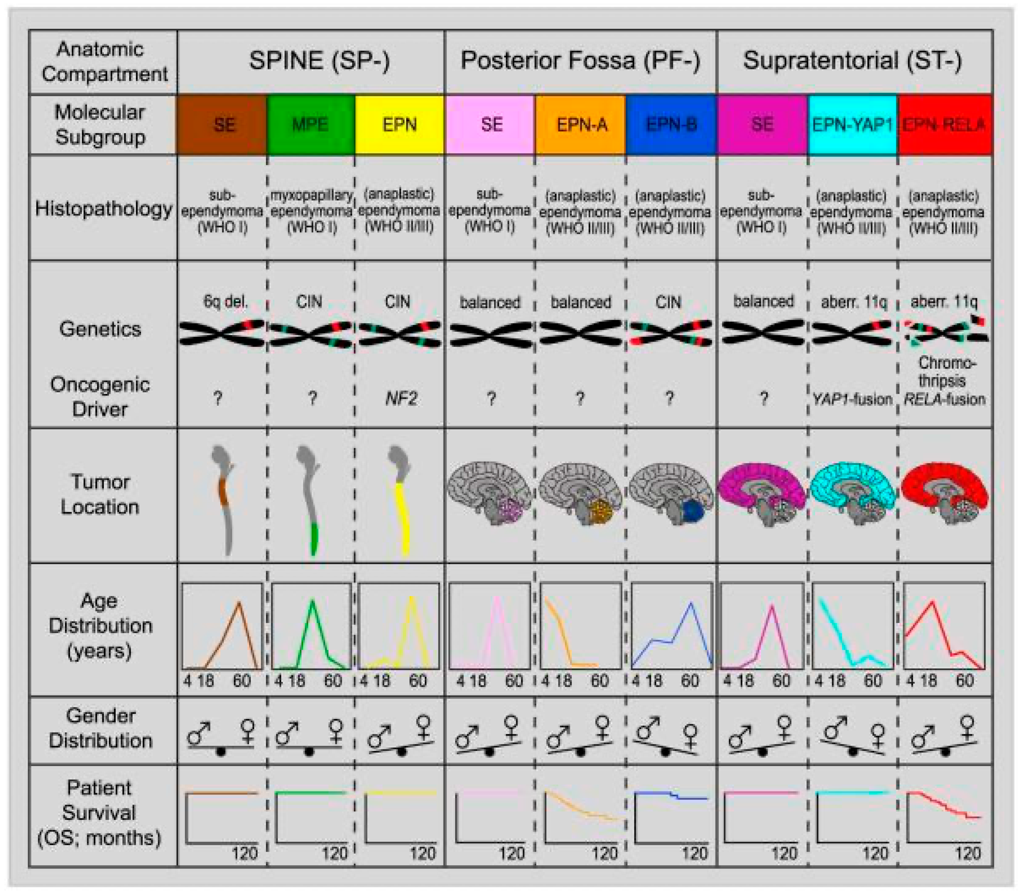
Historically, ependymomas were classified based on distinct hallmark histopathologic features identified through microscopy, with grading predominantly reliant on morphological criteria. Traditional diagnostic methods relied on features such as perivascular pseudorosettes, glial fibrillary acidic protein (GFAP) positivity, and, less commonly, ependymal rosettes [8]. These histopathological markers, along with cellularity, mitotic activity, microvascular proliferation, and presence of necrosis, were used to assign a World Health Organization (WHO) grade to the tumor to reflect its aggressiveness and potential clinical outcomes [9]. Although ependymomas arising from different anatomical locations may share similar histopathological features, they exhibit distinct site-specific genetic and epigenetic alterations, transcriptional profiles, and DNA copy number variations [10].
One of the most impactful tools in the molecular classification of ependymomas has been DNA methylation profiling. This approach has proven especially valuable in cases where traditional histopathologic features are insufficient or ambiguous, as it enables the identification of specific molecular signatures that distinguish between different subtypes [11]. As a result, the 2021 update to the World Health Organization Classification of Tumors of the Central Nervous System adopted a comprehensive molecular framework for categorizing ependymomas [5,12]. In this updated system, ependymomas are now classified into three main anatomical compartments based on the combination of histopathological features, molecular features, and location: supratentorial, posterior fossa, and spinal [5]. A notable exception to this compartmentalized classification is subependymomas, which can occur in all three anatomical regions, unlike the others (Figure 1) [4]. This shift recognizes that tumors in different parts of the CNS regions exhibit distinct genetic and epigenetic profiles, influencing their prognosis and treatment response (Figure 2 and Figure 3). The revised classification improves the precision of ependymoma diagnosis and prognosis by incorporating molecular data to more accurately reflect the underlying biology of these tumors.
Figure 2. Clinical, molecular, and demographic features of ependymoma subgroups across CNS compartments. This figure summarizes key characteristics of molecularly defined ependymoma subtypes categorized by anatomic location: spinal (SP), posterior fossa (PF), and supratentorial (ST). Each subgroup displays distinct histopathological patterns, genetic alterations, oncogenic drivers, and tumor localization. For instance, spinal ependymomas (SE, MPE, and EPN) frequently exhibit chromosomal instability (CIN) and NF2 loss, while supratentorial RELA- and YAP1-fused tumors are associated with chromothripsis and fusion-driven oncogenic activation. Age and gender distribution patterns vary by subgroup, with PF-EPN-A typically presenting in younger children and ST-RELA tumors affecting slightly older pediatric populations. Survival outcomes also differ markedly, with PF-A and ST-RELA subtypes associated with poorer prognoses compared to other groups. These distinctions underscore the importance of molecular classification in guiding diagnosis, prognosis, and therapeutic strategies [6].
Jmp 06 00023 g002

3. Grading of Ependymal Tumors

The grading system for ependymomas has been a subject of ongoing debate due to challenges in reproducibility and its clinical utility [1]. Notably, the 2021 WHO classification de-emphasized grading for many ependymoma subtypes compared to the 2016 edition, resulting in a less defined grading scale [5]. Pathologists may assign a grade 2 or grade 3 designation to ependymal tumors based on histopathological features rather than molecular status, except for myxopapillary ependymomas, which are explicitly classified as WHO grade 2. However, due to the lack of robust clinical trial data correlating molecular subtypes with patient outcomes, WHO grading has not been assigned to ependymoma subtypes defined by molecular alterations [11].

4. Supratentorial Ependymomas

Supratentorial ependymomas are classified into two subtypes based on gene fusions: Zinc Finger Translocation Associated (ZFTA) and Yes-associated protein 1 (YAP1) [5]. Ependymomas arising in the supratentorial compartment that lack these gene fusions are designated as “not otherwise specified” or “not elsewhere classified,” with grading determined histologically [13].

5. ZFTA-Fusion Positive Ependymomas

ST-ZFTA tumors are the most aggressive molecular subtype of supratentorial ependymomas and are associated with the worst clinical prognosis. Specifically, a 5-year progression-free survival (PFS) rate of 29% and overall survival (OS) rate of 75% have been reported for ZFTA-fusion supratentorial ependymomas [6]. These tumors predominantly arise in pediatric patients, with a median age of 8 years and a range from infancy to age 69; approximately 23% of cases occur in adults, with a peak in the sixth decade [14].
Molecularly, ZFTA-fusion ependymomas are identified by ZFTA-fusions, which activate oncogenic nuclear factor kappa-light-chain enhancer (NF-kB) signaling. Under physiological conditions, REL-associated protein (RELA) is sequestered in the cytoplasm and translocates to the nucleus under cellular stress to activate NF-kB [15]. In ZFTA-RELA fusions, this translocation occurs constitutively in neural stem cells, triggering NF-kB target gene activation and promoting ependymoma development. Therefore, sustained NF-kB activation is considered the primary oncogenic mechanism [16]. Initially classified based on RELA-ZFTA fusion, later studies revealed ZFTA (also known as C11orf95) is the central molecular driver in these tumors, regardless of its fusion partner, which may include RELA, mastermind like transcriptional coactivator 2/3 (MAML2/3), nuclear receptor coactivator 1/2 (NCOA1/2), Meningioma 1 (MN1), or Catenin Alpha 2 (CTNNA2). The recognition of the ZFTA-fusion subgroup, formerly referred to as RELA-fusion, was largely driven by Parker et al., who demonstrated that approximately two-thirds of supratentorial ependymomas harbor a RELA-ZFTA fusion linked to chromothripsis at chromosome 11q13.1 [4,16]. Pajtler et al. further identified the presence of ZFTA-fusion mutation in approximately 72% of supratentorial ependymomas [6].
While ZFTA-fusion is now recognized as the defining molecular hallmark of this subgroup, its exact oncogenic role remains to be fully elucidated. Nevertheless, its identification has spurred further research demonstrating that ZFTA-fusion proteins interact broadly across the genome, modifying chromatin structures to activate gene expression [17]. At the molecular level, recent studies have begun to illuminate ZFTA’s broader oncogenic role beyond simply acting as a fusion partner. ZFTA-fusion proteins act as chromatin modifiers, tethering transcriptional coactivators such as bromodomain-containing protein 4 (Brd4), histone acetyltransferase p300 (Ep300), and CREB-binding protein (Cbp) to thousands of genomic loci, creating a transcriptionally permissive environment for aberrant gene activation [18].
Given its aggressive clinical course and poor outcomes, ST-ZFTA has become a key focus for targeted therapeutic development. Aberrant activation of the NF-κB pathway and overexpression of receptor tyrosine kinases such as EPHB2 provide actionable targets in ZFTA-fusion tumors [16]. The multikinase inhibitor dasatinib has demonstrated potent preclinical activity by targeting EPHB2 and ABL1, resulting in tumor growth inhibition, immune reprogramming (M2-to-M1 macrophage polarization), and increased CD8+ T-cell activation. In syngeneic mouse models, dasatinib induced complete regression in 78% of tumors and prevented recurrence in a CD8+ T cell-dependent manner [19]. While preclinical data are compelling, clinical trials are needed to confirm efficacy and safety.
Additional tyrosine kinase inhibitors, including axitinib, imatinib, and pazopanib, have shown activity in high-risk pediatric ependymoma models by inhibiting PDGFRα/β signaling. Axitinib reduced mitotic gene expression and induced senescence in vitro [20]. EGFR inhibitors (e.g., gefitinib, AEE788) also impaired clonogenicity and prolonged survival in xenograft models [21]. Early-phase studies and preclinical screens suggest possible use of these drugs in high-risk and recurrent ependymomas, but clinical validation is pending. In addition to kinase inhibition, immune checkpoint blockade is a promising area of research. PD-L1 overexpression has been associated with T-cell exhaustion and poor prognosis in supratentorial ependymomas. These findings emphasize the importance of investigating PD-L1 inhibitors as adjunct therapies [22]. Continued advancement in understanding the molecular pathways driving aggressive ependymoma subtypes, such as ST-ZFTA, is essential for overcoming therapeutic barriers and developing targeted treatments that can improve outcomes for patients with historically poor prognoses.

6. YAP1-Fusion Ependymomas

In contrast, YAP1-fusion ependymomas represent a less aggressive molecular subtype, associated with a more favorable clinical outcome. A 5-year OS of 100% with gross total resection and a PFS of 66% have been reported [23]. These tumors primarily affect the pediatric population, with few occurrences documented in the adult population [6].
ST-YAP1 is characterized by recurrent gene fusions involving the YAP1 gene on chromosome 11, most commonly MAMLD1 on the X chromosome or, less commonly, with Family with Sequence Similarity 118 Member B (FAM118B). YAP1 encodes a transcriptional coactivator that plays a central role in the Hippo signaling pathway, which regulates cell proliferation, apoptosis, and tissue homeostasis. In the nucleus, YAP1 complexes with TEAD transcription factors to drive expression of pro-proliferative genes. Experimental studies have demonstrated that disrupting the YAP–TEAD interaction prevents tumor formation, while ectopic expression of YAP1 in mouse models induces ependymoma-like tumors, underscoring its central role in tumorigenesis and its potential as a therapeutic target [24,25]. Beyond Hippo signaling, YAP1 is involved in the Wnt/β-catenin pathway and interacts with Na+/H+ exchanger 3 regulating factor 1; ezrin–radixin–moesin (ERM) binding phosphoprotein 50 (NHERF1/EBP50), a tumor suppressor and organizer of ependymal cilia. NHERF1/EBP50 expression has also been identified as a diagnostic marker for ependymal tumors, aiding in differentiation from other CNS neoplasms. Furthermore, YAP1 activity has been implicated in therapeutic resistance, including resistance to radiation in glioblastoma and medulloblastoma, as well as immune evasion via upregulation of PD-L1, which inhibits T-cell activation. These mechanisms suggest broader relevance of YAP1 beyond ependymoma [26].
Therapeutic targeting of YAP1 is an emerging area of interest. In vitro and in vivo studies have shown that verteporfin, a photosensitizer approved for macular degeneration, inhibits YAP1/TEAD activity and suppresses tumor growth [27]. Additionally, compounds like dasatinib, fluvastatin, and pazopanib, all approved for other malignancies, have demonstrated inhibition of nuclear YAP/TAZ localization and reduction in cell proliferation in YAP-driven models. Pazopanib, in particular, induces proteasomal degradation of YAP/TAZ, offering a promising avenue for repurposing existing agents [28]. Despite the favorable prognosis and relatively well-defined molecular features, routine molecular testing for ST-YAP1 remains inconsistently applied in clinical practice.

7. Posterior Fossa Ependymomas

Posterior fossa ependymomas are now molecularly classified into posterior fossa group A (PF-A) and group B (PF-B) subtypes based on DNA methylation profiling. These subtypes are primarily distinguished by differences in histone H3 K27-trimethylation levels [5]. Immunohistochemical detection using an antibody against the trimethylated histone H3 at lysine 27 (H3K27me3) can differentiate tumors with high or low levels of this epigenetic marker [13,29]. Ependymomas in the posterior fossa that lack defining characteristics of either PF-A or PF-B are designated as “not otherwise specified” or “not elsewhere classified,” encompassing tumors without molecular analysis or those with other undefined alterations [4,13].

8. PF-A Ependymomas

PF-A ependymomas represent the more aggressive molecular subtype of posterior fossa ependymomas, primarily affecting children under five years of age, with a median age of diagnosis around 3 years. These tumors exhibit a marked male predominance (~65%) and represent the majority of pediatric PF ependymomas. Although rare, PF-A tumors have been identified in adults (1–18.5% of cases), often sharing molecular features with their pediatric counterparts [4,11]. Numerous studies have consistently demonstrated that PF-A tumors are associated with significantly poorer outcomes compared to PF-B counterparts, particularly in the presence of high-risk cytogenetic features such as chromosome 1q gain or 6q loss [6,11]. The 5-year overall survival (OS) rate is approximately 68%, while the progression-free survival (PFS) rate is significantly lower at 33%, with outcomes worsening in the presence of high-risk cytogenetic alterations such as chromosome 1q gain (17.3–25%) and 6q loss (6.4–8.6%) [6].
Molecularly, PF-A ependymomas are defined by a global reduction in histone H3 lysine 27 trimethylation (H3K27me3), with <80% H3K27me3-positive nuclei by immunohistochemistry serving as a diagnostic hallmark. The tumorigenic mechanisms of PF-A ependymomas are thought to resemble those of diffuse midline gliomas with H3K27M mutations, which result in the derepression of pro-oncogenic transcription factors through the reduction of histone trimethylation [29]. This reduction is strongly associated with Enhancer of Zeste Homologs Inhibitory Protein (EZHIP) overexpression, which occurs at high levels in PF-A tumors [30]. EZHIP functions as a potent inhibitor of polycomb repressive complex 2, blocking its ability to methylate H3K27 and thereby promoting tumorigenesis [13,31]. Less commonly, loss of Alpha-thalassemia, mental retardation, and X-linked (ATRX) protein have been reported in PF-A tumors, disrupting chromatin remodeling and contributing to DNA damage and genomic instability, further exacerbating tumor progression [13,32].
The tumor microenvironment also plays a significant role in PF-A pathobiology. Chronic activation of inflammatory markers, including Interleukin 6 (IL-6) and Signal Transducer and Activator of Transcription 3 (STAT3), as well as NF-kB signaling, has been shown to support tumor growth [13,33,34]. Additionally, PF-A tumors exhibit reduced expression of Leucine Zipper Down-regulated in Cancer 1 (LDOC1), a transcriptional repressor of NF-kB. Restoration of LDOC1 levels through DNA methylation inhibitors has been shown to reduce IL-6 secretion, thereby modulating this pro-tumorigenic signaling axis [13,35]. PF-A tumors are also influenced by hypoxic conditions, which alter histone methylation patterns and contribute to the emergence of a mesenchymal-like transcriptional signature. Hypoxia-induced genes involved in angiogenesis (e.g., VEGF, HIF-1α), wound healing, and cell adhesion/migration are enriched in PF-A, similar to mesenchymal glioblastoma [13,36]. Finally, telomerase activation contributes to PF-A tumorigenesis. Hypermethylation of the hTERT promoter and 1q gain are linked to increased telomerase activity in these tumors, supporting cellular immortality.
Despite its aggressive nature, targeted therapies for PF-A are lacking. However, insights into its molecular pathology have identified several promising avenues. Due to its epigenetic similarities, agents effective in DMG may also be beneficial in PF-A [13]. Notably, the combination of panobinostat (histone deacetylase inhibitor) and marizomib (proteasome inhibitor) has shown potent activity in preclinical models [37]. DNA methylation inhibitors like 5-azacytidine are under investigation but have shown limited clinical efficacy to date [35]. Given the critical role of EZHIP in PF-A oncogenesis, strategies aimed at disrupting the EZHIP–EZH2 interaction or detoxifying PRC2 target activation may offer targeted therapeutic options [38].
Additionally, PARP inhibitors, particularly when combined with radiotherapy, have demonstrated efficacy in preclinical models of EZHIP-overexpressing tumors, exploiting defects in DNA repair mechanisms [39]. Finally, Chimeric Antigen Receptor (CAR) T-cell therapy is being explored. Surface antigens such as EPHA2, HER2, and IL13Rα2, which are expressed in PF-A, are currently being targeted in preclinical models with intrathecal CAR-T cells, showing efficacy in both primary and metastatic settings [40].
Despite growing understanding of PF-A biology, integrating molecular profiling into standard-of-care clinical practice remains a significant challenge. Routine classification of ependymomas based on histopathology alone is insufficient to accurately diagnose PF-A, determine prognosis, or guide emerging targeted therapies. Immunohistochemical testing for H3K27me3 and, when feasible, DNA methylation profiling, are increasingly recommended to ensure accurate molecular subclassification. However, widespread implementation is hindered by issues of cost, limited availability of specialized testing platforms, and lack of access to centralized reference laboratories, particularly in resource-limited settings [41].

9. PF-B Ependymomas

In contrast, PF-B ependymomas are characterized by higher levels of H3 K27-trimethylation and occur more commonly in older children and adults, with a median age around 30 years [3,4]. These tumors are associated with a significantly better prognosis, with a reported 5-year OS of 100% and PFS of 73% [6]. PF-B tumors are rarely invasive or metastatic and exhibit a low likelihood of recurrence, particularly in patients older than 10 years [42].
Molecularly, PF-B tumors demonstrate substantial chromosomal instability, including frequent losses in 6p and 6q and gains in 15q, 18p, and 18q, but they do not harbor recurrent mutations [6,13,43]. They are biologically more differentiated than PF-A tumors, exhibiting features such as increased ciliogenesis, oxidative metabolism, and microtubule function [6,13,36]. Notably, FOXJ1, a key transcription factor involved in motile ciliogenesis and sonic hedgehog (Shh) signaling, is downregulated in PF-B tumors [44]. This supports an ependymal-like cellular trajectory, consistent with their less aggressive phenotype. Additionally, NHERF1/EBP50, a scaffold protein and diagnostic marker, regulates polarity structures and interacts with both β-catenin and YAP1 [26]. Dysregulation of the Wnt/β-catenin signaling pathway has been implicated in PF-B biology, and FOXJ1 overexpression in other tumor types has been shown to promote β-catenin nuclear translocation [13]. These interactions underscore the importance of β-catenin and NHERF1 pathways in tumor regulation of ependymomas.
Given their favorable prognosis and lower recurrence risk, PF-B ependymomas may be candidates for less intensive therapy following gross total resection. This raises the potential for treatment de-escalation in future clinical trials. Additionally, a novel NHERF1 PDZ1-domain inhibitor has emerged as a potential therapeutic for PF-B ependymomas. Originally developed for other indications, its combination with β-catenin inhibitors may offer a promising treatment avenue for this subtype [45]. Continued molecular investigation may enable more targeted treatment approaches for this subgroup.

10. Spinal Ependymomas

Ependymal tumors of the spinal cord include spinal ependymoma, spinal ependymoma with Myelocytomatosis-N (MYCN) amplification, myxopapillary ependymoma, and subependymoma. Both spinal ependymomas and those with specified MYCN amplification require spinal localization as diagnostic criteria, whereas myxopapillary and subependymomas can occur in other anatomical areas [4].
Spinal ependymomas represent a broad tumor category similar to the “not otherwise specified” or “not elsewhere classified” classifications. Diagnosis requires spinal localization and the absence of morphological features of myxopapillary or subependymomas [4]. These tumors primarily occur in the cervical spine but can also appear in the thoracic and lumbar regions, with a median diagnosis age of 41 [4,6]. Most exhibit chromosome 22q losses involving the neurofibromatosis type 2 (NF2) gene, and NF2 germline mutations are linked to NF2-related schwannomatosis (previously known as Neurofibromatosis Type 2). While the prevalence of these mutations in spinal ependymomas remains unclear, 33—53% of patients with NF2-related schwannomatosis show spinal ependymomas on imaging [46]. Despite potential recurrences, survival rates are favorable, though patients with NF2-related schwannomatosis often experience a high tumor burden, underscoring the need for safe, minimally invasive therapies [4,6].

11. Spinal Ependymomas with MYCN Amplification

One newly identified, high-risk spinal ependymoma subtype is characterized by MYCN amplification, a genomic alteration associated with aggressive behavior and poor clinical outcomes [11,47]. These tumors most commonly arise in the cervical and thoracic spine, with a median age at diagnosis of 32 years and a slight female predominance [4,47]. They are frequently present with intradural, extramedullary growth and demonstrate early leptomeningeal dissemination, recurrence, and metastasis. Histologically, they are high-grade, exhibiting pseudorosettes, papillary architecture, necrosis, microvascular proliferation, and elevated mitotic activity [4].
The defining oncogenic event is amplification of MYCN, a proto-oncogene on chromosome 2p24.3 that encodes a transcription factor essential for CNS development. MYCN amplification is consistently retained at relapse and is often accompanied by additional chromosomal alterations such as loss of 10q or focal 11q deletions, supporting its role in tumor progression [47]. Initial reports by Scheil et al. (2001) identified MYCN amplification in spinal ependymomas, and subsequent studies using DNA methylation profiling have confirmed its presence in a distinct molecular cluster of highly aggressive tumors [47,48,49]. These studies demonstrated median progression-free survival of just 17 months and overall survival of 7.3 years, the worst prognosis among spinal ependymoma subgroups [47].
While there are currently no approved therapies targeting MYCN, several experimental approaches are under investigation. MYCN inhibition by targeting histone deacetylase (HDAC), PARP, Auro A-kinase (AURKA), and Bromodomain and extra-terminal domain (BET) proteins are being explored in preclinical models, as well as immunotherapy targeting through DNA vaccination [13,50]. Although SPE-MYCN remains rare, its consistent molecular profile, aggressive behavior, and poor prognosis justify its classification as a distinct diagnostic and prognostic entity. Further research is needed to explore targeted therapies and validate MYCN amplification as a therapeutic vulnerability in this challenging ependymoma subtype.

12. Myxopapillary Ependymomas

Myxopapillary ependymoma is a distinct spinal tumor, most often found in the conus medullaris and filum terminale, though rare cases occur in the brain or beyond the CNS [4,11]. It primarily affects adults, with a median diagnosis age of 39 years. While overall survival exceeds 90% after surgery, about 20% of cases relapse, often linked to incomplete resection, large tumors, or multifocal disease [51,52]. Recognizing its potential for recurrence and metastasis, MPE was reclassified from WHO grade l to CNS WHO grade 2 [4]. It is characterized by GFAP positivity, papillary structures, and myxoid stoma, the latter being sufficient for diagnosis [4,11].
DNA methylation profiling has revealed chromosomal instability and widespread copy-number variations as defining, aiding in the diagnosis of challenging cases as this molecular subgroup occasionally includes tumors with classical ependymoma histology [53]. Emerging molecular data have identified recurrent overexpression of several genes, including HOXB13, a transcription factor normally involved in embryonic development of the lumbar spine but absent in adult filum terminale tissue, suggesting aberrant developmental gene reactivation in MPE tumorigenesis [54]. Recent studies in pediatric spinal ependymomas have confirmed HOXB13 overexpression and identified upregulation of genes involved in mitochondrial oxidative phosphorylation, such as COX2, a gene implicated in inflammation, tumor proliferation, and angiogenesis [55].
Despite these molecular insights, targeted therapies have not yet impacted clinical management due to the tumor’s favorable prognosis with surgery alone. As such, current research is focused on understanding the biological relevance of these findings rather than immediate therapeutic application. Larger, longitudinal studies are needed to determine whether molecular profiling can improve diagnostic precision or offer prognostic value beyond histopathology [4].

13. Subependymomas

Subependymomas are classified as supratentorial (ST-SE), spinal (SP-SE), or posterior fossa (PF-SE) in the 2021 CNS WHO classification, though they remain identified by morphological criteria without molecular classification. Subependymomas are slow-growing tumors with low cellularity and minimal proliferative activity [7]. The prognosis is favorable, often managed effectively with surgical resection alone, and the recurrence is rare even after partial removal [6].
SP-SE primarily affects the cervical or cervicothoracic spine, presenting with myelopathy or radicular pain, typically in adults (mean age 44), and with equal sex distribution [56,57]. Chromosome 6q deletion has emerged as a consistent and defining molecular feature, although its functional role in tumor biology and its prognostic implications remain uncertain [13]. Specifically, deletions at 6q25.3 have been associated with favorable outcomes, whereas 6q23 deletions have been linked to disease progression [58,59]. While the contribution of 6q loss in SE tumorigenesis and behavior requires further research, parallels in other malignancies suggest it may serve as a therapeutic target. For example, genes located in these regions, such as the proto-oncogene c-myeloblastosis gene and the tumor suppressor ephrin type-A receptor 7, are being explored for their roles in oncogenesis [13].
PF-SE, most often in the fourth ventricle, occurs mainly in older males (mean age 60) and is often asymptomatic. Gross total resection is generally curative, with high survival rates; radiotherapy is rarely used but considered for recurrences [6,60]. However, a subset of posterior fossa subependymomas with telomerase reverse transcriptase (TERT) promoter mutations or chromosome 6 loss has been linked to worse outcomes [61]. Although molecular alterations involving KIT and STAT signaling pathways have been identified, these currently lack actionable therapeutic relevance [6,13].
ST-SE, usually in the lateral ventricles, presents with CSF obstruction symptoms, mostly in middle-aged men [6,62,63]. The prognosis is excellent, with nearly 100% survival after gross total resection, which is often feasible due to its location; adjuvant radiotherapy is generally unnecessary [6,64]. Research has identified potential targets like p53, mouse double minute 2 homolog, and STAT3 inhibitors [13,65].
Although molecular studies have enhanced our understanding of subependymoma biology, they have yet to significantly impact clinical decision-making. Current treatment outcomes are excellent for all three subependymoma subtypes, reducing the immediate need for molecularly targeted therapies. As such, molecular profiling is typically reserved for diagnostically ambiguous cases rather than routine clinical management [11]. The presence of recurring molecular alterations, such as 6q deletions or pathway-level changes, highlights a gap in understanding whether these are true oncogenic drivers or incidental findings. Further large-scale, longitudinal studies are needed to determine their biological relevance and potential clinical utility in a tumor type where conventional treatment is already highly effective.
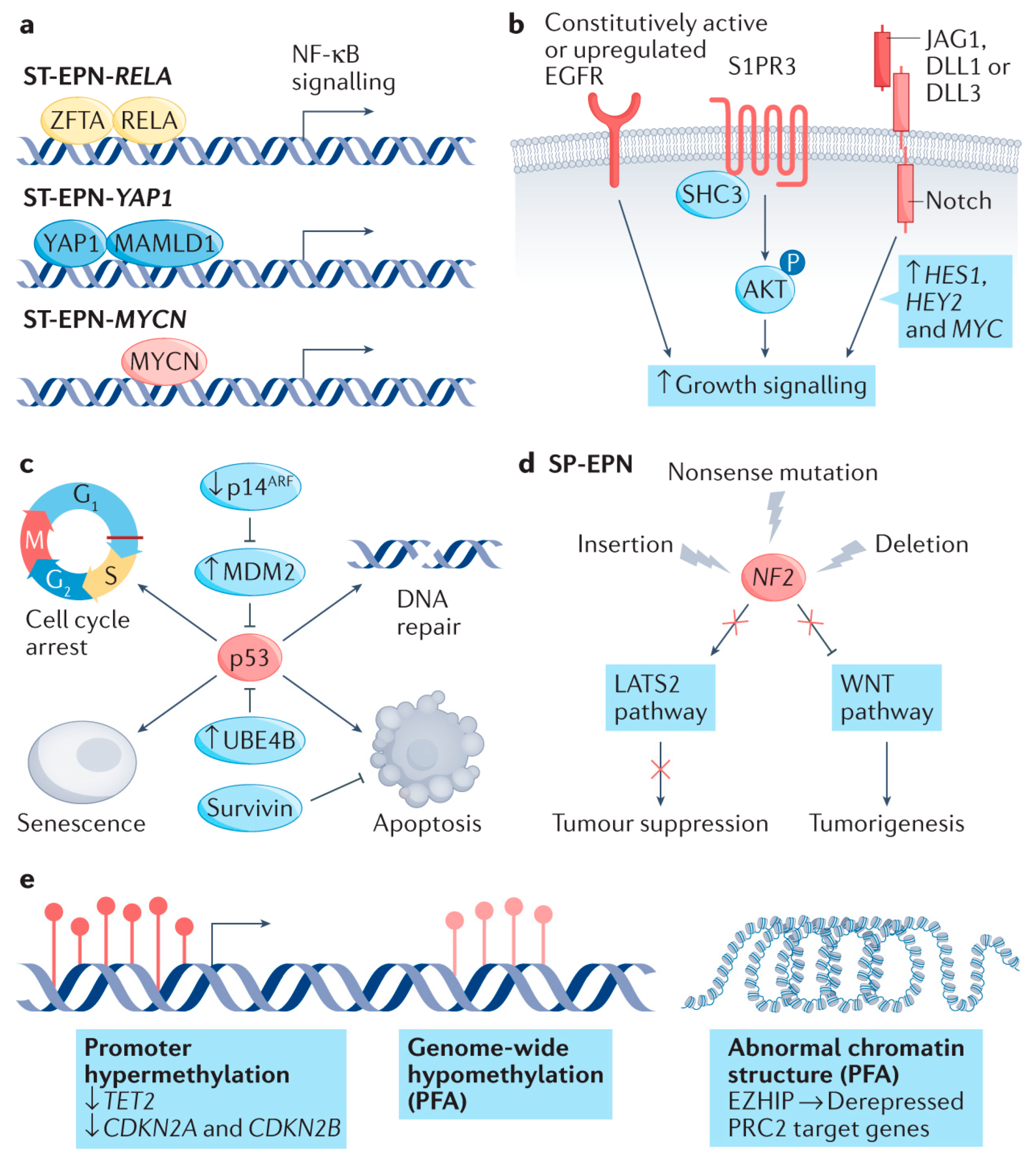
Figure 3. Molecular mechanisms contributing to ependymoma tumorigenesis. Multiple biological pathways contribute to ependymoma development, including dysregulated transcription, aberrant cell signaling, and epigenetic alterations. (a) Distinct oncogenic fusions such as ZFTA–RELA, YAP1–MAMLD1, and MYCN amplification activate subtype-specific transcriptional programs in supratentorial and spinal ependymoma subgroups. (b) Overactivation of receptor-mediated pathways—including EGFR, S1PR3, and Notch—facilitates unchecked cellular proliferation across various ependymoma types. (c) While TP53 mutations are rare, disruption of p53-associated signaling cascades promotes unchecked cell cycle activity and impairs programmed cell death leading to alterations as seen in cell cycle arrest, DNA repair, cell senescence, and apoptosis. (d) In spinal ependymomas, genetic alterations in the NF2 gene—commonly loss-of-function mutations—result in failure to suppress tumorigenic WNT signaling or LATS2 pathway leading to increased tumorigenesis and a decrease in tumor suppression. (e) Epigenetic disruptions, such as widespread DNA hypomethylation, promoter hypermethylation of tumor suppressors, and EZHIP-driven chromatin remodeling, are central to disease progression, especially in PFA tumors [66].
Jmp 06 00023 g003

14. Therapeutic Implications and Limitations to Current Standard of Care

Molecular stratification has revealed significant heterogeneity in the biology, clinical behavior, and therapeutic response of ependymomas. These distinctions challenge the adequacy of uniform treatment strategies across all subtypes. Yet, despite this growing molecular insight, maximal safe surgical resection followed by adjuvant radiation remains largely agnostic to tumor subtype. The variable response to this conventional approach reflects underlying biological complexity [13]. Contributing factors include limited drug penetration across the blood–brain barrier, intra-tumoral heterogeneity, and intrinsic resistance mechanisms [66]. For instance, the expression of apurinic/apyrimidinic endonuclease 1 (APE1) has been inversely correlated with radiosensitivity in pediatric ependymomas. Experimental suppression of APE1 through antisense oligonucleotides or siRNA has been shown to enhance radiation responsiveness [67].
Molecular profiling is increasingly being implemented in clinical practice, enabling the identification of key genetic and epigenetic alterations that drive ependymoma subtypes. These discoveries, outlined earlier in this paper, have laid the foundation for subtype-specific therapeutic strategies. However, most targeted approaches remain investigational and have yet to be incorporated into routine clinical care. As a result, chemotherapy continues to play a limited and often inconsistent role, with little subtype-specific evidence to guide its use.
The integration of molecular diagnostics into treatment planning is therefore not only timely but essential. Subtype-informed therapy has the potential to improve outcomes, minimize overtreatment in favorable-risk patients, and guide the development of novel targeted interventions. The following sections examine historical and ongoing efforts to refine therapeutic approaches through both cytotoxic agents and molecularly guided clinical trials.

15. Historical Clinical Trials

Cytotoxic agents have historically shown limited efficacy in ependymoma, particularly in recurrent or refractory cases. Temozolomide, a commonly used alkylating agent in glioblastoma, has had mixed results in adult ependymoma. A 2009 study reported limited benefit in recurrent intracranial cases, though a 2016 study noted partial or complete responses in a subset of patients [68,69]. A 2021 phase II trial investigating dose-dense temozolomide combined with daily lapatinib, a tyrosine kinase inhibitor, showed encouraging 6- and 12-month progression-free survival (PFS) rates of 55% and 38%, respectively, in recurrent supratentorial, infratentorial, and spinal ependymomas [70].
Resistance to chemotherapy in ependymoma is frequently linked to elevated expression of DNA repair enzymes such as O6-methylguanine-DNA methyltransferase (MGMT), particularly in recurrent pediatric tumors [71]. Preclinical studies suggest that temozolomide may suppress the tumor-initiating potential of stem-like ependymoma cells with low MGMT expression, supporting a potential role in selected molecular contexts [72]. Another resistance mechanism involves the efflux transporter ABCB1, which reduces intracellular drug accumulation. In vitro inhibition of ABCB1 using agents such as vardenafil and verapamil enhanced the cytotoxic effects of vincristine, etoposide, and methotrexate, while also impairing migration and invasion in pediatric ependymoma models [73]. Preliminary data from a randomized clinical trial of 325 pediatric patients (ages 1–21) showed that maintenance chemotherapy following surgery and radiotherapy improved 3-year event-free survival (EFS) from 71% to 80% [74].

16. Ongoing Research to Refine Treatment

The limited success of non-stratified chemotherapeutic approaches has fueled interest in molecularly targeted therapies and biologically guided clinical trials, especially for high-risk or recurrent disease. A summary of the key clinical trials and molecular studies is provided in Table 1. Initial efforts by the Collaborative Ependymoma Research Network (CERN) Foundation tested combinations such as carboplatin plus bevacizumab and lapatinib plus 5-FU across various subgroups, but these early trials (NCT01295944, NCT00883688, NCT01498783) often lacked molecular stratification, limiting their impact [75].
Subsequent trials have underscored the importance of integrating molecular classification into therapeutic design. The SIOP Ependymoma I trial (NCT00004224) and the Children’s Oncology Group ACNS0831 trial (NCT01096368) both reinforced the primacy of maximal safe resection and adjuvant radiotherapy, showing that the addition of chemotherapy offers limited benefit in unstratified populations [74,76]. These studies laid the groundwork for the SIOP Ependymoma II trial (NCT02265770), the first major prospective international effort to incorporate centralized molecular profiling into treatment algorithms. This trial tailors therapy based on age, tumor location, and molecular subtype, directly addressing key limitations from prior studies [77]. In parallel, translational efforts are increasingly focused on targeted agents and immunotherapies designed for specific molecular subtypes. Preclinical studies of kinase inhibitors, epigenetic modulators, and immune checkpoint blockers have shown promise in high-risk variants, such as ST-ZFTA and PF-A, although robust clinical efficacy data are still forthcoming.
Collectively, these findings highlight the limitations of a one-size-fits-all approach and emphasize the value of biology-driven treatment paradigms. As our understanding of ependymoma molecular subtypes continues to evolve, so too does the potential for more personalized, effective therapeutic strategies, particularly for aggressive variants with limited treatment options.
Table 1. Key clinical trials and molecular studies shaping the contemporary classification and management of ependymoma.
Trial/Study NameStatusYearsFocus/DesignMolecular Subtypes/FindingsImpact on Classification/ManagementReferences
Genome-wide Methylation ProfilingCompleted2011–2014Retrospective molecular profilingIdentified PFA, PFB, ST-ZFTA, ST-YAP1Established molecular subgroups, prognostic value[66,78,79]
ACNS0831 (COG)Completed2009–2019Randomized, maintenance chemo vs. observationNot molecularly stratified (retrospective analyses performed)No benefit of maintenance chemo; informed SIOP EPII designNCT01096368
[74]
Large-Scale Methylation StudiesCompleted2018–2023Multicenter, outcome correlationAll subtypesValidated molecular subgroups, improved risk stratification[66,78,79]
cIMPACT-NOW Update 7Completed2020Consensus classification updateZFTA, YAP1, PFA, PFB, MYCNFormalized molecular classification, dropped anaplastic grading[11]
WHO CNS5 (2021)Completed2021Classification updateTen molecularly defined subtypesStandardized molecular nomenclature, clinical relevance[4]
SIOP Ependymoma IIOngoing2014–Prospective, molecularly stratified, internationalCentralized molecular stratification (PFA, PFB, ST-ZFTA, ST-YAP1)Integrates molecular data into therapy, biomarker-driven managementNCT02265770
[77]
BIOMECA (SIOP EPII)Ongoing2020–Prospective biomarker validationPFA, PFB, ST-ZFTA, ST-YAP1Validated H3K27me3 IHC for PFA, fusion detection methodsNCT02265770
[80]
This table summarizes pivotal clinical trials and published research that have contributed to the evolving molecular classification and therapeutic strategies for ependymoma. Completed and ongoing efforts, including genome-wide methylation profiling, large-scale outcome studies, and prospective molecularly stratified trials (e.g., SIOP Ependymoma II and BIOMECA), have informed recent consensus classification systems (cIMPACT-NOW Update 7 and WHO CNS5, 2021). These efforts have delineated key molecular subtypes, such as PFA, PFB, ST-ZFTA, ST-YAP1, and MYCN-amplified variants, thereby enabling biomarker-driven risk stratification and guiding future clinical trial designs.

17. Discussion

The 2021 revision of the WHO classification of CNS tumors marked a pivotal evolution in the field of neuro-oncology by formally prioritizing molecular and genetic alterations over traditional histopathology. For ependymomas, this shift has refined diagnostic specificity and provided a framework for biologically informed risk stratification. The identification of distinct molecular subgroups has enabled clinicians and researchers to better predict clinical behavior and prognosis. However, while diagnostic taxonomy has advanced, therapeutic approaches remain largely stagnant. Despite recognition of biologically diverse subgroups, current treatment protocols continue to rely on maximal safe surgical resection and adjuvant radiation therapy, regardless of molecular classification. This one-size-fits-all model neglects the distinct oncogenic drivers and therapeutic vulnerabilities unique to each subtype. Chemotherapy, though used in various clinical contexts, still lacks robust, subtype-specific evidence of benefit. Furthermore, most therapeutic trials have enrolled molecularly heterogeneous populations, blurring outcome interpretation and hindering meaningful progress in targeted treatment development.
A key limitation in current practice is the underutilization of molecular profiling at disease recurrence. While initial molecular testing is now standard in many pediatric centers, it remains inconsistently applied in adult and resource-limited settings, where access to RNA sequencing, FISH, or methylation profiling may be restricted. Even when testing is performed at diagnosis, molecular evolution at relapse is rarely reassessed. This is a critical oversight, as secondary alterations may underlie treatment resistance and reveal novel, actionable targets. Routine re-biopsy and molecular re-characterization at progression must become standard practice to inform adaptive treatment strategies.
Beyond diagnostics, there is a concerning lag in translating molecular insights into rational therapeutic interventions. Many oncogenic drivers such as ZFTA fusions or EZHIP overexpression have been identified, yet few have yielded viable targeted therapies. The literature remains dominated by preclinical findings without clinical correlation, and functional studies elucidating the downstream effects of these alterations are limited. Without this mechanistic clarity, efforts to develop or repurpose drugs risk being misdirected or prematurely abandoned. Compounding this issue is the inadequate integration of molecular subgrouping into clinical trial design. Most trials continue to treat ependymoma as a unified entity, undermining the potential to detect subtype-specific efficacy signals. This not only diminishes trial sensitivity but may also obscure differential toxicity profiles and limit therapeutic refinement. Trials such as SIOP Ependymoma II represent an important step forward by incorporating molecular stratification prospectively, but broader adoption of such frameworks is urgently needed.
Moreover, logistical and economic barriers continue to impede the equitable implementation of molecular diagnostics, particularly in adult-focused or low-resource centers. Delays in turnaround time and prohibitive costs can limit access to timely molecular results, which are critical for tailoring treatment intensity, especially in differentiating indolent subtypes from aggressive ones. As the field shifts toward a precision medicine paradigm, investment in scalable, rapid, and cost-effective diagnostic infrastructure will be essential to ensure access for all patient populations.

18. Future Directions

To fully translate molecular insights into clinical impact, the field must adopt a more integrative and translationally focused research agenda. Key priorities include:
  • Routine longitudinal molecular profiling at diagnosis and recurrence to capture tumor evolution and guide treatment adaptation.
  • Functional validation of key oncogenic drivers to elucidate mechanisms of tumor initiation, progression, and resistance.
  • Development of robust, subtype-specific preclinical models to support rational drug development and testing.
  • Prospective, molecularly stratified clinical trials with clearly defined biologically relevant endpoints to identify targeted therapeutic opportunities.
  • Standardization and global accessibility of molecular diagnostics through investments in infrastructure, training, and cost-containment strategies.
Ultimately, the integration of molecular pathology into clinical decision-making must extend beyond diagnostics. With coordinated efforts spanning basic science, translational research, and health systems infrastructure, it will be possible to transform the molecular subclassification of ependymoma from a descriptive tool into a foundation for precision oncology.

Author Contributions

Concept and design: S.R. Drafting of the manuscript and Critical revision: A.S., A.C., A.K., E.V., S.R. All authors have read and agreed to the published version of the manuscript.

Funding

No funding was required for this publication.

Conflicts of Interest

The authors declare no conflicts of interest.

Abbreviations

The following abbreviations are used in this manuscript:
CNSCentral Nervous System
GFAPglial fibrillary acidic protein
SEsubependymoma
PF-APosterior fossa group A
PF-BPosterior fossa group B
MYCNMYCN-amplified
MPEMyxopapillary ependymomas
ZFTAZinc Finger Translocation Associated
YAP1 Yesassociated protein 1
H3K27me3trimethylated histone H3 at lysine 27
SP-SESpinal Subependymoma
PFPosterior Fossa Subependymoma
ST–SESupratentorial Subependymoma
CERNCollaborative Ependymoma Research Network
HDAChistone deacetylases

References

  1. Damodharan, S.; Puccetti, D. Pediatric Central Nervous System Tumor Overview and Emerging Treatment Considerations. Brain Sci. 2023, 13, 1106. [Google Scholar] [CrossRef]
  2. Jünger, S.T.; Timmermann, B.; Pietsch, T. Pediatric ependymoma: An overview of a complex disease. Child’s Nerv. Syst. 2021, 37, 2451–2463. [Google Scholar] [CrossRef]
  3. Pajtler, K.W.; Mack, S.C.; Ramaswamy, V.; Smith, C.A.; Witt, H.; Smith, A.; Hansford, J.R.; von Hoff, K.; Wright, K.D.; Hwang, E.; et al. The current consensus on the clinical management of intracranial ependymoma and its distinct molecular variants. Acta Neuropathol. 2017, 133, 5–12. [Google Scholar] [CrossRef]
  4. Kresbach, C.; Neyazi, S.; Schüller, U. Updates in the classification of ependymal neoplasms: The 2021 WHO Classification and beyond. Brain Pathol. 2022, 32, e13068. [Google Scholar] [CrossRef]
  5. Louis, D.N.; Perry, A.; Wesseling, P.; Brat, D.J.; Cree, I.A.; Figarella-Branger, D.; Hawkins, C.; Ng, H.K.; Pfister, S.M.; Reifenberger, G.; et al. The 2021 WHO Classification of Tumors of the Central Nervous System: A summary. Neuro-Oncology 2021, 23, 1231–1251. [Google Scholar] [CrossRef]
  6. Pajtler, K.W.; Witt, H.; Sill, M.; Jones, D.T.; Hovestadt, V.; Kratochwil, F.; Wani, K.; Tatevossian, R.; Punchihewa, C.; Johann, P.; et al. Molecular Classification of Ependymal Tumors across All CNS Compartments, Histopathological Grades, and Age Groups. Cancer Cell 2015, 27, 728–743. [Google Scholar] [CrossRef] [PubMed]
  7. Soni, N.; Ora, M.; Bathla, G.; Desai, A.; Gupta, V.; Agarwal, A. Ependymal Tumors: Overview of the Recent World Health Organization Histopathologic and Genetic Updates with an Imaging Characteristic. AJNR Am. J. Neuroradiol. 2024, 45, 1624–1634. [Google Scholar] [CrossRef] [PubMed]
  8. Bertero, L.; Ricci, A.A.; Tampieri, C.; Cassoni, P.; Modena, P. Ependymomas. Pathologica 2022, 114, 436–446. [Google Scholar] [CrossRef]
  9. Louis, D.N.; Ohgaki, H.; Wiestler, O.D.; Cavenee, W.K.; Burger, P.C.; Jouvet, A.; Scheithauer, B.W.; Kleihues, P. The 2007 WHO classification of tumours of the central nervous system. Acta Neuropathol. 2007, 114, 97–109. [Google Scholar] [CrossRef] [PubMed]
  10. Santi, M.; Viaene, A.N.; Hawkins, C. Ependymal Tumors. Pediatr. Dev. Pathol. 2022, 25, 59–67. [Google Scholar] [CrossRef]
  11. Ellison, D.W.; Aldape, K.D.; Capper, D.; Fouladi, M.; Gilbert, M.R.; Gilbertson, R.J.; Hawkins, C.; Merchant, T.E.; Pajtler, K.; Venneti, S.; et al. cIMPACT-NOW update 7: Advancing the molecular classification of ependymal tumors. Brain Pathol. 2020, 30, 863–866. [Google Scholar] [CrossRef]
  12. Louis, D.N.; Perry, A.; Reifenberger, G.; von Deimling, A.; Figarella-Branger, D.; Cavenee, W.K.; Ohgaki, H.; Wiestler, O.D.; Kleihues, P.; Ellison, D.W. The 2016 World Health Organization Classification of Tumors of the Central Nervous System: A summary. Acta Neuropathol. 2016, 131, 803–820. [Google Scholar] [CrossRef]
  13. Larrew, T.; Saway, B.F.; Lowe, S.R.; Olar, A. Molecular Classification and Therapeutic Targets in Ependymoma. Cancers 2021, 13, 6218. [Google Scholar] [CrossRef]
  14. Hübner, J.M.; Kool, M.; Pfister, S.M.; Pajtler, K.W. Epidemiology, molecular classification and WHO grading of ependymoma. J. Neurosurg. Sci. 2018, 62, 46–50. [Google Scholar] [CrossRef]
  15. Ozawa, T.; Kaneko, S.; Szulzewsky, F.; Qiao, Z.; Takadera, M.; Narita, Y.; Kondo, T.; Holland, E.C.; Hamamoto, R.; Ichimura, K. C11orf95-RELA fusion drives aberrant gene expression through the unique epigenetic regulation for ependymoma formation. Acta Neuropathol. Commun. 2021, 9, 36. [Google Scholar] [CrossRef] [PubMed]
  16. Parker, M.; Mohankumar, K.M.; Punchihewa, C.; Weinlich, R.; Dalton, J.D.; Li, Y.; Lee, R.; Tatevossian, R.G.; Phoenix, T.N.; Thiruvenkatam, R.; et al. C11orf95-RELA fusions drive oncogenic NF-κB signalling in ependymoma. Nature 2014, 506, 451–455. [Google Scholar] [CrossRef]
  17. Kupp, R.; Ruff, L.; Terranova, S.; Nathan, E.; Ballereau, S.; Stark, R.; Sekhar Reddy Chilamakuri, C.; Hoffmann, N.; Wickham-Rahrmann, K.; Widdess, M.; et al. Translocations Constitute Ependymoma Chromatin Remodeling and Transcription Factors. Cancer Discov. 2021, 11, 2216–2229. [Google Scholar] [CrossRef]
  18. Lötsch, D.; Kirchhofer, D.; Englinger, B.; Jiang, L.; Okonechnikov, K.; Senfter, D.; Laemmerer, A.; Gabler, L.; Pirker, C.; Donson, A.M.; et al. Targeting fibroblast growth factor receptors to combat aggressive ependymoma. Acta Neuropathol. 2021, 142, 339–360. [Google Scholar] [CrossRef] [PubMed]
  19. Ren, J.; Amoozgar, Z.; Uccello, T.P.; Lei, P.J.; Zhao, Y.; Ho, W.W.; Huang, P.; Kardian, A.; Mack, S.C.; Duda, D.G.; et al. Targeting EPHB2/ABL1 restores antitumor immunity in preclinical models of ependymoma. Proc. Natl. Acad. Sci. USA 2025, 122, e2319474122. [Google Scholar] [CrossRef]
  20. Donson, A.M.; Amani, V.; Warner, E.A.; Griesinger, A.M.; Witt, D.A.; Levy, J.M.M.; Hoffman, L.M.; Hankinson, T.C.; Handler, M.H.; Vibhakar, R.; et al. Identification of FDA-Approved Oncology Drugs with Selective Potency in High-Risk Childhood Ependymoma. Mol. Cancer Ther. 2018, 17, 1984–1994. [Google Scholar] [CrossRef] [PubMed]
  21. Servidei, T.; Meco, D.; Trivieri, N.; Patriarca, V.; Vellone, V.G.; Zannoni, G.F.; Lamorte, G.; Pallini, R.; Riccardi, R. Effects of epidermal growth factor receptor blockade on ependymoma stem cells in vitro and in orthotopic mouse models. Int. J. Cancer 2012, 131, E791–E803. [Google Scholar] [CrossRef] [PubMed]
  22. Wang, L.; Han, S.; Yan, C.; Yang, Y.; Li, Z.; Yang, Z. The role of clinical factors and immunocheckpoint molecules in the prognosis of patients with supratentorial extraventricular ependymoma: A single-center retrospective study. J. Cancer Res. Clin. Oncol. 2021, 147, 1259–1270. [Google Scholar] [CrossRef] [PubMed]
  23. Andreiuolo, F.; Varlet, P.; Tauziède-Espariat, A.; Jünger, S.T.; Dörner, E.; Dreschmann, V.; Kuchelmeister, K.; Waha, A.; Haberler, C.; Slavc, I.; et al. Childhood supratentorial ependymomas with YAP1-MAMLD1 fusion: An entity with characteristic clinical, radiological, cytogenetic and histopathological features. Brain Pathol. 2019, 29, 205–216. [Google Scholar] [CrossRef]
  24. Eder, N.; Roncaroli, F.; Domart, M.C.; Horswell, S.; Andreiuolo, F.; Flynn, H.R.; Lopes, A.T.; Claxton, S.; Kilday, J.P.; Collinson, L.; et al. YAP1/TAZ drives ependymoma-like tumour formation in mice. Nat. Commun. 2020, 11, 2380. [Google Scholar] [CrossRef]
  25. Pajtler, K.W.; Wei, Y.; Okonechnikov, K.; Silva, P.B.G.; Vouri, M.; Zhang, L.; Brabetz, S.; Sieber, L.; Gulley, M.; Mauermann, M.; et al. YAP1 subgroup supratentorial ependymoma requires TEAD and nuclear factor I-mediated transcriptional programmes for tumorigenesis. Nat. Commun. 2019, 10, 3914. [Google Scholar] [CrossRef]
  26. Georgescu, M.M.; Yell, P.; Mobley, B.C.; Shang, P.; Georgescu, T.; Wang, S.H.; Canoll, P.; Hatanpaa, K.J.; White, C.L.; Raisanen, J.M. NHERF1/EBP50 is an organizer of polarity structures and a diagnostic marker in ependymoma. Acta Neuropathol. Commun. 2015, 3, 11. [Google Scholar] [CrossRef] [PubMed]
  27. Liu-Chittenden, Y.; Huang, B.; Shim, J.S.; Chen, Q.; Lee, S.J.; Anders, R.A.; Liu, J.O.; Pan, D. Genetic and pharmacological disruption of the TEAD-YAP complex suppresses the oncogenic activity of YAP. Genes Dev. 2012, 26, 1300–1305. [Google Scholar] [CrossRef]
  28. Oku, Y.; Nishiya, N.; Shito, T.; Yamamoto, R.; Yamamoto, Y.; Oyama, C.; Uehara, Y. Small molecules inhibiting the nuclear localization of YAP/TAZ for chemotherapeutics and chemosensitizers against breast cancers. FEBS Open Bio 2015, 5, 542–549. [Google Scholar] [CrossRef]
  29. Bayliss, J.; Mukherjee, P.; Lu, C.; Jain, S.U.; Chung, C.; Martinez, D.; Sabari, B.; Margol, A.S.; Panwalkar, P.; Parolia, A.; et al. Lowered H3K27me3 and DNA hypomethylation define poorly prognostic pediatric posterior fossa ependymomas. Sci. Transl. Med. 2016, 8, 366ra161. [Google Scholar] [CrossRef]
  30. Pajtler, K.W.; Wen, J.; Sill, M.; Lin, T.; Orisme, W.; Tang, B.; Hübner, J.M.; Ramaswamy, V.; Jia, S.; Dalton, J.D.; et al. Molecular heterogeneity and CXorf67 alterations in posterior fossa group A (PFA) ependymomas. Acta Neuropathol. 2018, 136, 211–226. [Google Scholar] [CrossRef]
  31. Nambirajan, A.; Sharma, A.; Rajeshwari, M.; Boorgula, M.T.; Doddamani, R.; Garg, A.; Suri, V.; Sarkar, C.; Sharma, M.C. EZH2 inhibitory protein (EZHIP/Cxorf67) expression correlates strongly with H3K27me3 loss in posterior fossa ependymomas and is mutually exclusive with H3K27M mutations. Brain Tumor Pathol. 2021, 38, 30–40. [Google Scholar] [CrossRef] [PubMed]
  32. Dyer, M.A.; Qadeer, Z.A.; Valle-Garcia, D.; Bernstein, E. ATRX and DAXX: Mechanisms and Mutations. Cold Spring Harb. Perspect. Med. 2017, 7, a026567. [Google Scholar] [CrossRef]
  33. Griesinger, A.M.; Josephson, R.J.; Donson, A.M.; Mulcahy Levy, J.M.; Amani, V.; Birks, D.K.; Hoffman, L.M.; Furtek, S.L.; Reigan, P.; Handler, M.H.; et al. Interleukin-6/STAT3 Pathway Signaling Drives an Inflammatory Phenotype in Group A Ependymoma. Cancer Immunol. Res. 2015, 3, 1165–1174. [Google Scholar] [CrossRef]
  34. Griesinger, A.M.; Witt, D.A.; Grob, S.T.; Georgio Westover, S.R.; Donson, A.M.; Sanford, B.; Mulcahy Levy, J.M.; Wong, R.; Moreira, D.C.; DeSisto, J.A.; et al. NF-κB upregulation through epigenetic silencing of LDOC1 drives tumor biology and specific immunophenotype in Group A ependymoma. Neuro-Oncology 2017, 19, 1350–1360. [Google Scholar] [CrossRef]
  35. Michealraj, K.A.; Kumar, S.A.; Kim, L.J.Y.; Cavalli, F.M.G.; Przelicki, D.; Wojcik, J.B.; Delaidelli, A.; Bajic, A.; Saulnier, O.; MacLeod, G.; et al. Metabolic Regulation of the Epigenome Drives Lethal Infantile Ependymoma. Cell 2020, 181, 1329–1345.e1324. [Google Scholar] [CrossRef]
  36. Witt, H.; Mack, S.C.; Ryzhova, M.; Bender, S.; Sill, M.; Isserlin, R.; Benner, A.; Hielscher, T.; Milde, T.; Remke, M.; et al. Delineation of two clinically and molecularly distinct subgroups of posterior fossa ependymoma. Cancer Cell 2011, 20, 143–157. [Google Scholar] [CrossRef]
  37. Lin, G.L.; Wilson, K.M.; Ceribelli, M.; Stanton, B.Z.; Woo, P.J.; Kreimer, S.; Qin, E.Y.; Zhang, X.; Lennon, J.; Nagaraja, S.; et al. Therapeutic strategies for diffuse midline glioma from high-throughput combination drug screening. Sci. Transl. Med. 2019, 11, eaaw0064. [Google Scholar] [CrossRef]
  38. Jain, S.U.; Do, T.J.; Lund, P.J.; Rashoff, A.Q.; Diehl, K.L.; Cieslik, M.; Bajic, A.; Juretic, N.; Deshmukh, S.; Venneti, S.; et al. PFA ependymoma-associated protein EZHIP inhibits PRC2 activity through a H3 K27M-like mechanism. Nat. Commun. 2019, 10, 2146. [Google Scholar] [CrossRef] [PubMed]
  39. Han, J.; Yu, M.; Bai, Y.; Yu, J.; Jin, F.; Li, C.; Zeng, R.; Peng, J.; Li, A.; Song, X.; et al. Elevated CXorf67 Expression in PFA Ependymomas Suppresses DNA Repair and Sensitizes to PARP Inhibitors. Cancer Cell 2020, 38, 844–856.e847. [Google Scholar] [CrossRef] [PubMed]
  40. Donovan, L.K.; Delaidelli, A.; Joseph, S.K.; Bielamowicz, K.; Fousek, K.; Holgado, B.L.; Manno, A.; Srikanthan, D.; Gad, A.Z.; Van Ommeren, R.; et al. Locoregional delivery of CAR T cells to the cerebrospinal fluid for treatment of metastatic medulloblastoma and ependymoma. Nat. Med. 2020, 26, 720–731. [Google Scholar] [CrossRef]
  41. de Sousa, G.R.; Lira, R.C.P.; de Almeida Magalhães, T.; da Silva, K.R.; Nagano, L.F.P.; Saggioro, F.P.; Baroni, M.; Marie, S.K.N.; Oba-Shinjo, S.M.; Brandelise, S.; et al. A coordinated approach for the assessment of molecular subgroups in pediatric ependymomas using low-cost methods. J. Mol. Med. 2021, 99, 1101–1113. [Google Scholar] [CrossRef]
  42. Zhao, F.; Wu, T.; Wang, L.M.; Zhang, J.; Zhang, H.; Li, S.W.; Zhang, S.; Li, P.; Wang, B.; Luo, L.; et al. Survival and Prognostic Factors of Adult Intracranial Ependymoma: A Single-institutional Analysis of 236 Patients. Am. J. Surg. Pathol. 2021, 45, 979–987. [Google Scholar] [CrossRef]
  43. Mack, S.C.; Pajtler, K.W.; Chavez, L.; Okonechnikov, K.; Bertrand, K.C.; Wang, X.; Erkek, S.; Federation, A.; Song, A.; Lee, C.; et al. Therapeutic targeting of ependymoma as informed by oncogenic enhancer profiling. Nature 2018, 553, 101–105. [Google Scholar] [CrossRef]
  44. Abedalthagafi, M.S.; Wu, M.P.; Merrill, P.H.; Du, Z.; Woo, T.; Sheu, S.H.; Hurwitz, S.; Ligon, K.L.; Santagata, S. Decreased FOXJ1 expression and its ciliogenesis programme in aggressive ependymoma and choroid plexus tumours. J. Pathol. 2016, 238, 584–597. [Google Scholar] [CrossRef]
  45. Coluccia, A.; La Regina, G.; Naccarato, V.; Nalli, M.; Orlando, V.; Biagioni, S.; De Angelis, M.L.; Baiocchi, M.; Gautier, C.; Gianni, S.; et al. Drug Design and Synthesis of First in Class PDZ1 Targeting NHERF1 Inhibitors as Anticancer Agents. ACS Med. Chem. Lett. 2019, 10, 499–503. [Google Scholar] [CrossRef] [PubMed]
  46. Patronas, N.J.; Courcoutsakis, N.; Bromley, C.M.; Katzman, G.L.; MacCollin, M.; Parry, D.M. Intramedullary and spinal canal tumors in patients with neurofibromatosis 2: MR imaging findings and correlation with genotype. Radiology 2001, 218, 434–442. [Google Scholar] [CrossRef] [PubMed]
  47. Ghasemi, D.R.; Sill, M.; Okonechnikov, K.; Korshunov, A.; Yip, S.; Schutz, P.W.; Scheie, D.; Kruse, A.; Harter, P.N.; Kastelan, M.; et al. MYCN amplification drives an aggressive form of spinal ependymoma. Acta Neuropathol. 2019, 138, 1075–1089. [Google Scholar] [CrossRef]
  48. Scheil, S.; Brüderlein, S.; Eicker, M.; Herms, J.; Herold-Mende, C.; Steiner, H.H.; Barth, T.F.; Möller, P. Low frequency of chromosomal imbalances in anaplastic ependymomas as detected by comparative genomic hybridization. Brain Pathol. 2001, 11, 133–143. [Google Scholar] [CrossRef] [PubMed]
  49. Raffeld, M.; Abdullaev, Z.; Pack, S.D.; Xi, L.; Nagaraj, S.; Briceno, N.; Vera, E.; Pittaluga, S.; Lopes Abath Neto, O.; Quezado, M.; et al. High level MYCN amplification and distinct methylation signature define an aggressive subtype of spinal cord ependymoma. Acta Neuropathol. Commun. 2020, 8, 101. [Google Scholar] [CrossRef]
  50. Stermann, A.; Huebener, N.; Seidel, D.; Fest, S.; Eschenburg, G.; Stauder, M.; Schramm, A.; Eggert, A.; Lode, H.N. Targeting of MYCN by means of DNA vaccination is effective against neuroblastoma in mice. Cancer Immunol. Immunother. 2015, 64, 1215–1227. [Google Scholar] [CrossRef]
  51. Bates, J.E.; Choi, G.; Milano, M.T. Myxopapillary ependymoma: A SEER analysis of epidemiology and outcomes. J. Neuro-Oncol. 2016, 129, 251–258. [Google Scholar] [CrossRef]
  52. Montero, A.S.; Tran, S.; Amelot, A.; Berriat, F.; Lot, G.; Gaillard, S.; Villa, C.; Polivka, M.; Adam, C.; Idbaih, A.; et al. Clinical characteristics and long-term surgical outcome of spinal myxopapillary ependymoma: A French cohort of 101 patients. J. Neuro-Oncol. 2021, 152, 491–499. [Google Scholar] [CrossRef]
  53. Witt, H.; Gramatzki, D.; Hentschel, B.; Pajtler, K.W.; Felsberg, J.; Schackert, G.; Löffler, M.; Capper, D.; Sahm, F.; Sill, M.; et al. DNA methylation-based classification of ependymomas in adulthood: Implications for diagnosis and treatment. Neuro-Oncology 2018, 20, 1616–1624. [Google Scholar] [CrossRef]
  54. Barton, V.N.; Donson, A.M.; Kleinschmidt-DeMasters, B.K.; Birks, D.K.; Handler, M.H.; Foreman, N.K. Unique molecular characteristics of pediatric myxopapillary ependymoma. Brain Pathol. 2010, 20, 560–570. [Google Scholar] [CrossRef]
  55. Ahmad, O.; Chapman, R.; Storer, L.C.; Luo, L.; Heath, P.R.; Resar, L.; Cohen, K.J.; Grundy, R.G.; Lourdusamy, A. Integrative molecular characterization of pediatric spinal ependymoma: The UK Children’s Cancer and Leukaemia Group study. Neurooncol. Adv. 2021, 3, vdab043. [Google Scholar] [CrossRef]
  56. Shimada, S.; Ishizawa, K.; Horiguchi, H.; Shimada, T.; Hirose, T. Subependymoma of the spinal cord and review of the literature. Pathol. Int. 2003, 53, 169–173. [Google Scholar] [CrossRef]
  57. Krishnan, S.S.; Panigrahi, M.; Pendyala, S.; Rao, S.I.; Varma, D.R. Cervical Subependymoma: A rare case report with possible histogenesis. J. Neurosci. Rural. Pract. 2012, 3, 366–369. [Google Scholar] [CrossRef] [PubMed]
  58. Monoranu, C.M.; Huang, B.; Zangen, I.L.; Rutkowski, S.; Vince, G.H.; Gerber, N.U.; Puppe, B.; Roggendorf, W. Correlation between 6q25.3 deletion status and survival in pediatric intracranial ependymomas. Cancer Genet. Cytogenet. 2008, 182, 18–26. [Google Scholar] [CrossRef]
  59. Rajaram, V.; Gutmann, D.H.; Prasad, S.K.; Mansur, D.B.; Perry, A. Alterations of protein 4.1 family members in ependymomas: A study of 84 cases. Mod. Pathol. 2005, 18, 991–997. [Google Scholar] [CrossRef] [PubMed]
  60. Nguyen, H.S.; Doan, N.; Gelsomino, M.; Shabani, S. Intracranial Subependymoma: A SEER Analysis 2004–2013. World Neurosurg. 2017, 101, 599–605. [Google Scholar] [CrossRef] [PubMed]
  61. Thomas, C.; Thierfelder, F.; Träger, M.; Soschinski, P.; Müther, M.; Edelmann, D.; Förster, A.; Geiler, C.; Kim, H.Y.; Filipski, K.; et al. TERT promoter mutation and chromosome 6 loss define a high-risk subtype of ependymoma evolving from posterior fossa subependymoma. Acta Neuropathol. 2021, 141, 959–970. [Google Scholar] [CrossRef]
  62. Rushing, E.J.; Cooper, P.B.; Quezado, M.; Begnami, M.; Crespo, A.; Smirniotopoulos, J.G.; Ecklund, J.; Olsen, C.; Santi, M. Subependymoma revisited: Clinicopathological evaluation of 83 cases. J. Neuro-Oncol. 2007, 85, 297–305. [Google Scholar] [CrossRef] [PubMed]
  63. Kweh, B.T.S.; Rosenfeld, J.V.; Hunn, M.; Tee, J.W. Tumor characteristics and surgical outcomes of intracranial subependymomas: A systematic review and meta-analysis. J. Neurosurg. 2022, 136, 736–748. [Google Scholar] [CrossRef] [PubMed]
  64. Bi, Z.; Ren, X.; Zhang, J.; Jia, W. Clinical, radiological, and pathological features in 43 cases of intracranial subependymoma. J. Neurosurg. 2015, 122, 49–60. [Google Scholar] [CrossRef]
  65. Kong, L.Y.; Wei, J.; Haider, A.S.; Liebelt, B.D.; Ling, X.; Conrad, C.A.; Fuller, G.N.; Levine, N.B.; Priebe, W.; Sawaya, R.; et al. Therapeutic targets in subependymoma. J. Neuroimmunol. 2014, 277, 168–175. [Google Scholar] [CrossRef]
  66. Saleh, A.H.; Samuel, N.; Juraschka, K.; Saleh, M.H.; Taylor, M.D.; Fehlings, M.G. The biology of ependymomas and emerging novel therapies. Nat. Rev. Cancer 2022, 22, 208–222. [Google Scholar] [CrossRef]
  67. Bobola, M.S.; Jankowski, P.P.; Gross, M.E.; Schwartz, J.; Finn, L.S.; Blank, A.; Ellenbogen, R.G.; Silber, J.R. Apurinic/apyrimidinic endonuclease is inversely associated with response to radiotherapy in pediatric ependymoma. Int. J. Cancer 2011, 129, 2370–2379. [Google Scholar] [CrossRef]
  68. Chamberlain, M.C.; Johnston, S.K. Temozolomide for recurrent intracranial supratentorial platinum-refractory ependymoma. Cancer 2009, 115, 4775–4782. [Google Scholar] [CrossRef]
  69. Rudà, R.; Bosa, C.; Magistrello, M.; Franchino, F.; Pellerino, A.; Fiano, V.; Trevisan, M.; Cassoni, P.; Soffietti, R. Temozolomide as salvage treatment for recurrent intracranial ependymomas of the adult: A retrospective study. Neuro-Oncology 2016, 18, 261–268. [Google Scholar] [CrossRef]
  70. Gilbert, M.R.; Yuan, Y.; Wu, J.; Mendoza, T.; Vera, E.; Omuro, A.; Lieberman, F.; Robins, H.I.; Gerstner, E.R.; Wen, P.Y.; et al. A phase II study of dose-dense temozolomide and lapatinib for recurrent low-grade and anaplastic supratentorial, infratentorial, and spinal cord ependymoma. Neuro-Oncology 2021, 23, 468–477. [Google Scholar] [CrossRef] [PubMed]
  71. Buccoliero, A.M.; Castiglione, F.; Rossi Degl’Innocenti, D.; Paglierani, M.; Maio, V.; Gheri, C.F.; Garbini, F.; Moncini, D.; Taddei, A.; Sardi, I.; et al. O6-Methylguanine-DNA-methyltransferase in recurring anaplastic ependymomas: PCR and immunohistochemistry. J. Chemother. 2008, 20, 263–268. [Google Scholar] [CrossRef]
  72. Meco, D.; Servidei, T.; Lamorte, G.; Binda, E.; Arena, V.; Riccardi, R. Ependymoma stem cells are highly sensitive to temozolomide in vitro and in orthotopic models. Neuro-Oncology 2014, 16, 1067–1077. [Google Scholar] [CrossRef]
  73. Sabnis, D.H.; Storer, L.C.D.; Liu, J.F.; Jackson, H.K.; Kilday, J.P.; Grundy, R.G.; Kerr, I.D.; Coyle, B. A role for ABCB1 in prognosis, invasion and drug resistance in ependymoma. Sci. Rep. 2019, 9, 10290. [Google Scholar] [CrossRef] [PubMed]
  74. Smith, A. EPEN-54. ACNS0831, phase III randomized trial of post-radiation chemotherapy in patients with newly diagnosed ependymoma ages 1 to 21 year. Neuro-Oncology 2020, 22, iii318–iii319. [Google Scholar] [CrossRef]
  75. CERN Foundation. CERN Clinical Trials. Available online: https://www.cern-foundation.org/education/clinical-trials/cern-clinical-trials (accessed on 26 March 2025).
  76. Ritzmann, T.A.; Chapman, R.J.; Kilday, J.P.; Thorp, N.; Modena, P.; Dineen, R.A.; Macarthur, D.; Mallucci, C.; Jaspan, T.; Pajtler, K.W.; et al. SIOP Ependymoma I: Final results, long-term follow-up, and molecular analysis of the trial cohort-A BIOMECA Consortium Study. Neuro-Oncology 2022, 24, 936–948. [Google Scholar] [CrossRef]
  77. Leblond, P.; Massimino, M.; English, M.; Ritzmann, T.A.; Gandola, L.; Calaminus, G.; Thomas, S.; Pérol, D.; Gautier, J.; Grundy, R.G.; et al. Toward Improved Diagnosis Accuracy and Treatment of Children, Adolescents, and Young Adults With Ependymoma: The International SIOP Ependymoma II Protocol. Front. Neurol. 2022, 13, 887544. [Google Scholar] [CrossRef]
  78. Horbinski, C.; Solomon, D.A.; Lukas, R.V.; Packer, R.J.; Brastianos, P.; Wen, P.Y.; Snuderl, M.; Berger, M.S.; Chang, S.; Fouladi, M.; et al. Molecular Testing for the World Health Organization Classification of Central Nervous System Tumors: A Review. JAMA Oncol. 2025, 11, 317–328. [Google Scholar] [CrossRef]
  79. Khatua, S.; Ramaswamy, V.; Bouffet, E. Current therapy and the evolving molecular landscape of paediatric ependymoma. Eur. J. Cancer 2017, 70, 34–41. [Google Scholar] [CrossRef] [PubMed]
  80. Chapman, R.J.; Ghasemi, D.R.; Andreiuolo, F.; Zschernack, V.; Espariat, A.T.; Buttarelli, F.R.; Giangaspero, F.; Grill, J.; Haberler, C.; Paine, S.M.L.; et al. Optimizing biomarkers for accurate ependymoma diagnosis, prognostication, and stratification within International Clinical Trials: A BIOMECA study. Neuro-Oncology 2023, 25, 1871–1882. [Google Scholar] [CrossRef]
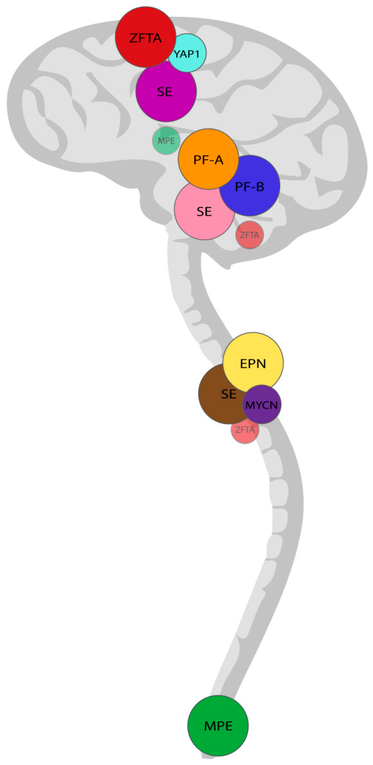
Figure 1. Anatomical localization of ependymoma subtypes according to the 2021 WHO classification. Tumor location is a key diagnostic criterion for nearly all ependymoma variants, except for subependymoma (SE), which may arise in the supratentorial, infratentorial/posterior fossa, or spinal compartments. ZFTA-fusion (ZFTA) and YAP1-fusion (YAP1) positive ependymomas typically occur in the supratentorial region. Posterior fossa group A (PF-A) and group B (PF-B) ependymomas are located infratentorially. Spinal ependymoma (EPN), spinal subependymoma (SE), and MYCN-amplified (MYCN) spinal ependymomas primarily involve the cervical and thoracic spinal cord, whereas myxopapillary (MPE) ependymomas are most commonly found in the caudal spinal cord. Although rare, infratentorial and spinal ZFTA-fusion positive ependymomas have been documented. Myxopapillary ependymomas may also present intracranially, despite their typical spinal origin [4].
Jmp 06 00023 g001
Disclaimer/Publisher’s Note: The statements, opinions and data contained in all publications are solely those of the individual author(s) and contributor(s) and not of MDPI and/or the editor(s). MDPI and/or the editor(s) disclaim responsibility for any injury to people or property resulting from any ideas, methods, instructions or products referred to in the content.