Article

Evaluation of a New Ethanol-Based Preservative Medium for Liquid-Based Cervical Cytology: A Performance Pilot Study for Molecular Applications

1
Department of Public Health, Federico II University of Naples, Via S. Pansini, 5, 80131 Naples, Italy
2
Department of Translational Medical Sciences, University of Naples Federico II, 80131 Naples, Italy
3
Innovation Department, Diapath S.p.A., Via Savoldini, 71, 24057 Martinengo, Italy
4
Engineering and Applied Science Department, University of Bergamo, Viale Marconi, 5, 24044 Dalmine, Italy
*
Author to whom correspondence should be addressed.
J. Mol. Pathol. 2025, 6(3), 22; https://doi.org/10.3390/jmp6030022
Submission received: 8 May 2025 / Revised: 7 July 2025 / Accepted: 1 September 2025 / Published: 2 September 2025
(This article belongs to the Collection Feature Papers in Journal of Molecular Pathology)

Abstract

Background: Since liquid-based cytology (LBC) has replaced the conventional Papanicolaou test in cervical cancer screening programs, pre-analytical procedures—particularly the choice of LBC collection media—have become crucial to ensure the accuracy of high-risk (HR) HPV DNA testing. This study aims to evaluate whether the newly developed CytoPath® LBC medium can serve as a reliable alternative to standard solutions. Methods: This study exploited cell lines to evaluate the stability, integrity, and recovery rate of genomic DNA at different fixation time points (1, 7, 14 and 40 days) and serial dilutions (1:5, 1:10 and 1:20) extracted from cell lines. These samples have also undergone quantitative Real-Time PCR (qPCR) based HR-HPV test to assess the relative performance of the new preservative solution in detecting viral DNA with respect to the standard reference. Results: Cervical cell lines preserved in both media demonstrated consistent DNA stability over time. DNA yields were comparable between the two media. Notably, the DNA Integrity Number (DIN) was higher in samples fixed with the CytoPath® solution. HR-HPV detection by qPCR showed equivalent performance, regardless of the fixative used. Conclusions: The CytoPath® fixative solution represents a valid alternative to standard preservation media, offering improved DNA integrity while maintaining equivalent performance in HR-HPV qPCR testing.

1. Introduction

Human papillomavirus (HPV) is associated with a wide spectrum of skin and mucosal lesions, including squamous cell carcinoma (SCC) of the genital tract, skin, and aerodigestive tract, as well as cervical adenocarcinoma [1,2]. In recent years, the incidence of HPV infection in the female genital tract has increased significantly [3]. To date, over 200 HPV genotypes have been identified. While many are linked to lesions with little or no malignant potential, others—particularly types 16, 18, 31, 35, 45, and 51—are strongly associated with varying degrees of dysplasia and malignancy. The strong association of these high-risk types with anogenital cancers has driven efforts to develop practical methods for HPV detection and genotyping [4,5].
Traditional cervical smear techniques have long been employed for early detection of cervical cancer and have proven effective in reducing the morbidity and mortality associated with invasive disease. However, these methods are hindered by high false-negative rates due to sampling errors, the presence of obscuring material, and limitations in screening and interpretation [6,7]. To address these shortcomings—particularly issues related to inadequate sampling and the detection of high-risk HPV—liquid-based cytology (LBC) has largely replaced conventional smears in routine clinical practice [6,7,8]. Unlike traditional smears where cells are spread directly onto a slide, LBC involves suspending cells in a liquid preservative medium, allowing for both manual and automated sample processing.
LBC offers several advantages: a reduction in inadequate or unsatisfactory samples, improved visualization of cellular and nuclear detail due to reduced background material, and the availability of residual material for ancillary molecular and immunohistochemical analyses [1,9]. The most widely used fixatives in clinical settings are alcohol-based solutions, which ensure cellular stability and preserve both morphology and nucleic acid integrity [10,11,12]. However, to further enhance the preservation of nucleic acids, the development and validation of alternative fixatives remains a key area of interest [13].
Any novel fixative, however, must undergo thorough validation before clinical adoption [14,15,16]. This is especially relevant given the expanding role of molecular testing beyond cervical cytology, including applications for endometrial and ovarian cancers [2]. Morphological assessment alone is insufficient to determine which HPV-related lesions will progress to malignancy, underlining the importance of accurate HPV genotyping in patient management.
Several diagnostic approaches have been developed for HPV typing, primarily based on the detection of DNA or RNA in cervical scrapings or biopsies. Certain types—such as HPV-16 and HPV-18—are frequently found in high-grade dysplasias and carcinomas and are classified as “high-risk” (HR), whereas others—such as HPV-6 and HPV-11—are considered “low-risk” (LR) due to their association with benign lesions. HPV genotyping thus contributes both to early diagnosis and prognostication by improving sensitivity in detecting patients at higher risk [1].
There are three primary classes of HPV-derived diagnostic markers: (1) serological markers detecting antibodies against viral proteins; (2) cellular markers reflecting the expression of viral or tumor-associated proteins; and (3) molecular markers based on the detection of viral nucleic acids—the latter currently being the preferred method. In this context, the application of next-generation sequencing (NGS) technologies is also emerging as a promising future direction [17,18].
Given these advancements, there is an increasing demand for standardized tools and innovative reagents that enhance the analytical sensitivity and specificity of HPV detection assays. Within the pre-analytical phase, fixative solutions play a crucial role in preserving cellular and molecular features as close as possible to their native state. An ideal fixative should be non-toxic, cost-effective, and capable of preserving morphology, antigenicity for immunostaining, and macromolecules—including nucleic acids—for downstream molecular analyses [16,19].
CytoPath® is a new, ethanol-based, formalin-free fixative solution [14] that has not yet been validated for molecular applications. The aim of this study is to conduct a pilot evaluation of the CytoPath® medium, assessing its performance in comparison to the standard reference fixative in terms of nucleic acid stability, integrity, recovery, and compatibility with HPV testing.

2. Material and Methods

2.1. Study Design

The study was carried out on cervical cell lines, that were fixed in Preservcyt® Solution (Hologic Inc., Marlborough, MA, USA) [20], and CytoPath® fixative (Diapath S.p.A., Martinengo, Italy) to evaluate potential differences in terms of DNA stability, integrity and recovery rate. Moreover, it evaluated the effect of both fixatives on the suitability for HPV detection through molecular analysis was evaluated in terms of concordance and success rate.

2.2. Cell Lines

In this study, two cell lines were employed. Cell lines were chosen to reproduce samples routinely undergoing HPV test. In particular, HeLa cell line was used as a positive control, featuring immortalized HPV+ cells derived from cervical cancer, whereas C33A cell line was used as a negative control, consisting of HPV epithelial cells from healthy cervical tissue.
HeLa (CCL-2™) cells and C33A (HTB-31™) cells were obtained from the American Type Culture Collection (ATCC®; Manassas, VA, USA). HeLa cells were grown in Minimum Essential Medium (MEM; Microgem Laboratory Research, Milan, Italy) supplemented with 10% Fetal Bovine Serum (FBS; Microgem Laboratory Research, Milan, Italy), 2 mM l-glutamine, 100 IU/mL penicillin G, and 100 µg/mL streptomycin; C33A cells were grown in Eagle’s Minimum Essential Medium (EMEM; ATCC® 30-2003, Manassas, VA, USA) supplemented with 10% Fetal Bovine Serum (FBS; Microgem Laboratory Research, Milan, Italy), 100 IU/mL penicillin G, and 100 µg/mL streptomycin in a humidified incubator at 37 °C under a 5% CO2 atmosphere. Cells were split and seeded in plates (75 cm2) every 3 days and used for assays during the exponential growth phase.
Both cell lines were collected by gently scraping from the plates, transferred to a Bürker counting chamber and counted by phase-contrast microscope (Carl Zeiss, Jena, Germany, HBO 50/ac model). After centrifugation, cell pellets were split in two and re-suspended in both fixatives. HeLa and C33A cells suspension were mixed at different ratios (1:5, 1:10 and 1:20) to obtain approximately 600,000 cells for each sample.
For qualitative and quantitative evaluation at different time points after fixation (1, 7, 14 and 40 days), 1 mL of each solution was picked up to perform the genomic DNA extraction. Briefly, for each sample, 1 mL of solution was centrifuged (13,000 r.p.m. for 5 min) to obtain the cell pellet. Then 180 µL of buffer and 20 µL of proteinase K enzyme were added and incubated overnight at 56 °C (Figure 1).

2.3. gDNA Extraction, Quality and Quantity DNA Evaluation

For genomic DNA extraction, 1 mL of solution was picked up from each sample from both fixative solutions (n = 24 for cell lines). Briefly, QIAamp DNA Minikit® Kit (Qiagen, Hilden, Germany) was used, and gDNA was extracted following the manufacturer’s instruction. gDNA was eluted in 30 µL nuclease free water. DNA quantity and quality were assessed by Genomic DNA Assay® kit (Agilent, Santa Clara, CA, USA) on 4200 TapeStation® Platform (Agilent), according to manufacturer’s instruction.
In addition, after DNA extraction, the residual material into the vials was sent to the Microbiology and Virology Unit, University of Naples Federico II, for HPV testing.

2.4. The Abbott Real-Time High-Risk HPV Test

The Abbott Real-Time High-Risk HPV test is a real-time PCR-based assay for concurrent detection and individual genotyping of HPV-16 and HPV-18 and pooled detection of 12 other hrHPV genotypes: HPV-31, HPV-33, HPV-35, HPV-39, HPV-45, HPV-51, HPV-52, HPV-56, HPV-58, HPV-59, HPV-66, and HPV-68 [19,21]. Real-Time PCR is performed on the m2000rt real-time PCR instrument using a modified GP5+/6+ primer mix consisting of three forward primers and two reverse primers designed to hybridize to an approximately 150-base HPV consensus region [21].
A primer set targeting a region of 136 bases in the human beta-globin gene was used as internal process control (IC), for monitoring sample adequacy, DNA extraction quality, and yield. The HPV and internal control probes are single-stranded DNA oligonucleotides modified with a fluorescent moiety covalently linked to one end of the probe and a quenching moiety to the other end [21]. Through the distinct labels, signals for HPV-16, HPV-18, non-HPV-16/18 types, and IC can be simultaneously detected and distinguished in a single reaction. The assay also has a proprietary algorithm for the amplification curve validation [21].

2.5. Statistical Methods

The data obtained were evaluated by statistical analysis in order to compare the performance of PreservCyt® and CytoPath® fixative solutions in terms of preserving the nucleic acids (concentration and integrity) and to perform the Real-Time High-Risk HPV DNA. The statistical analyses were performed using R (version 4.0.2, R Foundation for Statistical Computing, Vienna, Austria) and statistical significance was set at p < 0.05.

3. Results

3.1. Quality and Quantity Genomic DNA Evaluation: Tape Station Cell Lines Samples Analysis

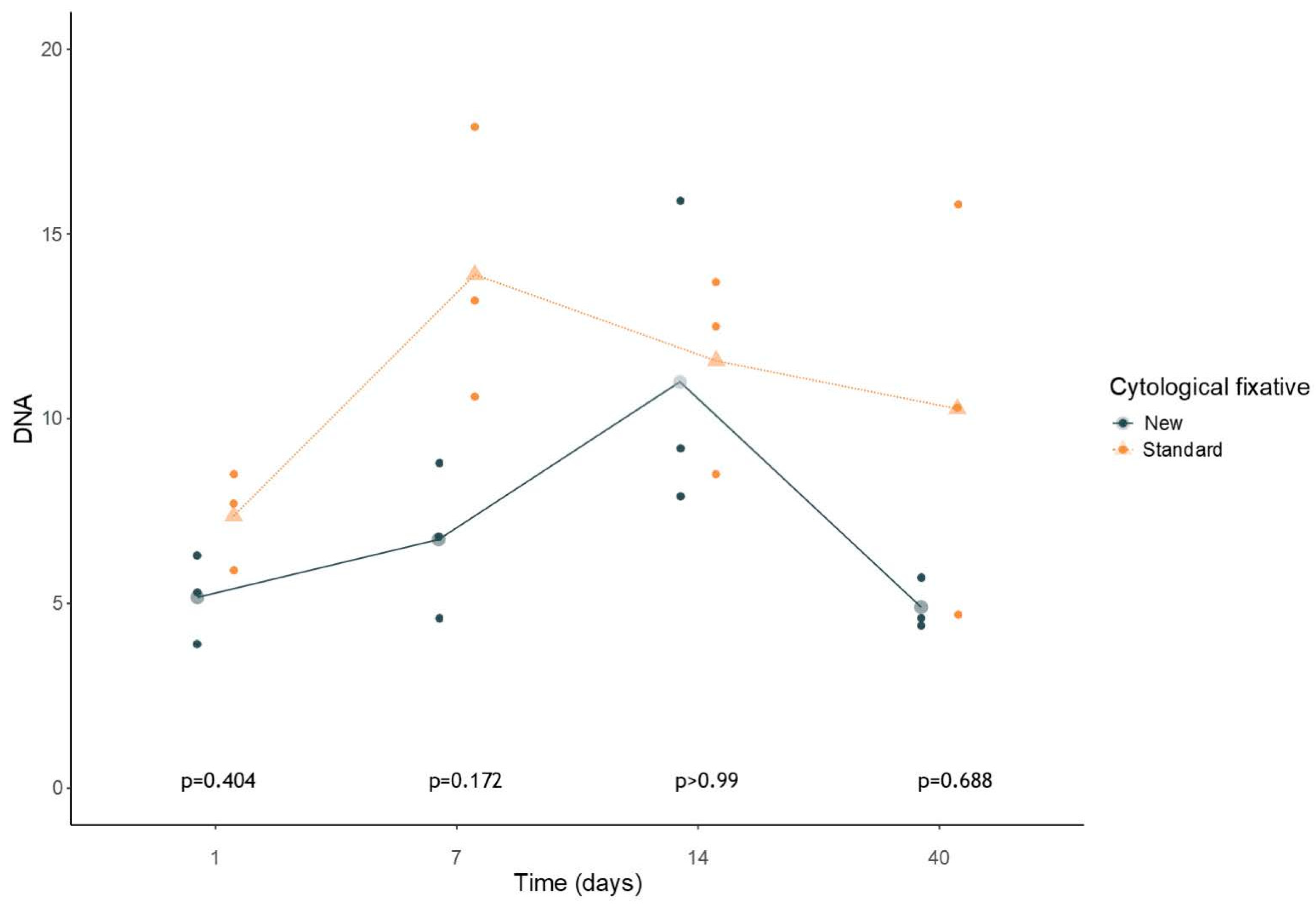
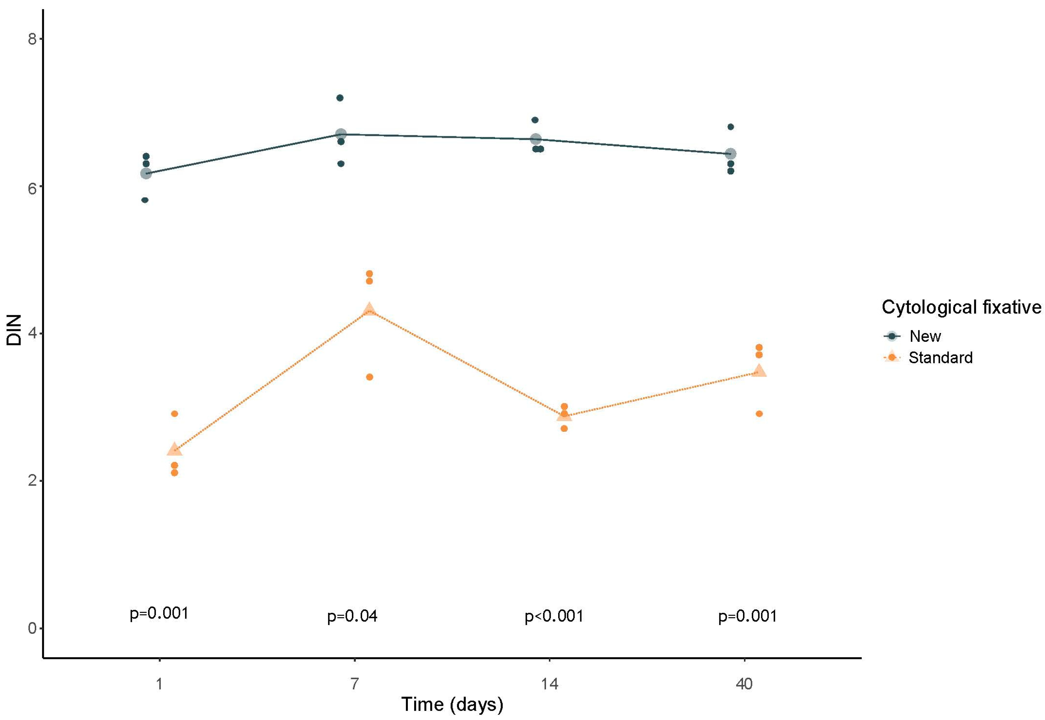
Overall, the analysis on cell line samples on the three serial dilutions (1:5; 1:10 and 1:20) showed a significantly higher (p < 0.05) quality of extracted nucleic acid using the new CytoPath® fixative compared with those fixed with the standard reference for all data points with no difference in terms of concentrations (Figure 2 and Figure 3). Detailed results about the analysis on cell line samples are shown in Supplementary Material Table S1.

3.2. HPV Real-Time PCR Analysis in Cell Lines

The Real-Time High-Risk HPV assay’s turnaround time for 96 samples is 6 to 8 h for 96 samples, depending on the method used for DNA extraction. The fully automated high-throughput instrument m2000sp or the smaller m24sp instrument can be used for DNA extraction or, alternatively, DNA can be prepared manually. The assay is validated by the manufacturer for use with cervical specimens collected with PreservCyt® Solution, SurePath Preservative Fluid, and the Abbott Cervi-Collect Specimen Collection Kit [19,21,22]. In this study, we evaluated the feasibility to use the CytoPath® medium preserving the nucleic acids to perform the Real-Time High-Risk HPV DNA assay by using cell lines samples. Moreover, as expected, since the Hela cell lines derived from cervical cancer, the HPV test showed positive results, and the output was similar in samples preserved in both fixative solutions.
In particular, the results were carried out on the three different serial dilutions of the cell line cultures (HeLa and C33A) fixed with both fixative solutions as follows: regarding the standard reference, we observed a Cycle threshold (CT) range of 19.7–22.09 for the 1:5 dilution, a CT range of 20.93–23 for the 1:10 dilution, and a CT range of 22.66–23.96 for 1:20 dilution. Whereas, regarding the cells preserved in the new fixative solution, a CT range of 21.55–24.07, of 22.35–23.79 and of 22.64–25.16 was achieved for the 1:5, 1:10, and 1:20 point dilutions, respectively. Detailed data are reported in Table 1.

3.3. Statistical Analysis

Differences in longitudinal variation in DIN and in DNA concentrations were evaluated using a mixed model ANOVA with time as the within factor and medium as the between factor. The model included the two main effects and the interaction between time and medium. In case of significance of the interaction term, post hoc comparisons were based on T-test for unpaired samples with Bonferroni correction.

4. Discussion

Liquid-based cytology (LBC) media provide an effective platform for improved specimen storage, supporting co-testing strategies such as high-risk HPV (HR-HPV) testing for cervical cancer screening [23]. Cervical swab specimens preserved in LBC media can also be utilized for additional molecular analyses, including DNA methylation profiling and HR-HPV E6/E7 mRNA testing [24]. Therefore, an ideal LBC medium should not only preserve cellular morphology but also maintain the integrity of nucleic acids [14,25,26]. Specimens stored in appropriate preservative solutions can yield extensive biological information, potentially leading to new diagnostic and therapeutic insights [8,10,11].
In the past, various alternative fixatives have been proposed for optimal cytological sample preservation. For instance, Zhao et al. evaluated VersaMedium, a novel ethanol-based LBC fixative. Their findings indicated that cervical exfoliated cells preserved in VersaMedium retained good morphological quality, although no further molecular analyses were conducted [13].
More recently, Casatta et al. investigated the performance of CytoPath®, a new ethanol-based, formalin-free LBC fixative, by comparing it with the standard reference solution. Their study evaluated both gynecological and non-gynecological samples and found no statistically significant differences in diagnostic adequacy between the two media. These results support the non-inferiority of CytoPath® in preserving cellular morphology for routine cytological assessments [14].
Building upon these findings, the present study aimed to evaluate the capacity of CytoPath® to preserve nucleic acids and to support HPV DNA detection, in direct comparison with the standard fixative routinely used in cytology laboratories. To achieve this, we initially focused on cervical cell line models, which allowed for controlled and reproducible assessment of DNA stability, integrity, and recovery rate over different time points and serial dilutions.
DNA extracted from these cell lines was analyzed using nanocapillary electrophoresis and a qPCR-based HR-HPV detection assay. The nanocapillary electrophoresis data were subjected to statistical analysis to compare the two fixatives in terms of DNA yield and integrity. The analysis revealed no significant difference in DNA concentration or extraction efficiency between the two media in cell line samples (Figure 2). Furthermore, results across various serial dilutions (Table 1) confirmed the consistency and adequacy of CytoPath® for handling cytological samples at varying concentrations.
Importantly, a statistically significant difference emerged in the quality of the extracted nucleic acids. Samples fixed in CytoPath® demonstrated a higher DNA Integrity Number (DIN) compared to those fixed in the standard solution. As shown in Figure 3, this improvement in DNA quality was consistent across all time intervals, from day 1 up to 40 days post-fixation (p < 0.05). This enhanced preservation of nucleic acids is particularly relevant given the increasing integration of molecular diagnostics into routine cytopathology workflows. It suggests that the use of CytoPath® could support not only conventional cytological evaluation but also high-quality molecular testing—essential for accurate diagnosis, prognosis, and patient stratification.
Therefore, the introduction of this new fixative into routine clinical laboratory workflows could offer a valuable diagnostic tool, providing preservation performance comparable to, or better than, that of currently adopted cytopathology standards.

5. Conclusions

CytoPath® is a promising fixative solution that is non-toxic, cost-effective, and effective in preserving both cellular morphology and nucleic acid integrity. The results of this pilot study support its potential for clinical implementation. However, larger-scale clinical studies involving patient-derived samples are warranted to confirm these findings and evaluate the reproducibility of the results obtained in cell lines. Our validation study represents an important first step, demonstrating the superior DNA preservation offered by CytoPath®, which in turn facilitates more accurate molecular analysis for HPV DNA detection.

Supplementary Materials

The following supporting information can be downloaded at https://www.mdpi.com/article/10.3390/jmp6030022/s1. Table S1. DNA quality (DIN) and quantity ([ ]) evaluation in cell lines samples at different fixation time points (1, 7, 14, and 40 days) and serial dilutions (1:5, 1:10, and 1:20) with 4200 TapeStation® Platform (Agilent).

Author Contributions

Conceptualization, G.T.; data curation, D.B., G.T., C.D.L., F.C., P.P., A.I., M.S., A.V., G.G., L.V., R.S., G.P. and C.L.; writing—original draft preparation, C.D.L., F.C. and P.P.; writing—review and editing; funding acquisition, N.C., D.B., G.T., C.D.L., F.C., P.P., A.I., M.S., A.V., G.G., L.V., R.S., G.P. and C.L. All authors have read and agreed to the published version of the manuscript.

Funding

This research was funded by: CN3: The National Center for Gene Therapy and Drugs based on RNA Technology MUR-CN3 CUP E63C22000940007. POS3: Italian Ministry of Health (Piano Operativo Salute Traiettoria 3, T3-AN-09, “Genomed”). ANTHEM: “NExt generation Omics-NEON” bando a cascata Anthem—prot. n. 0001983—CUP B53C22006700001. PRIN: Project ID: P2022L4CK4, funding code 000018 PRIN_PNRR_2022, CUP E53D23015450001. HERA-ORION: D3 4 Health, PNC—CUP B53C22005980001, PNC0000001_UNINA_HERA_ORION.

Institutional Review Board Statement

Ethical review and approval were waived for this study due to its retrospective nature.

Informed Consent Statement

Written informed consent was obtained from all patients and all subjects included in the study.

Data Availability Statement

The data presented in this study are available on request from the corresponding author due to (specify the reason for the restriction).

Conflicts of Interest

Diapath S.p.A. provided part of the reagents for the execution of this work. The other authors declare no conflicts of interest.

References

  1. Cancer Institute NSW. HPV and Cervical Cancer; Borruto, F., De Ridder, M., Eds.; Springer: Berlin/Heidelberg, Germany, 2012. [Google Scholar]
  2. Parks, P. HPV; Reference Point Press: San Diego, CA, USA, 2015. [Google Scholar]
  3. Cancer Today. Available online: https://gco.iarc.fr/today/en (accessed on 20 March 2024).
  4. Nayar, R.; Wilbur, D.C. The Bethesda System for Reporting Cervical Cytology: A Historical Perspective. Acta Cytol. 2017, 61, 359–372. [Google Scholar] [CrossRef] [PubMed]
  5. Pangarkar, M.A. The Bethesda System for Reporting Cervical Cytology. Cytojournal 2022, 19, 28. [Google Scholar] [CrossRef] [PubMed]
  6. Safaeian, M.; Solomon, D.; Castle, P.E. Cervical Cancer Prevention—Cervical Screening: Science in Evolution. Obs. Gynecol. Clin. N. Am. 2007, 34, 739. [Google Scholar] [CrossRef]
  7. Hoda, R.S.; Loukeris, K.; Abdul-Karim, F.W. Gynecologic Cytology on Conventional and Liquid-Based Preparations: A Comprehensive Review of Similarities and Differences. Diagn. CytoPathol. 2013, 41, 257–278. [Google Scholar] [CrossRef] [PubMed]
  8. Klinkhamer, P.J.J.M.; Meerding, W.J.; Rosier, P.F.W.M.; Hanselaar, A.G.J.M. Liquid-Based Cervical Cytology. Cancer CytoPathol. 2003, 99, 263–271. [Google Scholar] [CrossRef]
  9. Jeong, H.; Hong, S.R.; Chae, S.W.; Jin, S.Y.; Yoon, H.K.; Lee, J.; Kim, E.K.; Ha, S.T.; Kim, S.N.; Park, E.J.; et al. Comparison of Unsatisfactory Samples from Conventional Smear versus Liquid-Based Cytology in Uterine Cervical Cancer Screening Test. J. Pathol. Transl. Med. 2017, 51, 314–319. [Google Scholar] [CrossRef]
  10. Van Hemel, B.M.; Suurmeijer, A.J.H. Effective Application of the Methanol-Based PreservCyt(TM) Fixative and the Cellient(TM) Automated Cell Block Processor to Diagnostic CytoPathology, Immunocytochemistry, and Molecular Biology. Diagn. CytoPathol. 2013, 41, 734–741. [Google Scholar] [CrossRef]
  11. Perry, C.; Chung, J.Y.; Ylaya, K.; Choi, C.H.; Simpson, A.; Matsumoto, K.T.; Smith, W.A.; Hewitt, S.M. A Buffered Alcohol-Based Fixative for Histomorphologic and Molecular Applications. J. Histochem. Cytochem. 2016, 64, 425–440. [Google Scholar] [CrossRef]
  12. Bianchi, A.; Moret, F.; Desrues, J.M.; Champenois, T.; Dervaux, Y.; Desvouas, O.; Oursin, A.; Quinzat, D.; Dachez, R.; Bathelier, C.; et al. PreservCyt Transport Medium Used for the ThinPrep Pap Test Is a Suitable Medium for Detection of Chlamydia Trachomatis by the COBAS Amplicor CT/NG Test: Results of a Preliminary Study and Future Implications. J. Clin. Microbiol. 2002, 40, 1749–1754. [Google Scholar] [CrossRef]
  13. Zhao, G.; Liu, Z.; Tian, Y.; Zhu, M.; Wang, S.; Wang, H.; Gao, S.; Ma, Y.; Zheng, M. Stability, Integrity, and Recovery Rate of Cellular Nucleic Acids Preserved in a New Liquid-Based Cytology Medium. Diagn. CytoPathol 2018, 46, 213–220. [Google Scholar] [CrossRef]
  14. Casatta, N.; Poli, A.; Bassani, S.; Veronesi, G.; Rossi, G.; Ferrari, C.; Lupo, C. Evaluation of a Novel Fixative Solution for Liquid-Based Cytology in Diagnostic CytoPathology. Diagnostics 2023, 13, 3601. [Google Scholar] [CrossRef]
  15. De Luna, R.; Eloubeidi, M.A.; Sheffield, M.V.; Eltoum, I.; Jhala, N.; Jhala, D.; Chen, V.K.; Chhieng, D.C. Comparison of ThinPrep and Conventional Preparations in Pancreatic Fine-Needle Aspiration Biopsy. Diagn. CytoPathol. 2004, 30, 71–76. [Google Scholar] [CrossRef]
  16. Chung, J.Y.; Song, J.S.; Ylaya, K.; Sears, J.D.; Choi, L.; Cho, H.; Rosenberg, A.Z.; Hewitt, S.M. Histomorphological and Molecular Assessments of the Fixation Times Comparing Formalin and Ethanol-Based Fixatives. J. Histochem. Cytochem. 2018, 66, 121–135. [Google Scholar] [CrossRef] [PubMed]
  17. Clery, E.; Pisapia, P.; Migliatico, I.; Pepe, F.; De Luca, C.; Russo, M.; De Rosa, F.; Smeraglia, F.; Insabato, L.; Vigliar, E.; et al. Cytology Meets next Generation Sequencing and Liquid Biopsy: A Case of Lung Adenocarcinoma Presenting as Metastasis to the Phalanx. Diagn. CytoPathol 2020, 48, 759–764. [Google Scholar] [CrossRef]
  18. Pisapia, P.; Pepe, F.; Sgariglia, R.; Nacchio, M.; Russo, G.; Conticelli, F.; Girolami, I.; Eccher, A.; Bellevicine, C.; Vigliar, E.; et al. Next Generation Sequencing in Cytology. CytoPathology 2021, 32, 588–595. [Google Scholar] [CrossRef] [PubMed]
  19. Agreda, P.M.; Beitman, G.H.; Gutierrez, E.C.; Harris, J.M.; Koch, K.R.; LaViers, W.D.; Leitch, S.V.; Maus, C.E.; McMillian, R.A.; Nussbaumer, W.A.; et al. Long-Term Stability of Human Genomic and Human Papillomavirus DNA Stored in BD SurePath and Hologic PreservCyt Liquid-Based Cytology Media. J. Clin. Microbiol. 2013, 51, 2702–2706. [Google Scholar] [CrossRef] [PubMed]
  20. ThinPrep® PreservCyt® Solution Instructions for Use. 2019. Available online: https://www.hologic.com/sites/default/files/2020-07/AW-19166-002_001_02.pdf (accessed on 7 May 2025).
  21. Huang, S.; Tang, N.; Mak, W.B.; Erickson, B.; Salituro, J.; Li, Y.; Krumpe, E.; Schneider, G.; Yu, H.; Robinson, J.; et al. Principles and Analytical Performance of Abbott RealTime High Risk HPV Test. J. Clin. Virol. 2009, 45, S13–S17. [Google Scholar] [CrossRef]
  22. Zhao, F.H.; Hu, S.Y.; Bian, J.J.; Liu, B.; Peck, R.B.; Bao, Y.P.; Pan, Q.J.; Frappart, L.; Sellors, J.; Qiao, Y.L. Comparison of ThinPrep and SurePath Liquid-Based Cytology and Subsequent Human Papillomavirus DNA Testing in China. Cancer CytoPathol. 2011, 119, 387–394. [Google Scholar] [CrossRef]
  23. von Karsa, L.; Arbyn, A.; Arbyn, M.; Anttila, A.; Jordan, J.; Ronco, G.; Schenck, U.; Segnan, N.; Wiener, H. Executive Summary. In European Guidelines for Quality Assurance in Cervical Cancer Screening; Publications Office of the European Union: Luxembourg, 2015; ISBN 9789279485398. [Google Scholar]
  24. Zhang, L.; Tan, W.; Yang, H.; Zhang, S.; Dai, Y. Detection of Host Cell Gene/HPV DNA Methylation Markers: A Promising Triage Approach for Cervical Cancer. Front. Oncol. 2022, 12, 831949. [Google Scholar] [CrossRef]
  25. Dey, P.; Luthra, U.A.C.K.; Path, F.R.C.; George, J.; Zuhairy, F.; George, S.S.; Haji, B.I. Comparison of ThinPrep and Conventional Preparations on Fine Needle Aspiration Cytology Material. Acta Cytol. 2000, 44, 46–50. [Google Scholar] [CrossRef]
  26. Matsuo, Y.; Yoshida, T.; Yamashita, K.; Satoh, Y. Reducing DNA Damage by Formaldehyde in Liquid-Based Cytology Preservation Solutions to Enable the Molecular Testing of Lung Cancer Specimens. Cancer CytoPathol. 2018, 126, 1011–1021. [Google Scholar] [CrossRef]
Figure 1. Study design. Cell lines employed for this study were split and seeded in plates and they were collected by gently scraping from the plates. After centrifugation, cell pellets were split in two and re-suspended in both fixatives. HeLa and C33A cells suspension were mixed at different ratios (1:5, 1:10 and 1:20). For qualitative and quantitative evaluation at different time points after fixation (1, 7, 14 and 40 days), 1 mL of each solution was picked up to perform the genomic DNA extraction.
Jmp 06 00022 g001
Figure 2. Comparison between DNA concentration at different time points in cell lines (Hela and C33A) by using new and standard fixative. The statistical analysis showed no significant difference regarding the DNA concentration of cell lines samples on the three serial dilutions (1:5; 1:10 and 1:20) fixed in the new fixative solution and standard reference solution at different time points (1, 7, 14, 40 days).
Jmp 06 00022 g002
Figure 3. Comparison between DNA concentration at different time points in cell lines (Hela and C33A) by using new and standard fixative. The statistical analysis showed that the DIN value in samples extracted from cell line samples on the three serial dilutions (1:5; 1:10, and 1:20) maintained in the new fixative solution was significantly higher than samples derived from cells preserved in the standard reference solution (p < 0.05). Furthermore, the quality of the nucleic acids was increased in all considered time intervals, from the first day up to 40 days after the cell samples were immersed in the fixative solution (p < 0.05).
Jmp 06 00022 g003
Table 1. Cycle threshold (CT) carried out performing the HPV test in PCR Real-Time on extracted DNA from cell line (Hela and C33A).
Fissative TypeDiluitionTimingCT
Preservcyt®1:51 day22.09
1:1022.56
1:2023.54
Cytopath®1:522.71
1:1023.57
1:2023.94
Preservcyt®1:57 days21.63
1:1023
1:2023.96
Cytopath®1:522.59
1:1022.35
1:2023.91
Preservcyt®1:514 days19.7
1:1020.93
1:2022.66
Cytopath®1:521.55
1:1022.63
1:2025.16
Preservcyt®1:540 days20.47
1:1022.09
1:2022.85
Cytopath®1:524.07
1:1023.79
1:2022.64
The table shows three different point of serial dilutions (1:5; 1:10, and 1:20) of the cell lines (Hela and C33A) fixed by Preservcyt® and by Cytopath® with the relative Cycle threshold (CT) reported performing the HPV test in PCR Real-Time on extracted DNA samples at different timings (1, 7, 14, 40 days).
Disclaimer/Publisher’s Note: The statements, opinions and data contained in all publications are solely those of the individual author(s) and contributor(s) and not of MDPI and/or the editor(s). MDPI and/or the editor(s) disclaim responsibility for any injury to people or property resulting from any ideas, methods, instructions or products referred to in the content.