Review

Dysfunctional Astrocyte Metabolism: A Driver of Imbalanced Excitatory/Inhibitory Tone and Support for Therapeutic Intervention Targets

1
Department of Medical Laboratory Science, Faculty of Basic Medical Sciences, Adeleke University, Ede 232101, Osun State, Nigeria
2
Department of Human Physiology, Faculty of Basic Medical Sciences, Delta State University of Science and Technology, Ozoro 334111, Delta State, Nigeria
3
DELSU Joint Canada-Israel Neuroscience and Biopsychiatry Laboratory, Department of Pharmacology, Faculty of Basic Medical Sciences, College of Health Sciences, Delta State University, Abraka 330105, Delta State, Nigeria
4
School of Medical Sciences, Faculty of Health, University of Victoria, Victoria, BC V8P 5C2, Canada
*
Authors to whom correspondence should be addressed.
J. Mol. Pathol. 2025, 6(2), 12; https://doi.org/10.3390/jmp6020012
Submission received: 21 March 2025 / Revised: 7 May 2025 / Accepted: 21 May 2025 / Published: 11 June 2025

Abstract

A balanced excitatory/inhibitory (E/I) tone is crucial for proper brain function, and disruptions can lead to neurological disorders. This review explores the role of astrocytes in maintaining a balanced E/I tone in the brain, which is crucial for proper functioning. It highlights the potential for dysfunctional astrocyte metabolism to disrupt E/I balance, leading to neuronal dysfunction and potentially causing neurological disease pathogenesis. The review focuses on glucose, lactate shuttling, and glutamate metabolism. This review synthesizes findings from in vitro, in vivo, and human studies examining the interplay between astrocyte metabolism, neuronal activity, and E/I balance. Literature searches were conducted using keywords including “astrocyte metabolism”, “excitatory/inhibitory balance”, “glutamate”, “lactate shuttle”, “neurometabolic coupling”, and “neurological disorders” in databases such as PubMed and Web of Science. Disruptions in astrocyte glucose uptake or glycolysis can impair lactate production, reducing neuronal energy supply and affecting neuronal excitability. Impaired glutamate uptake and conversion to glutamine within astrocytes leads to elevated extracellular glutamate, promoting excitotoxicity. Altered glycogen metabolism and other metabolic impairments within astrocytes can also affect neuronal health and contribute to imbalances between excitation and inhibition. Dysfunctional astrocyte metabolism represents a significant contributor to E/I imbalance in the brain. Understanding the specific metabolic vulnerabilities of astrocytes and their impact on neuronal function provides potential therapeutic targets for neurological disorders characterized by E/I dysregulation. Targeting astrocyte metabolism may offer a novel approach to restoring E/I balance and improving neurological outcomes.

1. Introduction

Astrocytes are a subtype and specialized type of glial cells, abundantly distributed in the central nervous system (CNS), integral to the function and health of the brain. These cells perform a range of critical functions, including maintaining ion homeostasis, regulating neurotransmitters, influencing synapse formation and plasticity, upholding the integrity of the blood–brain barrier (BBB), and providing essential metabolic support [1,2,3,4,5]. Their contributions are particularly vital in maintaining the delicate balance between excitatory and inhibitory neurotransmission, a balance crucial for proper neuronal communication. Disruptions to this balance can lead to neuronal hyperactivity or hypoactivity, potentially contributing to a variety of neurological disorders. Astrocytes actively regulate the concentrations of ions like potassium (K+) and pH levels within the extracellular space, an essential function for ensuring optimal neuronal activity [6,7]. By buffering potassium levels, astrocytes prevent excessive neuronal excitability, and by maintaining appropriate pH, they support the proper functioning of ion channels and receptors on neuronal membranes. These actions directly influence neuronal firing patterns and synaptic transmission [6,7].
Beyond ion regulation, astrocytes play a crucial role in managing neurotransmitter levels in the synaptic cleft. They efficiently clear neurotransmitters such as glutamate and GABA, preventing excessive or prolonged receptor activation [8,9]. This clearance is achieved through specialized transporters located on astrocyte membranes, which rapidly remove neurotransmitters from the synaptic space. Furthermore, astrocytes participate in the recycling of neurotransmitters. For example, glutamate taken up by astrocytes is converted into glutamine, which is then shuttled back to neurons for glutamate resynthesis. This intricate process ensures efficient neurotransmission and prevents excitotoxicity, a condition caused by the excessive accumulation of glutamate. The influence of astrocytes extends to synapse formation and plasticity, which are fundamental to processes of learning and memory [10,11]. Astrocytes secrete various factors that promote synapse formation, stabilize synaptic connections, and modulate synaptic strength. They physically interact with synapses, ensheathing them and influencing the availability of neurotransmitters. Through these interactions, astrocytes contribute to the dynamic remodeling of neural circuits that underlie learning and adaptation. Astrocytes are also essential components of the BBB, a selective barrier that protects the delicate neural tissue from harmful substances circulating in the bloodstream [12,13]. Astrocytes surround brain capillaries, providing structural support and releasing factors that promote the tight junctions between endothelial cells, the cells that form the lining of the capillaries. By maintaining the integrity of the BBB, astrocytes prevent the entry of toxins, pathogens, and inflammatory molecules that could disrupt neuronal function and cause damage [12,13].
Metabolically, astrocytes provide crucial support to neurons. They store glycogen, a readily available source of glucose, and can release glucose to neurons during periods of high energy demand [14,15]. In addition, astrocytes metabolize glucose to produce lactate, which they then supply to neurons as an alternative energy source. This metabolic cooperation between astrocytes and neurons is essential for sustaining neuronal activity, particularly during periods of intense synaptic transmission. The metabolic functions of astrocytes are closely linked to their role in maintaining the excitatory/inhibitory (E/I) balance within the brain [16,17]. Impaired astrocyte metabolism can disrupt neurotransmitter homeostasis, leading to imbalances in glutamatergic and GABAergic signaling. For instance, if astrocyte metabolism is compromised, their ability to uptake and recycle glutamate may be reduced, leading to excessive glutamate accumulation in the synaptic cleft [18]. This, in turn, can result in excitotoxicity and neuronal damage. Conversely, impaired astrocyte metabolism may also affect the synthesis and release of gamma-aminobutyric acid (GABA), the primary inhibitory neurotransmitter in the brain, leading to a reduction in inhibitory tone [19]. Additionally, dysfunctional astrocyte metabolism can lead to a cascade of consequences on neuronal activity. Excessive glutamate release or inadequate uptake, resulting from impaired astrocyte function, can cause excitotoxicity or alter inhibitory signals mediated by GABA. These disruptions compromise network stability by altering the E/I tone. Such imbalances have been implicated in a range of neurological disorders, including epilepsy, Alzheimer’s disease (AD), schizophrenia and autism spectrum disorders (ASDs). Therefore, thoroughly understanding how impaired astrocyte metabolism affects neurotransmitter homeostasis is crucial for addressing neurological conditions associated with E/I imbalances. By targeting astrocyte function, novel therapeutic strategies to restore the E/I balance and alleviate the symptoms of these debilitating disorders can be developed.

2. Astrocyte Metabolism: The Foundation of E/I Balance

While excess metabolite accumulation in the brain does not automatically signify astrocyte dysfunction, it becomes a critical indicator in pathological conditions like metabolic syndrome, obesity, neurodevelopmental disorders and neurodegenerative diseases [20,21]. In these contexts, astrocytes exhibit reduced expression of key metabolic enzymes, such as lipoprotein lipase, hindering their ability to process metabolites like triglycerides and fatty acids [21]. This leads to morphological changes, including increased lipid and cholesterol content, indicating cellular stress [21]. Furthermore, disruptions in calcium signaling impair astrocyte function, affecting neurotransmitter regulation and neuron–glia communication, as seen in AD with amyloid–beta (Aβ) accumulation [22]. These combined factors suggest that excess metabolite accumulation fundamentally compromises astrocyte function, making astrocyte dysfunction a key element in the pathogenesis of neurodevelopmental disorders and neurodegenerative diseases [21,22].

2.1. Glucose Metabolism

Astrocytes are critical for maintaining the brain’s delicate balance between neuronal excitation and inhibition, a balance essential for proper neurological function. Central to this role is astrocytes’ metabolic activities, most notably their handling of glucose [22]. Astrocytes actively absorb glucose from the bloodstream via GLUT1 transporters located on their cell membranes (Figure 1) [22]. This glucose uptake is crucial, providing the fuel for various processes within astrocytes that ultimately influence neuronal health and neurotransmission. A primary pathway for glucose utilization in astrocytes is glycolysis, a process that breaks down glucose into pyruvate, generating ATP, the energy currency of the cell [23]. In addition to ATP production, glycolysis yields lactate. This lactate is not simply a waste product but serves as an alternative energy source for neurons, particularly during periods of high metabolic demand or stress [24,25]. While glycolysis is vital for direct energy production, astrocytes also utilize the pentose phosphate pathway (PPP). Unlike glycolysis, the PPP is not primarily involved in ATP production but generates NADPH, a crucial reducing agent. NADPH is critical for protecting against oxidative stress by neutralizing ROS [26]. Consequently, impaired PPP activity can leave both astrocytes and neurons more susceptible to oxidative damage. Astrocytes also have the unique ability to store glucose as glycogen, acting as a buffer during periods of either increased energy demand or when the blood glucose supply is limited [27,28]. The storage and release of glycogen are dynamically regulated, responding to factors such as neuronal activity levels and hormonal signals [28]. Therefore, astrocytes play a crucial role in neuronal function. Disruptions in astrocytic glycolysis can impair the neuronal energy supply, leading to compromised neuronal function and altered neuronal excitability [29]. In addition, reduced NADPH production due to impaired PPP activity can increase vulnerability to oxidative damage, which has cascade effects, notably affecting neurotransmitter synthesis and release [30]. Impaired glycogen storage or mobilization may lead to inadequate support for neuronal energy needs, potentially causing hyperexcitability or reduced inhibitory neurotransmission [31]. Overall, these reports highlight the role of impaired energy homeostasis in astrocyte dysfunction, notably reinforcing the possibility of altered calcium signaling in neurological and neuropsychiatric diseases.

2.2. Glutamate–Glutamine Cycle

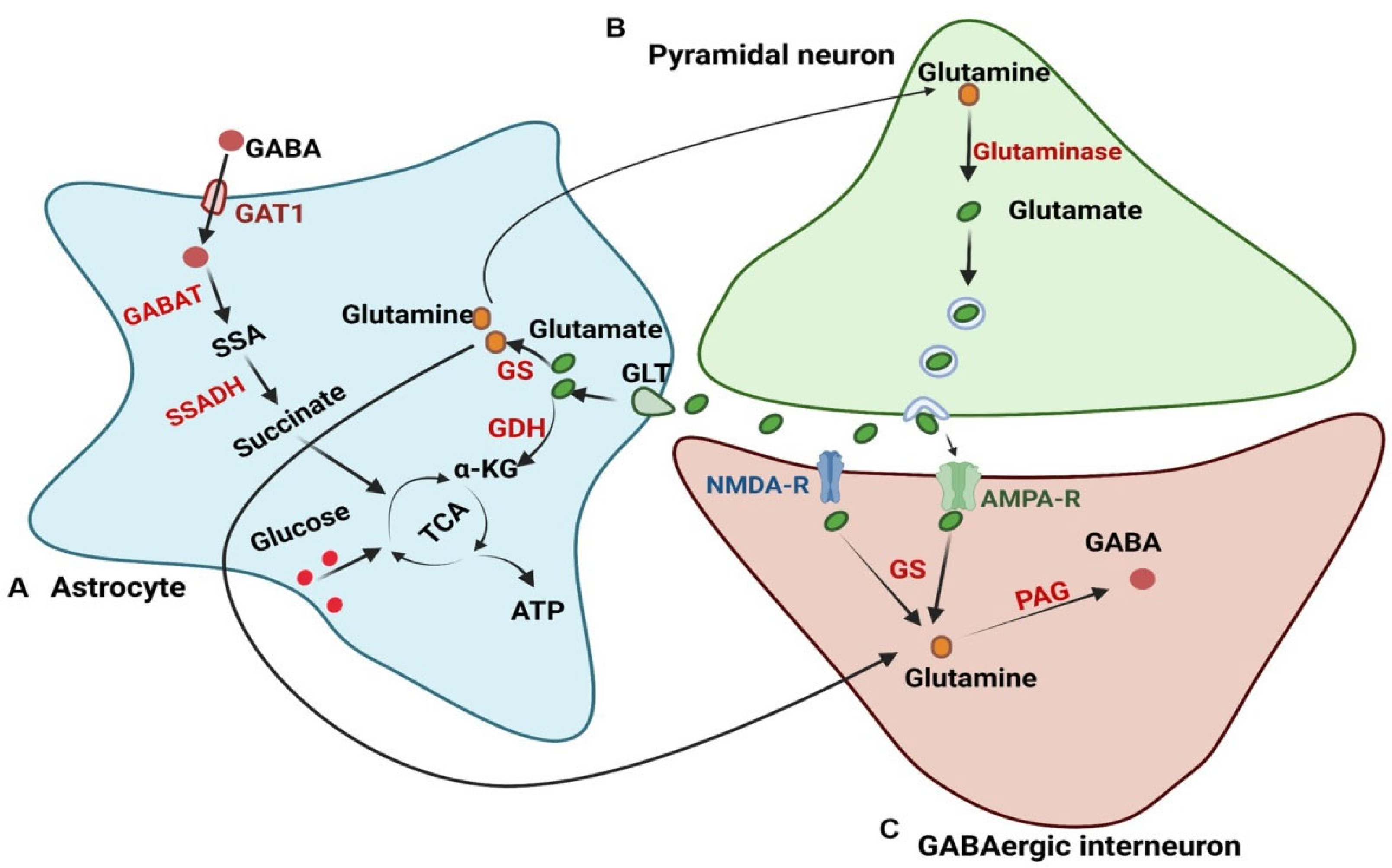
The glutamate–glutamine cycle is a vital metabolic pathway in the brain, primarily orchestrated by astrocytes, to maintain a delicate balance between excitatory and inhibitory neurotransmission [32,33]. This cycle is essential for the continuous recycling of glutamate and GABA, the brain’s principal excitatory and inhibitory neurotransmitters, respectively. At its core, the cycle prevents excitotoxicity by efficiently clearing excess glutamate from the synaptic cleft while providing the necessary precursors for synthesizing both glutamate and GABA [32,33]. Astrocytes play a central role in the operation of this cycle, performing several key functions that are crucial to its overall success. One of the primary roles of astrocytes is the efficient uptake of glutamate from the synaptic cleft (Figure 2). To accomplish this, astrocytes express excitatory amino acid transporters (EAATs), notably EAAT2 (also known as GLT-1) and EAAT1 (also known as GLAST) [34]. These transporters actively remove glutamate from the synapse, preventing the overstimulation of postsynaptic neurons and the subsequent risk of excitotoxic damage [34]. Once inside the astrocyte, glutamate undergoes a crucial transformation: it is converted into glutamine by the enzyme glutamine synthetase [34]. This conversion serves a dual purpose: it effectively removes potentially toxic levels of glutamate from the astrocyte while also creating a safe reservoir of neurotransmitter precursor [34]. The glutamine produced is then released by astrocytes into the extracellular space, where it becomes available for uptake by neurons [35]. Neurons, in turn, can convert glutamine back into either glutamate for excitatory neurotransmission or GABA for inhibitory neurotransmission, thus completing the cycle [34,35].
The importance of the glutamate–glutamine cycle in maintaining the E/I balance within the brain cannot be overstated. By efficiently removing excess glutamate from the synaptic cleft, astrocytes prevent the damaging effects of overexcitation, thereby protecting neurons from excitotoxicity [36]. Furthermore, the release of glutamine by astrocytes ensures that both glutamatergic (excitatory) and GABAergic (inhibitory) neurons have an adequate supply of the building blocks needed for neurotransmitter synthesis [37]. However, disruptions or dysregulation within the glutamate–glutamine cycle can have profound consequences for brain function. For example, a reduction in glutamate uptake by astrocytes, whether due to impaired EAAT function or decreased astrocyte activity, can lead to elevated levels of extracellular glutamate, thus increasing the risk of excitotoxicity. Similarly, reduced activity of glutamine synthetase within astrocytes can result in a buildup of glutamate and a deficiency of glutamine [38]. Impaired release or transport of glutamine from astrocytes to neurons can also limit the availability of neurotransmitter precursors, potentially leading to a decrease in the synthesis of both glutamate and GABA. Dysregulation of the glutamate–glutamine cycle, potentially stemming from genetic predispositions, environmental factors, or altered expression of key enzymes involved in glutamate metabolism, can lead to imbalances in the E/I ratio. Such imbalances in the E/I ratio, resulting from dysregulation of the glutamate–glutamine cycle, have been implicated in various neurological disorders by disrupting normal neuronal activity patterns [38].
Figure 2. Illustration of the intricate interplay of glutamate and GABA metabolism, highlighting the crucial role of astrocytes in this process. (A) Astrocytes regulate glutamate levels via glutamate transporters (GLTs). Once inside, glutamate is converted to glutamine by glutamine synthetase or used in the TCA cycle for ATP production. Astrocytes release glutamine via specific transporters such as system N transporter SN1 and/or system A transporter SAT1, facilitating its transfer to neurons [39]. (B) In neurons, glutamine, taken up via glutamine transporters such as the system A transporter SAT2, is converted back to glutamate by glutaminase. (C) For GABA synthesis, neurons obtain glutamine from astrocytes (via SAT2) or synthesize it from released glutamate. Phosphate-activated glutaminase (PAG) then converts glutamine into GABA. The figure also depicts enzymes like GABAT and SSADH, involved in GABA degradation, and transporters like GAT1, responsible for GABA reuptake. These processes maintain the balance between excitatory and inhibitory neurotransmission.
Jmp 06 00012 g002

2.3. Lactate Shuttle

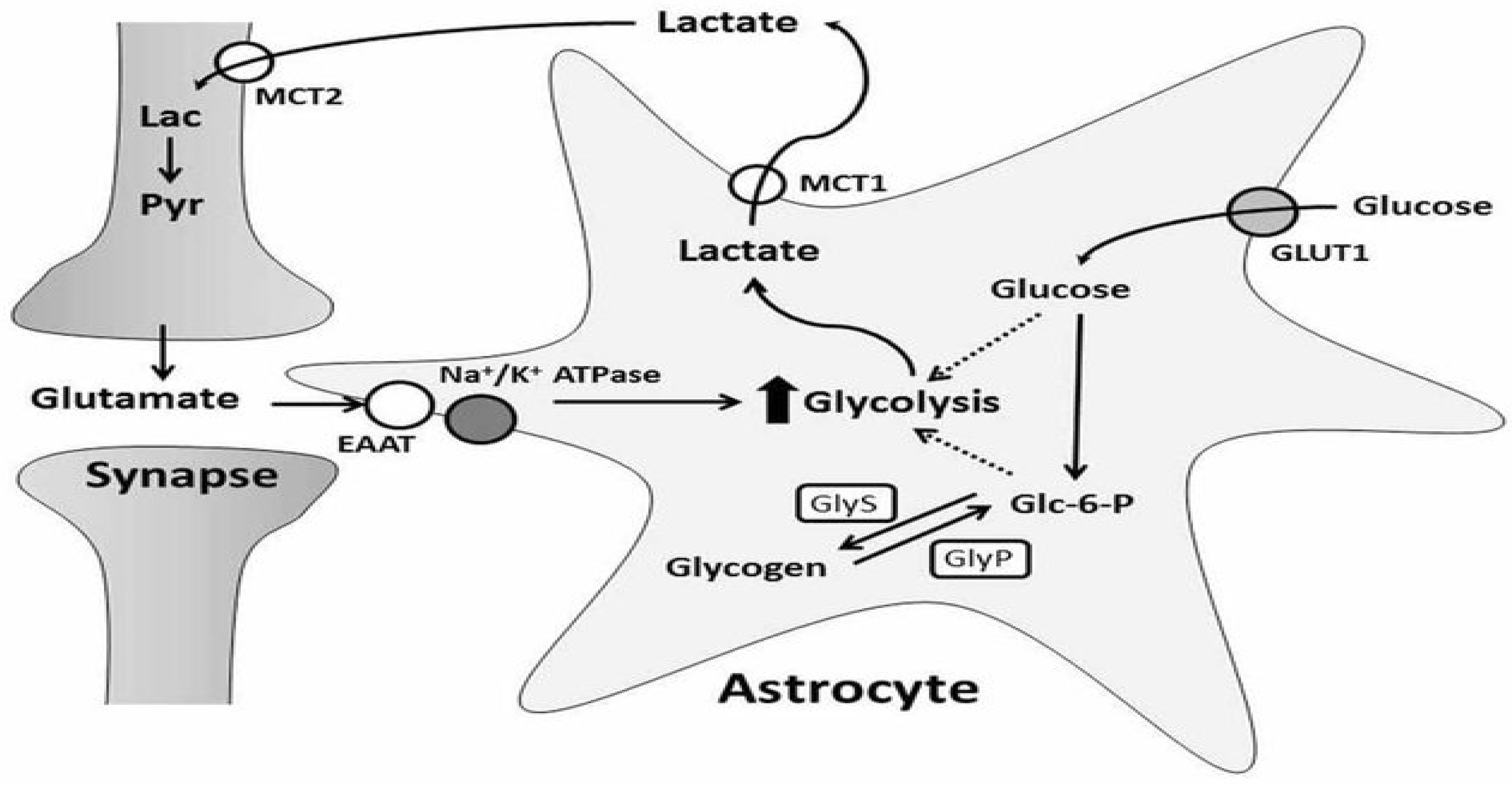
The astrocyte–neuron lactate shuttle hypothesis (ANLSH) proposes a critical role for astrocytes in neuronal energy metabolism, asserting that these glial cells produce lactate, which is then shuttled to neurons as an energy substrate. This process, according to the ANLSH, is vital for supporting neuronal function, especially when energy demands are elevated due to increased neuronal activity, stress, or injury [40]. At the heart of this hypothesis lies the production of lactate within astrocytes. This occurs through anaerobic glycolysis, where glucose is broken down into pyruvate, subsequently converted into lactate by lactate dehydrogenase (LDH) [40]. This lactate production is often coupled with glutamate uptake and the sodium-dependent activation of Na+-K+-ATPase [41] (Figure 3). Once produced, lactate is released from astrocytes and transported to neurons via monocarboxylate transporters (MCTs), with MCT2 being the primary transporter involved [41]. Neurons then utilize this lactate as an energy source through oxidative phosphorylation in the mitochondria, generating ATP, which is crucial for maintaining neuronal excitability and synaptic plasticity [42,43].
The implications of the ANLSH extend to the maintenance of the E/I balance within the brain. By guaranteeing an adequate energy supply to neurons during periods of increased activity, the ANLSH contributes to this delicate balance. Furthermore, dysregulation of the lactate shuttle mechanism has been linked to various neurological disorders, including AD, Parkinson’s disease (PD), and epilepsy [44]. Therefore, understanding the intricacies of the ANLSH could offer valuable insights into potential therapeutic interventions for these debilitating conditions. However, despite its significance, the ANLSH has faced criticism regarding the robustness of its experimental support. Some studies propose that glucose may remain the primary energy substrate for neurons, rather than relying exclusively on astrocyte-derived lactate [45].

2.4. Other Metabolic Pathways

Astrocytes, critical for neural function, maintain the delicate balance of E/I neurotransmission through intricate metabolic processes. Disruptions within these astrocytic metabolic pathways can trigger significant E/I imbalances, consequently impacting neural circuit function. At the core of astrocytic metabolic function lies energy metabolism, with oxidative phosphorylation (OXPHOS) and the Krebs cycle playing pivotal roles. Astrocytes possess high energy demands, primarily sustained by OXPHOS. Disruptions in OXPHOS compromise ATP production, affecting essential astrocyte functions such as glutamate uptake and potassium buffering [46]. Reduced ATP levels directly impair glutamate transporter activity, leading to the accumulation of extracellular glutamate and subsequent excitotoxicity. Furthermore, impaired mitochondrial function elevates levels of ROS, compounding neuronal damage [46]. Beyond energy production, astrocytes participate actively in amino acid metabolism, extending beyond the well-known glutamate–glutamine cycle. These cells are involved in synthesizing and metabolizing various amino acids, including GABA, which directly modulates neuronal excitability. Dysregulation of amino acid metabolism can alter neurotransmitter pools, leading to an E/I imbalance, which may manifest as either excessive excitation or inhibition, depending on the specific pathways affected [47]. The importance of astrocytic metabolism extends to lipid metabolism, where astrocytes synthesize lipids essential for synapse formation, membrane integrity, and signal transduction. Synaptic formation and function are compromised when lipid metabolism is dysfunctional, fundamentally altering neuronal excitability. Specific lipid metabolites act as signaling molecules that can affect synaptic plasticity [48]. Therefore, it is evident that disruptions in astrocytic energy, amino acid, and lipid metabolism can have profound consequences on E/I balance and overall neural circuit function.

3. Mechanisms Linking Astrocyte Metabolic Dysfunction to E/I Imbalance

3.1. Altered Glutamate Homeostasis

Astrocytes are critical for regulating glutamate levels in the synaptic cleft, primarily through EAATs [49]. Specifically, EAAT2 (also known as GLT-1) is responsible for the majority of glutamate uptake in the brain. In AD models, astrocytes expressing mutations in Amyloid Precursor Protein (APP) or Presenilin 1 (PSEN1) exhibit reduced EAAT2 expression, resulting in impaired glutamate clearance and elevated extracellular glutamate concentrations [18,50]. This accumulation of glutamate can lead to excitotoxicity, characterized by the overactivation of NMDA receptors on neurons and a subsequent disruption of the E/I balance [51]. Furthermore, studies have shown that astrocytes with AD mutations can have increased glycolysis and glutamate synthesis, further exacerbating the excessive extracellular glutamate levels that promote neuronal death [18,50].
Molecular Mechanism Example:
APP or PSEN1 mutations in astrocytes → ↓ EAAT2 expression → impaired glutamate clearance → ↑ extracellular glutamate → neuronal hyperexcitability and excitotoxicity [50,51].

3.2. Disrupted GABA Synthesis and Transport

Astrocytes are also involved in the synthesis of GABA, the primary inhibitory neurotransmitter in the brain. Astrocytes supply neurons with glutamine, a crucial precursor for both glutamate and GABA synthesis [52,53]. Deficient astrocyte metabolism, as observed in AD models, impairs the de novo synthesis of glutamine, thereby directly reducing the availability of this substrate for neuronal GABA synthesis [54]. Consequently, this leads to decreased inhibitory neurotransmission, which significantly contributes to the E/I imbalance. Even with maintained glutamine synthetase activity, impaired astrocytic metabolism can limit the overall glutamine availability, hindering GABA production in neurons [53,55].
Molecular Mechanism Example:
Impaired astrocyte metabolism → ↓ glutamine synthesis (despite maintained glutamine synthetase activity) → ↓ neuronal GABA synthesis → reduced inhibitory tone and E/I imbalance [54,56,57,58].
It is important to note that while a proportional reduction in both Glu and GABA would theoretically maintain the overall E/I ratio, the impact on neuronal function is more nuanced. Firstly, our review suggests that the observed reductions in Glu and GABA may not be perfectly proportional. The mechanisms underlying the alterations in Glu and GABA synthesis, release, or reuptake could be distinct, leading to disproportionate concentration changes. For example, Glu and GABA operate through distinct mechanisms despite their metabolic link. Their differences in synthesis, release, and reuptake can lead to disproportionate changes in their concentrations under physiological or pathological conditions [53,54]. Glutamate synthesis involves the conversion of glutamine to glutamate by glutaminase in neurons, with glutamine shuttled between astrocytes and neurons via specific transporters (ASCT2, SN1 in astrocytes; GlnT in neurons) [55]. GABA is synthesized from glutamate by glutamic acid decarboxylase (GAD) in GABAergic neurons [55]. Their release mechanisms differ: Glutamate is packaged into vesicles by vesicular glutamate transporters (vGLUT) and released into the synaptic cleft after neuronal depolarization. GABA, on the other hand, is packaged by the vesicular inhibitory amino acid transporter (VIAAT) and released via vesicular exocytosis, though astrocytes can release it through non-vesicular mechanisms like transporter reversal [55]. Glutamate reuptake and clearance occur mainly through EAATs on astrocytes, which are essential for preventing excitotoxicity and are tightly coupled to sodium gradients [55]. GABA uptake primarily happens via GABA transporters (GATs) in neurons, while astrocytes also express GAT-2/3, which may release GABA in response to increased intracellular sodium after glutamate uptake [55].
The distinct mechanisms regulating Glu and GABA at several levels, including transporter expression and localization, astrocyte–neuron interactions, enzymatic regulation, and variable pathway coupling and decoupling, lead to their disproportionate changes. For example, EAATs and GATs have different cellular and regional distributions affecting neurotransmitter clearance or release [55]. Astrocytic uptake of glutamate can trigger GABA release via transporter reversal, a process not mirrored in glutamate handling, leading to non-parallel changes in extracellular levels [55]. Similarly, the differing regulation of rate-limiting enzymes (glutaminase for Glu, GAD for GABA) results in them being independently affected by metabolic or pathological states [58]. Finally, even though GABA synthesis depends on glutamate, alterations in glutamate clearance or synthesis do not necessarily produce proportional changes in GABA due to the additional regulatory steps and compartmentalization [55]. Even with a relatively stable E/I ratio, a generalized reduction in both excitation and inhibition can decrease overall neuronal excitability and dampen network activity [59]. Therefore, the observed reductions in both Glu and GABA, even if not dramatically shifting the E/I ratio, likely contribute to significant alterations in neural circuit function.

3.3. Energy Deprivation and Neuronal Vulnerability

Beyond neurotransmitter metabolism, astrocytes provide essential metabolic support to neurons by supplying lactate and other energy substrates. In neurodegenerative conditions, astrocytic mitochondrial dysfunction results in brain hypometabolism, energetic failure, and oxidative stress [54]. This energy deprivation impairs neuronal function and survival, disproportionately affecting inhibitory interneurons, which are highly energy dependent. The selective vulnerability of these interneurons further shifts the E/I balance toward excitation [59,60]. The lactate shuttle, a key mechanism for energy transfer from astrocytes to neurons, is compromised when astrocytes suffer from mitochondrial deficits [61]. Our previous study shows that altered energy metabolism contributes to the alcohol exacerbation of psychological stress, notably indicated by significant glucose depletion and glial fibrillary acidic protein (GFAP), contributing to altered neuronal E/I balance. However, pharmacological intervention with naturally occurring flavonoids such as geraniol elevated GFAP expression in cortical brain regions, alongside improving energy homeostasis [62].
Molecular Mechanism Example:
Mitochondrial dysfunction in astrocytes → ↓ ATP production and impaired lactate shuttle → neuronal energy deprivation → selective vulnerability of inhibitory neurons → E/I imbalance [54].

3.4. Neuroinflammation and Metabolic Crosstalk

Astrocyte metabolic dysfunction can initiate and exacerbate neuroinflammatory responses. Damaged mitochondria in astrocytes may release damage-associated molecular patterns (DAMPs), which activate inflammatory signaling pathways [51]. In addition, impairments in astrocyte–neuron metabolic crosstalk, such as defective fatty acid (FA) metabolism and Apoe-dependent lipid handling, lead to oxidative stress and inflammation [63,64,65]. These inflammatory processes further disrupt neurotransmitter balance and synaptic function, contributing to E/I imbalance. Specifically, the ApoE4 variant, a major genetic risk factor for AD, has been shown to impair astrocytic lipid metabolism, leading to increased oxidative stress in neurons [66,67,68].
Molecular Mechanism Example:
ApoE4 variant in astrocytes → disrupted FA uptake and metabolism → accumulation of peroxidized FAs in neurons → oxidative stress and DAMP release → neuroinflammation → altered synaptic transmission and E/I imbalance [51].

3.5. Impact on Synaptic Function and Plasticity

Astrocytes play a critical role in regulating synaptic plasticity through Ca2⁺-dependent release of gliotransmitters, such as glutamate, D-serine, and ATP [69,70,71,72,73,74,75]. Disruptions in astrocyte Ca2⁺ signaling or metabolic support can impair the release of these modulators, affecting long-term potentiation (LTP) and long-term depression (LTD), which are essential for learning and memory [76,77]. For example, a reduced supply of D-serine from astrocytes impairs NMDA receptor-dependent synaptic plasticity, while altered glutamate release can either enhance or dampen excitatory transmission [78]. Both scenarios contribute to E/I imbalance [69]. Gliotransmitters, released in response to neuronal activity, fine-tune synaptic transmission and plasticity [79,80].
Molecular Mechanism Example:
Impaired astrocyte Ca2⁺ signaling or metabolic dysfunction → ↓ D-serine and glutamate release → impaired NMDAR-dependent LTP/LTD → abnormal synaptic plasticity and E/I imbalance [69].

4. Astrocyte Metabolic Dysfunction and E/I Imbalance in Neurological Disorders

4.1. Epilepsy

Astrocyte dysfunction is a significant contributor to the imbalance of E/I neurotransmission observed in various neurological disorders, most notably epilepsy. This dysfunction is critically involved in both epileptogenesis, the development of epilepsy, and ictogenesis, the generation of seizures [81,82]. Astrocytes play a vital role in maintaining glutamate homeostasis by efficiently uptaking and metabolizing glutamate released into the synaptic cleft. However, in epilepsy, astrocytes often fail to perform this function effectively, leading to elevated extracellular glutamate levels. This increase results in excessive stimulation of neurons, promoting hyperexcitability and seizure activity [80]. The closure of astrocytic gap junctions during epileptogenesis further exacerbates this issue by limiting the trafficking of energy metabolites and impairing the clearance of potassium ions from the extracellular space, which is essential for neuronal stability [80]. Complementing their role in glutamate regulation, astrocytes are also crucial for the synthesis of GABA (gamma-aminobutyric acid), the primary inhibitory neurotransmitter in the brain. A reduction in GABA production or release from astrocytes contributes to the E/I imbalance characteristic of epilepsy, as decreased GABAergic inhibition allows for heightened neuronal excitability [81,82]. The tonic release of GABA from astrocytes is important in maintaining this balance, and its dysregulation can lead to increased seizure susceptibility [82]. Beyond regulating neurotransmitters, astrocytes play a crucial role in controlling brain energy metabolism through processes such as glycolysis and glycogen metabolism. In epilepsy, metabolic dysfunction can impair astrocytes’ ability to provide neurons with adequate energy substrates during periods of heightened activity. This lack of energy support can exacerbate seizures by failing to buffer the increased demands placed on neuronal networks during excitatory events [80,83]. Astrocytic glycogen stores are critical for meeting energy demands, and their disruption can hinder the brain’s capacity to cope with increased neuronal activity, further promoting seizure activity [82,84]. Highlighting the importance of astrocyte function in epilepsy, several genetic mutations affecting astrocytic metabolic pathways have been implicated in the disorder. These mutations underscore the direct causal relationship between astrocyte dysfunction and the pathogenesis of epilepsy. For instance, alterations in ion channel expression within astrocytes can impair their potassium buffering capacity, leading to increased neuronal excitability and a greater likelihood of seizure generation [82,85].

4.2. Alzheimer’s Disease

Astrocyte metabolic dysfunction is increasingly recognized as a critical driver in the pathogenesis of AD, significantly contributing to the E/I imbalance characteristic of the disease and ultimately fostering the development of hallmark AD pathologies, such as amyloid plaques, tau tangles, and neurodegeneration. A key aspect of this dysfunction centers on the role of astrocytes in Aβ clearance. Typically, astrocytes efficiently remove Aβ, a protein central to AD pathology, but compromised metabolic function impairs this ability. This impairment can manifest as a reduced expression of Aβ-degrading enzymes or a decline in endocytosis, both of which result in the accumulation of Aβ and the subsequent formation of amyloid plaques [86]. Further compounding the issue, the metabolic state of astrocytes directly influences the processing of APP, potentially shifting it towards the amyloidogenic pathway and increasing Aβ production, thus exacerbating plaque formation [87,88]. Beyond their role in Aβ clearance, astrocytes also contribute to tau hyperphosphorylation, a process that leads to the formation of neurofibrillary tangles, another defining pathological feature of AD [89]. When astrocytic metabolic pathways are disrupted, cellular energy homeostasis is compromised. This leads to heightened oxidative stress and inflammatory signaling, which, in turn, activate kinases responsible for phosphorylating tau [89]. This hyperphosphorylation causes tau protein to detach from microtubules, its normal binding site, and aggregate into tangles within neurons [89].
The impairments in glucose metabolism within astrocytes represent another crucial aspect of astrocytic dysfunction in AD [88]. Astrocytes are vital for the brain’s glucose handling, and in AD, reduced glucose uptake and utilization by these cells significantly hinder their ability to provide adequate energy and metabolic support to neurons. The lactate shuttle, a process where astrocytes supply lactate as an energy source to neurons, is also disrupted when astrocytes are metabolically compromised [90]. This reduced energy supply increases neuronal vulnerability to excitotoxicity and oxidative stress, further exacerbating the E/I imbalance and contributing to neurodegeneration [88,91]. The vulnerability arises because neurons struggle to maintain proper ion gradients and neurotransmitter handling with insufficient energy, further destabilizing the excitatory/inhibitory balance and disrupting normal brain function [91]. The intimate relationship between astrocytes and neurons is essential for maintaining overall brain health, and disruptions in this relationship, driven by astrocytic dysfunction, ultimately translate to the significant cognitive decline that characterizes AD.

4.3. Autism Spectrum Disorder (ASD)

The emerging understanding of ASD highlights the crucial role of dysfunctional astrocyte metabolism in the context of E/I imbalance. Astrocytes, vital glial cells in the brain, are increasingly recognized for their contribution to maintaining neural homeostasis, particularly through the regulation of neurotransmitter signaling in glutamatergic and GABAergic pathways [92,93]. Alterations in these neurotransmitter systems are frequently observed in individuals with ASD, resulting in an elevated E/I ratio believed to underlie many of the cognitive and behavioral challenges associated with the disorder [92,93]. The influence of astrocytes on the E/I balance is multifaceted. One key mechanism involves the regulation of glutamate, the primary excitatory neurotransmitter. Astrocytes actively uptake excess glutamate from the synapse, preventing excitotoxicity and maintaining optimal levels for neural signaling. However, in ASD, astrocytic dysfunction can impair this glutamate uptake, leading to elevated extracellular glutamate concentrations and, consequently, heightened excitatory signaling. Studies have directly linked this impaired glutamate homeostasis to the manifestation of ASD symptoms [93,94].
Furthermore, astrocytes contribute significantly to GABA transmission, the primary inhibitory neurotransmitter system. They express GABA receptors and transporters, enabling them to modulate GABAergic signaling within neural circuits. Disruptions in GABAergic transmission are implicated in the pathogenesis of ASD, underscoring the importance of astrocyte health in maintaining inhibitory control and overall neural balance [93,94]. Additionally, aberrant calcium signaling within astrocytes represents another critical factor. Calcium signaling plays a crucial role in various astrocytic functions, including the release of gliotransmitters that influence synaptic transmission and plasticity. In ASD models, dysregulation of calcium signaling in astrocytes has been linked to neuroinflammation and behavioral deficits. This dysregulation can further exacerbate the E/I imbalance by impacting neurotransmitter release and synaptic plasticity [95,96].
The etiology of astrocyte dysfunction in ASD is thought to be complex, involving both genetic and environmental factors. Genetic studies have identified numerous autism-associated genes that are expressed in astrocytes, indicating a potential heritable component to their dysfunction [94,97]. These genetic predispositions may render astrocytes more susceptible to metabolic disturbances, thereby compromising their ability to support neuronal function and maintain neurotransmitter balance. Additionally, environmental factors, such as exposure to toxins or inflammatory agents during critical periods of brain development, can further exacerbate metabolic disturbances within astrocytes, resulting in compromised support for neuronal function and neurotransmitter balance [98].

4.4. Traumatic Brain Injury (TBI)

Traumatic brain injury initiates a complex cascade of metabolic events that profoundly impact astrocytes and contribute significantly to secondary brain injury [99]. This cascade is characterized by a series of interconnected processes, including impaired glucose metabolism, glutamate excitotoxicity, and neuroinflammation, ultimately leading to a disruption in the delicate balance of neurotransmission within the brain [99,100,101,102]. Following TBI, the brain’s ability to utilize glucose effectively is hampered, exacerbating the initial trauma’s effects, as glucose serves as the primary energy source for the brain. While there is an initial increase in glycolysis, this is often followed by metabolic diaschisis, a state where energy metabolism is compromised due to cellular dysfunction and oxidative stress [103,104,105]. Astrocytes, which normally play a crucial role in maintaining energy balance, are particularly affected by this metabolic dysfunction.
In parallel with impaired glucose metabolism, TBI triggers a surge in glutamate release. Under normal circumstances, astrocytes efficiently clear excess glutamate from the synaptic cleft [104]. However, after injury, their compromised metabolic state impairs their ability to regulate glutamate levels effectively. This leads to glutamate excitotoxicity, where excessive glutamate overstimulates neurons, resulting in neuronal damage and further disruption of brain function [104,106]. The excitotoxic effects are compounded by mitochondrial dysfunction and oxidative stress, which impair ATP production and lead to increased intracellular calcium levels, further contributing to neuronal injury [104,105,107]. These factors collectively exacerbate neuronal damage and contribute to secondary brain injury.
Neuroinflammation also plays a critical role in the secondary injury process following TBI. Damaged cells release inflammatory signals, activating microglia and reactive astrocytes [107]. While reactive astrocytes attempt to aid recovery and clear debris, their altered expression of transporters and metabolic enzymes can exacerbate the problem by further impairing glutamate regulation and energy supply [107,108]. This reactivity can lead to an E/I imbalance within the brain, recognized as a significant factor contributing to long-term neurological deficits following TBI [108]. The combined effects of impaired glucose metabolism, glutamate excitotoxicity, and neuroinflammation create a detrimental cycle that disrupts the brain’s delicate neurotransmitter balance. This E/I imbalance is associated with various long-term consequences for individuals recovering from TBI, including cognitive impairments, seizures, emotional disturbances, and motor deficits. The failure of astrocyte metabolism to adequately regulate neurotransmitter levels and maintain energy supply is a key driver of these secondary injuries. Therefore, a comprehensive understanding of these intricate mechanisms is crucial for developing effective therapeutic strategies that aim to mitigate secondary brain injury and ultimately improve outcomes for TBI patients.

4.5. Other Neurological Disorders

Dysfunctional astrocyte metabolism is increasingly recognized as a key contributor to a range of neurological disorders, disrupting the delicate balance between E/I neurotransmission, which is crucial for proper brain function [109,110]. Traditionally viewed as supportive cells, astrocytes are now understood to play a critical role in maintaining metabolic homeostasis within the brain, impacting neuronal health and contributing to the pathogenesis of diseases such as stroke, Huntington’s disease (HD), schizophrenia, and amyotrophic lateral sclerosis (ALS) [109,110,111]. Astrocytes are metabolic hubs, essential for providing neurons with vital energy substrates, such as lactate, and for facilitating the synthesis of neurotransmitters [109]. They also play a crucial role in clearing potentially toxic metabolic byproducts, further contributing to a stable and functional neuronal environment. However, when astrocytic metabolism is compromised, it can have profound consequences, disrupting neuronal function and leading to an E/I imbalance that underlies many neurological disorders.
In the context of stroke, the sudden loss of blood flow initiates a cascade of events culminating in ischemia and severe impairment of astrocyte metabolism [112]. This impairment often manifests as a reduction in both glucose uptake and lactate production, critical energy sources for neurons [113]. Simultaneously, an accumulation of toxic metabolites can occur, further stressing the already vulnerable neuronal population. Collectively, these metabolic disruptions render neurons susceptible to excitotoxicity, thereby exacerbating the E/I imbalance in affected brain regions and contributing to neuronal damage. Huntington’s disease also demonstrates the critical role of astrocytic metabolism in neurological health [114,115]. The expression of the mutant HD within astrocytes disrupts their normal metabolic pathways. This dysfunction impairs the ability of astrocytes to adequately meet the energy demands of surrounding neurons and compromises their efficiency in clearing glutamate. This, in turn, contributes to the excitotoxicity and progressive neuronal damage that are hallmarks of HD.
While schizophrenia has historically been understood through the lens of neuronal circuitry abnormalities, emerging evidence increasingly suggests that astrocyte dysfunction plays a significant contributing role [116]. Astrocytes in individuals with schizophrenia may exhibit alterations in glucose metabolism and glutamate handling, further disrupting the E/I balance and potentially exacerbating the symptoms associated with the disorder [116]. These findings underscore the importance of considering glial–neuronal interactions in the pathophysiology of schizophrenia. Similarly, in ALS, astrocyte dysfunction has been implicated in the degeneration of motor neurons, the hallmark of the disease [117,118]. Mutant superoxide dismutase 1 (SOD1), a protein associated with familial ALS, can impair astrocytic metabolism and compromise glutamate transport mechanisms. This leads to excitotoxicity, contributing to the progressive loss of motor neurons. This highlights how metabolic dysregulation in astrocytes can directly contribute to neuronal dysfunction and ultimately lead to neurodegeneration in devastating neurological conditions.

5. Therapeutic Strategies Targeting Astrocyte Metabolism to Restore E/I Balance

5.1. Modulation of Glutamate Homeostasis

Dysfunctional astrocyte metabolism is a significant contributor to imbalances between E/I signaling in the brain, a phenomenon implicated in various neurological disorders [119]. Because of their central role in brain homeostasis, modulating astrocyte metabolism presents a promising avenue for therapeutic intervention. A critical aspect of this modulation is the regulation of glutamate homeostasis, a process for which astrocytes are fundamentally responsible [120]. They maintain appropriate glutamate levels at the synapse, which is crucial for proper neuronal signaling and the prevention of excitotoxicity. One primary strategy for restoring E/I balance is to enhance glutamate uptake by astrocytes. This can be achieved by targeting EAATs, specifically EAAT2 (GLT1), the major player in clearing glutamate from the synaptic cleft [121]. Increasing EAAT2 activity can effectively reduce excess glutamate levels, minimizing excitotoxicity and potentially increasing inhibitory tone relative to excitation. Impaired astrocytic glutamate uptake can lead to neuronal excitotoxicity and neurodegeneration, underscoring the importance of effective glutamate regulation by astrocytes in maintaining brain health [122,123]. Another approach to modulating glutamate homeostasis involves inhibiting glutamine synthetase, the enzyme that converts glutamate into glutamine within astrocytes. Slowing this conversion process may decrease the availability of glutamine for neurons to convert back into glutamate. While seemingly counterintuitive, controlled GS inhibition can be beneficial in situations where excessive glutamate release stems from astrocyte dysregulation. In certain disease contexts where astrocyte function is impaired, this strategy could help re-establish a healthy E/I balance [123].

5.2. Enhancement of GABAergic Signaling

The therapeutic enhancement of GABAergic signaling presents a promising strategy for addressing imbalances in E/I neurotransmission, particularly those associated with dysfunctional astrocyte metabolism. Several approaches can be employed to modulate GABAergic activity, each with its mechanisms and potential benefits. One strategy involves increasing GABA production or release. This can be achieved through supplementation with GABA precursors like glutamate or glutamine, potentially boosting GABA synthesis, especially when astrocytes are involved in metabolizing these precursors into GABA [124]. Alternatively, directly enhancing the activity of GAD, the enzyme responsible for GABA synthesis, may augment GABA production. Promoting GABA release through modulation of receptors or transport proteins is another avenue for enhancing GABAergic signaling, potentially facilitating GABA release from astrocytes or GABAergic neurons. Pharmacological interventions provide additional methods for manipulating GABAergic neurotransmission. Benzodiazepines and barbiturates enhance GABA’s effects at GABA-A receptors, promoting inhibition by increasing the duration of chloride channel opening [125]. Medications targeting metabotropic GABA-B receptors can modulate neuronal excitability, contributing to the overall inhibitory tone. Additionally, inhibiting GABA transaminase or reuptake can increase synaptic GABA levels, prolonging its inhibitory effects [126].
Astrocytes play a crucial role in maintaining E/I balance by metabolizing GABA precursors and modulating GABAergic signaling. These glial cells can sense GABAergic activity and influence neuronal excitability by releasing gliotransmitters, such as glutamate and ATP [127]. Therefore, understanding astrocyte function is essential for tailoring therapeutic approaches. For instance, if astrocytes are deficient in producing GABA precursors, supplementation with these precursors may be beneficial. Conversely, if the issue is excessive glutamate release from astrocytes, targeting neuronal GABAergic signaling directly might be more appropriate [128]. The therapeutic potential of enhancing GABAergic signaling extends to various conditions, including neurological disorders characterized by E/I imbalances. In ASDs, for example, enhancing GABAergic signaling with benzodiazepines or selective modulators of GABA-A receptors can improve social and cognitive deficits [129]. Similarly, in conditions like AD, addressing GABAergic dysfunction may help restore E/I balance [129].

5.3. Targeting Neuroinflammation

Astrocytes are essential for maintaining brain health, serving as key regulators of neurotransmitter levels, energy provision, and immune responses. However, these critical functions can be severely compromised by neuroinflammation, leading to a state of reactive astrocytosis and a disruption in the delicate balance between E/I within the brain [109,110]. This imbalance is implicated in the pathology of numerous neurological disorders [109,110,111]. When neuroinflammation occurs, it significantly alters astrocyte metabolism, causing a shift away from the efficient process of oxidative phosphorylation towards glycolysis. This metabolic switch impacts the overall energy supply available to neurons and disrupts the carefully maintained homeostasis of neurotransmitters [130,131]. Furthermore, this inflammatory environment induces astrocytes to become reactive, altering their physical structure and functional capabilities. Reactive astrocytes often exhibit increased secretion of pro-inflammatory cytokines and chemokines, further perpetuating the inflammatory cycle [132,133]. The cumulative effect of these disruptions is a significant imbalance in the E/I tone, which can exacerbate conditions like epilepsy and depression [134]. Therapeutic strategies aimed at mitigating these effects include the use of broad-spectrum anti-inflammatory agents to generally reduce inflammation, thereby lessening astrocyte reactivity and promoting the restoration of normal metabolic function. A more targeted approach involves specifically modulating inflammatory mediators, such as TNF-α and IL-1β, known to be involved in astrocyte activation [135]. Ultimately, the goal is to normalize astrocyte metabolism and re-establish a healthy balance between excitation and inhibition in neuronal networks. Promising therapeutic targets include calcium channels, such as Orai1, where modulation has shown potential in reducing astrocyte reactivity and inflammation-induced behaviors [136]. Additionally, targeting specific metabolic pathways altered in reactive astrocytes, like glycolysis and lipid metabolism, could offer therapeutic benefits [137].

5.4. Metabolic Support and Energy Restoration

Astrocytes are central to maintaining the brain’s metabolic equilibrium, primarily by managing glucose uptake and processing, and the subsequent distribution of lactate to neurons. Compromised astrocyte metabolism can disrupt the delicate E/I balance, contributing to a range of neurological disorders. Therapeutic strategies aimed at reinforcing astrocyte energetics hold promise for rectifying these metabolic deficits. Several avenues are being explored to achieve this goal, with a focus on both improving glucose metabolism and exploring alternative energy sources [138]. One key approach involves the development of pharmacological agents designed to enhance glucose uptake by astrocytes. This can be achieved through optimizing the glycolytic pathway itself or by improving the expression and function of crucial transporters, such as GLUT1 and MCTs, which are essential for lactate shuttling [138]. Enhancing the efficiency of glycolysis within astrocytes is also critical for improving the energy supply to neurons. This necessitates a deeper understanding of specific metabolic bottlenecks that arise in different disease states, allowing for targeted interventions [139]. Simultaneously, improving MCT function can facilitate the efficient transfer of lactate from astrocytes to neurons, ensuring a consistent and reliable neuronal energy supply [138,140].
Beyond glucose-centric strategies, alternative metabolic interventions are gaining traction. Ketogenic diets, which promote the production of ketone bodies, offer an alternative energy source that can bypass impaired glucose metabolism, particularly beneficial when astrocyte glucose utilization is compromised [141]. Furthermore, supplementing with specific amino acids or antioxidants can support mitochondrial function and reduce oxidative stress within astrocytes, thereby enhancing their overall metabolic efficiency [142]. Modulating the insulin signaling pathway within astrocytes is another avenue being explored to improve their glucose handling and overall metabolic function [143]. However, the implementation of drastic metabolic changes, such as ketogenic diets, requires careful consideration of safety and appropriate patient selection, as the long-term effects of these interventions are still under investigation [143]. Ultimately, the development of tailored interventions based on a comprehensive understanding of the specific metabolic bottlenecks in astrocytes across different diseases is crucial. These targeted therapies hold the potential to improve metabolic efficiency, restore a healthy E/I balance, and ultimately alleviate the neurological consequences of astrocyte dysfunction [144].

5.5. Emerging Therapeutic Targets

The emerging understanding of dysfunctional astrocyte metabolism as a significant contributor to the E/I imbalance observed in numerous neurological disorders presents a compelling new direction for therapeutic strategies. This paradigm shift moves beyond the traditional neuron-centric view, recognizing astrocytes as active participants in disease pathogenesis. The central concept involves pinpointing specific metabolic pathways within astrocytes that, when disrupted, precipitate E/I imbalance, and subsequently designing interventions to rectify these dysfunctions [145]. Several potential therapeutic targets are surfacing within these astrocyte metabolic pathways. Given that astrocytes primarily utilize glucose to generate lactate, which is then transferred to neurons as an energy source, the glycolysis and lactate shuttle pathway emerges as a prime target. Modulating lactate production by inhibiting specific glycolytic enzymes within astrocytes, such as particular hexokinase or pyruvate kinase isoforms, could influence neuronal energy availability [145,146]. Simultaneously, enhancing neuronal lactate uptake via MCTs could bolster neuronal energy metabolism and resilience [145]. Furthermore, astrocytes play a critical role in the glutamate–glutamine cycle, clearing glutamate from the synapse and converting it to glutamine, which is then shuttled back to neurons. Disruptions in this cycle can lead to altered glutamate levels and E/I imbalance. Enhancing glutamate uptake by upregulating the expression or activity of glutamate transporters, such as GLT-1 (EAAT2 in humans), on astrocytes remains a crucial therapeutic objective [147]. Modulating glutaminase activity and augmenting glutamine synthetase activity within astrocytes could also yield beneficial outcomes [148]. While astrocytes predominantly rely on glycolysis, their TCA cycle and oxidative phosphorylation pathways remain essential, particularly for generating antioxidants and maintaining cellular redox balance. Augmenting antioxidant capacity by targeting the expression of antioxidant enzymes, such as SOD or glutathione peroxidase (GPx), could shield astrocytes from oxidative stress and enhance their functionality [149]. Other metabolic pathways, including glycogen metabolism and amino acid metabolism, such as the kynurenine pathway, also represent relevant targets for modulating astrocyte function and energy availability [150]. Gene therapy offers considerable promise for directly addressing astrocyte dysfunction at the genetic level. Approaches such as gene augmentation could restore normal function by delivering a functional copy of a deficient or mutated gene in astrocytes (e.g., the GLT1 gene) using viral vectors like AAV [151]. Gene editing technologies, such as CRISPR-Cas9, offer a precise approach to correcting specific genetic mutations within astrocytes [152]. Additionally, RNA interference (RNAi) can be employed to silence gene expression that contributes to astrocyte dysfunction [152]. However, several considerations and challenges must be addressed. Ensuring the specificity of therapeutic interventions to target astrocytes without affecting other cell types is paramount [153]. The effective and safe delivery of therapeutic agents or gene therapy vectors to astrocytes remains a significant hurdle due to the BBB [154]. Thorough evaluation of the long-term effects of modulating astrocyte metabolism or employing gene therapy is crucial to avoid unintended consequences [154]. The intricate and interconnected nature of astrocyte metabolism necessitates a systems-level understanding to prevent unforeseen effects on other pathways [155].

5.6. Nanoparticle Drug Delivery

Astrocytes play a vital role in maintaining a delicate balance between excitatory and inhibitory neurotransmission within the brain, a balance crucial for proper neurological function. These glial cells actively regulate the concentration of neurotransmitters such as glutamate and GABA by facilitating their uptake from the synaptic cleft, effectively preventing neuronal over- or under-stimulation [156]. Beyond neurotransmitter regulation, astrocytes provide essential metabolic support to neurons, supplying energy substrates like lactate, which neurons utilize for their energy demands [157]. Additionally, astrocytes are instrumental in maintaining ion homeostasis, carefully regulating the concentrations of various ions in the extracellular space surrounding neurons. This is essential for ensuring proper neuronal excitability and signaling [157]. However, when astrocyte metabolism becomes dysfunctional, the consequences can be significant, disrupting the finely tuned excitatory/inhibitory balance. This imbalance has been implicated in the pathogenesis of various neurological disorders, including conditions like depression and epilepsy [158]. For instance, when the glutamate uptake processes in astrocytes are compromised, it may lead to a buildup of glutamate in the synaptic cleft, triggering neuron overstimulation and ultimately resulting in excitotoxicity [159]. Furthermore, when astrocytes fail to provide adequate metabolic support to neurons, neuronal firing patterns can become disrupted, making neurons more vulnerable to damage and dysfunction [160,161,162].
To address these challenges associated with dysfunctional astrocyte metabolism, nanoparticle drug delivery systems have emerged as a promising therapeutic strategy. The core principle is to target therapeutic agents directly to astrocytes, aiming to restore the disrupted excitatory/inhibitory balance and alleviate the underlying causes of neurological disorders. Several concrete strategies can be employed using targeted nanoparticle delivery:
Enhancing Glutamate Uptake: One strategy focuses on improving glutamate uptake by astrocytes [163,164,165]. This can be achieved by delivering drugs that upregulate the expression or activity of astrocyte glutamate transporters, such as GLT-1 (EAAT2) or GLAST (EAAT1) [163]. For example, nanoparticles could be loaded with ceftriaxone, a beta-lactam antibiotic known to increase GLT-1 expression [166], or with activators of the Nrf2 pathway, which can also promote GLT-1 expression [167]. The nanoparticles would be surface-modified with ligands, such as glutamate or specific antibodies recognizing astrocyte-specific surface markers, to ensure targeted delivery.
Modulating Lactate Production: Another approach involves modulating lactate production by astrocytes. In conditions where astrocyte lactate production is impaired, nanoparticles could deliver compounds that enhance glycolysis or promote the activity of lactate dehydrogenase (LDH). Conversely, in situations where excessive lactate production contributes to neuronal dysfunction, nanoparticles could deliver LDH inhibitors. For instance, dichloroacetate (DCA) is an inhibitor of pyruvate dehydrogenase kinase (PDK), which indirectly increases the flux of glucose towards lactate production [168].
Restoring GABA Synthesis or Transport: In some neurological disorders, impaired GABA synthesis or transport in astrocytes contributes to E/I imbalance. Nanoparticles could be used to deliver precursors of GABA synthesis, such as glutamine, or compounds that enhance the activity of glutamic acid decarboxylase (GAD), the enzyme responsible for GABA synthesis [169]. Another therapeutic approach is to enhance GABA transport by the use of nipecotic acid [170]. The nanoparticle surface would be modified with ligands which preferentially binds to astrocytes.
Nanoparticles offer several key advantages in this context [162]. They can be designed to selectively bind to or be taken up by astrocytes, ensuring that therapeutic agents are delivered precisely to the cells where they are needed most. By concentrating drugs within astrocytes, nanoparticles can achieve a higher therapeutic effect with a lower overall drug dose, enhancing efficacy. Moreover, targeted delivery minimizes exposure to other brain cells and tissues, potentially reducing the risk of unwanted side effects [163]. This targeted approach is particularly relevant for treating neurological conditions that arise from or are exacerbated by astrocyte dysfunction, such as depression and epilepsy [164,165].

6. Astrocyte Heterogeneity and Disease

Astrocytes are not a single, uniform cell type. Rather, they exhibit significant heterogeneity along several axes, including developmental origin, morphology, molecular expression profiles, electrophysiological properties, and functional outputs [166,167]. This diversity is observed both between different brain regions (regional heterogeneity) and within a given brain region (local heterogeneity). Astrocytes in different areas may arise from different progenitor cells or at different times during development, influencing their properties. Notably, single-cell RNA sequencing (scRNA-seq) and other omics approaches have revealed distinct gene expression signatures that define astrocyte subtypes [167,168].
It is important to note that different astrocyte subtypes may have unique roles in synaptic transmission, ion homeostasis, and other functions. Given the functional diversity of astrocytes, it is not surprising that specific astrocyte subtypes may be differentially affected in neurological disorders. For example, Furman et al. [169] found that a particular astrocyte subtype was selectively vulnerable in a mouse model of AD, suggesting that targeting this subtype could be a potential therapeutic strategy. Dysfunctional astrocyte metabolism contributes to imbalanced E/I tone, a common feature of many neurological disorders. Researchers are working on mapping astrocyte subtypes across brain regions and identifying changes in disease states in order to develop targeted therapies that selectively modulate specific astrocyte subpopulations. This approach holds the potential to restore E/I balance and improve patient outcomes in conditions such as neurodegenerative diseases and psychiatric disorders [168,169].
Astrocyte Subtypes
Astrocytes, once viewed as a homogenous population of supportive cells, are now understood to be a heterogeneous group with specialized functions critical for both the normal physiology and pathology of the CNS. Recent advances in transcriptomic profiling have unveiled the existence of distinct astrocyte subtypes, exhibiting regional specificity and functional diversity that significantly influence healthy brain function and contribute to disease mechanisms. Studies have identified region-specific transcription factors that play a crucial role in defining these astrocyte subtypes.
Pan-astrocyte transcription factors: Transcription factors like NFIB and NFIX are universally expressed across various brain regions, including the olfactory bulb, hippocampus, cortex, and brainstem, and are responsible for regulating core astrocytic functions [170].
Region-specific transcription factors: Transcription factors such as Nkx6-1 in the brainstem, Nkx3-1 in the olfactory bulb, and Pgr in the cortex contribute to localized gene regulatory networks [170]. These transcription factors influence key metabolic functions, such as glucose metabolism via glycolysis and oxidative phosphorylation [171], as well as neurotransmitter homeostasis, including the regulation of the glutamate/GABA-glutamine cycle [172,173].
The functional relevance of these astrocyte subtypes extends to several critical areas:
Blood–brain barrier maintenance: Astrocytes contribute to maintaining the BBB through interactions with vascular endfeet [172].
Synaptic modulation: Astrocytes modulate synaptic activity through glutamate uptake, mediated by EAAT1/2 transporters [172,173].
Immune regulation: Astrocytes participate in immune regulation through cytokine signaling and antigen presentation [174,175].
Regional Susceptibility to Neurological Diseases
The distribution of astrocyte subtypes has been found to correlate with regional vulnerability to specific neurological diseases.
Alzheimer’s disease: Cortical astrocytes exhibit dysregulation in genes such as apoE, clusterin, and cystatin C, which are involved in cholesterol metabolism and protein aggregation [173,174].
Parkinson’s disease: Midbrain astrocytes show impaired antioxidant mechanisms, such as GST and peroxiredoxin, which exacerbate the loss of dopaminergic neurons [173].
Multiple sclerosis: Inflammatory astrocytes in white matter upregulate MHC-II, contributing to autoimmune demyelination [175].
Stroke: Brainstem astrocytes with high Nkx6-1 expression may influence the recovery of autonomic functions [170], while cortical astrocytes modulate excitotoxicity through glutamate clearance [171].
Primary vs. Secondary Roles in Disease
Astrocytes can play both primary and secondary roles in the pathogenesis of neurological diseases.
Primary drivers:
Astrocytic SOD1 mutations in amyotrophic lateral sclerosis directly cause motor neuron degeneration [173].
Hepatic encephalopathy arises from astrocyte-specific failures in ammonia detoxification [174].
Secondary responders:
Reactive astrocytes in traumatic injury can adopt pro-inflammatory (A1) or neuroprotective (A2) phenotypes [175].
Alzheimer’s-associated astrocytes show upregulated β-amyloid processing enzymes only after neuronal pathology emerges [173,174].
Addressing Contradictions and Limitations
Several factors contribute to discrepancies and limitations in understanding astrocyte function in the context of disease.
Methodological variability: Bulk RNA sequencing approaches may obscure subtype-specific signals compared to single-cell approaches [170,173].
Disease models:
In vitro models often fail to replicate regional TF gradients, such as Nkx6-1 in the brainstem [170].
Transgenic mice overexpressing human apoE4 exhibit stronger Alzheimer’s pathology than wild-type mice [173], which can complicate cross-species comparisons.
Temporal factors: Pro-inflammatory astrocyte signatures may peak early in multiple sclerosis but dominate late-stage AD [175], leading to conflicting therapeutic interpretations.

7. Future Directions and Challenges

Future research efforts should prioritize the use of advanced imaging and genetic tools, such as live super-resolution imaging, to monitor astrocyte dynamics in real time. Furthermore, the exploitation of genetic model organisms (e.g., C. elegans, Drosophila, Zebrafish) can drive advances in understanding astrocyte heterogeneity and the development of therapies that target specific astrocyte subtypes to modulate neural circuit function in disease states.

7.1. Development of Astrocyte-Specific Tools and Techniques

The advancement of astrocyte-specific tools and techniques represents a critical area for progress in understanding and targeting astrocyte dysfunction in neurological disorders. Significant obstacle currently hindering research is the limited availability of methods that selectively manipulate and monitor astrocyte activity and metabolism in vivo within the brain’s intricate environment. Existing techniques often lack the specificity to isolate astrocytes’ unique contributions, making it challenging to differentiate their roles from those of neurons and other glial cells in the development of imbalanced E/I tone [176]. A key challenge lies in ensuring that manipulations are indeed astrocyte-specific, avoiding confounding effects from other cell types.
The future of astrocyte research relies on developing and implementing more advanced methodologies. Optogenetics and chemogenetics, including designer receptors exclusively activated by designer drugs (DREADDs), offer exciting possibilities. These techniques allow for precise temporal and spatial control over astrocyte activity. However, the effective use of these techniques depends critically on astrocyte-specific targeting.
Optogenetics utilizes light to activate or inhibit genetically modified astrocytes, providing high temporal resolution [176]. Chemogenetics, specifically DREADDs, involves engineered receptors activated by synthetic ligands, enabling longer-lasting modulation of astrocyte function [176]. To achieve astrocyte specificity, researchers commonly employ cell-type specific promoters, such as GFAP, Gfa2, or Aldh1l1, to drive the expression of the light-sensitive proteins (in optogenetics) or DREADDs specifically in astrocytes. These promoters are chosen for their preferential, though not always exclusive, expression in astrocytes. These tools allow researchers to selectively activate or inhibit specific metabolic pathways or signaling cascades within astrocytes, and subsequently observing the resulting changes in neuronal activity and E/I balance. This degree of control is essential for establishing causal relationships between specific astrocyte functions and network-level dysfunction [176]. Despite the use of astrocyte-specific promoters, careful validation is crucial to confirm the target cell specificity. This includes the following:
Immunohistochemical analysis: Confirming the expression of the introduced protein (e.g., channelrhodopsin or DREADD) is primarily localized to astrocytes based on the presence of astrocyte markers (e.g., GFAP, S100β, Aldh1l1).
Quantitative PCR (qPCR) or RNA sequencing (RNA-seq): Assessing the expression levels of the introduced gene in different cell types to quantify specificity.
Electrophysiological recordings: Confirming that activation (inhibition) occurs primarily in astrocytes upon stimulation (e.g., light stimulation in optogenetics or CNO administration in DREADD experiments).
Consideration of potential compensatory mechanisms: It is imperative to acknowledge that even with specific targeting, downstream processes involving other cell types can occur.
It is also paramount to consider potential off-target effects and compensatory mechanisms that may arise from manipulating astrocytes. For instance, some astrocyte promoters might exhibit low-level expression in other glial cell types or even neurons. Therefore, it is imperative to utilize multiple validation methods to ascertain the specificity of the manipulation and the observed effects.
Astrocytes are essential in neurological disorders, such as AD, PD, and HD. Their involvement in glutamate and ion homeostasis, cholesterol and sphingolipid metabolism, and tripartite synapses highlights their importance [176,177]. Recent studies emphasize astrocyte secretome profiling in understanding their role in neurodegenerative diseases, identifying potential therapeutic targets, such as FAM3C and KITLG [178]. Furthermore, the molecular diversity of astrocytes and their regional expression patterns highlight their complex roles in CNS disorders [179]. Consequently, the refinement and broader application of astrocyte-specific tools are vital for translating fundamental research findings into effective therapeutic strategies for neurological disorders characterized by E/I imbalance.

7.2. Longitudinal Studies and Biomarker Discovery

Understanding the role of dysfunctional astrocyte metabolism in driving excitatory/inhibitory imbalance in neurological disorders necessitates a shift towards dynamic perspectives, achievable through longitudinal studies. These studies, which track individuals over extended periods, are vital for illuminating how astrocyte function evolves throughout the course of disease progression. Moving beyond static snapshots of astrocyte activity, longitudinal investigations allow researchers to correlate changes in astrocyte metabolism with clinical outcomes and specific disease stages. For example, studies following patients with AD have demonstrated a significant link between the decline in astrocyte function and the development of progressive hypometabolism, underscoring the critical role of astrocytes in the advancement of the disease [180,181].
However, our ability to fully leverage the insights gained from longitudinal studies is currently hampered by the lack of reliable and specific biomarkers for astrocyte dysfunction. The absence of such biomarkers severely limits our capacity to achieve early diagnosis of astrocyte-related pathologies, monitor the effectiveness of therapeutic interventions targeting astrocyte metabolism, and personalize treatment strategies for individuals with neurological disorders [182]. An ideal biomarker would be readily accessible, for instance, through blood or cerebrospinal fluid, and precisely reflect the underlying metabolic alterations occurring within astrocytes that contribute to the excitatory/inhibitory imbalance observed in many neurological conditions [183]. The discovery and rigorous validation of these biomarkers are essential steps in translating basic research findings regarding astrocyte metabolism into tangible benefits for patients. By identifying and validating biomarkers that can be easily measured and correlated with astrocyte function, we can improve diagnostic accuracy, facilitate the development of targeted therapies, and monitor the effectiveness of interventions aimed at restoring astrocyte function. The convergence of longitudinal studies and biomarker development holds promise for unraveling the complexities of astrocyte-mediated disease mechanisms and the development of effective, personalized treatments for neurological disorders [184].

7.3. Clinical Translation

Despite the compelling preclinical promise of targeting astrocyte metabolism to restore the delicate balance between excitation and inhibition in neurological disorders, significant obstacles impede the successful translation of these findings into effective clinical therapies (Challenges and Opportunities of Targeting Astrocytes to Halt Neurodegeneration [185]). The journey from promising results in animal models to tangible benefits for patients is fraught with complexities. A primary hurdle lies in the fundamental differences between simplified animal models and the intricate reality of the human brain. While animal models offer a controlled environment for studying disease mechanisms, they often fail to fully capture the multifaceted nature of human neurological disorders. The heterogeneity of the disease itself, the varying genetic backgrounds of patients, and the influence of diverse environmental factors are often inadequately represented, leading to discrepancies in therapeutic outcomes between preclinical studies and clinical trials [185].
A further significant challenge is the BBB, which restricts the delivery of potential astrocyte-targeted therapies to the specific brain regions where they are needed [186]. This protective barrier, while essential for maintaining brain homeostasis, significantly limits the access of many drugs to the central nervous system, thereby reducing their therapeutic efficacy. Given the crucial role of astrocytes in maintaining brain homeostasis and supporting neuronal health, targeting them holds immense promise for treating neurodegenerative diseases such as AD, PD, and ALS [187]. However, achieving this promise requires overcoming the delivery challenges posed by the BBB.
Accurately assessing astrocyte function in the living human brain presents another considerable difficulty. Current imaging techniques often lack the necessary resolution and specificity to directly monitor astrocyte metabolism and its complex interplay with neuronal activity. This limitation makes it challenging to identify suitable patient populations for targeted therapies and to effectively monitor treatment responses [188]. The development of reliable biomarkers that can reflect astrocyte dysfunction in vivo is therefore essential for the successful implementation of astrocyte-targeted therapies. To navigate these challenges, well-designed clinical trials are of paramount importance. These trials must incorporate rigorous methodologies, including stringent patient selection criteria, appropriate placebo controls, and comprehensive outcome measures capable of assessing both clinical symptoms and relevant changes in neuronal and astrocyte function [182]. Adaptive trial designs, which allow for modifications based on emerging data, can further enhance efficiency and increase the probability of success. Moreover, thorough safety profiling is crucial; off-target effects on astrocytes or disruption of their essential homeostatic functions could lead to unintended and potentially detrimental consequences.

8. Conclusions

Dysfunctional astrocyte metabolism emerges as a critical factor in the E/I imbalance characteristic of many neurological disorders. These star-shaped glial cells are essential for maintaining metabolic homeostasis within the brain, influencing energy levels and redox balance. When astrocyte metabolism falters, it disrupts key processes such as glucose metabolism, lactate shuttling, and the crucial recycling of neurotransmitters like glutamate and GABA. Consequently, the ability of astrocytes to effectively regulate neuronal excitability is compromised. This impairment directly impacts synaptic transmission and the overall stability of neural networks, leading to neurodegeneration and excitotoxicity as a result of, among other things, impaired glutamate uptake. The E/I imbalance contributes to conditions like AD, depression, and epilepsy. Targeting astrocyte metabolism may restore E/I balance and improve neurological outcomes. Future research should focus on specific metabolic pathways, develop targeted interventions, and understand the relationship between astrocytes and neurons. These advancements could revolutionize treatment for various neurological diseases.

Author Contributions

Conceptualization, U.G.J. and M.O.O.; writing—original draft preparation, U.G.J., D.T.E., B.B.-A. and E.J., writing—review and editing, U.G.J., D.T.E., B.B.-A. and E.J. All authors have read and agreed to the published version of the manuscript.

Funding

This research was supported by the DELSU Joint Canada-Israel Project funded by the Canadian Institutes of Health Research (CIHR), the International Development Research Center (IDRC), the Israel Science Foundation (ISF), and the Azrieli Foundation, as well as and an Early Principal Investigator Grant supported by the International Brain Research Organization (IBRO), both awarded to B.B.-A at Delta State University.

Acknowledgments

We thank the reviewers for their helpful comments.

Conflicts of Interest

The authors declare no conflicts of interest.

References

  1. Chiareli, R.A.; Carvalho, G.A.; Marques, B.L.; Mota, L.S.; Oliveira-Lima, O.C.; Gomes, R.M.; Birbrair, A.; Gomez, R.S.; Simão, F.; Klempin, F.; et al. The role of astrocytes in the neurorepair process. Front. Cell Dev. Biol. 2021, 9, 665795. [Google Scholar] [CrossRef] [PubMed]
  2. Aharoni, R.; Eilam, R.; Arnon, R. Astrocytes in multiple sclerosis—Essential constituents with diverse multifaceted functions. Int. J. Mol. Sci. 2021, 22, 5904. [Google Scholar] [CrossRef] [PubMed]
  3. Soares, É.N.; Costa, A.C.; Ferrolho, G.D.; Ureshino, R.P.; Getachew, B.; Costa, S.L.; Da Silva, V.D.; Tizabi, Y. Nicotinic Acetylcholine Receptors in Glial Cells as Molecular Target for Parkinson’s Disease. Cells 2024, 13, 474. [Google Scholar] [CrossRef]
  4. Sufleţel, R.T.; Mihu, C.M.; Boşca, A.B.; Melincovici, C.S.; Mărginean, M.V.; Jianu, E.M.; Onofrei, M.M.; Constantin, A.M.; Moldovan, I.M.; Coneac, A.; et al. Short histological kaleidoscope–recent findings in histology. Part IV Rom. J. Morphol. Embryol. 2024, 65, 377. [Google Scholar] [CrossRef] [PubMed]
  5. Yuan, W.Q.; Huang, W.P.; Jiang, Y.C.; Xu, H.; Duan, C.S.; Chen, N.H.; Liu, Y.J.; Fu, X.M. The function of astrocytes and their role in neurological diseases. Eur. J. Neurosci. 2023, 58, 3932–3961. [Google Scholar] [CrossRef]
  6. Theparambil, S.M.; Begum, G.; Rose, C.R. pH regulating mechanisms of astrocytes: A critical component in physiology and disease of the brain. Cell Calcium 2024, 120, 102882. [Google Scholar] [CrossRef] [PubMed]
  7. Walz, W. The Brain as an Organ. In The Gliocentric Brain: Phenotype Plasticity of the Damaged Brain; Springer International Publishing: Cham, Switzerland, 2024; pp. 7–27. [Google Scholar]
  8. Syvänen, V.; Koistinaho, J.; Lehtonen, Š. Identification of the abnormalities in astrocytic functions as potential drug targets for neurodegenerative disease. Expert Opin. Drug Discov. 2024, 19, 603–616. [Google Scholar] [CrossRef]
  9. Yu, Y.; Chen, R.; Mao, K.; Deng, M.; Li, Z. The role of glial cells in synaptic dysfunction: Insights into Alzheimer’s disease mechanisms. Aging Dis. 2024, 15, 459. [Google Scholar] [CrossRef]
  10. Escalada, P.; Ezkurdia, A.; Ramírez, M.J.; Solas, M. Essential role of astrocytes in learning and memory. Int. J. Mol. Sci. 2024, 25, 1899. [Google Scholar] [CrossRef]
  11. Squadrani, L.; Wert-Carvajal, C.; Müller-Komorowska, D.; Bohmbach, K.; Henneberger, C.; Verzelli, P.; Tchumatchenko, T. Astrocytes enhance plasticity response during reversal learning. Commun. Biol. 2024, 7, 852. [Google Scholar] [CrossRef]
  12. Chen, T.; Dai, Y.; Hu, C.; Lin, Z.; Wang, S.; Yang, J.; Zeng, L.; Li, S.; Li, W. Cellular and molecular mechanisms of the blood–brain barrier dysfunction in neurodegenerative diseases. Fluids Barriers CNS 2024, 21, 60. [Google Scholar] [CrossRef] [PubMed]
  13. Che, J.; Sun, Y.; Deng, Y.; Zhang, J. Blood-brain barrier disruption: A culprit of cognitive decline? Fluids Barriers CNS 2024, 21, 63. [Google Scholar] [CrossRef] [PubMed]
  14. Dienel, G.A.; Schousboe, A.; McKenna, M.C.; Rothman, D.L. A tribute to Leif Hertz: The historical context of his pioneering studies of the roles of astrocytes in brain energy metabolism, neurotransmission, cognitive functions, and pharmacology identifies important, unresolved topics for future studies. J. Neurochem. 2024, 168, 461–495. [Google Scholar] [CrossRef]
  15. Calì, C.; Cantando, I.; Veloz Castillo, M.F.; Gonzalez, L.; Bezzi, P. Metabolic reprogramming of astrocytes in pathological conditions: Implications for neurodegenerative diseases. Int. J. Mol. Sci. 2024, 25, 8922. [Google Scholar] [CrossRef]
  16. Vivi, E.; Di Benedetto, B. Brain stars take the lead during critical periods of early postnatal brain development: Relevance of astrocytes in health and mental disorders. Mol. Psychiatry 2024, 29, 2821–2833. [Google Scholar] [CrossRef]
  17. Tang, Y.; Wang, C.; Li, Q.; Liu, G.; Song, D.; Quan, Z.; Yan, Y.; Qing, H. Neural Network Excitation/Inhibition: A Key to Empathy and Empathy Impairment. Neuroscientist 2024, 30, 644–665. [Google Scholar] [CrossRef] [PubMed]
  18. Salcedo, C.; Pozo Garcia, V.; García-Adán, B.; Ameen, A.O.; Gegelashvili, G.; Waagepetersen, H.S.; Freude, K.K.; Aldana, B.I. Increased glucose metabolism and impaired glutamate transport in human astrocytes are potential early triggers of abnormal extracellular glutamate accumulation in hiPSC-derived models of Alzheimer’s disease. J. Neurochem. 2024, 168, 822–840. [Google Scholar] [CrossRef]
  19. Koh, W.; Lee, C.J. Diagnostic and therapeutic potential of tonic gamma-aminobutyric acid from reactive astrocytes in brain diseases. Clin. Transl. Med. 2024, 14, e1642. [Google Scholar] [CrossRef]
  20. Li, Z.; Jiang, Y.Y.; Long, C.; Peng, X.; Tao, J.; Pu, Y.; Yue, R. Bridging metabolic syndrome and cognitive dysfunction: Role of astrocytes. Front. Endocrinol. 2024, 15, 1393253. [Google Scholar] [CrossRef]
  21. Zhang, Y.M.; Qi, Y.B.; Gao, Y.N.; Chen, W.G.; Zhou, T.; Zang, Y.; Li, J. Astrocyte metabolism and signaling pathways in the CNS. Front. Neurosci. 2023, 17, 1217451. [Google Scholar] [CrossRef]
  22. Beard, E.; Lengacher, S.; Dias, S.; Magistretti, P.J.; Finsterwald, C. Astrocytes as key regulators of brain energy metabolism: New therapeutic perspectives. Front. Physiol. 2022, 12, 825816. [Google Scholar] [CrossRef] [PubMed]
  23. Sharma, K.; Tapadia, M.G. Dysregulated Peripheral Metabolism in Neurodegenerative Disorders. In Altered Metabolism: A Major Contributor of Comorbidities in Neurodegenerative Diseases; Springer Nature: Singapore, 2024; pp. 157–172. [Google Scholar]
  24. Brooks, G.A.; Arevalo, J.A.; Osmond, A.D.; Leija, R.G.; Curl, C.C.; Tovar, A.P. Lactate in contemporary biology: A phoenix risen. J. Physiol. 2022, 600, 1229–1251. [Google Scholar] [CrossRef]
  25. Vavřička, J.; Pavel, B.R.; Follprecht, D.; Novák, J.; Kroužeckŷ, A. Modern perspective of lactate metabolism. Physiol. Res. 2024, 73, 499. [Google Scholar] [CrossRef]
  26. Ben-Azu, B.; Aderibigbe, A.O.; Ajayi, A.M.; Eneni, A.O.; Omogbiya, I.A.; Owoeye, O.; Umukoro, S.; Iwalewa, E.O. Morin decreases cortical pyramidal neuron degeneration via inhibition of neuroinflammation in mouse model of schizophrenia. Int. Immunopharmacol. 2019, 70, 338–353. [Google Scholar] [CrossRef] [PubMed]
  27. Markussen, K.H.; Corti, M.; Byrne, B.J.; Vander Kooi, C.W.; Sun, R.C.; Gentry, M.S. The multifaceted roles of the brain glycogen. J. Neurochem. 2024, 168, 728–743. [Google Scholar] [CrossRef]
  28. Beltran-Velasco, A.I. Brain Glycogen—Its Metabolic Role in Neuronal Health and Neurological Disorders—An Extensive Narrative Review. Metabolites 2025, 15, 128. [Google Scholar] [CrossRef] [PubMed]
  29. Shichkova, P.; Coggan, J.S.; Markram, H.; Keller, D. Brain Metabolism in Health and Neurodegeneration: The Interplay Among Neurons and Astrocytes. Cells 2024, 13, 1714. [Google Scholar] [CrossRef]
  30. Prasad, S.K.; Acharjee, A.; Singh, V.V.; Trigun, S.K.; Acharjee, P. Modulation of brain energy metabolism in hepatic encephalopathy: Impact of glucose metabolic dysfunction. Metab. Brain Dis. 2024, 39, 1649–1665. [Google Scholar] [CrossRef]
  31. Colpaert, M.; Singh, P.K.; Donohue, K.J.; Pires, N.T.; Fuller, D.D.; Corti, M.; Byrne, B.J.; Sun, R.C.; Vander Kooi, C.W.; Gentry, M.S. Neurological glycogen storage diseases and emerging therapeutics. Neurotherapeutics 2024, 21, e00446. [Google Scholar] [CrossRef]
  32. Hertz, L.; Rothman, D.L. Glutamine-glutamate cycle flux is similar in cultured astrocytes and brain and both glutamate production and oxidation are mainly catalyzed by aspartate aminotransferase. Biology 2017, 6, 17. [Google Scholar] [CrossRef]
  33. Limón, I.D.; Angulo-Cruz, I.; Sánchez-Abdon, L.; Patricio-Martínez, A. Disturbance of the glutamate-glutamine cycle, secondary to hepatic damage, compromises memory function. Front. Neurosci. 2021, 15, 578922. [Google Scholar] [CrossRef] [PubMed]
  34. Sidoryk-Węgrzynowicz, M.; Adamiak, K.; Strużyńska, L. Astrocyte–Neuron Interaction via the Glutamate–Glutamine Cycle and Its Dysfunction in Tau-Dependent Neurodegeneration. Int. J. Mol. Sci. 2024, 25, 3050. [Google Scholar] [CrossRef] [PubMed]
  35. Zhang, D.; Hua, Z.; Li, Z. The role of glutamate and glutamine metabolism and related transporters in nerve cells. CNS Neurosci. Ther. 2024, 30, e14617. [Google Scholar] [CrossRef] [PubMed]
  36. Satarker, S.; Bojja, S.L.; Gurram, P.C.; Mudgal, J.; Arora, D.; Nampoothiri, M. Astrocytic glutamatergic transmission and its implications in neurodegenerative disorders. Cells 2022, 11, 1139. [Google Scholar] [CrossRef]
  37. Onaolapo, A.Y.; Onaolapo, O.J. Peripheral and central glutamate dyshomeostasis in neurodegenerative disorders. Curr. Neuropharmacol. 2021, 19, 1069–1089. [Google Scholar] [CrossRef]
  38. Liang, S.L.; Carlson, G.C.; Coulter, D.A. Dynamic regulation of synaptic GABA release by the glutamate-glutamine cycle in hippocampal area CA1. J. Neurosci. 2006, 26, 8537–8548. [Google Scholar] [CrossRef]
  39. Andersen, J.V.; Markussen, K.H.; Jakobsen, E.; Schousboe, A.; Waagepetersen, H.S.; Rosenberg, P.A.; Aldana, B.I. Glutamate metabolism and recycling at the excitatory synapse in health and neurodegeneration. Neuropharmacology 2021, 196, 108719. [Google Scholar] [CrossRef]
  40. Kann, O.; Söder, L.; Khodaie, B. Lactate is a potentially harmful substitute for brain glucose fuel: Consequences for metabolic restoration of neurotransmission. Neural Regen. Res. 2025, 20, 1403–1404. [Google Scholar] [CrossRef]
  41. Erlichman, J.S.; Hewitt, A.; Damon, T.L.; Hart, M.; Kurascz, J.; Li, A.; Leiter, J.C. Inhibition of monocarboxylate transporter 2 in the retrotrapezoid nucleus in rats: A test of the astrocyte–neuron lactate-shuttle hypothesis. J. Neurosci. 2008, 28, 4888–4896. [Google Scholar] [CrossRef]
  42. Bhatti, M.S.; Frostig, R.D. Astrocyte-neuron lactate shuttle plays a pivotal role in sensory-based neuroprotection in a rat model of permanent middle cerebral artery occlusion. Sci. Rep. 2023, 13, 12799. [Google Scholar] [CrossRef]
  43. Kim, Y.; Dube, S.E.; Park, C.B. Brain energy homeostasis: The evolution of the astrocyte-neuron lactate shuttle hypothesis. Korean J. Physiol. Pharmacol. Off. J. Korean Physiol. Soc. Korean Soc. Pharmacol. 2025, 29, 1–8. [Google Scholar] [CrossRef] [PubMed]
  44. Kuang, X.; Chen, S.; Ye, Q. The lactate metabolism and protein lactylation in epilepsy. Front. Cell. Neurosci. 2025, 18, 1464169. [Google Scholar] [CrossRef] [PubMed]
  45. Mason, S. Lactate shuttles in neuroenergetics—Homeostasis, allostasis and beyond. Front. Neurosci. 2017, 11, 43. [Google Scholar] [CrossRef]
  46. Rose, J.; Brian, C.; Pappa, A.; Panayiotidis, M.I.; Franco, R. Mitochondrial Metabolism in Astrocytes Regulates Brain Bioenergetics, Neurotransmission and Redox Balance. Front. Neurosci. 2020, 14, 536682. [Google Scholar] [CrossRef] [PubMed]
  47. Hasel, P.; Aisenberg, W.H.; Bennett, F.C.; Liddelow, S.A. Molecular and metabolic heterogeneity of astrocytes and microglia. Cell. Metab. 2023, 35, 555–570. [Google Scholar] [CrossRef]
  48. Barber, C.N.; Raben, D.M. Lipid Metabolism Crosstalk in the Brain: Glia and Neurons. Front. Cell. Neurosci. 2019, 13, 212. [Google Scholar] [CrossRef]
  49. Passlick, S.; Rose, C.R.; Petzold, G.C.; Henneberger, C. Disruption of glutamate transport and homeostasis by acute metabolic stress. Front. Cell. Neurosci. 2021, 15, 637784. [Google Scholar] [CrossRef]
  50. Bell, S.M.; Wareing, H.; Capriglia, F.; Hughes, R.; Barnes, K.; Hamshaw, A.; Adair, L.; Shaw, A.; Olejnik, A.; De, S.; et al. Increasing hexokinase 1 expression improves mitochondrial and glycolytic functional deficits seen in sporadic Alzheimer’s disease astrocytes. Mol. Psychiatry. 2025, 30, 1369–1382. [Google Scholar] [CrossRef]
  51. Mulica, P.; Grünewald, A.; Pereira, S.L. Astrocyte-neuron metabolic crosstalk in neurodegeneration: A mitochondrial perspective. Front. Endocrinol. 2021, 12, 668517. [Google Scholar] [CrossRef]
  52. Andersen, J.V.; Schousboe, A. Milestone Review: Metabolic dynamics of glutamate and GABA mediated neurotransmission—The essential roles of astrocytes. J. Neurochem. 2023, 166, 109–137. [Google Scholar] [CrossRef]
  53. Andersen, J.V.; Schousboe, A.; Wellendorph, P. Astrocytes regulate inhibitory neurotransmission through GABA uptake, metabolism, and recycling. Essays Biochem. 2023, 67, 77–91. [Google Scholar] [PubMed]
  54. Andersen, J.V.; Christensen, S.K.; Westi, E.W.; Diaz-delCastillo, M.; Tanila, H.; Schousboe, A.; Aldana, B.I.; Waagepetersen, H.S. Deficient astrocyte metabolism impairs glutamine synthesis and neurotransmitter homeostasis in a mouse model of Alzheimer’s disease. Neurobiol. Dis. 2021, 148, 105198. [Google Scholar] [CrossRef] [PubMed]
  55. Chatton, J.Y.; Pellerin, L.; Magistretti, P.J. GABA uptake into astrocytes is not associated with significant metabolic cost: Implications for brain imaging of inhibitory transmission. Proc. Natl. Acad. Sci. USA 2003, 100, 12456–12461. [Google Scholar] [CrossRef]
  56. Brezolin, É.C.; Gayger-Dias, V.; Da Silva, V.F.; Cigerce, A.; Schultz, B.; Sobottka, T.M.; Nardin, P.; de Assis, A.M.; Leite, M.C.; Quincozes-Santos, A.; et al. Astrocyte dysfunction alters GABAergic communication and ammonia metabolism in the streptozotocin-induced sporadic Alzheimer’s disease model. J. Alzheimer’s Dis. Rep. 2024, 8, 1381–1393. [Google Scholar] [CrossRef]
  57. Andersen, J.V.; Marian, O.C.; Qvist, F.L.; Westi, E.W.; Aldana, B.I.; Schousboe, A.; Don, A.S.; Skotte, N.H.; Wellendorph, P. Deficient brain GABA metabolism leads to widespread impairments of astrocyte and oligodendrocyte function. Glia 2024, 72, 1821–1839. [Google Scholar] [CrossRef] [PubMed]
  58. Chen, L.; Li, X.; Tjia, M.; Thapliyal, S. Homeostatic plasticity and excitation-inhibition balance: The good, the bad, and the ugly. Curr. Opin. Neurobiol. 2022, 75, 102553. [Google Scholar] [CrossRef]
  59. Sukenik, N.; Vinogradov, O.; Weinreb, E.; Segal, M.; Levina, A.; Moses, E. Neuronal circuits overcome imbalance in excitation and inhibition by adjusting connection numbers. Proc. Natl. Acad. Sci. USA 2021, 118, e2018459118. [Google Scholar] [CrossRef]
  60. Chen, Y.; Hu, J.; Zhang, Y.; Peng, L.; Li, X.; Li, C.; Wu, X.; Wang, C. Epilepsy therapy beyond neurons: Unveiling astrocytes as cellular targets. Neural Regen. Res. 2026, 21, 23–38. [Google Scholar] [CrossRef]
  61. Espinelli Amorim, F.; Rye, C.S.; Milton, A.L. Mitochondrial dysfunction in PTSD: A mechanism to understand trauma susceptibility? MedRxiv 2025. [Google Scholar] [CrossRef]
  62. Ben-Azu, B.; Adebesin, A.; Moke, G.E.; Ojiokor, V.O.; Olusegun, A.; Jarikre, T.A.; Akinluyi, E.T.; Olukemi, O.A.; Omeiza, N.A.; Nkenchor, P.; et al. Alcohol exacerbates psychosocial stress-induced neuropsychiatric symptoms: Attenuation by geraniol. Neurochem. Int. 2024, 177, 105748. [Google Scholar] [CrossRef]
  63. Frisardi, V. Apolipoprotein E genotype: The innocent bystander or active bridge between metabolic syndrome and cognitive impairment? J. Alzheimer’s Dis. 2012, 30, S283–S304. [Google Scholar] [CrossRef] [PubMed]
  64. Cerasuolo, M.; Papa, M.; Colangelo, A.M.; Rizzo, M.R. Alzheimer’s disease from the amyloidogenic theory to the puzzling crossroads between vascular, metabolic and energetic maladaptive plasticity. Biomedicines 2023, 11, 861. [Google Scholar] [CrossRef] [PubMed]
  65. Lin, Y.T.; Seo, J.; Gao, F.; Feldman, H.M.; Wen, H.L.; Penney, J.; Cam, H.P.; Gjoneska, E.; Raja, W.K.; Cheng, J.; et al. APOE4 causes widespread molecular and cellular alterations associated with Alzheimer’s disease phenotypes in human iPSC-derived brain cell types. Neuron 2018, 98, 1141–1154. [Google Scholar] [CrossRef]
  66. Jabeen, K.; Rehman, K.; Akash, M.S. Genetic mutations of APOEε4 carriers in cardiovascular patients lead to the development of insulin resistance and risk of Alzheimer’s disease. J. Biochem. Mol. Toxicol. 2022, 36, e22953. [Google Scholar] [CrossRef]
  67. Cho, S.; Lee, H.; Seo, J. Impact of genetic risk factors for Alzheimer’s disease on brain glucose metabolism. Mol. Neurobiol. 2021, 58, 2608–2619. [Google Scholar] [CrossRef]
  68. Liu, X.; Ying, J.; Wang, X.; Zheng, Q.; Zhao, T.; Yoon, S.; Yu, W.; Yang, D.; Fang, Y.; Hua, F. Astrocytes in neural circuits: Key factors in synaptic regulation and potential targets for neurodevelopmental disorders. Front. Mol. Neurosci. 2021, 14, 729273. [Google Scholar] [CrossRef]
  69. Gąssowska-Dobrowolska, M.; Chlubek, M.; Kolasa, A.; Tomasiak, P.; Korbecki, J.; Skowrońska, K.; Tarnowski, M.; Masztalewicz, M.; Baranowska-Bosiacka, I. Microglia and astroglia—The potential role in neuroinflammation induced by pre-and neonatal exposure to lead (Pb). Int. J. Mol. Sci. 2023, 24, 9903. [Google Scholar] [CrossRef] [PubMed]
  70. Novakovic, M.M.; Korshunov, K.S.; Grant, R.A.; Martin, M.E.; Valencia, H.A.; Budinger, G.S.; Radulovic, J.; Prakriya, M. Astrocyte reactivity and inflammation-induced depression-like behaviors are regulated by Orai1 calcium channels. Nat. Commun. 2023, 14, 5500. [Google Scholar] [CrossRef]
  71. Xiong, Y.; Chen, J.; Li, Y. Microglia and astrocytes underlie neuroinflammation and synaptic susceptibility in autism spectrum disorder. Front. Neurosci. 2023, 17, 1125428. [Google Scholar] [CrossRef]
  72. Purushotham, S.S.; Buskila, Y. Astrocytic modulation of neuronal signalling. Front. Netw. Physiol. 2023, 3, 1205544. [Google Scholar] [CrossRef]
  73. Goenaga, J.; Araque, A.; Kofuji, P.; Chao, D.H.M. Calcium signaling in astrocytes and gliotransmitter release. Front. Synaptic Neurosci. 2023, 15, 1138577. [Google Scholar] [CrossRef] [PubMed]
  74. Chung, W.S.; Baldwin, K.T.; Allen, N.J. Astrocyte regulation of synapse formation, maturation, and elimination. Cold Spring Harb. Perspect. Biol. 2024, 16, a041352. [Google Scholar] [CrossRef] [PubMed]
  75. Navarrete, M.; Cuartero, M.I.; Palenzuela, R.; Draffin, J.E.; Konomi, A.; Serra, I.; Colié, S.; Castaño-Castaño, S.; Hasan, M.T.; Nebreda, Á.R.; et al. Astrocytic p38α MAPK drives NMDA receptor-dependent long-term depression and modulates long-term memory. Nat. Commun. 2019, 10, 2968. [Google Scholar] [CrossRef]
  76. Manninen, T.; Saudargiene, A.; Linne, M.L. Astrocyte-mediated spike-timing-dependent long-term depression modulates synaptic properties in the developing cortex. PLoS Comput. Biol. 2020, 16, e1008360. [Google Scholar] [CrossRef]
  77. Pereira, M.F.; Amaral, I.M.; Lopes, C.; Leitão, C.; Madeira, D.; Lopes, J.P.; Gonçalves, F.Q.; Canas, P.M.; Cunha, R.A.; Agostinho, P. l-α-aminoadipate causes astrocyte pathology with negative impact on mouse hippocampal synaptic plasticity and memory. FASEB J. 2021, 35, e21726. [Google Scholar] [CrossRef]
  78. Veiga, A.; Abreu, D.S.; Dias, J.D.; Azenha, P.; Barsanti, S.; Oliveira, J.F. Calcium-Dependent Signaling in Astrocytes: Downstream Mechanisms and Implications for Cognition. J. Neurochem. 2025, 169, e70019. [Google Scholar] [CrossRef]
  79. Mathure, D.; Kumar, D.; Ranpise, H.; Awasthi, R. Reactive Astrogliosis and Neuronal Functions of Astrocytes in Neurological Disorders. Ind. J. Pharm. Edu. Res. 2024, 58, 386–400. [Google Scholar] [CrossRef]
  80. Boison, D.; Steinhäuser, C. Epilepsy and astrocyte energy metabolism. Glia 2018, 66, 1235–1243. [Google Scholar] [CrossRef]
  81. Banerjee, S.; Jirsa, V. A review of epileptic markers: From ion channels, astrocytes, synaptic imbalance to whole brain network dynamics. Explor. Neurosci. 2024, 3, 478–492. [Google Scholar] [CrossRef]
  82. Çarçak, N.; Onat, F.; Sitnikova, E. Astrocytes as a target for therapeutic strategies in epilepsy: Current insights. Front. Mol. Neurosci. 2023, 16, 1183775. [Google Scholar] [CrossRef]
  83. Chen, P.; Chen, F.; Zhou, B. Understanding the role of glia-neuron communication in the pathophysiology of epilepsy: A review. J. Integr. Neurosci. 2022, 21, 102. [Google Scholar] [CrossRef] [PubMed]
  84. Vezzani, A.; Ravizza, T.; Bedner, P.; Aronica, E.; Steinhäuser, C.; Boison, D. Astrocytes in the initiation and progression of epilepsy. Nat. Rev. Neurol. 2022, 18, 707–722. [Google Scholar] [CrossRef]
  85. Yang, F.; Sun, X.; Ding, Y.; Ma, H.; Yang, T.O.; Ma, Y.; Wei, D.; Li, W.; Xu, T.; Jiang, W. Astrocytic Acid-Sensing Ion Channel 1a Contributes to the Development of Chronic Epileptogenesis. Sci. Rep. 2016, 6, 31581. [Google Scholar] [CrossRef]
  86. Cai, Z.; Wan, C.Q.; Liu, Z. Astrocyte and Alzheimer’s disease. J. Neurol. 2017, 264, 2068–2074. [Google Scholar] [CrossRef] [PubMed]
  87. Rodríguez-Giraldo, M.; González-Reyes, R.E.; Ramírez-Guerrero, S.; Bonilla-Trilleras, C.E.; Guardo-Maya, S.; Nava-Mesa, M.O. Astrocytes as a therapeutic target in alzheimer’s disease–comprehensive review and recent developments. Int. J. Mol. Sci. 2022, 23, 13630. [Google Scholar] [CrossRef]
  88. Thal, D.R. The role of astrocytes in amyloid β-protein toxicity and clearance. Exp. Neurol. 2012, 236, 1–5. [Google Scholar] [CrossRef] [PubMed]
  89. Botella Lucena, P.; Heneka, M.T. Inflammatory aspects of Alzheimer’s disease. Acta Neuropathol. 2024, 148, 31. [Google Scholar] [CrossRef]
  90. González-Reyes, R.E.; Nava-Mesa, M.O.; Vargas-Sánchez, K.; Ariza-Salamanca, D.; Mora-Muñoz, L. Involvement of astrocytes in Alzheimer’s disease from a neuroinflammatory and oxidative stress perspective. Front. Mol. Neurosci. 2017, 10, 427. [Google Scholar] [CrossRef]
  91. Zulfiqar, S.; Garg, P.; Nieweg, K. Contribution of astrocytes to metabolic dysfunction in the Alzheimer’s disease brain. Biol. Chem. 2019, 400, 1113–1127. [Google Scholar] [CrossRef]
  92. Uzunova, G.; Pallanti, S.; Hollander, E. Excitatory/inhibitory imbalance in autism spectrum disorders: Implications for interventions and therapeutics. World J. Biol. Psychiatry 2016, 17, 174–186. [Google Scholar] [CrossRef]
  93. Canitano, R.; Palumbi, R. Excitation/inhibition modulators in autism spectrum disorder: Current clinical research. Front. Neurosci. 2021, 15, 753274. [Google Scholar] [CrossRef] [PubMed]
  94. Allen, M.; Huang, B.S.; Notaras, M.J.; Lodhi, A.; Barrio-Alonso, E.; Lituma, P.J.; Wolujewicz, P.; Witztum, J.; Longo, F.; Chen, M.; et al. Astrocytes derived from ASD individuals alter behavior and destabilize neuronal activity through aberrant Ca2+ signaling. Mol. Psychiatry. 2022, 27, 2470–2484. [Google Scholar] [CrossRef]
  95. Cano, A.C.; Santos, D.; Beltrão-Braga, P.C. The Interplay of Astrocytes and Neurons in Autism Spectrum Disorder. Adv. Neurobiol. 2024, 269–284. [Google Scholar]
  96. Novakovic, M.M.; Prakriya, M. Calcium signaling at the interface between astrocytes and brain inflammation. Curr. Opin. Neurobiol. 2025, 90, 102940. [Google Scholar] [CrossRef] [PubMed]
  97. Colomar, L.; San José Cáceres, A.; Álvarez-Linera, J.; González-Peñas, J.; Huertas Patón, A.; Martín de Blas, D.; Polo Arrondo, A.P.; Solís, A.; Jones, E.; Parellada, M. Role of cortical excitatory/inhibitory imbalance in autism spectrum disorders from a symptom severity trajectories framework: A study protocol. BMC Psychiatry 2023, 23, 213. [Google Scholar] [CrossRef]
  98. Cantando, I.; Centofanti, C.; D’Alessandro, G.; Limatola, C.; Bezzi, P. Metabolic dynamics in astrocytes and microglia during post-natal development and their implications for autism spectrum disorders. Front. Cell. Neurosci. 2024, 18, 1354259. [Google Scholar] [CrossRef]
  99. Jalloh, I.; Carpenter, K.L.; Helmy, A.; Carpenter, T.A.; Menon, D.K.; Hutchinson, P.J. Glucose metabolism following human traumatic brain injury: Methods of assessment and pathophysiological findings. Metab. Brain Dis. 2015, 30, 615–632. [Google Scholar] [CrossRef]
  100. Ben-Azu, B.; Oritsemuelebi, B.; Oghorodi, A.M.; Adebesin, A.; Isibor, H.; Eduviere, A.T.; Otuacha, O.S.; Akudo, M.; Ekereya, S.; Maidoh, I.F.; et al. Psychopharmacological interaction of alcohol and posttraumatic stress disorder: Effective action of naringin. Eur. J. Pharmacol. 2024, 978, 176791. [Google Scholar] [CrossRef]
  101. Kurtz, P.; Rocha, E.E. Nutrition therapy, glucose control, and brain metabolism in traumatic brain injury: A multimodal monitoring approach. Front. Neurosci. 2020, 14, 190. [Google Scholar] [CrossRef]
  102. Freire, M.A.; Rocha, G.S.; Bittencourt, L.O.; Falcao, D.; Lima, R.R.; Cavalcanti, J.R. Cellular and molecular pathophysiology of traumatic brain injury: What have we learned so far? Biology 2023, 12, 1139. [Google Scholar] [CrossRef]
  103. Hovda, D.A.; Lee, S.M.; Smith, M.L.; Von Stuck, S.; Bergsneider, M.; Kelly, D.; Shalmon, E.; Martin, N.; Caron, M.; Mazziotta, J.; et al. The neurochemical and metabolic cascade following brain injury: Moving from animal models to man. J. Neurotrauma 1995, 12, 903–906. [Google Scholar] [CrossRef] [PubMed]
  104. Jurcău, M.C.; Andronie-Cioara, F.L.; Jurcău, A.; Marcu, F.; Ţiț, D.M.; Pașcalău, N.; Nistor-Cseppentö, D.C. The Link between Oxidative Stress, Mitochondrial Dysfunction and Neuroinflammation in the Pathophysiology of Alzheimer’s Disease: Therapeutic Implications and Future Perspectives. Antioxidants 2022, 11, 2167. [Google Scholar] [CrossRef] [PubMed]
  105. Fesharaki-Zadeh, A.; Datta, D. An overview of preclinical models of traumatic brain injury (TBI): Relevance to pathophysiological mechanisms. Front. Cell. Neurosci. 2024, 18, 1371213. [Google Scholar] [CrossRef] [PubMed]
  106. Karve, I.P.; Taylor, J.M.; Crack, P.J. The contribution of astrocytes and microglia to traumatic brain injury. Br. J. Pharmacol. 2016, 173, 692–702. [Google Scholar] [CrossRef]
  107. Giza, C.C.; Hovda, D.A. The neurometabolic cascade of concussion. J. Athl. Train. 2001, 36, 228. [Google Scholar] [CrossRef]
  108. Burda, J.E.; Bernstein, A.M.; Sofroniew, M.V. Astrocyte roles in traumatic brain injury. Exp. Neurol. 2016, 275 Pt 3, 305–315. [Google Scholar] [CrossRef] [PubMed]
  109. Bonvento, G.; Bolaños, J.P. Astrocyte-neuron metabolic cooperation shapes brain activity. Cell. Metab. 2021, 33, 1546–1564. [Google Scholar] [CrossRef]
  110. Ben-Azu, B.; Fokoua, A.R.; Annafi, O.S.; Adebayo, O.G.; Del Re, E.C.; Okuchukwu, N.; Aregbesola, G.J.; Ejenavi, A.C.; Isiwele, D.M.; Efezino, A.J.; et al. Effective action of silymarin against ketamine-induced schizophrenia in male mice: Insight into the biochemical and molecular mechanisms of action. J. Psychiatr. Res. 2024, 179, 141–155. [Google Scholar] [CrossRef]
  111. Lee, H.G.; Wheeler, M.A.; Quintana, F.J. Function and therapeutic value of astrocytes in neurological diseases. Nat. Rev. Drug Discov. 2022, 21, 339–358. [Google Scholar] [CrossRef]
  112. Nuszkiewicz, J.; Kukulska-Pawluczuk, B.; Piec, K.; Jarek, D.J.; Motolko, K.; Szewczyk-Golec, K.; Woźniak, A. Intersecting pathways: The role of metabolic dysregulation, gastrointestinal microbiome, and inflammation in acute ischemic stroke pathogenesis and outcomes. J. Clin. Med. 2024, 13, 4258. [Google Scholar] [CrossRef]
  113. Bélanger, M.; Allaman, I.; Magistretti, P.J. Brain energy metabolism: Focus on astrocyte-neuron metabolic cooperation. Cell. Metab. 2011, 14, 724–738. [Google Scholar] [CrossRef]
  114. Chang, C.P.; Wu, C.W.; Chern, Y. Metabolic dysregulation in Huntington’s disease: Neuronal and glial perspectives. Neurobiol. Dis. 2024, 201, 106672. [Google Scholar] [CrossRef] [PubMed]
  115. Khakh, B.S.; Beaumont, V.; Cachope, R.; Munoz-Sanjuan, I.; Goldman, S.A.; Grantyn, R. Unravelling and Exploiting Astrocyte Dysfunction in Huntington’s Disease. TINS 2017, 40, 422–437. [Google Scholar] [CrossRef]
  116. Rawani, N.S.; Chan, A.W.; Dursun, S.M.; Baker, G.B. The underlying neurobiological mechanisms of psychosis: Focus on neurotransmission dysregulation, neuroinflammation, oxidative stress, and mitochondrial dysfunction. Antioxidants 2024, 13, 709. [Google Scholar] [CrossRef] [PubMed]
  117. Jensen, B.K. Astrocyte-Neuron Interactions Contributing to Amyotrophic Lateral Sclerosis Progression. Adv. Neurobiol. 2024, 285–318. [Google Scholar]
  118. Jagaraj, C.J.; Shadfar, S.; Kashani, S.A.; Saravanabavan, S.; Farzana, F.; Atkin, J.D. Molecular hallmarks of ageing in amyotrophic lateral sclerosis. Cell. Mol. Life Sci. 2024, 81, 111. [Google Scholar] [CrossRef]
  119. Pathak, D.; Sriram, K. Neuron-astrocyte omnidirectional signaling in neurological health and disease. Front. Mol. Neurosci. 2023, 16, 1169320. [Google Scholar] [CrossRef]
  120. Mahmoud, S.; Gharagozloo, M.; Simard, C.; Gris, D. Astrocytes Maintain Glutamate Homeostasis in the CNS by Controlling the Balance between Glutamate Uptake and Release. Cells 2019, 8, 184. [Google Scholar] [CrossRef]
  121. Todd, A.C.; Hardingham, G.E. The Regulation of Astrocytic Glutamate Transporters in Health and Neurodegenerative Diseases. Int. J. Mol. Sci. 2020, 21, 9607. [Google Scholar] [CrossRef]
  122. Magdaleno Roman, J.Y.; Chapa González, C. Glutamate and excitotoxicity in central nervous system disorders: Ionotropic glutamate receptors as a target for neuroprotection. Neuroprotection 2024, 2, 137–150. [Google Scholar] [CrossRef]
  123. Sears, S.M.; Hewett, S.J. Influence of glutamate and GABA transport on brain excitatory/inhibitory balance. Exp. Biol. Med. 2021, 246, 1069–1083. [Google Scholar] [CrossRef] [PubMed]
  124. Qiao, Y.N.; Li, L.; Hu, S.H.; Yang, Y.X.; Ma, Z.Z.; Huang, L.; An, Y.P.; Yuan, Y.Y.; Lin, Y.; Xu, W.; et al. Ketogenic diet-produced β-hydroxybutyric acid accumulates brain GABA and increases GABA/glutamate ratio to inhibit epilepsy. Cell Discov. 2024, 10, 17. [Google Scholar] [CrossRef] [PubMed]
  125. Zhu, W.; Huang, L.; Cheng, H.; Li, N.; Zhang, B.; Dai, W.; Wu, X.; Zhang, D.; Feng, W.; Li, S.; et al. GABA and its receptors’ mechanisms in the treatment of insomnia. Heliyon 2024, 10, e40665. [Google Scholar] [CrossRef]
  126. Reyes-Haro, D.; Cisneros-Mejorado, A.; Arellano, R.O. Therapeutic potential of GABAergic signaling in myelin plasticity and repair. Front. Cell Dev. Biol. 2021, 9, 662191. [Google Scholar] [CrossRef]
  127. Vélez-Fort, M.; Audinat, E.; Angulo, M.C. Central role of GABA in neuron–glia interactions. Neurosci. 2012, 18, 237–250. [Google Scholar] [CrossRef]
  128. Bi, D.; Wen, L.; Wu, Z.; Shen, Y. GABAergic dysfunction in excitatory and inhibitory (E/I) imbalance drives the pathogenesis of Alzheimer’s disease. Alzheimer’s Dement. 2020, 16, 1312–1329. [Google Scholar] [CrossRef]
  129. Ben-Azu, B.; Aderibigbe, A.O.; Ajayi, A.M.; Eneni, A.O.; Umukoro, S.; Iwalewa, E.O. Involvement of GABAergic, BDNF and Nox-2 mechanisms in the prevention and reversal of ketamine-induced schizophrenia-like behavior by morin in mice. Brain Res. Bull. 2018, 139, 292–306. [Google Scholar] [CrossRef]
  130. Robb, J.L.; Hammad, N.A.; Weightman Potter, P.G.; Chilton, J.K.; Beall, C.; Ellacott, K.L. The metabolic response to inflammation in astrocytes is regulated by nuclear factor-kappa B signaling. Glia 2020, 68, 2246–2263. [Google Scholar] [CrossRef] [PubMed]
  131. Liu, Z.; Guo, Y.; Zhang, Y.; Gao, Y.; Ning, B. Metabolic reprogramming of astrocytes: Emerging roles of lactate. Neural. Regen. Res. 2026, 21, 421–432. [Google Scholar] [CrossRef]
  132. Rama Rao, K.V.; Kielian, T. Neuron–astrocyte interactions in neurodegenerative diseases: Role of neuroinflammation. Clin. Exp. Neuroimmunol. 2015, 6, 245–263. [Google Scholar] [CrossRef]
  133. Zhang, H.; Zhang, X.; Chai, Y.; Wang, Y.; Zhang, J.; Chen, X. Astrocyte-mediated inflammatory responses in traumatic brain injury: Mechanisms and potential interventions. Front. Immunol. 2025, 16, 1584577. [Google Scholar] [CrossRef] [PubMed]
  134. Wetherington, J.; Serrano, G.; Dingledine, R. Astrocytes in the epileptic brain. Neuron. 2008, 58, 168–178. [Google Scholar] [CrossRef] [PubMed]
  135. Edison, P. Astroglial activation: Current concepts and future directions. Alzheimer’s Dement. 2024, 20, 3034–3053. [Google Scholar] [CrossRef]
  136. Lawrence, J.M.; Schardien, K.; Wigdahl, B.; Nonnemacher, M.R. Roles of neuropathology-associated reactive astrocytes: A systematic review. Acta Neuropathol. Commun. 2023, 11, 42. [Google Scholar] [CrossRef]
  137. Iglesias, J.; Morales, L.; Barreto, G.E. Metabolic and inflammatory adaptation of reactive astrocytes: Role of PPARs. Mol. Neurobiol. 2017, 54, 2518–2538. [Google Scholar] [CrossRef] [PubMed]
  138. Zhang, H.; Zheng, Q.; Guo, T.; Zhang, S.; Zheng, S.; Wang, R.; Deng, Q.; Yang, G.; Zhang, S.; Tang, L.; et al. Metabolic reprogramming in astrocytes results in neuronal dysfunction in intellectual disability. Mol. Psychiatry 2024, 29, 1569–1582. [Google Scholar] [CrossRef]
  139. Clemente-Suárez, V.J.; Martín-Rodríguez, A.; Redondo-Flórez, L.; López-Mora, C.; Yáñez-Sepúlveda, R.; Tornero-Aguilera, J.F. New insights and potential therapeutic interventions in metabolic diseases. Int. J. Mol. Sci. 2023, 24, 10672. [Google Scholar] [CrossRef]
  140. Descalzi, G.; Gao, V.; Steinman, M.Q.; Suzuki, A.; Alberini, C.M. Lactate from astrocytes fuels learning-induced mRNA translation in excitatory and inhibitory neurons. Commun. Biol. 2019, 2, 247. [Google Scholar] [CrossRef]
  141. Jang, J.; Kim, S.R.; Lee, J.E.; Lee, S.; Son, H.J.; Choe, W.; Yoon, K.S.; Kim, S.S.; Yeo, E.J.; Kang, I. Molecular mechanisms of neuroprotection by ketone bodies and ketogenic diet in cerebral ischemia and neurodegenerative diseases. Int. J. Mol. Sci. 2023, 25, 124. [Google Scholar] [CrossRef]
  142. Chen, Z.; Yuan, Z.; Yang, S.; Zhu, Y.; Xue, M.; Zhang, J.; Leng, L. Brain energy metabolism: Astrocytes in neurodegenerative diseases. CNS Neurosci. Ther. 2023, 29, 24–36. [Google Scholar] [CrossRef]
  143. González-García, I.; Gruber, T.; García-Cáceres, C. Insulin action on astrocytes: From energy homeostasis to behaviour. J. Neuroendocrinol. 2021, 33, e12953. [Google Scholar] [CrossRef] [PubMed]
  144. Perez-Catalan, N.A.; Doe, C.Q.; Ackerman, S.D. The role of astrocyte-mediated plasticity in neural circuit development and function. Neural Dev. 2021, 16, 1. [Google Scholar] [CrossRef] [PubMed]
  145. Kang, B.S.; Choi, B.Y.; Kho, A.R.; Lee, S.H.; Hong, D.K.; Park, M.K.; Lee, S.H.; Lee, C.J.; Yang, H.W.; Woo, S.Y.; et al. Effects of Pyruvate Kinase M2 (PKM2) Gene Deletion on Astrocyte-Specific Glycolysis and Global Cerebral Ischemia-Induced Neuronal Death. Antioxidants 2023, 12, 491. [Google Scholar] [CrossRef]
  146. Falkowska, A.; Gutowska, I.; Goschorska, M.; Nowacki, P.; Chlubek, D.; Baranowska-Bosiacka, I. Energy metabolism of the brain, including the cooperation between astrocytes and neurons, especially in the context of glycogen metabolism. Int. J. Mol. Sci. 2015, 16, 25959–25981. [Google Scholar] [CrossRef]
  147. Verkhratsky, A.; Butt, A.; Li, B.; Illes, P.; Zorec, R.; Semyanov, A.; Tang, Y.; Sofroniew, M.V. Astrocytes in human central nervous system diseases: A frontier for new therapies. Signal Transduct. Target. Ther. 2023, 8, 396. [Google Scholar] [CrossRef]
  148. Matés, J.M.; Campos-Sandoval, J.A.; de Los Santos-Jiménez, J.; Márquez, J. Dysregulation of glutaminase and glutamine synthetase in cancer. Cancer Lett. 2019, 467, 29–39. [Google Scholar] [CrossRef]
  149. Raj Rai, S.; Bhattacharyya, C.; Sarkar, A.; Chakraborty, S.; Sircar, E.; Dutta, S.; Sengupta, R. Glutathione: Role in oxidative/nitrosative stress, antioxidant defense, and treatments. ChemistrySelect 2021, 6, 4566–4590. [Google Scholar] [CrossRef]
  150. Maszka, P.; Kwasniak-Butowska, M.; Cysewski, D.; Slawek, J.; Smolenski, R.T.; Tomczyk, M. Metabolomic footprint of disrupted energetics and amino acid metabolism in neurodegenerative diseases: Perspectives for early diagnosis and monitoring of therapy. Metabolites 2023, 13, 369. [Google Scholar] [CrossRef]
  151. Fontana, A.C. Current approaches to enhance glutamate transporter function and expression. J. Neurochem. 2015, 134, 982–1007. [Google Scholar] [CrossRef]
  152. Meneghini, V.; Peviani, M.; Luciani, M.; Zambonini, G.; Gritti, A. Delivery platforms for CRISPR/Cas9 genome editing of glial cells in the central nervous system. Front. Genome Ed. 2021, 3, 644319. [Google Scholar] [CrossRef]
  153. Seifert, G.; Schilling, K.; Steinhäuser, C. Astrocyte dysfunction in neurological disorders: A molecular perspective. Nat. Rev. Neurosci. 2006, 7, 194–206. [Google Scholar] [CrossRef] [PubMed]
  154. Terstappen, G.C.; Meyer, A.H.; Bell, R.D.; Zhang, W. Strategies for delivering therapeutics across the blood–brain barrier. Nat. Rev. Drug Discov. 2021, 20, 362–383. [Google Scholar] [CrossRef] [PubMed]
  155. Dey, A.; Ghosh, S.; Majhi, S.; Hoque, A.N.; Chakrabarti, A. Systems biology approaches as a tool in understanding neurodevelopmental disorders: Some case studies. In Systems Biology Approaches: Prevention, Diagnosis, and Understanding Mechanisms of Complex Diseases; Springer: Singapore, 2024; pp. 511–536. [Google Scholar]
  156. Ben-Azu, B.; Adebayo, O.G.; Fokoua, A.R.; Onuelu, J.E.; Asiwe, J.N.; Moke, E.G.; Omogbiya, I.A.; Okpara, O.L.; Okoro, J.E.; Oghenevwerutevwe, O.M.; et al. Containment of neuroimmune challenge by diosgenin confers amelioration of neurochemical and neurotrophic dysfunctions in ketamine-induced schizophrenia in mice. Brain Disorders. 2024, 13, 100122. [Google Scholar] [CrossRef]
  157. Héja, L.; Barabás, P.; Nyitrai, G.; Kékesi, K.A.; Lasztóczi, B.; Tőke, O.; Tárkányi, G.; Madsen, K.; Schousboe, A.; Dobolyi, Á.; et al. Glutamate uptake triggers transporter-mediated GABA release from astrocytes. PLoS ONE 2009, 4, e7153. [Google Scholar] [CrossRef] [PubMed]
  158. Kabała, K.; Janicka, M. Relationship between the GABA pathway and signaling of other regulatory molecules. Int. J. Mol. Sci. 2024, 25, 10749. [Google Scholar] [CrossRef]
  159. Balkhi, H.M.; Gul, T.; Banday, M.Z.; Haq, E. Glutamate excitotoxicity: An insight into the mechanism. Int. J. Adv. Res. 2014, 2, 361–373. [Google Scholar]
  160. Papa, M.; De Luca, C.; Petta, F.; Alberghina, L.; Cirillo, G. Astrocyte–neuron interplay in maladaptive plasticity. Neurosci. Biobehav. Rev. 2014, 42, 35–54. [Google Scholar] [CrossRef] [PubMed]
  161. Brandebura, A.N.; Paumier, A.; Onur, T.S.; Allen, N.J. Astrocyte contribution to dysfunction, risk and progression in neurodegenerative disorders. Nat. Rev. Neurosci. 2023, 24, 23–39. [Google Scholar] [CrossRef]
  162. Yang, Y.; Wang, X. Nano-drug delivery systems (NDDS) in metabolic dysfunction-associated steatotic liver disease (MASLD): Current status, prospects and challenges. Front. Pharmacol. 2024, 15, 1419384. [Google Scholar] [CrossRef]
  163. Surnar, B.; Shah, A.S.; Park, M.; Kalathil, A.A.; Kamran, M.Z.; Ramirez Jaime, R.; Toborek, M.; Nair, M.; Kolishetti, N.; Dhar, S. Brain-accumulating nanoparticles for assisting astrocytes to reduce human immunodeficiency virus and drug abuse-induced neuroinflammation and oxidative stress. ACS Nano 2021, 15, 15741–15753. [Google Scholar] [CrossRef]
  164. Seifert, G.; Carmignoto, G.; Steinhäuser, C. Astrocyte dysfunction in epilepsy. Brain Res. Rev. 2010, 63, 212–221. [Google Scholar] [CrossRef] [PubMed]
  165. Pekny, M.; Pekna, M.; Messing, A.; Steinhäuser, C.; Lee, J.M.; Parpura, V.; Hol, E.M.; Sofroniew, M.V.; Verkhratsky, A. Astrocytes: A central element in neurological diseases. Acta Neuropathol. 2016, 131, 323–345. [Google Scholar] [CrossRef]
  166. Holt, M.G. Astrocyte heterogeneity and interactions with local neural circuits. Essays Biochem. 2023, 67, 93–106. [Google Scholar]
  167. Batiuk, M.Y.; Martirosyan, A.; Wahis, J.; de Vin, F.; Marneffe, C.; Kusserow, C.; Koeppen, J.; Viana, J.F.; Oliveira, J.F.; Voet, T.; et al. Identification of region-specific astrocyte subtypes at single cell resolution. Nat. Commun. 2020, 11, 1220. [Google Scholar] [CrossRef]
  168. Xin, W.; Bonci, A. Functional astrocyte heterogeneity and implications for their role in shaping neurotransmission. Front. Cell. Neurosci. 2018, 12, 141. [Google Scholar] [CrossRef]
  169. Furman, J.L.; Sama, D.M.; Gant, J.C.; Beckett, T.L.; Murphy, M.P.; Bachstetter, A.D.; Van Eldik, L.J.; Norris, C.M. Targeting astrocytes ameliorates neurologic changes in a mouse model of Alzheimer’s disease. J. Neurosci. 2012, 32, 16129–16140. [Google Scholar] [CrossRef]
  170. Lozzi, B.; Huang, T.W.; Sardar, D.; Huang, A.Y.; Deneen, B. Regionally distinct astrocytes display unique transcription factor profiles in the adult brain. Front. Neurosci. 2020, 14, 61. [Google Scholar] [CrossRef] [PubMed]
  171. Ivanov, A.I.; Malkov, A.E.; Waseem, T.; Mukhtarov, M.; Buldakova, S.; Gubkina, O.; Zilberter, M.; Zilberter, Y. Glycolysis and oxidative phosphorylation in neurons and astrocytes during network activity in hippocampal slices. J. Cereb. Blood Flow Metab. 2014, 34, 397–407. [Google Scholar] [CrossRef] [PubMed]
  172. Bachoo, R.M.; Kim, R.S.; Ligon, K.L.; Maher, E.A.; Brennan, C.; Billings, N.; Chan, S.; Li, C.; Rowitch, D.H.; Wong, W.H.; et al. Molecular diversity of astrocytes with implications for neurological disorders. Proc. Natl. Acad. Sci. USA 2004, 101, 8384–8389. [Google Scholar] [CrossRef]
  173. Montgomery, D.L. Astrocytes: Form, functions, and roles in disease. Vet. Pathol. 1994, 31, 145–167. [Google Scholar] [CrossRef]
  174. Reid, J.K.; Kuipers, H.F. She doesn’t even go here: The role of inflammatory astrocytes in CNS disorders. Front. Cell. Neurosci. 2021, 15, 704884. [Google Scholar] [CrossRef] [PubMed]
  175. Li, D.; Agulhon, C.; Schmidt, E.; Oheim, M.; Ropert, N. New tools for investigating astrocyte-to-neuron communication. Front. Cell. Neurosci. 2013, 7, 193. [Google Scholar] [CrossRef] [PubMed]
  176. Parkin, G.M.; Udawela, M.; Gibbons, A.; Dean, B. Glutamate transporters, EAAT1 and EAAT2, are potentially important in the pathophysiology and treatment of schizophrenia and affective disorders. World J. Psychiatry 2018, 8, 51–63. [Google Scholar] [CrossRef]
  177. Booth, H.D.E.; Hirst, W.D.; Wade-Martins, R. The Role of Astrocyte Dysfunction in Parkinson’s Disease Pathogenesis. Trends Neurosci. 2017, 40, 358–370. [Google Scholar] [CrossRef]
  178. Liu, J.; Gao, J.; Guo, L.; Ma, G.; Guan, M.; Xia, C.; He, J.; Yang, Y.; Wu, Y.; Xu, J.; et al. Astrocyte-specific secretome profiling reveals its correlation with neurological disorders. bioRxiv 2024. [Google Scholar] [CrossRef]
  179. Carter, S.F.; Chiotis, K.; Nordberg, A.; Rodriguez-Vieitez, E. Longitudinal association between astrocyte function and glucose metabolism in autosomal dominant Alzheimer’s disease. Eur. J. Nucl. Med. Mol. Imaging 2019, 46, 348–356. [Google Scholar] [CrossRef]
  180. Sarkar, S.; Guha, S.; Biswas, S.C. Role of Reactive Astrocytes in Alzheimer’s Disease. In The Biology of Glial Cells: Recent Advances; Springer: Singapore, 2022; pp. 199–242. [Google Scholar]
  181. Serrano-Pozo, A.; Li, H.; Li, Z.; Muñoz-Castro, C.; Jaisa-Aad, M.; Healey, M.A.; Welikovitch, L.A.; Jayakumar, R.; Bryant, A.G.; Noori, A.; et al. Astrocyte transcriptomic changes along the spatiotemporal progression of Alzheimer’s disease. Nat. Neurosci. 2024, 27, 2384–2400. [Google Scholar] [CrossRef] [PubMed]
  182. Valori, C.F.; Possenti, A.; Brambilla, L.; Rossi, D. Challenges and Opportunities of Targeting Astrocytes to Halt Neurodegenerative Disorders. Cells 2021, 10, 2019. [Google Scholar] [CrossRef]
  183. Afridi, R.; Rahman, M.H.; Suk, K. Implications of glial metabolic dysregulation in the pathophysiology of neurodegenerative diseases. Neurobiol. Dis. 2022, 174, 105874. [Google Scholar] [CrossRef]
  184. Hamby, M.E.; Sofroniew, M.V. Reactive astrocytes as therapeutic targets for CNS disorders. Neurotherapeutics 2010, 7, 494–506. [Google Scholar] [CrossRef]
  185. Tan, R.; Hong, R.; Sui, C.; Yang, D.; Tian, H.; Zhu, T.; Yang, Y. The role and potential therapeutic targets of astrocytes in central nervous system demyelinating diseases. Front. Cell. Neurosci. 2023, 17, 1233762. [Google Scholar] [CrossRef] [PubMed]
  186. Yue, Q.; Hoi, M.P.M. Emerging roles of astrocytes in blood-brain barrier disruption upon amyloid-beta insults in Alzheimer’s disease. Neural. Regen. Res. 2023, 18, 1890–1902. [Google Scholar] [PubMed]
  187. Finsterwald, C.; Magistretti, P.J.; Lengacher, S. Astrocytes: New targets for the treatment of neurodegenerative diseases. Curr. Pharm. Des. 2015, 21, 3570–3581. [Google Scholar] [CrossRef]
  188. Pereira, M.J.; Ayana, R.; Holt, M.G.; Arckens, L. Chemogenetic manipulation of astrocyte activity at the synapse—A gateway to manage brain disease. Front. Cell Dev. Biol. 2023, 11, 1193130. [Google Scholar] [CrossRef] [PubMed]
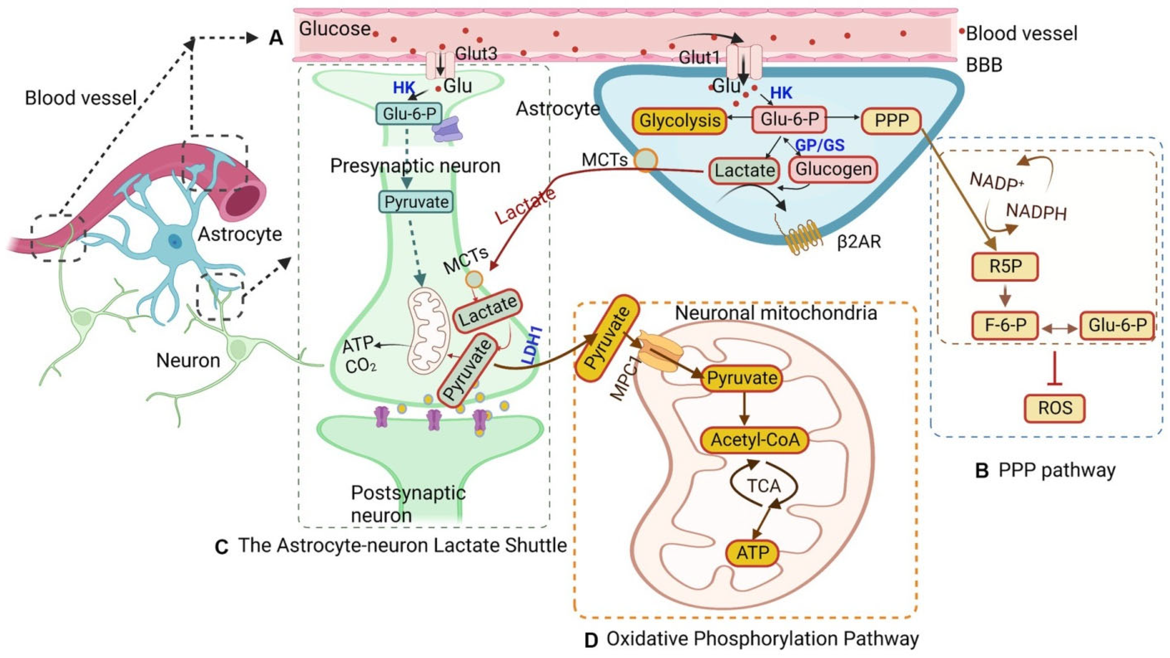
Figure 1. Astrocytic glucose metabolism and energy transfer. This figure illustrates the intricate glucose metabolism within astrocytes, highlighting their vital role in brain energy supply. (A) Glucose uptake and initial metabolism: Glucose enters astrocytes from the bloodstream via GLUTs (glucose transporters) and is converted to glucose-6-phosphate (G6P), a pivotal molecule intersecting multiple pathways. Glucose uptake via GLUTs can be modulated by signaling cascades initiated by the β2-adrenergic receptor (β2AR). Once inside the astrocyte, glucose is phosphorylated by hexokinase (HK) to form G6P. G6P then serves as a branch point directing glucose flux towards glycolysis, the astrocyte–neuron lactate shuttle, the pentose phosphate pathway (PPP), and oxidative phosphorylation. Glycogen synthesis and breakdown are regulated by glycogen phosphorylase (GP) and glycogen synthase (GS), respectively. (B) Pentose phosphate pathway (PPP): The PPP sees G6P transformed into ribulose-5-phosphate (R5P), concurrently reducing NADP+ to NADPH, which is crucial for cellular redox balance. R5P can also produce glyceraldehyde-3-phosphate and fructose-6-phosphate (F6P), with F6P able to revert to G6P, indicating cyclical interconversion. (C) Astrocyte–neuron lactate shuttle: Lactate, produced in astrocytes, primarily via the enzyme lactate dehydrogenase 1 (LDH1), is transported to neurons via MCTs (monocarboxylate transporters). Neurons convert lactate back to pyruvate, which fuels ATP production in mitochondria, supporting neuronal activity. (D) Oxidative phosphorylation: G6P can enter the glycolytic pathway, generating pyruvate. Pyruvate is then converted to acetyl-CoA, which enters the tricarboxylic acid (TCA) cycle in the mitochondria to generate ATP.
Jmp 06 00012 g001
Figure 3. The astrocyte–neuron lactate shuttle. Astrocytes, using GLUT1 transporters, absorb glucose and convert it to glucose-6-phosphate (Glc-6-P), storing it as glycogen via glycogen synthase (GlyS). When energy demands rise, glycogen phosphorylase (GlyP) mediates glycogenolysis, creating Glc-6-P for glycolysis. Synaptic transmission triggers astrocyte glycolysis and lactate production via glutamate uptake, boosting glucose consumption or glycogen breakdown. Lactate, produced by astrocytes, is transported into the extracellular space by MCT1 and taken up by neurons through MCT2. Neurons can then convert this lactate into pyruvate, which fuels oxidative phosphorylation. This astrocyte–neuron lactate shuttle highlights a vital metabolic partnership, ensuring neurons receive the necessary energy support during periods of high activity.
Jmp 06 00012 g003
Disclaimer/Publisher’s Note: The statements, opinions and data contained in all publications are solely those of the individual author(s) and contributor(s) and not of MDPI and/or the editor(s). MDPI and/or the editor(s) disclaim responsibility for any injury to people or property resulting from any ideas, methods, instructions or products referred to in the content.