Review

The Current Landscape of Molecular Pathology for the Diagnosis and Treatment of Pediatric Medulloblastoma

1
Department of Pediatrics, Cincinnati Children’s Hospital Medical Center, Cincinnati, OH 45229-3039, USA
2
Department of Pediatrics, University of Cincinnati College of Medicine, Cincinnati, OH 45229-3039, USA
3
Cancer and Blood Diseases Institute, The Cure Starts Now Foundation Brain Tumor Center, Division of Oncology, Cincinnati Children’s Hospital Medical Center, 3333 Burnet Avenue, MLC 7018, Cincinnati, OH 45229-3039, USA
*
Author to whom correspondence should be addressed.
J. Mol. Pathol. 2025, 6(2), 11; https://doi.org/10.3390/jmp6020011
Submission received: 11 April 2025 / Revised: 27 May 2025 / Accepted: 4 June 2025 / Published: 11 June 2025
(This article belongs to the Collection Feature Papers in Journal of Molecular Pathology)

Abstract

:
Medulloblastoma (MB) is a malignant brain tumor that requires intense multimodal treatment. There is significant treatment-related morbidity associated with MB, and overall prognosis varies between the subgroups of the disease. These tumors were previously risk-stratified based solely on histopathological features. However, advancements in oncologic molecular research have led to novel changes to MB tumor classification, which also affects the prognosis and treatment strategies for individual patients. The WHO CNS5 now recognizes four main molecular subgroups of MB. Each subgroup contains its own genomic heterogeneity that correlates with a unique way to risk stratify patients, determine overall prognosis, and inform treatment. These discoveries have already impacted the implications and outcomes of current treatments based on the subgroup of patients. Ongoing research to better understand this classification system has paved the way for the development of molecular targeted therapy.

1. Introduction

The most common solid neoplasms in the pediatric population are central nervous system (CNS) tumors [1]. Medulloblastoma (MB) is the most prevalent malignant brain tumor in the pediatric population [2]. MB is an embryonal neuroepithelial tumor that arises from the cerebellum and accounts for roughly 20% of pediatric CNS tumors and almost half of all posterior fossa neoplasms [3,4]. It has a slight male predominance with a 1.5:1 male-to-female ratio [4]. While MB can affect all age groups, the highest incidence rate is seen in children aged 4–9 years old at 44%, then adolescents aged 10–16 years old at 23%, and lastly infants 0–3 years old at 12% [4]. It has a high propensity for local invasion to surrounding structures and can disseminate via cerebrospinal fluid (CSF) to distant regions in the brain and spinal cord resulting in metastatic disease [4]. The mainstay of treatment is multimodal and compromises a combination of maximal safe resection, chemotherapy, and radiation. Although these therapies and appropriate risk stratification have improved overall survival, many patients still face significant morbidity secondary to treatment that impacts their quality of life. Many patients face long-term treatment-related complications including neurologic deficits, cognitive impairments, and endocrinologic abnormalities, as well as chemotherapy related toxicities [4]. Previously, the subgroups of MB were determined based on histopathology findings. Advances in MB research have led to a classification system that also incorporates methylomic profiling and molecular characteristics. While the extent of resection, presence of metastatic disease, and histology findings were previously the key drivers in determining outcomes, these molecular studies demonstrate the genetic heterogeneity of MB and are now recognized as a critical component to dictate risk stratification and prognosis, and thus, inform therapeutic treatment [5].

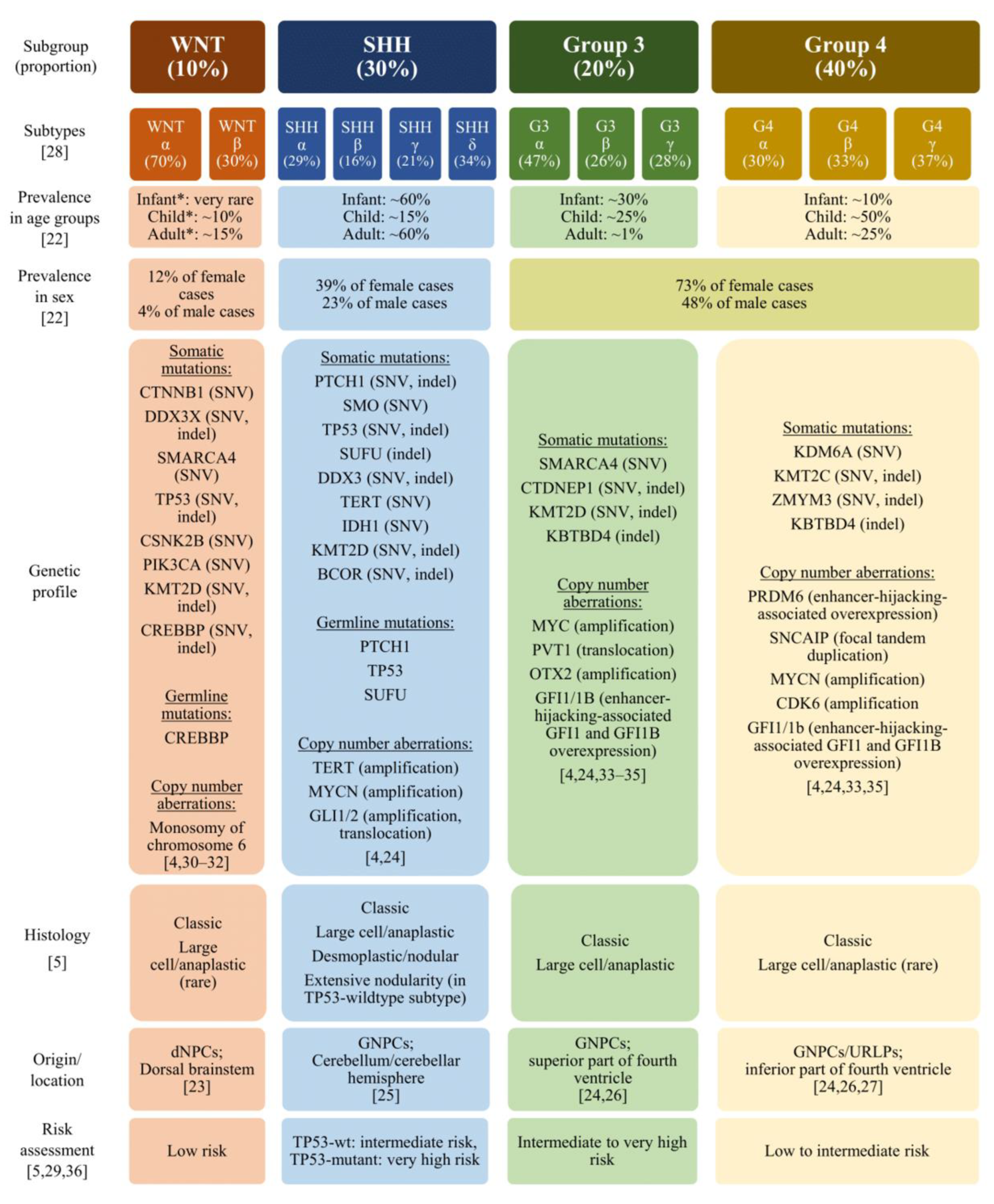
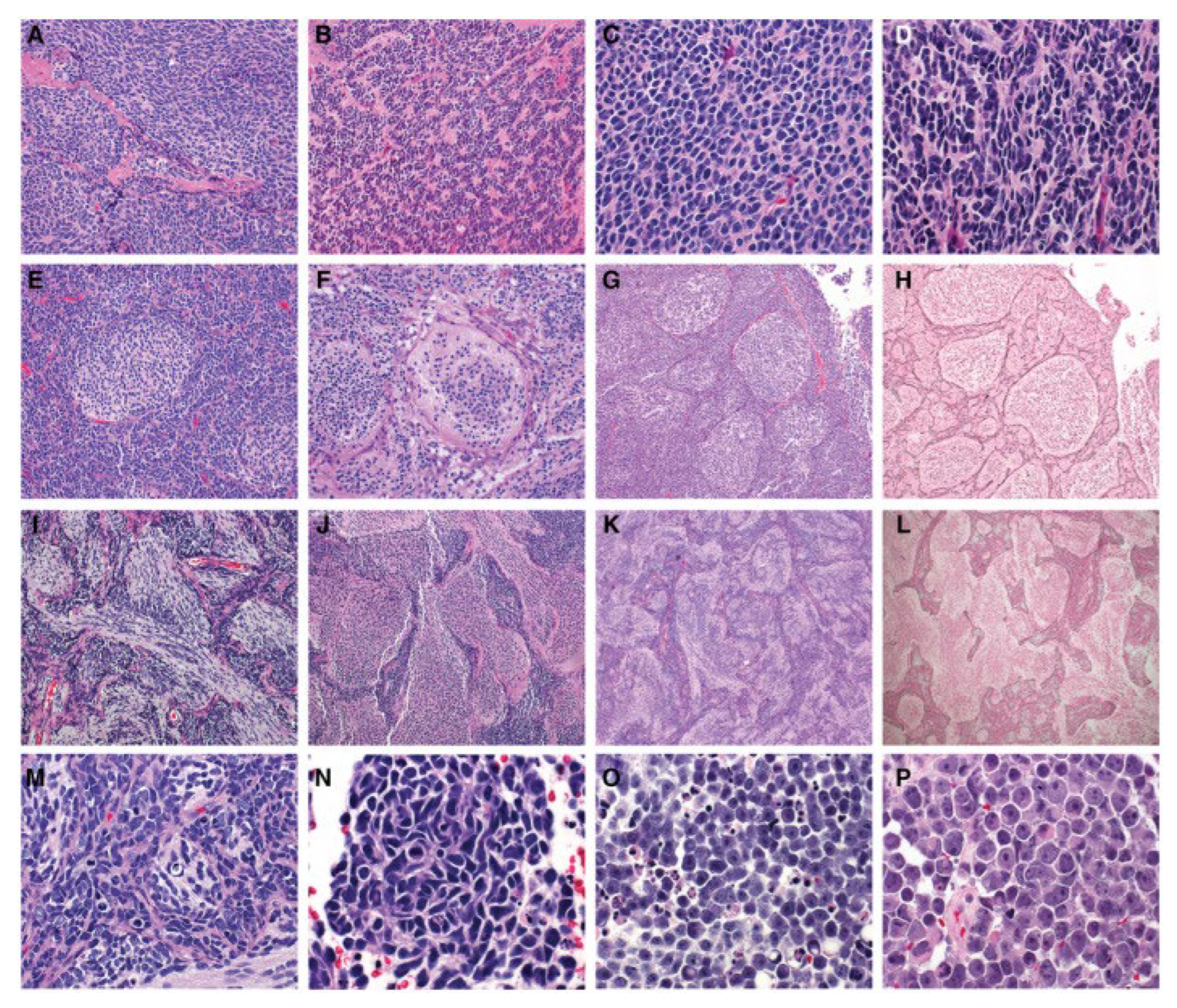
2. Histopathology and Molecular Pathology in the Diagnosis of Medulloblastoma

Previously, according to the WHO Classification of CNS tumors, MB was divided into four subgroups based on histological features. These included the classic, desmoplastic/nodular, extensive nodularity, and large cell/anaplastic subgroups (Figure 1) [6]. Classic morphology demonstrates small round blue cells, mitotic figures, apoptotic bodies, and sometimes Homer Wright rosettes [7]. Approximately 70% of all MB tumors have classic morphology [3]. Desmoplastic/nodular morphology consists of nodular zones with moderate cellularity surrounded by densely packed undifferentiated cells that produce an intercellular reticulin fiber network [7]. Elevated Ki-67 within internodular areas and reticulin staining are useful to help identify this subgroup. The extensive nodularity subgroup appears similar to the desmoplastic/nodular subgroup but with larger nodules and more differentiated cells [7]. This is the least common subgroup. Both the desmoplastic/nodular and extensive nodularity subgroups are more commonly seen in younger children and are associated with better outcomes in this age group [7]. Lastly, the large cell/anaplastic subgroup consists of two distinct histologic variants but has been combined into one histologic subgroup because the variants are usually seen together. Histopathology of this subgroup reveals a large cell with prominent nucleoli and/or anaplasia with cytologic pleomorphism, nuclear wrapping/molding, and a high mitotic and apoptotic count [7]. Large cell/anaplastic MB is more commonly associated with metastatic disease at time of diagnosis and recurrence and therefore has inferior outcomes. Other high risk oncogenomic features likely contribute to the poor outcomes seen within this subgroup [7].
Ongoing research in cancer genomics has led to advancements in MB molecular classification. Specifically, methylation profiling has led to the identification of various subgroups of MB. DNA methylation is a cellular process involved in regulation of gene expression and each cell type in the body has a unique methylation profile [8]. The WHO CNS5 recognizes four major molecular subgroups of MB using DNA methylation profiles: wingless (WNT), sonic hedgehog (SHH), group 3, and group 4 (Figure 2) [7,9]. Defining the molecular subgroup at diagnosis by methylation profiling is now the standard of care for MB. Each subgroup has varying implications on clinical outcomes, risk stratification, and intensity of treatment selection.
WNT-activated tumors’ nomenclature stems from their essential diagnostic criterion of WNT pathway activation. While the WNT tumors are the least common subgroup, accounting for about 10% of all MB, they are associated with an excellent prognosis with an overall five-year survival of nearly 100% [7,9,10]. This example highlights the importance of methylomic and molecular classification in determining the diagnosis of MB and deciding on the best treatment strategies for patients. Patients who die usually succumb to other complications rather than the disease itself. It is rarely associated with metastatic disease at time of diagnosis. However, patients with metastatic WNT MB still have improved outcomes when compared to other groups with metastatic disease [11,12]. MB is more common in males, but WNT MB affects both sexes equally [9,10,13]. This subgroup can be seen at all ages, with a peak from 6 to 10 years old, but is not commonly seen in infants [9,10]. Patients with Turcot syndrome type I have a mutation in the WNT pathway inhibitor APC, which increases the risk for MB in these patients. From a histology standpoint, the classic and, rarely, the large/anaplastic morphologies are seen in WNT MB tumors [10]. WNT tumors typically reveal CTNNB1 mutations, nuclear B-catenin on immunohistochemical staining, and monosomy 6. Research suggests that WNT tumors’ favorable prognosis is due to their leaky vasculature that disrupts the blood–brain barrier, which allows for easier targeted delivery of chemotherapy agents [9]. Current clinical trials are aiming to leverage these concepts to decrease the intensity of chemotherapy and/or radiation to improve long-term treatment-associated morbidity while maintaining excellent outcomes.
Similarly to WNT tumors, SHH tumors are named for their diagnostic criterion, with SHH signaling pathway activation. The SHH tumors account for approximately 25% of all MB [7,9,10]. From a histology standpoint, almost all desmoplastic/nodular types are seen within the SHH tumors, as well as some classic and large cell/anaplastic morphologies [10]. This subgroup of MB is largely impacted by age and genetics. Age can be subcategorized into infancy (≤3 years old), childhood (4–15 years old), and adulthood (≥16 years old) [7,9,14]. Infant SHH tumors are the least mutated compared to the other age groups. The two most common mutations in infants are PTCH1 and SUFU [7]. SHH tumors in children aged 4–17 are usually driven by mutations in TP53 and PTCH1, and commonly in conjunction with GLI2 and MCYN amplification [9]. This age group has the worst prognosis within this subgroup; therefore, these patients are often treated as high-risk [9]. Specifically, MYCN amplification combined with GLI2 amplification and 14q loss is associated with worse overall survival percentages [15]. The adult SHH tumors are typically driven by PTCH1 or SMO mutations [9]. Additionally, the WHO criteria identify two additional subtypes within the SHH group: SHH with TP53 wildtype or SHH with TP53 mutant. Prognosis in this subset significantly depends upon TP53 status. The five-year survival rates are about 40% in TP53 mutant tumors versus about 80% in TP53 wildtype tumors [9]. The TP53 wildtype SHH tumors have a bimodal age distribution and are more commonly seen in infants and adults, while theTP53 mutant SHH tumors are more prevalent in middle childhood [7]. There is an increased risk in developing a SHH tumor in various conditions, including Li Fraumeni syndrome and Gorlin syndrome. Li Fraumeni syndrome is the result of a germline mutation in TP53, which increases one’s one lifetime risk for cancer and is therefore seen in TP53 mutant tumors. Gorlin syndrome results from a mutation in PTCH or SUFU, which leads to a gain of function mutation in SHH signaling [16]. The patients tend to present with multiple basal cell carcinomas, odontogenic keratocysts of the jaw, palmar and plantar pits, skeletal abnormalities (especially bifid, fused or splayed ribs), and with a family history of Gorlin syndrome [16]. MB affects both sexes equally in patients with Gorlin syndrome and tends to occur during infancy between 1 and 2 years of age [16]. Given the role of TP53 in DNA repair and cell apoptosis, and the role of PTCH1 in preventing uncontrolled cell proliferation, radiation treatment should be avoided or used with extreme caution due to the risk of developing secondary malignancies within the radiation field for patients with Li Fraumeni and Gorlin syndrome [16,17].
Group 3 tumors account for approximately 25% of all MB and tend to occur in infancy and childhood, with a peak incidence between 3 and 5 years of age, with a 2:1 predilection for male patients [9,10,18]. Most group 3 tumors demonstrate classic histology [10]. However, most large cell/anaplastic tumors are found within this subgroup [10]. This subgroup has a dismal prognosis, with an approximate 50% chance of overall survival, and is very frequently metastatic [9,10]. These are non-WNT and non-SHH signaling tumors. Two common high-risk genetic features that are identified within this subgroup and associated with its poor outcomes are MYC amplification and isochrome 17q (mainly when linked to MYC amplification) [9,10]. These tumors may also express other chromosomal aberrations including loss of 16q, 10q, and 9q or gain of 7 and 1q, as well as GFI1/GFI1B activation and SMARCA4 mutations [19]. While it is accepted that molecular findings associated with MYC amplification and isochrome 17q portend a worse prognosis, the data is not clear regarding how the other abnormalities listed affect group 3 MB outcomes.
Lastly, group 4 tumors are the most common and account for 35% of all MB [9]. They can occur at all ages and have a male predominance with a 2:1 ratio of male to female patients [10,18]. The large cell/anaplastic and classic histology are typically seen in this subgroup [10]. Metastatic disease and young age at the time of diagnosis are poor prognostic indicators [7,18]. These are also non-WNT and non-SHH signaling tumors. They can express various chromosomal aberrations including isochrome or 17q gain, chromosome 11 loss or 7 gain, and 8p or 10q loss, as well as KDM6A loss of function mutation or overexpression of EZH2 [9,19]. An important differentiation is that the group 4 tumors with isochrome 17q have improved outcomes compared with those seen in group 3 tumors with isochrome 17q [9]. Additionally, chromosome 11 loss and 17q gain are recognized as good prognostic indicators [7].
Figure 2. Molecular subgroups of MB as seen in Voskamp, M.J. et al.—Immunotherapy in Medulloblastoma: Current State of Research, Challenges, and Future Perspectives, Cancers (Basel), 2021 [20]. * Risk assessment varies depending on genetic subtype.
Jmp 06 00011 g002

3. Considerations for Molecular Targets of Therapy

There have been historical and current, ongoing clinical trials to investigate the efficacy of molecular targeted therapy for treatment of the various subgroups of MB. The WNT subgroup proves challenging for molecular targeted therapy for a variety of reasons. The leaky vasculature of WNT-MB disrupts the blood–brain barrier and allows for increased chemotherapy penetration at the tumor site [21]. Thus, disrupting this signaling pathway would potentially eliminate the tumors robust response to chemotherapy agents [21,22]. Additionally, WNT signaling plays a critical role in embryonal development, cell proliferation, and bone formation [21]. Therefore, inhibitors of this pathway could have negative implications on normal development and tissue regeneration, as well as resultant osteoporosis in skeletally immature patients [21]. While there have not been any clinical trials for molecular targeted therapies for the WNT subgroup, there are trials underway to investigate decreased radiation dosing and chemotherapy treatment regimens to maintain the WNT subgroup’s excellent overall survival with decreased acute and long-term side effects.
There is a breadth of research investigating molecular targeted therapy for the SHH-MB subgroup. Smoothened (SMO) is a protein involved in the SHH signaling pathway. Vismodegib, a SMO antagonist, was studied in phase II trials PBTC-032 and PBTC-025B in both pediatric and adult recurrent MB patients. Sonidegib, a SMO inhibitor, was used in a small phase II study (NCT01708174) and proposed for the Alliance AMBUSH trial as part of maintenance therapy for newly diagnosed SHH-MB in young adults and adult patients [21,23]. However, these SMO modulators exhibit early onset treatment resistance and growth plate fusion in skeletally immature patients [21]. Other previous clinical trials incorporated these agents for specific populations based on these known side effects. Specifically, the SJMB12 protocol incorporated Vismodegib for patients with a mature skeletal system [24]. Additionally, the EORTC 1634 trial is a multi-national European protocol utilizing risk stratification based on molecular subgroups of MB with experimental arms including decreased craniospinal radiation and Sonidegib with standard chemotherapy in non-metastatic (M0) post-pubertal young adult and adult patients [25]. This trial has been difficult to implement and while there is not one clear answer as to why, there are several possible reasons. Some possibilities impacting implementation include various practices in different countries, access to care (radiation centers, SMO inhibitors, etc.), the comfort level of physicians to dose-reduce craniospinal radiation in adult patients where the long-term neurocognitive side effects are less of a risk than in pediatric patients, and the concern for increasing the risk of disease relapse by decreasing initial therapy intensity. Another treatment that targets the SHH pathway by binding GLI1/2 is arsenic trioxide, which is currently being explored in clinical trials [21]. Additionally, histone deacetylases (HDACs) are upregulated in SHH-MB tumors. Panobinostat, a HDAC inhibitor, is currently in a pilot phase I study (NCT04315064) for recurrent or progressed MB patients [21]. Interestingly, Panobinostat can be infused intracranially, which allows for increased bioavailability at the tumor site, thus mitigating adverse side effects [21]. These trials represent several examples of the molecular targeted therapies for SHH-MB. There are additional ongoing or proposed studies investigating molecular targets for CDK inhibitors, CK2 inhibitors, itraconazole analogs, and lithium for the TP-53 mutation subtype [21,22].
Group 3 molecular therapies tend to involve epigenetic factors that affect MYC expression. HDAC inhibitors and PI3K inhibitors both induce expression of the onco-suppressor gene FOXO1, and when used synergically, they have demonstrated inhibition of tumor proliferation and prolonged survival in mice models [22]. This subtype of MB tumors has been responsive to Palbociclib, a CDK inhibitor, in mice models. There is a phase I trial (NCT02255461) being conducted to investigate this treatment in refractory or recurrent tumors [21,22]. Other promising targets include bromodomain and SETD8 inhibitors that interrupt the gene processing mechanisms of MYC [22].
Group 4 MB rarely have common somatic mutations and preclinical/clinical studies for molecular targets within this subgroup are limited [19,21]. While this is the most prevalent subgroup of MB, its low rate of mutations is disadvantageous for targeted therapies.

4. Discussion

The identification of molecular subgroups and advancements in oncogenomic research have already led to key changes in the diagnosis of MB. It has significant implications for risk stratification and prognosis, which subsequently affects the multimodal treatment regimen. Several studies have demonstrated how this vast array of molecular-genomic data influences outcomes in MB patients. For instance, the Children’s Oncology Group average-risk MB study, ACNS0331 (NCT00085735), investigated outcomes in reduced versus standard craniospinal radiation (CSI) doses and posterior fossa radiation therapy (PFRT) or involved field radiation therapy (IFRT). There were notable molecular differences amongst the subgroups, including superior outcomes in SHH patients treated with IFRT versus PFRT and inferior outcomes in group 4 patients treated with lower dose CSI versus standard dose (especially in individuals with balanced chromosomes 11 and 17) [26]. The Children’s Oncology Group high-risk MB study, ACNS0332 (NCT00392327), found that therapy intensification during radiation therapy with carboplatin improves outcomes in high-risk group 3 MB patients only, underscoring the unique biology across molecular subgroups of MB [27]. The SJMB03 protocol was able to identify low-risk groups as WNT, low-risk SHH, and low-risk combined group 3 and 4 versus high-risk groups including high-risk SHH and high-risk combined groups 3 and 4 based on the various molecular characteristics seen within each group [12]. SJMB12 shows potential for the addition of pemetrexed and gemcitabine for treatment of non-WNT, non-SHH tumors given the significant anti-proliferative effects and increased survival time seen with this dual treatment regimen in mice models for group 3 MYC amplified MB [28,29]. Given that the WNT MB tumors have an excellent prognosis, there are several studies (ACNS1422, SIOP-PNET5, SJMB12) investigating outcomes with lower dose CSI and/or decreased chemotherapy [28]. Additionally, group 4 tumors with chromosome 11 loss are known to have better outcomes, and the upcoming Children’s Oncology Group study, ACNS2031 (NCT05382338), will study outcomes in this specific group with reduced CSI dosing [28].
While there are many ongoing or proposed studies investigating treatment strategies for initial therapy based on the molecular subgroups of MB, there are few treatment options and currently no standard of care for relapsed MB. About 30% of MB patients will relapse and face a dismal prognosis with less than a 5% overall survival rate [30]. Young children (0–5 years old) have a higher incidence of relapse compared to older children/adolescents, which is likely due to the lack of radiation during initial treatment, given the known detrimental neurocognitive side effects of radiation within this age group [31]. The timing of relapse varies based on the molecular subgroup. Faster times to relapse are reported in group 3 (0.9 years) and SHH (1.2 years) tumors, followed by slower times to relapse in group 4 (2.74 years) and WNT (2.77 years) tumors [31]. However, it is important to note that the WNT tumors rarely relapse and tend to occur in patients who received inferior initial treatment [31]. While there is no standard of care for relapsed MB at this time, there are a few studies that employ frequently practiced protocols for this patient population. The Children’s Oncology group ACNS0821 study, which includes a combination of temozolomide, irinotecan and bevacizumab, and the European MEMMAT trial, which utilizes an antiangiogenic metronomic approach, have both prolonged progression-free survival. It is important to note both regimens are well tolerated without many significant adverse events. Unfortunately, both studies did not have a significant impact on overall survival outcomes in relapsed MB patients [32,33].
These studies highlight that it is not only important to understand the molecular stratification of MB, but also to recognize the various genomic signature subtypes that compromise each of the subgroups. The evolution of this field has been a pivotal point for clinical understanding of MB tumors. The discovery of the intricacies of the molecular pathways and genetic mutations that compromise MB tumors has led to a more comprehensive understanding of the outcomes of previous protocols involving uniform treatment. This will guide future directions for more targeted clinical trials based on the subgroup of a MB tumor.

Future Directions

There has been immense progress to better understand the molecular and genetic heterogeneity of MB tumors. However, it is critical to identify new mutations/alterations that can continue to help with individualized risk stratification, prognosis, and treatment. The current treatments for MB tumors still carry significant treatment-related morbidity. Continued expansion within this field could prove beneficial if new targets for molecularly targeted therapy could be identified. Specifically, group 3 tumors carry the poorest prognosis; therefore, further studies are needed to develop effective targeted therapies for the subtypes within this subgroup. Additionally, although group 4 tumors are the most common MB tumors, there is an evident lack of research on directed therapies within this subgroup. Lastly, it is evident that more treatment options are desperately needed for relapsed MB patients. Current pre-clinical data will continue to inform future studies that target specific molecular alterations in relapsed MB and it is imperative that molecular subgroup is considered when creating new therapies. Future targeted therapies provide hope for either improved initial treatment that reduces the risk of recurrence and/or for more effective treatment options available at the time of relapse. There are continuous opportunities for novel discoveries that could revolutionize treatment for MB tumors, guided by the evolution in the field incorporating methylomic and molecular findings into the diagnostic criteria of MB.

Author Contributions

Concept and design: A.K. and S.R., drafting of the manuscript and critical revision: A.K., A.C., A.S., E.V. and S.R. All authors have read and agreed to the published version of the manuscript.

Funding

This research received no external funding.

Conflicts of Interest

The authors declare no conflicts of interest.

Abbreviations

The following abbreviations are used in this manuscript:
MBmedulloblastoma
CNScentral nervous system
CSFcerebrospinal fluid
WNTwingless
SHHsonic hedgehog
SMOsmoothened
HDAChistone deacetylases
CSIstandard craniospinal radiation
PFRTposterior fossa radiation therapy
IFRTinvolved field radiation therapy

References

  1. Kline, N.E.; Sevier, N. Solid tumors in children. J. Pediatr. Nurs. 2003, 18, 96–102. [Google Scholar] [CrossRef]
  2. Rossi, A.; Caracciolo, V.; Russo, G.; Reiss, K.; Giordano, A. Medulloblastoma: From molecular pathology to therapy. Clin. Cancer Res. 2008, 14, 971–976. [Google Scholar] [CrossRef]
  3. Orr, B.A. Pathology, diagnostics, and classification of medulloblastoma. Brain Pathol. 2020, 30, 664–678. [Google Scholar] [CrossRef]
  4. Mahapatra, S.; Amsbaugh, M.J. Medulloblastoma. In StatPearls [Internet]; StatPearls Publishing: Treasure Island, FL, USA, 2025. [Google Scholar] [PubMed]
  5. Kijima, N.; Kanemura, Y. Molecular Classification of Medulloblastoma. Neurol. Med. Chir. 2016, 56, 687–697. [Google Scholar] [CrossRef]
  6. Choi, J.Y. Medulloblastoma: Current Perspectives and Recent Advances. Brain Tumor Res. Treat. 2023, 11, 28–38. [Google Scholar] [CrossRef]
  7. Cotter, J.A.; Hawkins, C. Medulloblastoma: WHO 2021 and Beyond. Pediatr. Dev. Pathol. 2022, 25, 23–33. [Google Scholar] [CrossRef]
  8. Moore, L.D.; Le, T.; Fan, G. DNA methylation and its basic function. Neuropsychopharmacology 2013, 38, 23–38. [Google Scholar] [CrossRef]
  9. Archer, T.C.; Mahoney, E.L.; Pomeroy, S.L. Medulloblastoma: Molecular Classification-Based Personal Therapeutics. Neurotherapeutics 2017, 14, 265–273. [Google Scholar] [CrossRef]
  10. Taylor, M.D.; Northcott, P.A.; Korshunov, A.; Remke, M.; Cho, Y.J.; Clifford, S.C.; Eberhart, C.G.; Parsons, D.W.; Rutkowski, S.; Gajjar, A.; et al. Molecular subgroups of medulloblastoma: The current consensus. Acta Neuropathol. 2012, 123, 465–472. [Google Scholar] [CrossRef]
  11. Mani, S.; Chatterjee, A.; Dasgupta, A.; Shirsat, N.; Pawar, A.; Epari, S.; Sahay, A.; Sahu, A.; Moiyadi, A.; Prasad, M.; et al. Clinico-Radiological Outcomes in WNT-Subgroup Medulloblastoma. Diagnostics 2024, 14, 358. [Google Scholar] [CrossRef]
  12. Gajjar, A.; Robinson, G.W.; Smith, K.S.; Lin, T.; Merchant, T.E.; Chintagumpala, M.; Mahajan, A.; Su, J.; Bouffet, E.; Bartels, U.; et al. Outcomes by Clinical and Molecular Features in Children with Medulloblastoma Treated with Risk-Adapted Therapy: Results of an International Phase III Trial (SJMB03). J. Clin. Oncol. 2021, 39, 822–835. [Google Scholar] [CrossRef]
  13. Ellison, D.W.; Kocak, M.; Dalton, J.; Megahed, H.; Lusher, M.E.; Ryan, S.L.; Zhao, W.; Nicholson, S.L.; Taylor, R.E.; Bailey, S.; et al. Definition of disease-risk stratification groups in childhood medulloblastoma using combined clinical, pathologic, and molecular variables. J. Clin. Oncol. 2011, 29, 1400–1407. [Google Scholar] [CrossRef]
  14. Northcott, P.A.; Hielscher, T.; Dubuc, A.; Mack, S.; Shih, D.; Remke, M.; Al-Halabi, H.; Albrecht, S.; Jabado, N.; Eberhart, C.G.; et al. Pediatric and adult sonic hedgehog medulloblastomas are clinically and molecularly distinct. Acta Neuropathol. 2011, 122, 231–240. [Google Scholar] [CrossRef]
  15. Shih, D.J.; Northcott, P.A.; Remke, M.; Korshunov, A.; Ramaswamy, V.; Kool, M.; Luu, B.; Yao, Y.; Wang, X.; Dubuc, A.M.; et al. Cytogenetic prognostication within medulloblastoma subgroups. J. Clin. Oncol. 2014, 32, 886–896. [Google Scholar] [CrossRef]
  16. Onodera, S.; Nakamura, Y.; Azuma, T. Gorlin Syndrome: Recent Advances in Genetic Testing and Molecular and Cellular Biological Research. Int. J. Mol. Sci. 2020, 21, 7559. [Google Scholar] [CrossRef]
  17. Kolodziejczak, A.S.; Guerrini-Rousseau, L.; Planchon, J.M.; Ecker, J.; Selt, F.; Mynarek, M.; Obrecht, D.; Sill, M.; Autry, R.J.; Stutheit-Zhao, E.; et al. Clinical outcome of pediatric medulloblastoma patients with Li-Fraumeni syndrome. Neuro-Oncology 2023, 25, 2273–2286. [Google Scholar] [CrossRef]
  18. Hovestadt, V.; Ayrault, O.; Swartling, F.J.; Robinson, G.W.; Pfister, S.M.; Northcott, P.A. Medulloblastomics revisited: Biological and clinical insights from thousands of patients. Nat. Rev. Cancer 2020, 20, 42–56. [Google Scholar] [CrossRef]
  19. Menyhárt, O.; Giangaspero, F.; Győrffy, B. Molecular markers and potential therapeutic targets in non-WNT/non-SHH (group 3 and group 4) medulloblastomas. J. Hematol. Oncol. 2019, 12, 29. [Google Scholar] [CrossRef]
  20. Voskamp, M.J.; Li, S.; van Daalen, K.R.; Crnko, S.; Ten Broeke, T.; Bovenschen, N. Immunotherapy in Medulloblastoma: Current State of Research, Challenges, and Future Perspectives. Cancers 2021, 13, 5387. [Google Scholar] [CrossRef]
  21. Gatto, L.; Franceschi, E.; Tosoni, A.; Di Nunno, V.; Bartolini, S.; Brandes, A.A. Molecular Targeted Therapies: Time for a Paradigm Shift in Medulloblastoma Treatment? Cancers 2022, 14, 333. [Google Scholar] [CrossRef]
  22. Wen, J.; Hadden, M.K. Medulloblastoma drugs in development: Current leads, trials and drawbacks. Eur. J. Med. Chem. 2021, 215, 113268. [Google Scholar] [CrossRef]
  23. Mahajan, A.; Shih, H.; Penas-Prado, M.; Ligon, K.; Aldape, K.; Hu, L.S.; Loughan, A.R.; Basso, M.R.; Leeper, H.E.; Nahed, B.V.; et al. The Alliance AMBUSH Trial: Rationale and Design. Cancers 2022, 14, 414. [Google Scholar] [CrossRef]
  24. Gajjar, A.J.; Orr, B.; Ellison, D.; Lafay-Cousin, L.; Minturn, J.; Fisher, M.; Bendel, A.; Hwang, E.; Murray, J.; Landi, D.; et al. MDB-49. Outcomes of Sonic Hedgehog (Shh) Medulloblastoma (Mb) in Children and Adults Treated with Risk-Adapted Therapy on a Prospective International Protocol (SJMB12). Neuro-oncology 2024, 26. [Google Scholar] [CrossRef]
  25. Hau, P.; Frappaz, D.; Hovey, E.; McCabe, M.G.; Pajtler, K.W.; Wiestler, B.; Seidel, C.; Combs, S.E.; Dirven, L.; Klein, M.; et al. Development of Randomized Trials in Adults with Medulloblastoma-The Example of EORTC 1634-BTG/NOA-23. Cancers 2021, 13, 3451. [Google Scholar] [CrossRef] [PubMed]
  26. Michalski, J.M.; Janss, A.J.; Vezina, L.G.; Smith, K.S.; Billups, C.A.; Burger, P.C.; Embry, L.M.; Cullen, P.L.; Hardy, K.K.; Pomeroy, S.L.; et al. Children’s Oncology Group Phase III Trial of Reduced-Dose and Reduced-Volume Radiotherapy with Chemotherapy for Newly Diagnosed Average-Risk Medulloblastoma. J. Clin. Oncol. 2021, 39, 2685–2697. [Google Scholar] [CrossRef]
  27. Leary, S.E.S.; Packer, R.J.; Li, Y.; Billups, C.A.; Smith, K.S.; Jaju, A.; Heier, L.; Burger, P.; Walsh, K.; Han, Y.; et al. Efficacy of Carboplatin and Isotretinoin in Children with High-risk Medulloblastoma: A Randomized Clinical Trial From the Children’s Oncology Group. JAMA Oncol. 2021, 7, 1313–1321. [Google Scholar] [CrossRef]
  28. Lazow, M.A.; Palmer, J.D.; Fouladi, M.; Salloum, R. Medulloblastoma in the Modern Era: Review of Contemporary Trials, Molecular Advances, and Updates in Management. Neurotherapeutics 2022, 19, 1733–1751. [Google Scholar] [CrossRef]
  29. Morfouace, M.; Shelat, A.; Jacus, M.; Freeman, B.B.; Turner, D.; Robinson, S.; Zindy, F.; Wang, Y.D.; Finkelstein, D.; Ayrault, O.; et al. Pemetrexed and gemcitabine as combination therapy for the treatment of Group3 medulloblastoma. Cancer Cell 2014, 25, 516–529. [Google Scholar] [CrossRef]
  30. Hill, R.M.; Plasschaert, S.L.A.; Timmermann, B.; Dufour, C.; Aquilina, K.; Avula, S.; Donovan, L.; Lequin, M.; Pietsch, T.; Thomale, U.; et al. Relapsed Medulloblastoma in Pre-Irradiated Patients: Current Practice for Diagnostics and Treatment. Cancers 2021, 14, 126. [Google Scholar] [CrossRef]
  31. Holmberg, K.O.; Borgenvik, A.; Zhao, M.; Giraud, G.; Swartling, F.J. Drivers Underlying Metastasis and Relapse in Medulloblastoma and Targeting Strategies. Cancers 2024, 16, 1752. [Google Scholar] [CrossRef]
  32. Levy, A.S.; Krailo, M.; Chi, S.; Villaluna, D.; Springer, L.; Williams-Hughes, C.; Fouladi, M.; Gajjar, A. Temozolomide with irinotecan versus temozolomide, irinotecan plus bevacizumab for recurrent medulloblastoma of childhood: Report of a COG randomized Phase II screening trial. Pediatr. Blood Cancer 2021, 68, e29031. [Google Scholar] [CrossRef] [PubMed]
  33. Peyrl, A.; Chocholous, M.; Sabel, M.; Lassaletta, A.; Sterba, J.; Leblond, P.; Nysom, K.; Torsvik, I.; Chi, S.N.; Perwein, T.; et al. Sustained Survival Benefit in Recurrent Medulloblastoma by a Metronomic Antiangiogenic Regimen: A Nonrandomized Controlled Trial. JAMA Oncol. 2023, 9, 1688–1695. [Google Scholar] [CrossRef]
Figure 1. Histologic subgroups of MB as seen in Orr, B.A., pathology, diagnostics, and classification of medulloblastoma, Brain Pathology, 2020. Images (AD) represent classic histology, (EH) for desmoplastic/nodular, (IL) for extensive nodularity, and (MP) for large cell/anaplastic [3].
Jmp 06 00011 g001
Disclaimer/Publisher’s Note: The statements, opinions and data contained in all publications are solely those of the individual author(s) and contributor(s) and not of MDPI and/or the editor(s). MDPI and/or the editor(s) disclaim responsibility for any injury to people or property resulting from any ideas, methods, instructions or products referred to in the content.

