Article

DNA Takes Over on the Control of the Morphology of the Composite Self-Organized Structures of Barium and Calcium Silica–Carbonate Biomorphs, Implications for Prebiotic Chemistry on Earth

1
Departmento de Biología, División de Ciencias Naturales y Exactas, Campus Guanajuato, Universidad de Guanajuato, Noria Alta S/N, Col. Noria Alta, Guanajuato 36050, Mexico
2
Instituto de Ciencias Aplicadas y Tecnología, Universidad Nacional Autónoma de México, Circuito Exterior S/N, Ciudad Universitaria, Mexico City 04510, Mexico
3
Instituto de Química, Universidad Nacional Autónoma de México, Av. Universidad 3000, Ciudad Universitaria, Mexico City 04510, Mexico
*
Authors to whom correspondence should be addressed.
Earth 2024, 5(3), 293-310; https://doi.org/10.3390/earth5030016
Submission received: 22 June 2024 / Revised: 16 July 2024 / Accepted: 23 July 2024 / Published: 24 July 2024

Abstract

:
The origin of life is associated with the existing environmental factors of the Precambrian Era of the Earth. The minerals rich in sodium silicates, in aluminum and in other chemical elements, such as kaolinite, were among the factors present at that time. Kaolinite is an abundant mineral on our planet, which indicates that it possibly had an essential role in the origin of the first blocks that constructed life on Earth. Evidence of this is the cherts, which are rocks with a high concentration of silica that retain the vestiges of the most ancient life on our planet. There are also inorganic structures called biomorphs that are like the cherts of the Precambrian, which take on a morphology and crystalline structure depending on the chemical molecules that make up the reaction mixture. To evaluate the interaction of kaolinite with DNA, the objective of this work is to synthesize biomorphs in the presence of kaolinite and genomic DNA that comes from a prokaryote and a eukaryote microorganism. Our results show that the difference between the prokaryote DNA and the eukaryote DNA favors the morphology and the crystalline phase of the calcium silica–carbonate biomorphs, while in the case of the barium silica–carbonate biomorphs, the environmental factors participate directly in the morphology but not in the crystalline phase. Results show that when a mineral such as kaolinite is present in genomic DNA, it is precisely the DNA that controls both the morphology and the crystalline phase as well as the chemical composition of the structure. This fact is relevant as it shows that, independently of the morphology or the of size of the organism, it is the genomic DNA that controls all the chemical elements toward the most stable structure, therefore allowing the perpetuation, conservation and maintenance of life on our planet (since the origin of the genomic DNA in the Precambrian Era to the present day).

1. Introduction

Traveling through the history of life is a never-ending journey that can lead to many different paths. The very origin of Earth is frequently associated with the presence of microorganisms as the only existing form of life [1]. Currently, it is postulated that before the existence of microorganisms, there must have been a primitive organism with a primary metabolism [2]. However, various physical, chemical and atmospheric conditions at that time and space must have participated in the formation of that pre-cell [3,4]. Within these elements, banks of sand formed by minerals and accumulations of water were found. These sandbanks were rich mainly in sodium silicates, calcium, aluminum and in potassium which react with the carbon dioxide (CO2) present in water, favoring the lowering of CO2 considerably down to levels far below to those found at present [5]. Kaolin is found in the group of minerals that contain high amounts of silicates. These include kaolinite, dickite, nacrite, halloysite, antigorite, chamosite, chrysotile and cronstedtite [6,7]. Kaolinite is of special geologic relevance because can be found abundantly in soils and hydrothermally eroded rocks [8,9]. The geologic origin of kaolinite has been inferred through studies that include geomorphologic context, the analysis of trace elements and the associated mineralogy. Proof of hydrothermal context, or at least of the origin of acid–base on Earth, is when kaolinite is found associated with jarosite, alunite and opaline silica [10,11]. Similar phases to kaolinite have also been observed on the surface of Mars [12,13,14,15,16]. The fact that kaolinite is an abundant mineral on our planet that has also been found in similar phases on Mars indicates that this mineral possibly played an essential role in the origin of the first building blocks of life on Earth [17,18,19]. Evidence of this is the cherts, which are rocks rich in silica that preserve remnants of more ancient life on our planet [20,21,22,23]. Although some groups are opposed to accepting that Precambrian cherts are evidence of the first forms of life in the world [24,25,26,27,28,29], no other evidence of the origin of life has either been identified or reported. There are also inorganic structures called biomorphs that are morphologically and chemically similar to the microfossils identified in the Precambrian cherts, presenting epitaxial nucleation and growth [23,30,31,32,33,34,35,36,37,38,39,40,41,42,43,44,45,46,47]. Due to the characteristics shared between the cherts and the biomorphs, our work group has proposed that biomorphs are the remains of the cherts, though not as a fossil hallmark, and that they were the first inorganic framework where the formed protobiomolecules were concentrated, preserved, aligned and duplicated to give rise to the pre-cell [35,48]. We have previously evaluated the effect of various clays, ions and biomolecules independently, in dissolution in the synthesis of biomorphs [34,35,37,48], as well as the implication of various ions and biomolecules together with some clay in dissolution [3,4]. Recently, it has been shown that biomorphs, if obtained on a mineral, interact with the molecules of the mineral and the mixture of the synthesis of the biomorph [49]. Nevertheless, what has not been evaluated during the synthesis of the biomorphs is the way in which minerals rich in aluminum silicates such as kaolinite interact with a unique biomolecule such as DNA. Knowing this interaction will favor the comprehension of the way in which, being in dissolution in this first stage of the Earth, the mineral and the biomolecule could interact to give rise to the pre-cell formed by an inorganic and an organic part. For the purpose of evaluating the interaction between kaolinite and DNA in the formation of the pre-cell, the objective of this work is to synthetize biomorphs in the presence of kaolinite and DNA found both in a prokaryote microorganism and in a eukaryote microorganism. Our results showed that the difference between the prokaryotic genomic DNA and the eukaryotic DNA favored the morphology and the crystalline phase of the calcium silica–carbonate biomorphs. However, in the case of the barium silica–carbonate biomorphs, the environmental factors influenced the morphology directly but not the crystalline phase. Results show that when a mineral such as kaolinite is found in the presence of genomic DNA, it is the DNA, not the mineral, that controls the morphology, the crystalline phase and the chemical composition of the structure. This is a relevant fact as it shows that, independently of the morphology and of the size of the organism, it is the genomic DNA that controls all the chemical elements toward the most stable structure, allowing the perpetuation, conservation and maintenance of life on our planet (since the origin of the genomic DNA in the Precambrian Era until our current time).

2. Materials and Methods

2.1. Extraction of DNA

The microorganisms Escherichia coli and Candida albicans were used for the extraction of DNA. Below we describe the methodology that we carried out for obtaining the DNA that was unique to each species as the first was a bacterium and the second a yeast.

2.1.1. E. coli

The E. coli strain JM109 was grown in 10 mL of liquid Luria–Bertani medium for 16 h at 37 °C with orbital stirring at 180× g. The cellular culture was centrifuged at 3000× g for 10 min to precipitate the cells and the supernatant was discarded. The protocol previously described by the work group [35] was followed, according to the One-4-all genomic DNA kit from Bio Basic (Markham, Ontario, Canada), which is briefly described below. First, 300 µL of the lysis solution was added to the cellular pellet (20 mM Tris-Cl, pH 8.0, 2 mM sodium EDTA, 1.2% Triton X-100, lysozyme to 20 mg/mL), 180 µL of ACL buffer and 20 µL of proteinase K. The mixture was incubated for 60 min at 56 °C. The mixture was then centrifuged for 10 min at 12,000× g, and the supernatant was recovered. Then, 200 µL of 96% RNAse- and DNAse-free ethanol was added. The mixture was transferred to an EZ-10 column provided by the supplier where, by adding various solutions, the elimination of RNA and proteins was carried out, to allow for the DNA to precipitate. It was centrifuged at 14,000× g for 1 min and the ethanol was decanted carefully, allowing the total evaporation of ethanol for 15 min at room temperature. The DNA was hydrated with 50 µL of water free from nucleases for 1 h at 65 °C. The DNA was stored at −20 °C for later use in the synthesis of the biomorphs.

2.1.2. C. albicans

The original culture of C. albicans for obtaining DNA was carried out in 10 mL of liquid YPD medium (10 g/L yeast extract, 20 g/L peptone and 20 g/L glucose), which was incubated all night (approximately 16 h) at 28 °C with constant 220× g stirring. The protocol that was followed has been reported previously [35] and is described succinctly below. The culture of C. albicans was centrifuged at 3000× g for 10 min. The cellular pellet was lysed using four cycles of freezing–defrosting using liquid nitrogen. After the cellular break-up, 600 µL of a urea solution (urea 7 M, NaCl 0.35 M, Tris-HCl 50 mM pH 8.0, EDTA 20 mM, N-laurylsarcosine 1%) was mixed with the aid of a vortex for 2 min and was left to rest at room temperature for 30 min. At the end of this time, 600 μL of a solution of isoamyl phenol–chloroform–alcohol (25:24:1) was added and mixed by vortex for 2 to 10 min and centrifuged at 12,000× g for 10 min. The aqueous phase was recovered, and this step was repeated several times until the organic phase was no longer visible. Then, the DNA was precipitated by adding an equal volume of isopropanol to the supernatant from the previous step, centrifuging at 14,000× g for 10 min and discarding the supernatant. The pellet was resuspended in 500 μL of 70% cold ethanol, mixed in the vortex and centrifuged at 12,000× g for 10 min. The supernatant was discarded, and the pellet was left to dry on absorbent paper at room temperature. Finally, the DNA was hydrated with 50 µL of sterile water free of nucleases at 65 °C for 1 h. To eliminate RNA, 1 μL of RNAse (20 mg/mL) was added and incubated at 37 °C for 20 min. The genomic DNA obtained was used for the formation of the biomorphs.

2.2. Visualization of DNA in Agarose Gel

The DNAs obtained from E. coli and C. albicans were visualized in an 0.8% denatured agarose gel [50]. The 1 kb molecular weight marker was used (New England Biolabs; Ipswich, MA, USA). The migration of DNA through the agarose gel was carried out at 80 volts for 30–45 min. Once the electrophoresis had concluded, the gel was stained with 0.1% ethyl bromide and the bands were visualized with UV light (Gel Doc XR System, version 2.0. Bio-Rad; Hercules, CA, USA).

2.3. Spectrophotometer Analysis

The concentration of the DNAs was estimated using the NanoDrop 2000 spectrophotometer with a 260 nm wavelength (Thermo Fisher Scientific, Inc., Waltham, MA, USA). Purity was determined with the A260/A280 and A260/A230 relationships [51,52].

2.4. Biomorph Synthesis

The calcium or barium biomorph synthesis was carried out using the gas diffusion method adapted by our work group [37,42,53] where glass slides for all conditions were used: 5 mm long, 5 mm wide and 1 mm thick. The glass slide was placed in a crystallizing cell with a final volume of 200 µL. Synthesis was carried out in two different atmospheric conditions: one emulating the Precambrian Era (5% CO2, 50 °C) and the second at current temperatures and CO2 (STP). All the reagents used were from Sigma-Aldrich (St. Louis, MO, USA). The formation of the biomorphs was allowed for 24 h. The experiments were carried out in triplicate.
The synthesis mixtures were carried out as follows:

2.4.1. Control Biomorphs

The mixture for the synthesis of the control biomorphs contained 1000 ppm sodium metasilicate, 20 mM calcium or barium chloride, and the pH was adjusted to 11.0 with sodium hydroxide (NaOH).

2.4.2. Biomorphs with Kaolinite and Genomic DNA

The mixture for the synthesis of the calcium or barium silica–carbonate biomorphs with DNA and kaolinite contained 2 ng of genomic DNA of E. coli, or of C. albicans, 4200 ppm of kaolinite, 1000 ppm sodium metasilicate, calcium or barium chloride 20 mM. The pH of the mixture was adjusted to 11.0 with NAOH.

2.5. Characterization of the Biomorphs

The morphology of the biomorphs was visualized by scanning electron microscope. The chemical composition and the crystalline structure of the biomorphs were determined by Raman spectroscopy and Fourier transform infrared spectroscopy (FTIR).

2.5.1. Scanning Electron Microscopy (SEM)

The description of the synthesized biomorph morphology under the various conditions was carried out using a TESCAN scanning electron microscope (Brno, Kohoutovice, Czech Republic) model VEGA3 SB, with a secondary electron detector (SE) from 10 to 20 kV in high-vacuum conditions (work distance of 10 mm). Previous to the SEM analyses a sputter coater system (version 3.0) was used to perform a gold coating of 30 s (West Chester, Pennsylvania, USA. SPI Module Sputter Coater and Vaccum Base with Pump 110 V 50/60 Hz).

2.5.2. Raman Spectroscopy

The chemical analysis and structural determination of the biomorphs were carried out according to the methodology described previously by the work group. The Raman spectra were obtained using a Raman WiTec Alpha300RA spectrophotometer coupled to a confocal microscopy system (WITec GmbH, Ulm, Germany) and 532 nm laser light excitation originated from a NY:YVO4 green laser. The incident laser beam with a power of 14.4 mW focused using 20×, 50× and 100× (Zeiss, Germany) objectives, 0.4, 0.75 and 0.9 NA, respectively. The Raman map was obtained using 0.03 s of integration time. Processing and analysis of the data were carried out using WITec Project Version 5.1 software [37].

2.5.3. Fourier Transform Infrared Spectroscopy (FTIR)

The characterization of the biomorphs was additionally carried out by means of Fourier transform infrared spectroscopy (FTIR). This technique was carried out in a Nicolet iS50R Thermo Scientific (Waltham, MA, USA) spectrometer, equipped with an attenuated total reflectance (ATR) diamond crystal accessory (Smart-iTX), as has been carried out previously by the work group [15,16,17,18,19,20,21,22]. Spectra acquisitions were collected with 32 scans, 4 cm−1 of spectral resolution, in the range of 525 to 4000 cm−1. The data processing and analysis were performed with the OriginPro version 2021 software.

3. Results and Discussion

3.1. The Difference between the Prokaryotic and the Eukaryotic DNA Favors the Morphology and Crystalline Phase of the Silica–Carbonates of Calcium

Once the calcium silica–carbonate biomorphs were obtained, whether in the presence of kaolinite or DNA, the morphology that they adopted was visualized by SEM. The control biomorphs obtained in current temperature and CO2 atmospheric conditions (STP) presented polycrystalline aggregate morphology (Figure 1A). In the case of the biomorphs produced in the conditions that emulate the Precambrian, a polycrystalline arrangement was also found, as well as druse type spheres (Figure 1B).
This type of morphology observed in the control biomorphs has been found when synthetizing biomorphs of this type in atmospheric conditions [48]. The druse morphology was identified in the morphology of the calcium silica–carbonate biomorphs with E. coli DNA in STP conditions (Figure 1C) while the biomorphs synthesized with DNA from E. coli in the Precambrian conditions presented druse morphology (Figure 1D). In the presence of DNA from C. albicans in STP conditions, biomorphs with druse morphology were obtained, as well as with DNA from E. coli.
In conditions that emulate the Precambrian, biomorphs with a long thin leaf shape were visualized (Figure 1F). The morphologies identified in the biomorphs in the presence of genomic DNA of E. coli or of C. albicans are found to be in accord with the morphology found with the genomic DNA of these microorganisms [37]. These results also show the way in which the genomic DNA molecules direct the inorganic molecules to adopt morphologies that emulate flowers or leaves, as previously reported [37]. However, they also show that regardless of the origin of the genomic DNA (whether from a prokaryote or eukaryote), biomorphs emulate morphologies of living organisms, as we know them today. Biomorphs whose synthesis was carried out in STP conditions in the presence of kaolinite and the DNA of E. coli presented a morphology characteristic to that of a flower (Figure 1G). Meanwhile, the biomorphs that were obtained with these two components, but under conditions that emulate those of the Precambrian, were found as aggregates with the morphology of small flowers (Figure 1H). However, when using the mixture for the synthesis of the C. albicans DNA with kaolinite, in STP conditions, we observed a polycrystalline arrangement emulating an aggregate of leaves (Figure 1I). With this same mixture for synthesis, but in conditions that emulated the Precambrian, we observed a morphology emulating thin long leaves (Figure 1J), very similar to those observed with this DNA but without kaolinite (Figure 1F). The biomorph morphologies obtained both with DNA and with kaolinite and genomic DNA produced morphologies that emulate organisms such as leaves and flowers. This shows that the pre-cell or pioneer organism indeed must have been formed with one part mineral and a second part organic, just as Wächtershäuser proposed (2006) [2]. This union that took place from the first era of Earth, part mineral, with organic molecules, was what allowed the formation of the pioneer organism that gave rise to life. From then on, and up to the present, in the structure of living organisms, both parts co-exist, the mineral and the organic [54].
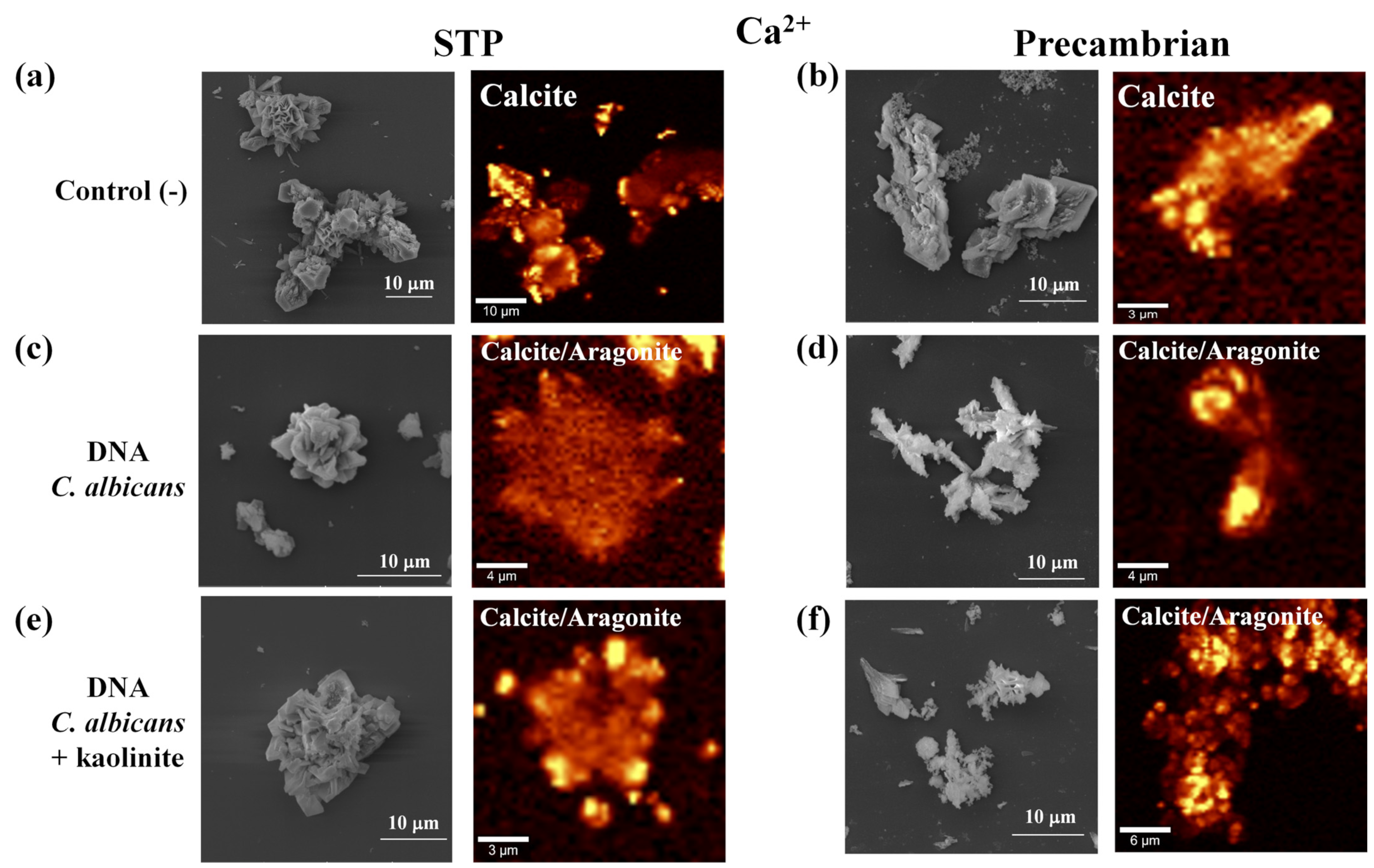
To find out the chemical composition as well as the crystalline phase of the synthesized calcium silica–carbonate biomorphs, these were analyzed with Raman spectroscopy and FTIR. For the control biomorph in STP conditions, Raman spectra identified bands at 157, 282, 712, 1086, 1750 cm−1 (Figure 2a(ii); Table 1), and by FTIR the bands were at 712.1, 871.1, 1399.9 cm−1 (Table 1).
In the case of the control biomorphs obtained in conditions emulating the Precambrian era, bands were identified by Raman spectroscopy at 157, 282, 1082 cm−1 (Figure 2b(ii); Table 1). These biomorphs, when analyzed by FTIR, identified bands at 792.0, 871.2, 950.1, 1073.3, 1387.2, 2980.7 cm−1 (Table 1). Both for the control biomorphs synthesized in STP conditions as well as in the Precambrian conditions, these vibrations corresponded to the calcite of the calcium carbonate (CaCO3) polymorph. The crystalline structure has previously been reported for the biomorphs obtained in this condition [34,35,36,37,38].
In the biomorphs obtained in the presence of E. coli genomic DNA, both in STP and Precambrian conditions, bands by Raman spectroscopy were identified at 157, 282, 283, 712, 1086 cm−1 (Figure 2c(ii),d(ii); Table 1), and by FTIR at 575.3, 604.7, 712.2, 750.5, 871.7, 1407.30, 2980.7 and 3420.8 cm−1 (Table 1). Vibrations correspond to the calcite polymorph, as well as to the control biomorphs.
The spectra of the biomorphs in both conditions of synthesis (STP and Precambrian), in the presence of E. coli DNA with kaolinite showed bands by Raman spectroscopy at 157, 282, 712, 1085, 1086 cm−1 (Figure 2e(ii),f(ii); Table 1). Bands found for these biomorphs by FTIR were at 544.0, 595.0, 626.0, 680.7, 797.5, 913.4, 939.0, 1008.3, 1031.0, 1386.9, 1407.0, 2980.7, 3482.6, 3619.6 and 3690.8 cm−1 (Table 1). These bands of the biomorphs synthesized with E. coli and kaolinite DNA also correspond to calcite. These data show that in the presence of genomic E. coli DNA with kaolinite, the polymorph obtained from the silica–carbonate biomorphs is calcite as the main component of the crystallites. It has been reported that calcite is the most frequent polymorph identified in the hard and support structures of organisms [55,56,57,58,59,60,61], being also the most thermodynamically stable polymorph, followed by aragonite and vaterite [62] (Carteret et al., 2009). Biomineralization is the chemical process that occurs during the synthesis of the biomorphs, through which the CaCO3 polymorphs are obtained on the surface or the internal part of the biomorphs. This is a mechanism that continues to occur in organisms [54,63,64,65,66,67]. It has been reported that biomineralization occurs practically in all forms of life, from bacteria to superior organisms, reporting, therefore, the formation of more than 60 different biological minerals [68,69]. When analyzing biomorphs with C. albicans genomic DNA by Raman spectroscopy and synthesized in both atmospheric conditions (STP and Precambrian), bands were identified at 111, 157, 282, 470, 711, 1086, 1480, 1760 and 2911 cm−1 (Figure 3c,d; Table 1). While by FTIR, bands were determined at 580, 711, 792.6, 871.20, 1073.1, 1387.4, 1406.2 cm−1 (Table 1). These bands correspond to the calcite and aragonite polymorphs as the main components of the crystallites inserted in the silica–carbonate surface.
In the case of the Raman spectra of the biomorphs in the presence of DNA with kaolinite, both in the STP and Precambrian conditions, bands were shown at 112, 156, 281, 711, 1085, 1437, 1470, 1750 and 2980 cm−1 (Figure 3e,f; Table 1). By FTIR, bands were identified at 599.4, 596.0, 576.9, 626.1, 680.8, 712.1, 757.3, 797.4, 871.3, 913.3, 939.2, 1008.2, 1031.0, 1394.1, 2980.8 and 3691.0 cm−1 (Table 1). Bands correspond to the calcite and aragonite polymorphs, as well as to the biomorphs obtained with C. albicans DNA. These results obtained both with the C. albicans genomic DNA as well as with kaolinite are interesting for various reasons. The first reason to consider is that in the biomorphs that were synthesized with E. coli genomic DNA, only the calcite polymorph was obtained, while with the C. albicans DNA, two polymorphs were obtained: calcite and aragonite. Whether the DNA came from a prokaryote or from a eukaryote, the DNA is capable of controlling the inorganic molecules present in the solution so that the biomorph adopts a morphology similar to that of a living organism. It seems that the main difference is at the molecular level, since the DNA that comes from a eukaryote, while capable of directing the synthesis toward the most stable crystalline habit, also plays an important role in the chemical composition and crystalline structure. This is possibly due to the differences between the genomic DNA of prokaryotes and eukaryotes, among which are the prokaryote DNAs that are circular chromosomes, and the eukaryote DNAs are linear chromosomes confined within a nucleus [70,71,72,73,74,75,76]. The prokaryote DNA is found in the cytoplasm, while the eukaryote DNA is packed into the nucleus of the cell [70,71,72,73,74,75,76]. The eukaryote genes contain introns that code for a single protein, while the prokaryote genes lack introns and are organized in operons, which code for various proteins. Prokaryote cells have only one point of origin and replication takes place in two opposing directions at the same time in the cell cytoplasm. The eukaryote cells have multiple origins and use unidirectional replication within the cell’s nucleus. The number of polymerases is different in these cell types, as is the amount of DNA, which is approximately twenty-five times greater in the eukaryote cells than in the prokaryotes [70,71,72,73,74,75,76]. The second reason to consider is that C. albicans genomic DNA is what directed the synthesis of the biomorphs since the same polymorphs were also obtained in the presence of kaolinite (Figure 3, Table 1).
The fact of having obtained calcite and aragonite is a result that agrees with what is found in various organisms since it has been described that the two polymorphs of CaCO3 are the most abundant crystalline structures in organisms, all the way from microorganisms to superior organisms [55,56,57,58,59,60,77]. There are organisms with structures formed both by calcite and aragonite. For example, the pearl oyster Pinctada fucata has an outer layer formed by calcite while the inner nacreous layer is formed by aragonite [78,79]. Additionally, our results show that the C. albicans DNA is what directs the crystalline structure of the biomorphs, which is also in agreement with works that have shown that macromolecules extracted from the aragonitic shell layers of some mollusks induce the formation of aragonite in vitro. The same effect is observed with the macromolecules from shell made of calcite layers that induce the formation of one of the polymorphs only. Data show that these macromolecules are responsible for the precipitation of calcite or aragonite in vivo [77]. In this way, the facts obtained here favor the hypothesis that the biomorphs might have been the first crystalline structures that isolated, protected and preserved the first biomolecules that were formed on Earth. Additionally, it is shown that the formation of the calcite and aragonite polymorphs must have occurred right from the first structures that originated life. From that moment until now, it seems that inorganic structures have preserved their crystalline structure.

3.2. Environmental Factors That Participate Directly in the Morphology of the BaCO3 Biomorphs but Not in the Crystalline Phase

The barium carbonate biomorphs are also of special relevance because biomorphs adopt morphologies associated with typical forms of life even without the presence of biomolecules [30,37,38,80,81,82] and, at the same time, some of their morphologies are reminiscent of fossils, which biomorphs might have very well been, though they had not been termed as such, keeping them invisible [35]. The morphology, chemical composition and crystalline structure of the BaCO3 biomorphs were studied and, in the presence of E. coli or C. albicans genomic DNA and kaolinite, the synthesis of these biomorphs was carried out.
The control barium silica–carbonate biomorphs synthesized in STP atmospheric conditions showed the aggregate that emulates the morphology of a sphere (Figure 4A). For the control biomorphs obtained in the Precambrian atmospheric conditions, the aggregate that emulates the morphology of a flower with elongated petals was observed (Figure 4B). The barium silica–carbonate biomorphs that were treated with E. coli genomic DNA in STP conditions presented polycrystalline morphology with a section that emulated the flower of the plant Taraxacum officinale (Figure 4C). In the case of the biomorphs obtained with E. coli DNA but under the atmospheric conditions that emulate the Precambrian, the morphology observed was that of flowers with short petals and leaves (Figure 4D).
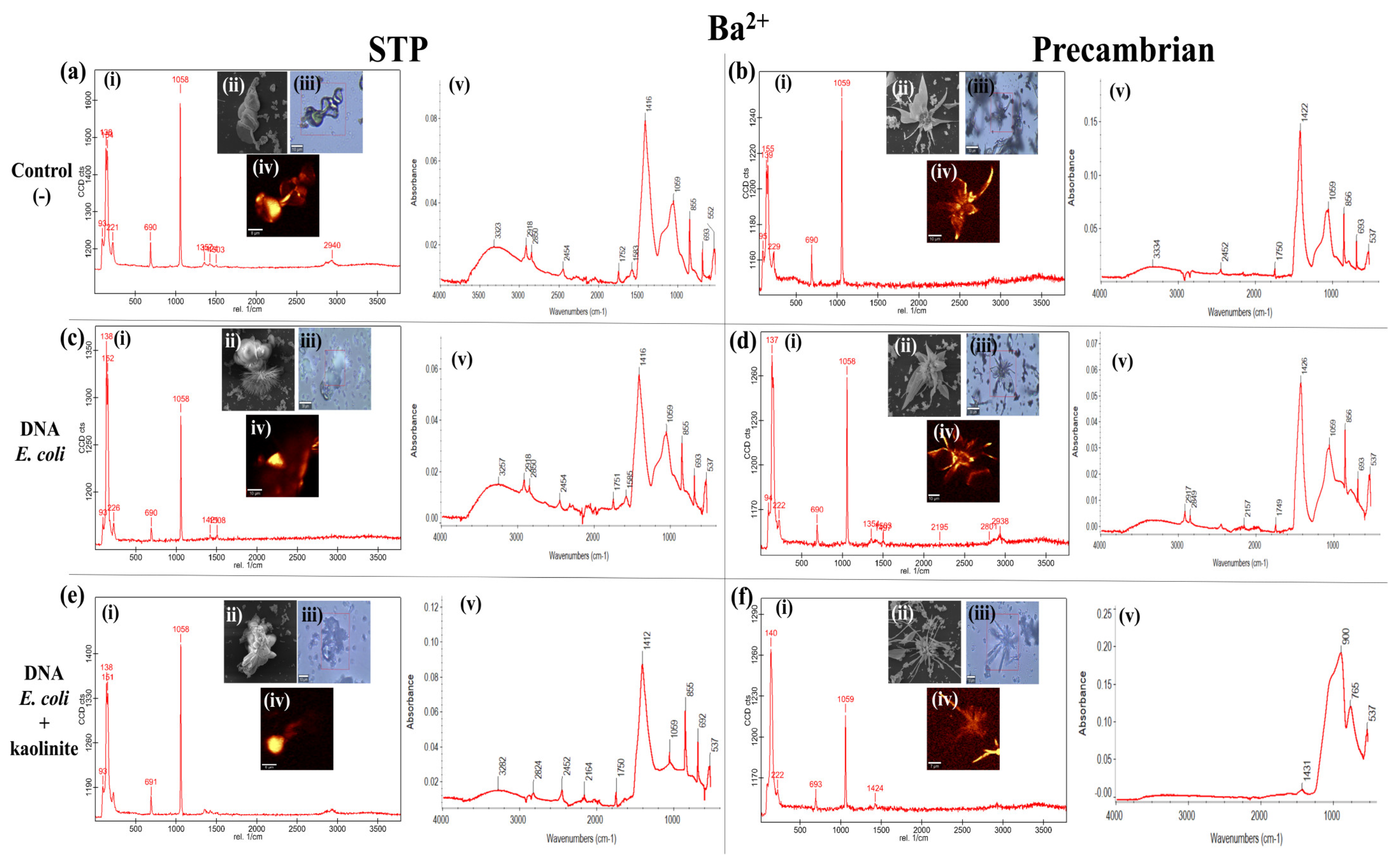
For the biomorphs that were synthesized with C. albicans genomic DNA, leaf and druse morphology was found (Figure 4E). The biomorphs that were synthesized with C. albicans genomic DNA in STP conditions presented flower with leaf morphology (Figure 4F). The biomorphs produced with DNA from E. coli with kaolinite presented polycrystal morphology (Figure 4G) similar to the morphology found only in the E. coli DNA (Figure 4C). The biomorphs synthesized with DNA from E. coli with kaolinite in Precambrian conditions presented leaves, flowers and stems (Figure 4H). The biomorphs that were obtained with C. albicans genomic DNA and kaolinite in STP atmosphere presented leaf and stem morphology (Figure 4I) while the biomorphs obtained in Precambrian atmosphere from C. albicans DNA and kaolinite showed flower, leaf and stem morphology (Figure 4J). This type of morphology that emulates flowers, stems and leaves in the presence of DNA has already been found in the presence of five types of DNA [37]. These results, as happened with calcium silica–carbonate biomorphs, show that the genomic DNA is what directs the morphology, the chemical composition and the crystalline structure. In addition, these morphologies that emulate various organs of plants that were identified in the barium silica–carbonate biomorphs with genomic DNA only or with kaolinite suggest that the barium, by being a chemical element present in the form of barite in the Earth’s crust [83] from the first epochs on Earth, formed part of some of the first organisms. In this way, barium from that time was possibly preserved to the present day in some organisms by being a part of some of the pioneer structures, as are some species of algae, invertebrates and fish from the Irish Sea [84]. When determining the crystalline structure of the control barium silica–carbonate biomorphs obtained in STP conditions by Raman spectroscopy, bands were identified at 93, 138, 154, 221, 690, 1058 cm−1 (Figure 5a(i), Table 2), and by FTIR at 549.7, 585.1, 604.3, 692.6, 768.5, 855.5, 953.2, 1059.0, 1416.9, 1577.7, 2980 cm−1 (Figure 5a(v), Table 2). The bands identified correspond to the polymorph of BaCO3, commonly known as witherite [85,86]. For the biomorphs synthesized in Precambrian conditions, the witherite polymorph was also identified both by Raman spectroscopy and by FTIR (Figure 5b(i,v), Table 2).
The barium silica–carbonate biomorphs that were synthesized in the presence of E. coli genomic DNA in both STP and Precambrian conditions showed the witherite band spectra shown by Raman spectroscopy and FTIR (Figure 5c(i,v),d(i,v), Table 2). The witherite polymorph was also identified in the biomorphs obtained with E. coli DNA and kaolinite in both evaluated atmospheric conditions (Figure 5e(i,v),f(i,v), Table 2).
In the case of barium silica–carbonate biomorphs synthesized with C. albicans genomic DNA, as well as with the biomorphs with C. albicans genomic DNA and kaolinite, in both STP and Precambrian conditions, the witherite phase was identified (Figure 6, Table 2).
These results show that in the case of the barium silica–carbonate biomorphs, the crystal form that is obtained is always witherite, whether they were synthesized with a biomolecule such as DNA or in the presence of a mineral, such as kaolinite. These data agree with those from other barium silica–carbonate biomorphs obtained previously in the presence of DNA or minerals [37,48]. This is because witherite is the most stable crystalline phase of BaCO3 [87]. Computer studies have proven that the orthorhombic phase with the space group Pmcn is the most stable at normal temperature and pressure [87] because when the pressures are modified, other phases of the BaCO3 are obtained [88,89].
It has also been observed that when witherite is synthesized in vitro, the morphology that is obtained is dependent on the environmental conditions, such as temperature and CO2 concentration [90,91,92,93]. However, this does not affect the BaCO3 crystalline phase since the orthorhombic phase is obtained from witherite. These observations are found to correlate with what we observed in our results because, as in the synthesis of the barium silica–carbonate biomorphs, the environmental conditions used, as well as the biomolecules and the mineral such as kaolinite, define the morphology they adopt (Figure 4). This has been a constant crystalline behavior that has been observed in the BaCO3 biomorphs [34,35,36,37,38,39,40]. These results altogether indicate on the one hand that the barium silica–carbonate biomorphs, as we have proposed, might be one of the first structures where the proto-molecules that gave rise to the pioneer organism were protected, aligned, duplicated and preserved [34,35]. On the other hand, it was a big advantage that BaCO3 could adopt one or the other morphology, depending on the environmental conditions. These conditions perhaps favored the existence of various morphologies of the pioneer organisms where, possibly, some organisms have conserved the morphology or mimicked it, as well as some part of the chemical composition [34,35]. This is based on the fact that today there are organisms which contain chemical elements that differ from the majority of organisms; for example, chemical elements such as barium (Ba), strontium (Sr), arsenic (As), cadmium (Cd), wolframium (W), among others, have been identified [94,95,96]. So, our results allow the inference that possibly some of the pioneer organisms that existed in the Precambrian Era may have been conserved up to the present, while other organisms evolved more complex and functional forms whereas others inevitably perished.

4. Conclusions

Even when minerals are considered a fundamental part of the origin of life on Earth due to their contribution in the formation of the first inorganic structures in which biomolecules have been conserved and perpetuated, our results show that when a mineral such as kaolinite is found in the presence of genomic DNA, it is precisely the DNA, not the mineral, that directs the morphology, the crystalline stage and the chemical composition of the structure. This is a relevant fact as it shows how, independently of the morphology and of the size of the organism, it is the DNA that directs all the chemical elements towards the most stable structure, allowing for the perpetuation, conservation and maintenance of life on our planet (since the origin of the genomic DNA in the Precambrian Era to the present day). The fact that the morphology of barium silica–carbonate biomorphs changes depending on the environmental conditions whereas their crystalline structure remains stable possibly indicates that, in a first stage in the origin of the proto-cell, the barium silica–carbonate biomorphs may have formed part of the inorganic structure. As time went on and the chemical composition of the Earth changed, these inorganic structures might have replaced or might have incorporated other elements, such as calcium, so that calcium might have replaced barium in the structure of the organisms. This opens a wide range of possibilities regarding morphologies that have arisen in the organisms that we know today.

Author Contributions

Conceptualization, M.C.-C. and A.M.; methodology, M.C.-C. and S.R.I.; validation, M.C.-C., S.R.I. and A.M.; formal analysis, M.C.-C. and A.M.; investigation, M.C.-C., S.R.I. and A.M.; writing—original draft preparation, M.C.-C.; writing—review and editing, M.C.-C. and A.M.; supervision, M.C.-C. and A.M.; project administration, M.C.-C.; funding acquisition, M.C.-C. All authors have read and agreed to the published version of the manuscript.

Funding

This research was carried out with the financial support granted to M. Cuéllar-Cruz by Project No. CF2019-39216 from the Consejo Nacional de Ciencias, Humanidades y Tecnologías (CONAHCYT) and Proyecto Institucional (Institutional Project) UGTO-005/2024 from Universidad de Guanajuato, México.

Data Availability Statement

Data are contained within the article.

Acknowledgments

The authors thank Laboratorio Universitario de Caracterización Espectroscópica of the Instituto de Ciencias Aplicadas y Tecnología, UNAM (Institute of Applied Science and Technology of the National Autonomous University of Mexico) for the Raman spectroscopy and FTIR measurements, and José Guadalupe Bañuelos for their technical support. One of the authors (A.M.) acknowledges Antonia Sánchez Marin for her English style corrections for this manuscript. The authors acknowledge Teresa Horn for the first revision and English corrections in the present manuscript.

Conflicts of Interest

The authors declare no conflicts of interest.

References

  1. Jenkins, R.J.F.; Ford, C.H.; Gehling, J.G. The Ediacara Member of the Rawnsley Quartzite: The context of the Ediacara assemblage (late Precambrian, Flinders Ranges). J. Geol. Soc. Aust. 1983, 30, 101–119. [Google Scholar] [CrossRef]
  2. Wächtershäuser, G. From volcanic origins of chemoautotrophic life to Bacteria, Archaea and Eukarya. Philos. Trans. R. Soc. Lond. B Biol. Sci. 2006, 361, 1787–1808. [Google Scholar] [CrossRef] [PubMed]
  3. Cuéllar-Cruz, M. The equation of life in the Universe: Biomorphs as reminiscence of the first forms of life. Prog. Cryst. Growth Charact. Mater. 2024, 70, 100624. [Google Scholar] [CrossRef]
  4. Cuéllar-Cruz, M. The equation of the origin in the Universe (Part II): The combination of chemical elements does not determine the emergence of life on Earth. Prog. Cryst. Growth Charact. Mater. 2024, 70, 100625. [Google Scholar] [CrossRef]
  5. Ulrich, B. Soil Acidity and its Relations to Acid Deposition. In Effects of Accumulation of Air Pollutants in Forest Ecosystems; Ulrich, B., Pankrath, J., Eds.; Springer: Dordrecht, The Netherlands, 1983; pp. 127–146. [Google Scholar]
  6. Dixon, J.B. Kaolin and Serpentine group minerals. In Minerals in Soil Environments, 2nd ed.; Dixon, J.B., Weed, S.B., Eds.; American Society of Agronomy, Crop Science Society of America, and Soil Science Society of America: Madison, WI, USA, 1989; Volume 1, pp. 467–525. [Google Scholar]
  7. Moore, D.M.; Reynolds, R.J. X-ray Diffraction and the Identification and Analysis of Clay Minerals. In State Geological Survey; Oxford University Press: Oxford, UK, 1989; p. 332. [Google Scholar]
  8. Lázaro, B.B. Halloysite and kaolinite: Two clay minerals with geological and technological importance. Rev. Acad. Cienc. Exactas Fis. Quim. Nat. Zaragoza 2015, 70, 7–38. [Google Scholar]
  9. Deer, W.A.; Howie, R.A.; Zussman, J. An Introduction to Rock-Forming Minerals; Longman Group Ltd.: London, UK, 1975; p. 528. [Google Scholar]
  10. Swindale, L.D. The crystallography of minerals of the kaolin group. In Soil Components; Inorganic Components; Springer: Berlin/Heidelberg, Germany, 1975; Volume 2, pp. 121–154. [Google Scholar]
  11. Ehlmann, B.L.; Swayze, G.A.; Milliken, R.E.; Mustard, J.F.; Clark, R.N.; Murchie, S.L.; Breit, G.N.; Wray, J.J.; Gondet, B.; Poulet, F.; et al. Discovery of alunite in Cross Crater, Terra Sirenum, Mars: Evidence for acidic, sulfurous waters. Am. Min. 2016, 101, 1527–1542. [Google Scholar] [CrossRef]
  12. Pineau, M.; Mathian, M.; Baron, F.; Rondeau, B.; Deit, L.L.; Allard, T.; Mangold, N. Estimating kaolinite crystallinity using near-infrared spectroscopy: Implications for its geology on Earth and Mars. Am. Min. 2022, 107, 1453–1469. [Google Scholar] [CrossRef]
  13. Murchie, S.; Arvidson, R.; Bedini, P.; Beisser, K.; Bibring, J.-P.; Bishop, J.; Boldt, J.; Cavender, P.; Choo, T.; Clancy, R.T.; et al. Compact Reconnaissance Imaging Spectrometer for Mars (CRISM) on Mars Reconnaissance Orbiter (MRO). J. Geophys. Res. 2007, 112, 1–57. [Google Scholar] [CrossRef]
  14. Mustard, J.F.; Murchie, S.L.; Pelkey, S.M.; Ehlmann, B.L.; Milliken, R.E.; Grant, J.A.; Bibring, J.-P.; Poulet, F.; Bishop, J.; Dobrea, E.N.; et al. Hydrated silicate minerals on Mars observed by the Mars Reconnaissance Orbiter CRISM instrument. Nature 2008, 454, 305–309. [Google Scholar] [CrossRef]
  15. Carter, J.; Quantin, C.; Thollot, P.; Loizeau, D.; Ody, A.; Lozach, L. Oxia Planum, a clay-laden landing site proposed for the Exomars Rover Mission: Aqueous mineralogy and alteration scenarios. In Proceedings of the 47th Annual Lunar and Planetary Science Conference, The Woodlands, TX, USA, 21–25 March 2016; p. 2064. [Google Scholar]
  16. Ehlmann, B.L.; Edwards, C.S. Mineralogy of the Martian surface. Annu. Rev. Earth Planet. Sci. 2014, 42, 291–315. [Google Scholar] [CrossRef]
  17. Carrascoza Mayén, J.F.; Rydzewski, J.; Szostak, N.; Blazewicz, J.; Nowak, W. Prebiotic Soup Components Trapped in Montmorillonite Nanoclay Form New Molecules: Car-Parrinello Ab Initio Simulations. Life 2019, 9, 46. [Google Scholar] [CrossRef] [PubMed]
  18. Bhushan, B.; Nayak, A. Kamaluddin Catalytic Role of Manganese Oxides in Prebiotic Nucleobases Synthesis from Formamide. Orig. Life Evol. Biosph. 2016, 46, 203–213. [Google Scholar] [CrossRef] [PubMed]
  19. Ritson, D.J.; Battilocchio, C.; Ley, S.V.; Sutherland, J.D. Mimicking the Surface and Prebiotic Chemistry of Early Earth Using Flow Chemistry. Nat. Commun. 2018, 9, 1821. [Google Scholar] [CrossRef] [PubMed]
  20. Rouillard, J.; Van Kranendonk, M.J.; Lalonde, S.; Gong, J.; van Zuilen, M.A. Correlating trace element compositions, petrology, and Raman spectroscopy data in the ∼3.46 Ga Apex chert, Pilbara Craton, Australia. Precambrian Res. 2021, 366, 106415. [Google Scholar] [CrossRef]
  21. Schopf, J.W.; Packer, B.M. Early Archean (3.3-Billion to 3.5-Billion-Year-Old) Microfossils from Warrawoona Group, Australia. Science 1987, 237, 70–73. [Google Scholar] [CrossRef] [PubMed]
  22. Schopf, J.W. Microfossils of the Early Archean Apex Chert: New Evidence of the Antiquity of Life. Science 1993, 260, 640–646. [Google Scholar] [CrossRef] [PubMed]
  23. Schopf, W.J.; Kudryavtsev, A.B. Biogenicity of Earth’s Earliest Fossils: A Resolution of the Controversy. Gondwana Res. 2012, 22, 761–771. [Google Scholar] [CrossRef]
  24. Brasier, M.D.; Green, O.R.; Jephcoat, A.P.; Kleppe, A.K.; Van Kranendonk, M.J.; Lindsay, J.F.; Steele, A.; Grassineau, N.V. Questioning the Evidence for Earth’s Oldest Fossils. Nature 2002, 416, 76–81. [Google Scholar] [CrossRef] [PubMed]
  25. Brasier, M.D.; McLoughlin, N.; Green, O.; Wacey, D. A Fresh Look at the Fossil Evidence for Early Archaean Cellular Life. Philos. Trans. R. Soc. Lond. Ser. B 2006, 361, 887–902. [Google Scholar] [CrossRef]
  26. Brasier, M.D.; Antcliffe, J.; Saunders, M.; Wacey, D. Changing the picture of Earth’s earliest fossils (3.5–1.9 Ga) with new approaches and new discoveries. Proc. Natl. Acad. Sci. USA 2015, 112, 4859–4864. [Google Scholar] [CrossRef]
  27. Pinti, D.L.; Mineau, R.; Clement, V. Hydrothermal Alteration and Microfossil Artefacts of the 3465-Million-Year-Old Apex Chert. Nat. Geosci. 2009, 2, 640–643. [Google Scholar] [CrossRef]
  28. Marshall, C.P.; Emry, J.R.; Olcott Marshall, A. Haematite Pseudomicrofossils Present in the 3.5-Billion-Year-Old Apex Chert. Nat. Geosci. 2011, 4, 240–243. [Google Scholar] [CrossRef]
  29. Sforna, M.C.; van Zuilen, M.A.; Philippot, P. Structural Characterization by Raman Hyperspectral Mapping of Organic Carbon in the 3.46 Billion-Year-Old Apex Chert, Western Australia. Geochim. Cosmochim. Acta 2014, 124, 18–33. [Google Scholar] [CrossRef]
  30. Garcia-Ruiz, J.M.; Hyde, S.T.; Carnerup, A.M.; Christy, A.G.; Van Kranendonk, V.M.J.; Welham, N.J. Self-assembled silicacarbonate structures and detection of ancient microfossils. Science 2003, 302, 1194–1197. [Google Scholar] [CrossRef] [PubMed]
  31. Opel, J.; Wimmer, F.P.; Kellermeier, M.; Colfen, H. Functionalisation of Silica-Carbonate Biomorphs. Nanoscale Horiz. 2016, 1, 144–149. [Google Scholar] [CrossRef] [PubMed]
  32. Voinescu, A.E.; Kellermeier, M.; Carnerup, A.M.; Larsson, A.K.; Touraud, D.; Hyde, S.T.; Kunz, W. Co-Precipitation of Silica and Alkaline-Earth Carbonates Using TEOS as Silica Source. J. Cryst. Growth 2007, 306, 152–158. [Google Scholar] [CrossRef]
  33. Voinescu, A.E.; Kellermeier, M.; Bartel, B.; Carnerup, A.M.; Larsson, A.K.; Touraud, D.; Kunz, W.; Kienle, L.; Pfitzner, A.; Hyde, S.T. Inorganic Self-Organized silica aragonite biomorphic composites. Cryst. Growth Des. 2008, 8, 1515–1521. [Google Scholar] [CrossRef]
  34. Cuéllar-Cruz, M. New insights on the origin of life: The role of silico-carbonates of Ba (II) to preserve DNA against highly intense UV radiation. ACS Omega 2023, 8, 29585–29594. [Google Scholar] [CrossRef] [PubMed]
  35. Cuéllar-Cruz, M.; Islas, S.R.; Ramírez-Ramírez, N.; Pedraza-Reyes, M.; Moreno, A. Protection of the DNA from selected species of five kingdoms in Nature by Ba(II), Sr(II) and Ca(II) silica-carbonates: Implications about biogenicity and evolving from prebiotic chemistry to biological chemistry. ACS Omega 2022, 7, 37410–37426. [Google Scholar] [CrossRef]
  36. Cuéllar-Arcos, J.C.; Islas, S.R.; Cuéllar-Cruz, M. Biomorphs possess mineral crystalline plasticity by adapting their morphology and chemical composition in the presence of organic biomolecules and inorganic ions. ACS Earth Space Chem. 2023, 7, 2535–2547. [Google Scholar] [CrossRef]
  37. Cuéllar-Cruz, M.; Islas, S.R.; González, G.; Moreno, A. Influence of nucleic acids on the synthesis of crystalline Ca (II), Ba (II), and Sr (II) silica-carbonate biomorphs: Implications for the chemical origin of life on primitive Earth. Cryst. Growth Des. 2019, 19, 4667–4682. [Google Scholar] [CrossRef]
  38. Cuéllar-Cruz, M.; Moreno, A. The role of calcium and strontium as the most dominant elements during combinations of different alkaline Earth metals in the synthesis of crystalline silica-carbonate biomorphs. Crystals 2019, 9, 381. [Google Scholar] [CrossRef]
  39. Islas, S.R.; Cuéllar-Cruz, M. Silica-carbonate of Ba(II) and Fe2+/Fe3+ Complex as Study Models to Understand Prebiotic Chemistry. ACS Omega 2021, 6, 35629–35640. [Google Scholar] [CrossRef] [PubMed]
  40. Cuéllar-Cruz, M. Influence of Abiotic Factors in the Chemical Origin of Life: Biomorphs as a Study Model. ACS Omega 2021, 6, 8754–8763. [Google Scholar] [CrossRef] [PubMed]
  41. Zhang, G.; Morales, J.; Garcia-Ruiz, J.M. Growth Behaviour of Silica/Carbonate Nanocrystalline Composites of Calcite and Aragonite. J. Mater. Chem. B 2017, 5, 1658–1663. [Google Scholar] [CrossRef] [PubMed]
  42. Bittarello, E.; Aquilano, D. Self-assembled nanocrystals of barium carbonate in biomineral-like structures. Eur. J. Mineral. 2007, 19, 345–351. [Google Scholar] [CrossRef]
  43. Bittarello, E.; Roberto Massaro, F.R.; Aquilano, D. The epitaxial role of silica groups in promoting the formation of silica/carbonate biomorphs: A first hypothesis. J. Cryst. Growth 2010, 312, 402–412. [Google Scholar] [CrossRef]
  44. Bittarello, E.; Massaro, F.R.; Rubbo, M.; Costa, E.; Aquilano, D. Witherite (BaCO3)/α-Quartz epitaxial nucleation and growth: Experimental findings and theorical implications on biomineralization. Cryst. Growth Des. 2009, 9, 971–977. [Google Scholar] [CrossRef]
  45. Bruno, M.; Rubbo, M.; Pastero, L.; Massaro, F.R.F.; Nestola, D.; Aquilano, D. Computational Approach to the Study of Epitaxy: Natural Occurrence in Diamond/Forsterite and Aragonite/Zabuyelite. Cryst. Growth Des. 2015, 15, 2979–2987. [Google Scholar] [CrossRef]
  46. Bruno, M.; Pastero, L.; Cotellucci, A.; Aquilano, D. Epitaxy: A methodological approach to the study of an old phenomenon. Cryst Eng Comm. 2022, 24, 4165–4173. [Google Scholar] [CrossRef]
  47. Aquilano, D.; Bruno, M.; Ghignone, S.; Pastero, L.; Cotellucci, A. Epitaxies of Ca sulfates on calcite. II. The main {010}, {001} and {100} forms of bassanite epi-deposited on the {10.4} substrate form of calcite. J. Appl. Crystallogr. 2022, 55, 1289–1296. [Google Scholar] [CrossRef]
  48. Cuéllar-Cruz, M.; Ramírez-Cardona, M.; Islas, S.R.; Moreno, A. Influence of different types of clay minerals on the shape and form of silica-carbonates (Biomorphs) of Ca(II), Ba(II), AND Sr(II). ACS Earth Space Chem. 2022, 6, 3054–3065. [Google Scholar] [CrossRef]
  49. Zúñiga-Estrada, E.A.; Zúñiga-Estrada, M.A.; Islas, S.R.; Moreno, A.; Cuéllar-Cruz, M. Minerals participated in the formation of the first inorganic structure that favored the synthesis of protomolecules: Biomorphs as models of study. ACS Omega 2024. submitted. [Google Scholar]
  50. Rickwood, D.; Hames, B.D. Gel Electrophoresis of Nucleic Acids: A practical Approach; Oxford University Press: New York, NY, USA, 1990; pp. 65–66. [Google Scholar]
  51. López-Mora, P.A.; López Gutiérrez, A.M.; Marulanda-Ángel, M.L. Standardizing genomic DNA extraction in Tabebuia rosea (Bertol.) DC. and Cordia alliodora (Ruiz and Pav.) Okén. Temas Agrar. 2011, 16, 28–41. [Google Scholar] [CrossRef]
  52. Demeke, T.; Jenkins, G.R. Influence of DNA extraction methods, PCR inhibitors and quantification methods on real-time PCR assay of biotechnology-derived traits. Anal. Bioanal. Chem. 2010, 396, 1977–1990. [Google Scholar] [CrossRef] [PubMed]
  53. Noorduin, W.L.; Grinthal, A.; Mahadevan, L.; Aiznberg, J. Rationally Designed Complex, Hierarchical Microarchitectures. Science 2013, 340, 832–837. [Google Scholar] [CrossRef] [PubMed]
  54. Cuéllar-Cruz, M. Synthesis of inorganic and organic crystals mediated by proteins in different biological organisms. A mechanism of biomineralization conserved throughout evolution in all living species. Prog. Cryst. Growth Charact. Mater. 2017, 63, 94–103. [Google Scholar] [CrossRef]
  55. Ruiz-Arellano, R.R.; Moreno, A. Obtainment of spherical-shaped calcite crystals induced by intramineral proteins isolated from eggshells of ostrich and emu. Cryst. Growth Des. 2014, 14, 5137–5143. [Google Scholar] [CrossRef]
  56. Ruiz-Arellano, R.R.; Medrano, F.J.; Moreno, A.; Romero, A. Crystal Structure of Struthiocalcin-1, an Intramineral Protein from Struthio camelus Eggshell, in two crystal forms. Acta Crystallogr. D Biol. Crystallogr. 2015, 71, 809–818. [Google Scholar] [CrossRef]
  57. Reyes-Grajeda, J.P.; Moreno, A.; Romero, A. Crystal structure of ovocleidin-17, a major protein of the calcified Gallus gallus eggshell: Implications in the calcite mineral growth pattern. J. Biol. Chem. 2004, 279, 40876–40881. [Google Scholar] [CrossRef]
  58. Cölfen, H. Biomineralization: A crystal-clear view. Nat. Mater. 2010, 9, 960–961. [Google Scholar] [CrossRef]
  59. Mann, K. The calcified eggshell matrix proteome of a songbird, the zebra finch (Taeniopygia guttata). Proteome Sci. 2015, 13, 29. [Google Scholar] [CrossRef] [PubMed]
  60. Mann, K.; Mann, M. The proteome of the calcified layer organic matrix of turkey (Meleagris gallopavo) eggshell. Proteome Sci. 2013, 11, 40. [Google Scholar] [CrossRef]
  61. Jáuregui-Zúñiga, D.; Reyes-Grajeda, J.P.; Sepúlveda-Sanchez, J.D.; Whitaker, J.R.; Moreno, A. Crystallochemical characterization of calcium oxalate crystals isolated from seed coats of Phaseolus vulgaris and leaves of Vitis vinifer. J. Plant Physiol. 2003, 160, 239–245. [Google Scholar] [CrossRef] [PubMed]
  62. Carteret, C.; Dandeu, A.; Moussaoui, S.; Muhr, H.; Humbert, B.; Plasari, E. Polymorphism Studied by Lattice Phonon Raman Spectroscopy and Statistical Mixture Analysis Method. Application to Calcium Carbonate Polymorphs during Batch Crystallization. Cryst. Growth Des. 2009, 9, 807–812. [Google Scholar] [CrossRef]
  63. Mann, S. Biomineralization: Principle and Concepts in Bioinorganic Materials Chemistry; Oxford University Press: New York, NY, USA, 2001. [Google Scholar]
  64. Lowenstam, H.A. Minerals formed by organisms. Science 1981, 211, 1126–1131. [Google Scholar] [CrossRef]
  65. Lowenstam, H.A.; Weiner, S. On Biomineralization; Oxford University Press: New York, NY, USA, 1989. [Google Scholar]
  66. Benzerara, K.; Miot, J.; Morin, G.; Ona-Nguema, G.; Skouri-Panet, F.; Ferard, C. Significance, mechanisms and environmental implications of microbial biomineralization. Comptes Rendus Geosci. 2011, 343, 160–167. [Google Scholar] [CrossRef]
  67. Phillips, A.J.; Gerlach, R.; Lauchnor, E.; Mitchell, A.C.; Cunningham, A.B.; Spangler, L. Engineered applications of ureolytic biomineralization: A review. Biofouling 2013, 29, 715–733. [Google Scholar] [CrossRef] [PubMed]
  68. Anbu, P.; Kang, C.H.; Shin, Y.J.; So, J.S. Formations of calcium carbonate minerals by bacteria and its multiple applications. Springerplus 2016, 5, 250. [Google Scholar] [CrossRef]
  69. Sarikaya, M. Biomimetics: Materials fabrication through biology. Proc. Natl. Acad. Sci. USA 1999, 96, 14183–14185. [Google Scholar] [CrossRef]
  70. Blattner, F.R.; Plunkett, G.; Bloch, C.A.; Perna, N.T.; Burland, V.; Riley, M.; Collado-Vides, J.; Glasner, J.D.; Rode, C.K.; Mayhew, G.F.; et al. The complete genome sequence of Escherichia coli K-12. Science 1997, 277, 1453–1462. [Google Scholar] [CrossRef] [PubMed]
  71. Adams, M.D.; Celniker, S.E.; Holt, R.A.; Evans, C.A.; Gocayne, J.D.; Amanatides, P.G.; Scherer, S.E.; Li, P.W.; Hoskins, R.A.; Galle, R.F.; et al. The Genome Sequence of Drosophila melanogaster. Science 2000, 287, 2185–2195. [Google Scholar] [CrossRef] [PubMed]
  72. Bult, C.J.; White, O.; Olsen, G.J.; Zhou, L.; Fleischmann, R.D.; Sutton, G.G.; Blake, J.A.; FitzGerald, L.M.; Clayton, R.A.; Gocayne, J.D.; et al. Complete genome sequence of the methanogenic archaeon Methanococcus jannaschii. Science 1996, 273, 1058–1073. [Google Scholar] [CrossRef] [PubMed]
  73. Ahnert, S.E.; Fink, T.M.; Zinovyev, A. How much non-coding DNA do eukaryotes require? J. Theor. Biol. 2008, 252, 587–592. [Google Scholar] [CrossRef] [PubMed]
  74. Bendich, A.J.; Drlica, K. Prokaryotic and eukaryotic chromosomes: What’s the difference? Bioessays 2000, 22, 481–486. [Google Scholar] [CrossRef]
  75. Cairns, J. The chromosome of Escherichia coli. In Cold Spring Harbor Symposia on Quantitative Biology; Cold Spring Harbor Laboratory Press: Long Island, NY, USA, 1963; Volume 28, pp. 43–46. [Google Scholar]
  76. Mason, D.J.; Powelson, D.M. Nuclear division as observed in live bacteria by a new technique. J. Bacteriol. 1956, 71, 474–479. [Google Scholar] [CrossRef]
  77. Falini, G.; Albeck, S.; Weiner, S.; Addadi, L. Control of aragonite or calcite polymorphism by mollusk shell macromolecules. Science 1996, 271, 67–69. [Google Scholar] [CrossRef]
  78. Marin, F.; Luquet, G.; Marie, B.; Medakovic, D. Molluscan shell proteins: Primary structure, origin, and evolution. Curr. Top. Dev. Biol. 2008, 80, 209–276. [Google Scholar] [PubMed]
  79. Xiang, L.; Su, J.; Zheng, G.; Liang, J.; Zhang, G.; Wang, H.; Xie, L.; Zhang, R. Patterns of expression in the matrix proteins responsible for nucleation and growth of aragonite crystals in flat pearls of Pinctada fucata. PLoS ONE 2013, 8, e66564. [Google Scholar] [CrossRef]
  80. Montalti, M.; Zhang, G.; Genovese, D.; Morales, J.; Kellermeier, M.; Garcia-Ruiz, J.M. Local pH oscillations witness autocatalytic self-organization of biomorphic nanostructures. Nat. Commun. 2017, 8, 14427. [Google Scholar] [CrossRef]
  81. Melero-García, E.; Santisteban-Bailón, R.; García-Ruiz, J.M. Role of bulk pH during witherite biomorph growth in silica gels. Cryst. Growth Des. 2009, 9, 4730–4734. [Google Scholar] [CrossRef]
  82. García-Ruiz, J.M.; Melero-García, E.; Hyde, S.T. Morphogenesis of self-assembled nanocrystalline materials of barium carbonate and silica. Science 2009, 323, 362–365. [Google Scholar] [CrossRef] [PubMed]
  83. Chang, L.L.Y.; Howie, R.A.; Zussman, J. Rock forming minerals. In Non-Silicates: Sulphates, Carbonates, Phosphates and Halides, 2nd ed.; Geological Society of London: London, UK, 1998. [Google Scholar]
  84. Mauchline, J.; Templeton, W.L. Strontium, calcium and barium in marine organisms from the Irish Sea. ICES J. Mar. Sci. 1966, 30, 161–170. [Google Scholar] [CrossRef]
  85. Lin, C.C.; Liu, L.G. High-pressure Raman spectroscopic study of post-aragonite phase transition in witherite (BaCO3). Eur. J. Mineral. 1997, 9, 785–792. [Google Scholar] [CrossRef]
  86. Buzgar, N.; Apopei, A.I. The Raman Study of Certain Carbonates. Geol. Tomul L 2009, 2, 97–112. [Google Scholar]
  87. Zaoui, A.; Shahrour, I. Molecular dynamics study of high-pressure polymorphs of BaCO3. Philos. Mag. Lett. 2010, 90, 689–697. [Google Scholar] [CrossRef]
  88. Lin, C.C.; Liu, L. High pressure phase transformations in aragonite-type carbonates. Phys. Chem. Miner. 1997, 24, 149–157. [Google Scholar] [CrossRef]
  89. Holl, C.M.; Smyth, J.R.; Laustsen, H.M.S.; Jacobsen, S.D.; Downs, R.T. Compression of witherite to 8 GPa and the crystal structure of BaCO3 II. Phys. Chem. Miner. 2000, 27, 467–473. [Google Scholar]
  90. Jiao, J.; Liu, X.; Gao, W.; Wang, C.; Feng, H.; Zhao, X.; Chen, L. Two-step synthesis of witherite and tunin g of morphology. Mater. Res. Bull. 2010, 45, 181–185. [Google Scholar] [CrossRef]
  91. Shen, X.F.; Yan, X.P. Facile Shape-Controlled Synthesis of Well-Aligned Nanowire Architectures in Binary Aqueous Solution. Angew. Chem. Int. Ed. 2000, 46, 7659–7663. [Google Scholar] [CrossRef]
  92. Yu, S.H.; Cölfen, H.; Xu, A.W.; Dong, W. Complex spherical BaCO3 superstructures self-assembled by a facile mineralization process under control of simple polyelectrolytes. Cryst. Growth Des. 2004, 4, 33–37. [Google Scholar] [CrossRef]
  93. Yu, S.H.; Cölfen, H.; Tauer, K.; Antonietti, M. Tectonic arrangement of BaCO3 nanocrystals into helices induced by a racemic block copolymer. Nat. Mater. 2005, 4, 51–55. [Google Scholar] [CrossRef] [PubMed]
  94. Williams, R.J.P.; Fraústo da Silva, J.J.R. Bringing Chemistry to Life: From Matter to Man; Oxford University Press: New York, NY, USA, 1999; pp. 293–298. [Google Scholar]
  95. Fraústo da Silva, J.J.R.; Williams, R.J.P. The Biological Chemistry of the Elements: The Inorganic Chemistry of Life, 2nd ed.; Oxford University Press: New York, NY, USA, 1991; pp. 7–10. [Google Scholar]
  96. Turner, D.R.; Whitfield, M.; Dickson, A.G. The Equilibrium Speciation of Dissolved Components in Fresh-Water and Seawater at 25-Degrees-C and 1 Atm Pressure. Geochim. Cosmochim. Acta 1981, 45, 855. [Google Scholar] [CrossRef]
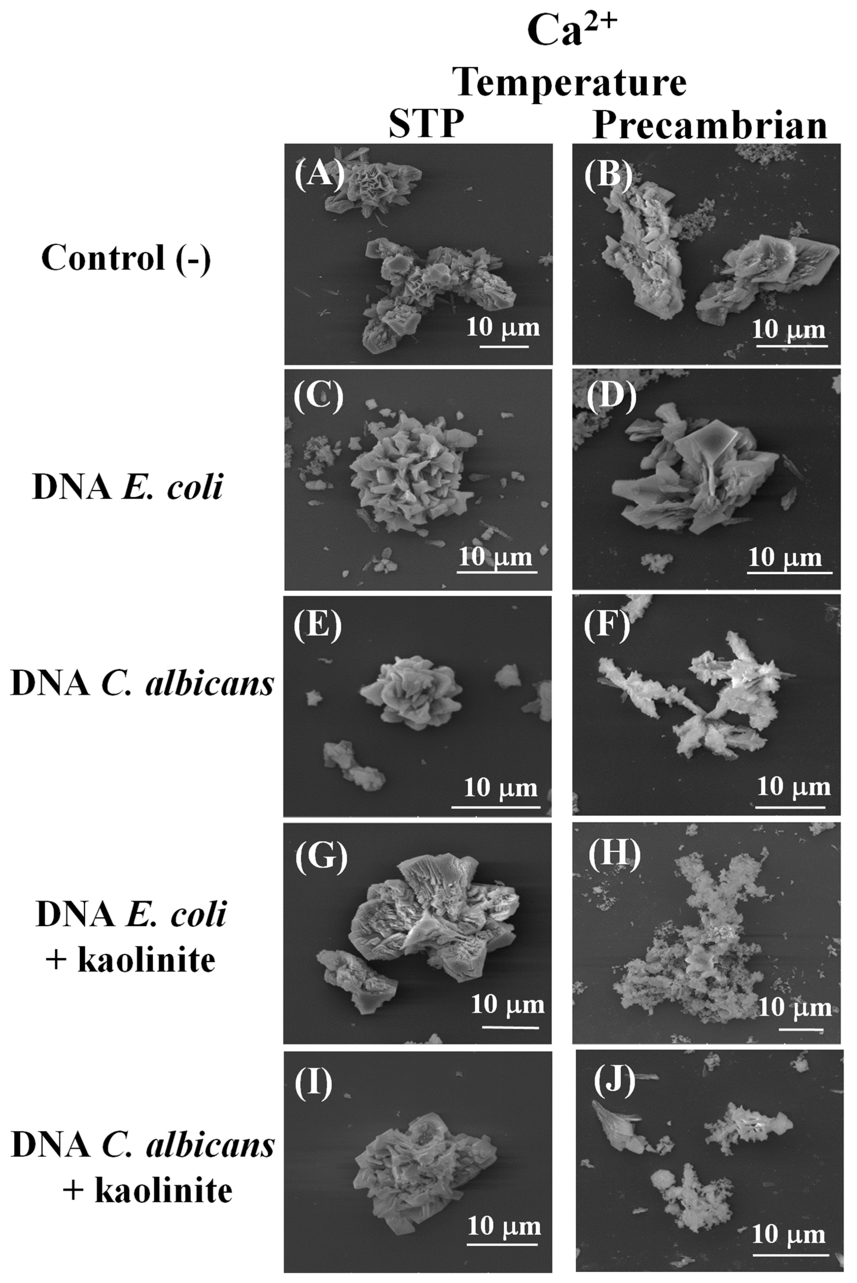
Figure 1. SEM microphotographs of the calcium silica–carbonate biomorphs synthesized in two atmospheric conditions. (A,B) Control, without DNA or kaolinite. (C,D) DNA from E. coli. (E,F) DNA from C. albicans. (G,H) DNA from E. coli with kaolinite. (I,J) DNA from C. albicans with kaolinite. STP: Current temperature and CO2 atmospheric conditions. Precambrian: 5% CO2, 50 °C.
Earth 05 00016 g001
Figure 2. Identification by Raman spectroscopy of the polymorphs obtained in the calcium silica–carbonate biomorphs synthesized in STP and Precambrian conditions. (a,b) Control. (c,d) DNA from E. coli. (e,f) DNA from E. coli and kaolinite. (i) SEM micrograph; (ii) Raman spectra; (iii) Raman map.
Earth 05 00016 g002
Figure 3. Characterization by Raman spectroscopy of the polymorphs synthesized in the calcium silica–carbonate biomorphs, in STP and Precambrian conditions. (a,b) Control. (c,d) DNA from C. albicans. (e,f) DNA from C. albicans, kaolinite.
Earth 05 00016 g003
Figure 4. SEM microphotographs of the barium silica–carbonate biomorphs synthesized in two atmospheric conditions. (A,B) Control, without DNA without kaolinite. (C,D) DNA from E. coli. (E,F) DNA from C. albicans. (G,H) DNA from E. coli with kaolinite. (I,J) DNA from C. albicans with kaolinite. STP: Current atmospheric conditions of temperature and CO2. Precambrian: 5% CO2, 50 °C.
Earth 05 00016 g004
Figure 5. Identification of the crystalline phase of the barium silica–carbonate biomorphs by Raman and FTIR spectroscopy. (a) Control biomorphs, STP. (b) Control biomorphs, Precambrian. (c) E. coli, genomic DNA, STP. (d) E. coli, genomic DNA, Precambrian. (e) E. coli genomic DNA and kaolinite, STP. (f) E. coli genomic DNA and kaolinite, Precambrian. (i) Raman spectra. (ii) SEM micrograph. (iii) Confocal image. (iv) Raman map. (v) FTIR spectra.
Earth 05 00016 g005
Figure 6. Identification of the crystalline phase of the barium silica–carbonate biomorphs by Raman spectroscopy and by FTIR (a) Control biomorphs, STP. (b) Control biomorphs, Precambrian. (c) C. albicans genomic DNA, STP. (d) C. albicans genomic DNA, Precambrian. (e) C. albicans genomic DNA and kaolinite, STP. (f) C. albicans genomic DNA and kaolinite, Precambrian. (i) Raman spectra. (ii) SEM micrograph. (iii) Raman map. (iv) FTIR spectra.
Earth 05 00016 g006
Table 1. Identification through Raman and IR spectroscopy of the polymorphs of calcium silica–carbonate biomorphs.
Type of DNAType of SynthesisRaman (cm−1)IR (cm−1)Composition
Control STP157, 282, 712, 1086, 1750712.1, 871.1, 1399.9Calcite
Precambrian157, 282, 1082792.0, 871.2, 950.1, 1073.3, 1387.2, 2980.7Calcite
E. coliSTP157, 282, 712, 1086575.3, 604.7, 712.2, 750.5, 871.7, 1407.30Calcite
Precambrian157, 283, 713, 1086711.6, 796.6, 871.1, 1076.9, 1394.1, 2980.7, 3420.8Calcite
E. coli
+ kaolinite
STP157, 282, 712, 1085560, 757.3, 873.0, 1407.0Calcite
Precambrian157, 282, 713, 1086544.0, 595.0, 626.0, 680.7, 797.5, 913.4, 939.0, 1008.3, 1031.0, 1386.9, 2980.7, 3482.6, 3619.6, 3690.8Calcite
C. albicansSTP111, 157, 282, 470, 711, 1086, 1480, 2911580, 711, 871.20, 1406.2Calcite, Aragonite
Precambrian114, 155, 281, 712, 1085, 1457, 1760711.5, 792.6, 870.6, 1073.1, 1387.4Calcite, Aragonite
C. albicans
+ kaolinite
STP112, 156, 281, 711, 1085, 1470, 2980712.1, 757.3, 871.3, 1394.1Calcite, Aragonite
Precambrian112, 156, 281, 712, 1085, 1437, 1750599.4, 596.0, 576.9, 626.1, 680.8, 797.4, 913.3, 939.2, 1008.2, 1031.0, 2980.8, 3691.0Calcite, Aragonite
Table 2. Identification through Raman spectroscopy and IR spectroscopy of the barium silica–carbonate biomorphs.
Type of DNAType of Synthesis Raman Spectroscopy (cm−1)FTIR (cm−1)Composition
Control STP93, 138, 154, 221, 690, 1058549.7, 585.1, 604.3, 692.6, 768.5, 855.5, 953.2, 1059.0, 1416.9, 1577.7, 2980Witherite
Precambrian95, 139, 155, 229, 690, 1059692.8, 798.4, 855.9, 1074.0, 1418.8, 1578.9, 1749.5, 2980.8Witherite
E. coliSTP93, 138, 152, 226, 690, 1058, 1412, 1508563.9, 575.4, 692.4, 855.2, 954.7, 1077.3, 1417.2, 1577.4, 2980.8Witherite
Precambrian94, 137, 222, 690, 1058, 1354, 1497, 1503, 2195, 2801, 2938576.4, 692.9, 791.6, 855.5, 945.5, 1069.7, 1420.9Witherite
E. coli + kaoliniteSTP93, 138, 151, 691, 1058, 1440, 2890 575.1, 599.3, 627.6, 692.4, 773.2, 855.5, 910.7, 1059.1, 1417.0Witherite
Precambrian96, 140, 222, 693, 1059, 1060, 1424, 2900626.3, 692.7, 779.5, 855.8, 911.7, 1072.9, 1420.0, 2980.0Witherite
C. albicansSTP93, 138, 155, 221, 689, 1058, 1356, 1503, 2840692.8, 764.6, 856.1, 888.0, 1059.3, 1417.2, 1578.1, 1979.2, 2160.8, 2980.8Witherite
Precambrian98, 141, 225, 692, 1009, 1060, 1470, 2800689.0, 764.2, 892.2, 1059.3, 1402.0, 1700.0, 2980.7Witherite
C. albicans + kaoliniteSTP138, 230, 693, 1059, 1425, 1504595.6, 626.8, 692.4, 752.9, 856.3, 911.1, 1072.9, 1417.3, 2980.7Witherite
Precambrian92, 138, 153, 690, 1059, 1420595.5, 626.4, 687.7, 776.6, 911.2, 1007.6, 1027.9, 2884.1, 2980.7Witherite
Disclaimer/Publisher’s Note: The statements, opinions and data contained in all publications are solely those of the individual author(s) and contributor(s) and not of MDPI and/or the editor(s). MDPI and/or the editor(s) disclaim responsibility for any injury to people or property resulting from any ideas, methods, instructions or products referred to in the content.