
Proceeding Paper

Hydrogel-like Biofilms of Candida tropicalis: Biofouling of Polymeric Prosthetic Materials and Emerging Antifungal Strategies †

by
Bindu Sadanandan
* and
Kavyasree Marabanahalli Yogendraiah
Department of Biotechnology, M S Ramaiah Institute of Technology, Bengaluru 560054, Karnataka, India
*
Author to whom correspondence should be addressed.
Presented at the 1st International Online Conference on Gels, 3–5 December 2025; Available online: https://sciforum.net/event/IOCG2025.
Mater. Proc. 2026, 29(1), 5; https://doi.org/10.3390/materproc2026029005
Published: 31 March 2026
(This article belongs to the Proceedings of The 1st International Online Conference on Gels)

Abstract

The non-albicans Candida species Candida tropicalis is an opportunistic fungal pathogen that forms a robust gel-like biofilm on polymeric prosthetic materials. These biofilms are embedded in an extracellular polymeric substance that retains large amounts of water, resulting in a hydrogel-like matrix that protects fungal cells, increases antifungal resistance, and contributes to the biofouling of these prosthetic materials. Biofouling is the unwanted colonization and accumulation of microbial communities on material surfaces, which alters their function and compromises clinical performance. Clinically, it is significant because it is linked to recurrent urinary tract infections, bloodstream infections, and persistent device-related infections, which often result in therapeutic failure and device malfunction. Polymers such as silicone elastomer, polypropylene, polystyrene, polyurethane, polyethylene, and polyvinyl chloride are widely used in catheters, surgical meshes, implants, and prostheses because of their durability, flexibility, and biocompatibility, yet their surface properties often encourage microbial adhesion and biofilm formation. This review emphasizes that the gel-like biofilm architecture of C. tropicalis underpins its persistence and resistance, while also highlighting promising antifungal strategies being developed to mitigate these infections. Notably, palmitic acid has been shown to disrupt mature biofilms by lowering ergosterol and inducing oxidative stress, whereas C-10 massoia lactone damages the extracellular matrix and suppresses hyphal growth. Drug repurposing approaches, such as combining minocycline with fluconazole, restore susceptibility in resistant isolates and demonstrate synergistic antibiofilm activity. Additionally, biomaterial-based interventions, such as chitosan coatings on silicone surfaces, significantly reduce fungal adhesion and biofilm formation. Together, these findings reflect a translational shift toward integrating natural products, repurposed drugs, and functionalized biomaterials into antifungal development. Understanding biofouling and these emerging strategies is crucial for developing effective control measures against C. tropicalis biofilms and for guiding the design of infection-resistant prosthetic devices.

1. Introduction

Candida tropicalis is a clinically important non-albicans Candida species that has emerged as a major opportunistic fungal pathogen, particularly in hospitalized and immunocompromised individuals. It is frequently implicated in invasive candidiasis, bloodstream infections, and device-associated infections and is often associated with higher mortality rates compared to other Candida species. The clinical significance of C. tropicalis is largely attributed to its strong biofilm-forming ability, high adaptability to diverse environmental conditions, and increasing resistance to commonly used antifungal agents. Unlike planktonic cells, C. tropicalis readily adheres to abiotic surfaces and undergoes a transition to a sessile lifestyle, forming structured biofilms that promote persistence and therapeutic failure [1,2].
A defining characteristic of C. tropicalis biofilms is the production of a dense extracellular polymeric substance (EPS) matrix that retains substantial amounts of water, resulting in a gel-like or hydrogel-like architecture [3,4]. This hydrated matrix provides mechanical stability and creates a protective microenvironment that shields embedded fungal cells from antifungal agents, host immune defences, and environmental stress. The hydrogel-like nature of the biofilm matrix also supports metabolic heterogeneity and facilitates long-term survival on inert surfaces. Consequently, C. tropicalis biofilms exhibit markedly reduced susceptibility to azoles and other antifungal drugs, contributing to recurrent and persistent infections [5].
Globally, invasive candidiasis represents one of the most common hospital-acquired fungal infections, with Candida species ranking among the leading causes of bloodstream infections in intensive care units worldwide. Recent global burden estimates indicate that approximately 1.5 million individuals develop invasive candidiasis or candidemia annually, resulting in nearly one million deaths each year, highlighting its substantial contribution to infection-related mortality. A significant proportion of these infections are device-associated, particularly involving central venous catheters and other implanted medical devices, where biofilm formation plays a critical role in persistence, antifungal resistance, and therapeutic failure. The global burden is unevenly distributed, with Asia accounting for nearly half of reported candidemia cases from countries with available surveillance data. Among non-albicans Candida species, C. tropicalis shows marked geographic predominance in Asia, Latin America, and parts of Africa, where it may constitute 15–40% of candidemia cases, and in several Asian settings, it ranks as a leading cause of bloodstream infections. Biofilm-associated C. tropicalis infections are increasingly reported among hospitalized, immunocompromised, and critically ill patients, contributing to prolonged hospital stays and escalating healthcare costs [6].
In India, a densely populated country with a high prevalence of established risk factors for invasive fungal disease, including prolonged ICU admission, extensive use of indwelling medical devices, broad-spectrum antibiotic exposure, and immunosuppression, the burden of candidemia is particularly pronounced. Data modelling studies and systematic reviews estimate that approximately 188,000 cases of candidemia occur annually based on clinical reporting and ICU surveillance. However, when accounting for substantial under-diagnosis due to the limited sensitivity of blood cultures (≈40%), the true annual incidence of invasive candidiasis in India is projected to reach ~470,000 cases. Notably, C. tropicalis has emerged as the predominant bloodstream isolate in India, accounting for ~42% of candidemia episodes, followed by C. albicans and the emerging multidrug-resistant C. auris, underscoring a regional shift toward non-albicans Candida species with significant clinical and therapeutic implications [7,8].
C. tropicalis biofilms lead to biofouling once they adhere to polymeric prosthetic materials, which is defined as the undesirable colonization and accumulation of microbial communities on material surfaces [9]. Biofouling alters the physicochemical and functional properties of medical devices, compromises their clinical performance, and is strongly associated with device malfunction and failure. Clinically, biofouling by C. tropicalis contributes to catheter-associated urinary tract infections, bloodstream infections, and persistent implant-related infections that are difficult to eradicate without device removal [4,10,11].
Polymeric materials commonly used in medical devices, including polyurethane, silicone, polyvinyl chloride (PVC), polymethyl methacrylate (PMMA), polyethylene, and polymer-coated metals, are highly susceptible to in vivo biofouling [12]. These materials are widely employed in central venous and urinary catheters, endotracheal tubes, prosthetic joints, heart valves, peritoneal dialysis catheters, ventricular shunts, and dental or voice prostheses. Their surface hydrophobicity, flexibility, and tendency to adsorb host proteins facilitate rapid surface conditioning after implantation, creating an ideal niche for fungal attachment and biofilm-driven fouling [13,14,15].
Biofouling of polymeric prosthetic materials by C. tropicalis leads to a spectrum of device-associated infections, most notably catheter-related bloodstream infections (candidemia), catheter-associated urinary tract infections, fungal peritonitis, prosthetic joint infections, and prosthetic valve endocarditis. Among these, catheter-associated infections are the most frequent, as intravascular and urinary devices provide direct access to nutrient-rich host fluids. Biofilm formation on device surfaces enables persistent fungal colonization, continuous shedding of cells into the bloodstream or surrounding tissues, and progression to invasive disease [16].
The mortality associated with device-related candidemia remains high, typically ranging from 20% to over 60% depending on patient comorbidities, timing of diagnosis, and adequacy of source control. Infections involving biofilm-coated prosthetic devices are associated with significantly worse outcomes compared to non-device-associated infections, as biofilms protect fungal cells from host immune responses and antifungal therapy. Prosthetic joint and cardiac device infections, although less common, are associated with particularly high morbidity and risk of relapse [17,18].
Biofouling is particularly problematic when caused by antifungal-resistant or drug-tolerant C. tropicalis strains. Biofilm-associated cells exhibit markedly reduced susceptibility to azoles, echinocandins, and polyenes due to extracellular matrix sequestration of drugs, altered metabolic states, and the presence of persister-like subpopulations. Fluconazole resistance is increasingly reported in C. tropicalis, especially in device-associated infections, further complicating treatment. The combination of intrinsic biofilm tolerance and acquired antifungal resistance often renders pharmacological therapy alone insufficient, making device removal unavoidable and underscoring the urgent need for antifungal–antifouling surface strategies [19].
Conventional antifungal therapies frequently fail to eliminate C. tropicalis biofilms formed on polymeric materials, highlighting the need for alternative control strategies [12]. Recent research has focused on emerging antifungal strategies, including natural bioactive compounds, drug repurposing combinations, and biomaterial-based surface modifications, and integrating antifungal antifouling approaches, which aim to disrupt biofilm architecture, reduce fungal adhesion, or enhance antifungal efficacy. Therefore, this study highlights the hydrogel-like biofilm behaviour of C. tropicalis, its role in the biofouling of polymeric prosthetic materials, and emerging antifungal strategies that offer promising avenues for preventing and controlling device-associated infections.

2. Hydrogel-like Biofilm Architecture of C. tropicalis

The three-dimensional architecture of a C. tropicalis biofilm is organized into a stratified structure comprising three distinct layers, surface, transitional, and basal, each contributing uniquely to the biofilm’s stability, virulence, and drug resistance, as shown in (Figure 1). The surface layer, or top stack, is rich in ECM components such as β-glucans, mannans, proteins, lipids, and extracellular DNA (eDNA). This layer harbours predominantly yeast-form and hyphal cells that are metabolically active due to greater exposure to oxygen and nutrients. It plays a pivotal role as a protective barrier, with eDNA and ECM components trapping antifungal agents and modulating host immune responses, thereby enhancing biofilm resilience [20,21].
Beneath this lies the middle layer, or intermediate stack, which serves as a transitional zone. It contains a mix of yeast and filamentous cells embedded in the ECM, where active morphogenetic switching occurs; yeast cells differentiate into hyphae or pseudohyphae. This layer exhibits moderate metabolic activity and functions as the structural backbone of the biofilm, facilitating communication and nutrient exchange between the superficial and basal layers. C. tropicalis often exhibits more loosely arranged cells with less extensive hyphal penetration, resulting in a comparatively thinner and less cohesive intermediate layer.
The bottom layer, or basal stack, is directly anchored to the surface of medical devices or host tissues and is primarily composed of quiescent yeast-form cells in close contact with the substratum. Though ECM is less abundant here, this layer is critical for biofilm adhesion and long-term persistence. It harbours persister cells, metabolically dormant variants that display remarkable tolerance to antifungal agents. This basal compartment acts as a reservoir for regrowth and is a key contributor to chronic and recurrent infections [22]. Together, these layers create a dynamic and fortified structure that enables C. tropicalis to evade host defences, resist therapy, and persist in clinical settings [23].

3. Biofouling of Prosthetic Polymeric Materials by C. tropicalis

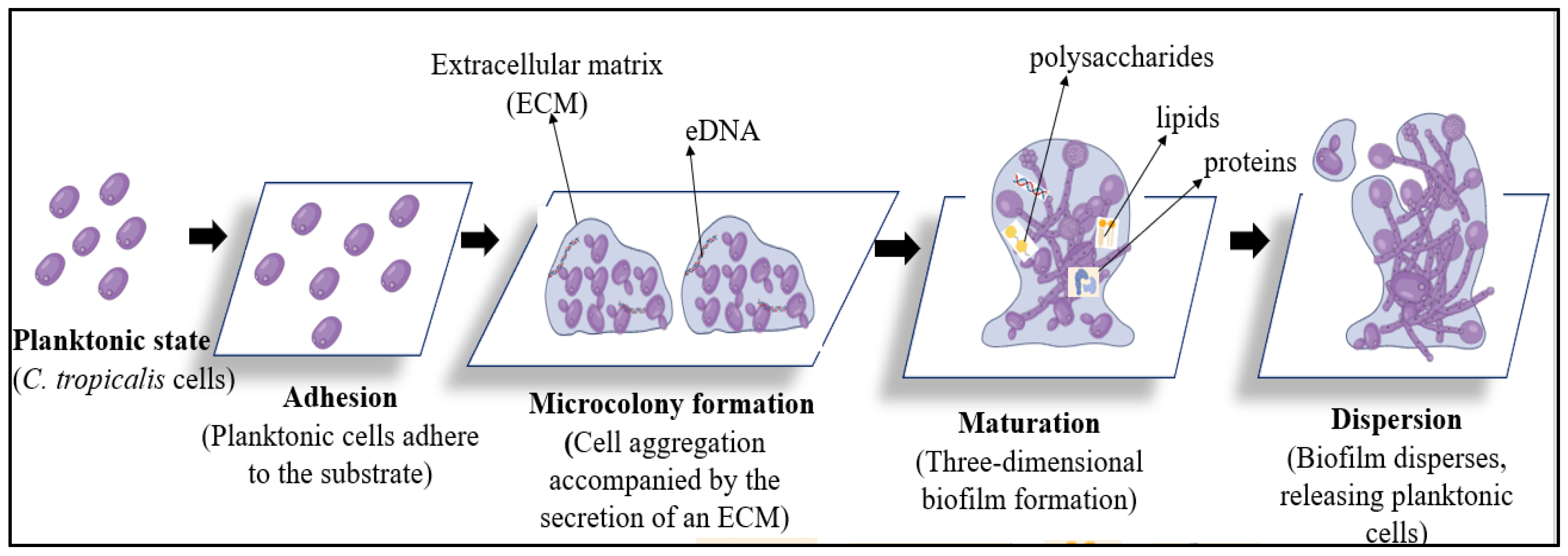
In vivo biofouling of prosthetic polymeric materials by C. tropicalis is a progressive, multistep process driven by surface-associated biofilm development under host conditions. Following implantation, polymeric devices become rapidly conditioned by host-derived biomolecules, creating a favourable interface for fungal attachment and colonization. Biofouling then proceeds through sequential stages involving initial adhesion to conditioned polymer surfaces, establishment of microcolonies and extracellular matrix production, maturation into stable biofilm-driven fouling layers, and eventual dispersion of fungal cells that promote secondary surface colonization and persistent infection. Each stage is influenced by interactions between the fungal cells, the physicochemical properties of the polymeric material, and the surrounding in vivo environment, as shown in (Figure 2).

3.1. Initial Surface Conditioning and Fungal Attachment

In vivo biofouling of prosthetic polymeric materials begins immediately after implantation, when device surfaces such as silicone elastomer, polypropylene, polyurethane, polyvinylchloride or polymethyl methacrylate become rapidly coated with host-derived biomolecules, including plasma proteins, glycoproteins, and cellular debris. This conditioning layer transforms the inert polymer surface into a biologically active interface that promotes microbial colonization. C. tropicalis planktonic yeast cells subsequently adhere to these conditioned polymeric surfaces through physicochemical interactions and surface-associated adhesins, marking the onset of biological fouling. Surface properties of the polymer, including hydrophobicity, roughness, and chemical composition, strongly influence the extent of fungal attachment under host-mimicking conditions [24,25].

3.2. Establishment of Irreversible Biofouling and Microcolony Development

Following initial attachment, C. tropicalis cells proliferate on the polymer surface and form localized microcolonies, leading to irreversible biofouling of the prosthetic material. During this phase, fungal cells secrete EPSs that anchor them firmly to the device surface and promote intercellular cohesion. The accumulation of extracellular matrix components stabilizes early fungal colonization and facilitates adaptation to host-derived fluids such as urine, blood, or vaginal secretions. This stage represents the transition from transient surface contamination to persistent device-associated biofouling [1].

3.3. Mature-Biofilm-Driven Fouling and Device Dysfunction

As biofouling progresses, C. tropicalis develops mature, three-dimensional biofilm structures on polymeric prosthetic materials. These biofilms are embedded within a dense extracellular matrix that confers mechanical stability and protects fungal cells from host immune defences and antifungal agents. In vivo, mature-biofilm-associated fouling contributes to functional impairment of medical devices, including surface degradation, lumen obstruction, and sustained inflammatory responses in surrounding tissues. The establishment of nutrient and oxygen gradients within the biofilm further enhances fungal persistence and antifungal tolerance on the prosthetic surface [26].

3.4. Biofilm Dispersion and Secondary Biofouling

Advanced biofouling culminates in biofilm dispersion, during which fungal cells detach from the prosthetic surface and disseminate within the host or device environment. Dispersed C. tropicalis cells retain enhanced adhesive capacity and can rapidly colonize adjacent polymeric surfaces or host tissues, leading to secondary biofouling and recurrent device-associated infections. This dissemination phase explains the persistence of candidiasis despite antifungal therapy and underscores the difficulty of eradicating biofouled prosthetic materials in clinical settings [27].
Several experimental studies have investigated the sequential stages of C. tropicalis biofilm development, including adhesion, microcolony formation, extracellular matrix production, maturation, and dispersion. Representative primary studies describing these processes are explained in (Table 1), whereas (Table 2) represents clinical biofouling of polymeric prosthetic materials by C. tropicalis: devices, infections, and outcomes.

4. Emerging Antifungal and Antifouling Strategies

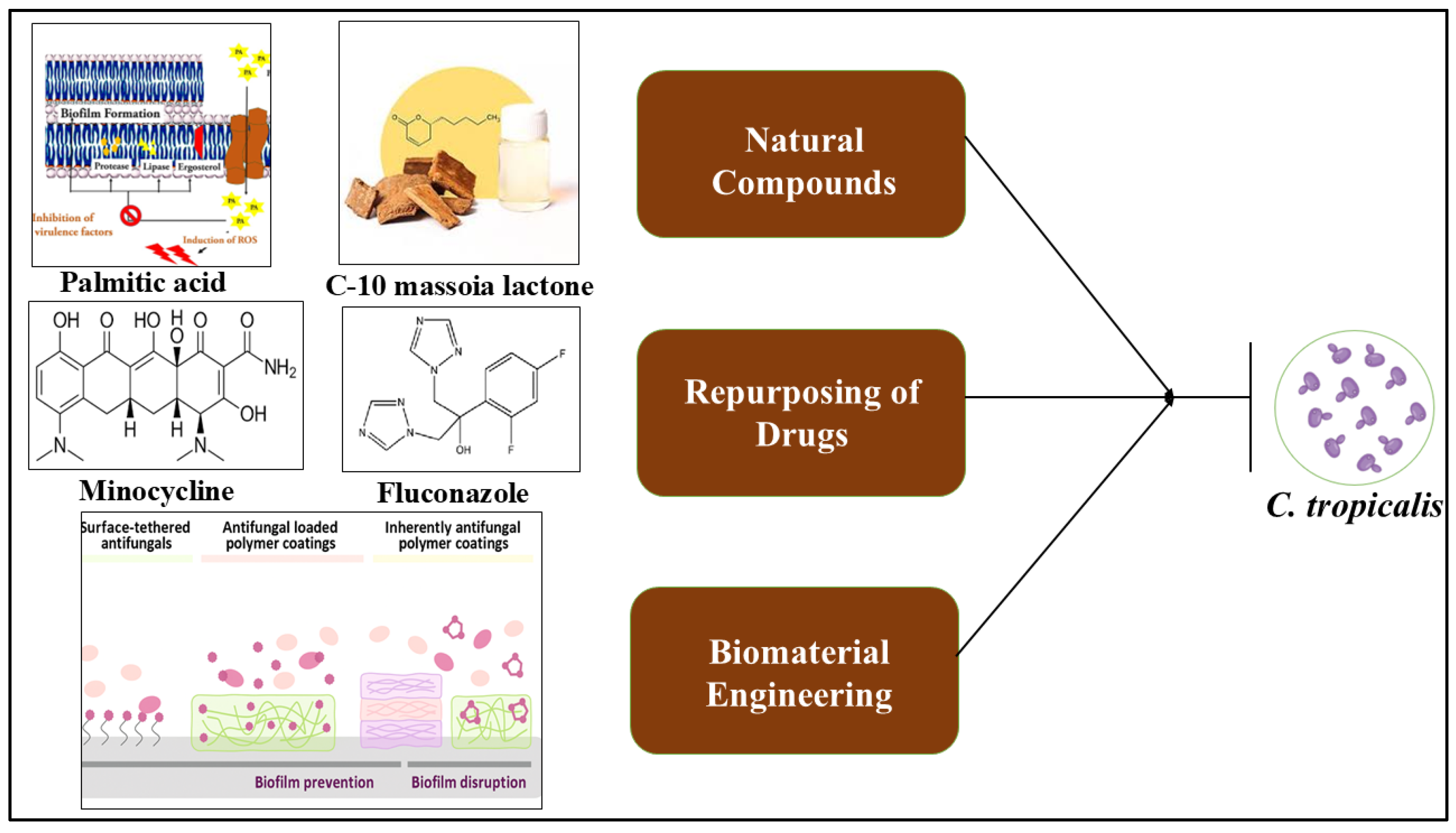
The intrinsic resistance of C. tropicalis biofilms and their ability to cause persistent biofouling on polymeric prosthetic materials have prompted the development of alternative and adjunctive antifungal strategies beyond conventional monotherapy. Recent research has increasingly focused on biofilm-targeted antifungal approaches, drug repurposing, and antifouling biomaterial modifications, aiming to disrupt biofilm architecture, inhibit fungal adhesion, and improve therapeutic outcomes as shown in (Figure 3 and Table 3).

4.1. Natural Bioactive Compounds Targeting Biofilms

Natural bioactive compounds have emerged as important alternative and adjunctive antifungal strategies for controlling C. tropicalis biofilms, particularly in the context of persistent biofouling on polymeric prosthetic materials. The intrinsic resistance of C. tropicalis biofilms is largely attributed to their complex, hydrogel-like extracellular matrix; their metabolic heterogeneity; and the reduced susceptibility of sessile cells to conventional antifungal agents. In response, recent research has increasingly focused on natural compounds because of their multi-target mechanisms, ability to interfere with multiple stages of biofilm development, and comparatively lower risk of resistance emergence.
A wide range of natural bioactive agents, including fatty acids, plant-derived lactones, spice-derived phytochemicals, phenolic and terpenoid compounds, and antimicrobial peptides, have demonstrated activity against C. tropicalis biofilms. Phenolic compounds (e.g., eugenol, cinnamaldehyde, and thymol), terpenoids (e.g., farnesol, geraniol, and α-terpineol), and antimicrobial peptides (e.g., histatin-5, LL-37, and defensins) exhibit significant antibiofilm activity against C. tropicalis by targeting membrane integrity, morphogenesis, oxidative balance, and extracellular matrix stability across different stages of biofilm development [41,42]. All these compounds are effective against both immature and mature biofilms, although their modes of action differ depending on the developmental stage. During early biofilm formation, natural compounds primarily inhibit fungal adhesion to abiotic surfaces, reduce cell surface hydrophobicity, interfere with pseudohyphal development, and disrupt quorum-regulated proliferation, thereby limiting microcolony formation and initial ECM deposition. Such effects are particularly valuable for antifouling applications, where preventing biofilm establishment on polymeric materials is critical.
In contrast, mature C. tropicalis biofilms pose a greater therapeutic challenge due to their dense ECM and reduced antifungal penetration. Several natural compounds have been shown to overcome these barriers by destabilizing the hydrogel-like matrix, inducing oxidative stress, and compromising fungal cell membrane integrity. For instance, palmitic acid disrupts mature C. tropicalis biofilms by reducing ergosterol levels, leading to membrane destabilization and loss of cellular homeostasis, ultimately decreasing biofilm viability [43,44]. Similarly, C-10 massoia lactone targets both the cellular and extracellular components of the biofilm by damaging the EPS matrix and suppressing filamentous development, resulting in weakened biofilm architecture and reduced adhesion to abiotic surfaces [42,45].
In addition to these well-defined compounds, spice-derived extracts and phytochemicals, such as those obtained from garlic, clove, cinnamon, turmeric, and other culinary plants, have been reported to exert antibiofilm effects through combined actions on membrane integrity, oxidative balance, and ECM synthesis [12]. These bioactives often act synergistically on multiple cellular targets, impairing both biofilm formation and maintenance. Their broad-spectrum activity and compatibility with surface functionalization strategies further support their potential incorporation into antifouling coatings and biomaterial modifications [2].
Overall, the ability of natural bioactive compounds to target adhesion, morphogenesis, matrix integrity, and stress tolerance across different stages of C. tropicalis biofilm development highlights their promise as biofilm-directed antifungal agents. Their integration into combination therapies and antifouling biomaterial designs represents a translational approach for mitigating device-associated infections caused by hydrogel-like C. tropicalis biofilms.

4.2. Drug Repurposing and Combination Therapies

Drug repurposing and combination-based antifungal strategies have emerged as promising approaches for overcoming the intrinsic resistance of C. tropicalis biofilms and their persistent biofouling of polymeric prosthetic materials. Drug repurposing involves the application of already approved non-antifungal drugs for antifungal purposes, thereby offering a rapid, cost-effective, and clinically translatable alternative to de novo antifungal development. In this context, the combination of minocycline and fluconazole represents a classic drug repurposing strategy, as minocycline is a tetracycline antibiotic originally developed for bacterial infections but has been repurposed here to enhance antifungal efficacy [46]. This combination has been shown to restore fluconazole susceptibility in fluconazole-resistant C. tropicalis isolates and significantly reduce biofilm biomass. Mechanistically, minocycline interferes with biofilm-associated defence mechanisms, including the suppression of efflux pump activity, disruption of oxidative stress response pathways, and alteration of metabolic homeostasis, thereby facilitating improved penetration and activity of fluconazole within the biofilm matrix [47,48].
Beyond minocycline, several other non-antifungal drugs have demonstrated synergistic antibiofilm activity against C. tropicalis when used in combination with conventional antifungals. Antibiotics such as doxycycline and rifampicin enhance antifungal penetration and reduce biofilm metabolic activity, while statins such as simvastatin and atorvastatin potentiate azole activity by interfering with ergosterol biosynthesis through the mevalonate pathway [49,50,51,52]. Calcineurin inhibitors, including cyclosporine A and tacrolimus, sensitize C. tropicalis biofilm cells to azoles by inhibiting stress-adaptation pathways critical for biofilm survival. In addition, nonsteroidal anti-inflammatory drugs such as aspirin and ibuprofen have been shown to impair adhesion, morphogenesis, and early biofilm development, whereas repurposed antidepressant and antiparasitic agents such as sertraline and chloroquine disrupt membrane integrity, mitochondrial function, and intracellular pH homeostasis. Selective serotonin reuptake inhibitors (SSRIs) have also shown synergistic interactions with fluconazole in biofilm models of multiple Candida species, indicating broader repurposing opportunities [53]. Collectively, these combination therapies act by simultaneously targeting fungal growth, biofilm architecture, and stress-response mechanisms, thereby overcoming biofilm-mediated drug tolerance. Such drug repurposing approaches are particularly attractive for managing device-associated C. tropicalis infections, where mature biofilms often limit the efficacy of standard antifungal monotherapy.

4.3. Biomaterial-Based Antifouling Strategies

Preventing the initial adhesion of C. tropicalis to biomaterial surfaces represents a pivotal strategy for controlling biofouling and reducing the incidence of device-associated infections. Biomaterial-based interventions focus on surface modification of prosthetic materials to inhibit microbial attachment and consequent biofilm formation. While silicone elastomers have been a primary substrate for such studies, other polymeric materials, including polyurethane, polyethylene, polypropylene, PVC, PMMA and polystyrene, also support fungal adhesion and biofilm development due to their hydrophobic surface properties and surface energy profiles, making them relevant targets for antifouling engineering [4,30].
Naturally derived chitosan and its derivatives have emerged as promising antifouling coatings across diverse polymer substrates. Chitosan-based coatings on catheter-related surfaces significantly reduce C. tropicalis adhesion and biofilm development by altering surface charge, increasing hydrophilicity, and exerting intrinsic antimicrobial effects. Carboxymethyl chitosan, a water-soluble chitosan derivative, has demonstrated inhibitory effects on both initial adhesion and later stages of biofilm formation for monomicrobial (including C. tropicalis) and polymicrobial biofilms on silicone and other surfaces, highlighting its potential as a broad-spectrum antibiofilm coating [54,55].
In addition to chitosan, antifungal nanocoatings and polymer composites have shown efficacy in reducing fungal colonization. Lignin-based sustainable nanocoatings doped with antimicrobial components (e.g., silver nanoparticles and reactive photoactive groups) significantly decreased C. tropicalis growth on coated surfaces, with enhanced activity under light-activated conditions owing to reactive oxygen species generation and associated cytotoxic effects [56]. Moreover, covalent grafting of antifungal agents such as echinocandin drugs (e.g., caspofungin) onto biomaterial surfaces via plasma polymer interlayers has been demonstrated to suppress fungal attachment and biofilm initiation while maintaining compatibility with mammalian cells, indicating long-term antifouling potential across varied polymer substrates [57].
Beyond specific polymer coatings, surface functionalization techniques that enhance hydrophilicity or introduce antifouling chemistries have been explored. Hydrophilic polymers such as polyethylene glycol and zwitterionic coatings are known to create highly hydrated surface layers that resist protein adsorption and fungal adhesion, thereby reducing the likelihood of biofilm formation on prosthetic materials. While much of the recent composite research reports on bacterial systems, principles of zwitterionic hydration and antifouling deterrence directly apply to fungal biofilms and inform the design of next-generation prosthetic surface chemistries with broad-spectrum resistance to colonization [58].
Additionally, hydrogel-based barriers that can be grafted or applied as surface layers on medical devices exhibit antifouling behaviour by presenting hydrated, low-adhesion interfaces that resist fungal settlement. Bioinspired hydrophilic polymer networks (incorporating lubricious moieties such as phosphorylcholine) have demonstrated antifouling proficiency in reducing microbial attachment across hydrophobic and hydrophilic counter surfaces, suggesting potential applicability for fungal biofilm control on diverse biomedical materials [2].
Collectively, these biomaterial strategies, ranging from chitosan and antimicrobial nanocoatings to advanced surface functionalization and hydrogel-based antifouling layers, provide a multifaceted approach to controlling C. tropicalis adhesion and biofilm formation on polymeric prosthetic surfaces. By preventing initial attachment and disrupting early biofilm establishment, these engineered surfaces can significantly reduce reliance on systemic antifungal therapy and prolong the functional lifespan of implanted medical devices.

4.4. Integrated Antifungal–Antifouling Approaches

Emerging evidence indicates that effective control of C. tropicalis biofilms requires integrated antifungal–antifouling strategies that simultaneously target fungal cells and the material–biofilm interface. Biofilm persistence on prosthetic materials is driven not only by antifungal tolerance of sessile cells but also by favourable surface properties that promote adhesion and extracellular matrix accumulation. As a result, combined approaches that incorporate antifungal activity within antifouling surface designs have demonstrated superior performance compared to either strategy alone.
One representative example is the integration of chitosan-based antifouling coatings with antifungal agents on polymeric catheter materials. Chitosan coatings reduce initial C. tropicalis adhesion by altering surface charge and hydrophilicity, while embedded or surface-bound antifungal agents such as fluconazole or amphotericin B actively suppress fungal growth within early biofilms. This dual-action system has been shown to significantly reduce both initial colonization and subsequent biofilm maturation, highlighting the advantage of combining surface resistance with localized antifungal delivery.
Another effective integrated strategy involves nanoparticle-functionalized polymer surfaces, such as polyurethane or silicone modified with silver, zinc oxide, or bioactive plant-derived nanoparticles. These materials exhibit antifouling behaviour by disrupting fungal attachment and extracellular matrix formation while simultaneously exerting fungicidal or fungistatic effects through oxidative stress induction and membrane damage. Such systems are particularly effective against hydrogel-like C. tropicalis biofilms, where matrix penetration is otherwise limited [59].
Hydrogel-based surface coatings provide an additional example of integration, where hydrated, low-adhesion polymer networks act as antifouling barriers while serving as reservoirs for controlled antifungal release. Hydrogels incorporating natural antifungal compounds (e.g., fatty acids, phenolics, or lactones) or repurposed drugs enable sustained local drug exposure at the material surface, reducing fungal adhesion, suppressing biofilm maturation, and minimizing systemic toxicity. These approaches are especially relevant for long-term indwelling devices, such as urinary catheters and implants, where prolonged antifungal protection is required.
More recently, zwitterionic polymer coatings combined with antifungal functionalization have been proposed as substrate-independent antifouling platforms. Zwitterionic surfaces resist protein adsorption and fungal attachment through strong hydration layers, while the addition of antifungal moieties enhances the killing of any adherent C. tropicalis cells. This integrated design addresses both early-stage adhesion and later-stage biofilm resilience, offering a robust solution for preventing biofouling across diverse prosthetic materials [60].
Table 3. Emerging strategies to mitigate C. tropicalis biofilm formation and biofouling on polymeric prosthetic materials.
Strategy
Category
Representative AgentsMechanismsBiofilm Development Stage TargetedKey SignificanceReferences
Natural Bioactive CompoundsPhenolics (eugenol, cinnamaldehyde, thymol); terpenoids (farnesol, geraniol, α-terpineol); fatty acids (palmitic acid); lactones (C-10 massoia lactone); antimicrobial peptides (histatin-5, LL-37, defensins); spice-derived extracts (garlic, clove, cinnamon, turmeric)Disrupt membranes and ergosterol; inhibit adhesion, morphogenesis, and quorum sensing; destabilize ECM; induce oxidative stressEarly and mature biofilmMulti-target activity; low resistance risk; suitable for antifouling coatings and combination therapies[41,61,62,63]
Drug Repurposing and CombinationsMinocycline + fluconazole; doxycycline, rifampicin; statins (simvastatin, atorvastatin); calcineurin inhibitors (cyclosporine A, tacrolimus); NSAIDs (aspirin, ibuprofen); SSRIs (sertraline); antiparasitics (chloroquine).Suppress efflux pumps and stress responses; enhance azole penetration; interfere with ergosterol and metabolismPredominantly mature biofilmRapid, cost-effective strategy; restores azole susceptibility; effective for resistant biofilms[64,65,66]
Biomaterial-Based AntifoulingChitosan and carboxymethyl chitosan coatings; antimicrobial nanocoatings (silver-, lignin-, and photoactive-based); PEG and zwitterionic polymers; plasma-polymerized antifungal grafts; hydrogel-based surface barriersReduce fungal adhesion via surface charge and hydration; inhibit ECM deposition and early biofilm formationInitial adhesion and early biofilm formationPrevents biofilm establishment; reduces need for systemic antifungals; prolongs device lifespan[54,67,68]
Integrated Antifungal–AntifoulingChitosan + fluconazole/amphotericin B coatings; nanoparticle-functionalized polymers with antifungal activity; drug-loaded hydrogels; zwitterionic–antifungal hybrid surfaces Combined antifouling surface resistance and localized antifungal activityAll stages (from adhesion to mature biofilm formation)Most effective strategy; limits recurrence; ideal for long-term prosthetic devices[59,60,69]
Overall, these integrated antifungal–antifouling strategies demonstrate that combining surface-engineered resistance to adhesion with localized antifungal activity provides a more comprehensive and durable approach to controlling C. tropicalis biofilms. Such systems reduce dependence on systemic antifungal therapy, limit biofilm-mediated resistance, and significantly improve the functional longevity of polymeric prosthetic devices. Continued interdisciplinary efforts spanning microbiology, material science, and pharmaceutical engineering are essential to translate these integrated strategies into clinically viable infection-resistant medical devices.

5. Conclusions

C. tropicalis poses a significant challenge in clinical settings due to its ability to form stable, hydrogel-like biofilms on polymeric prosthetic materials. The biofilm developmental process, supported by a complex extracellular matrix, promotes persistent adhesion, enhanced stress tolerance, and marked resistance to antifungal therapy, leading to biofouling and recurrent device-associated infections. Biofouling of prosthetic materials initiates with fungal attachment to polymer surfaces such as silicone, polyurethane, PVC and PMMA followed by microcolony formation and extracellular matrix deposition. Progressive biofouling alters surface properties, impairs device functionality, promotes long-term microbial persistence, and facilitates continuous fungal dissemination, often necessitating device removal or replacement. Recent advances demonstrate that effective control of C. tropicalis biofilms requires strategies beyond conventional antifungal monotherapy. Natural bioactive compounds, drug repurposing and combination therapies, and biomaterial-based antifouling modifications have shown strong potential by targeting biofilm formation, matrix integrity, and fungal adhesion. Notably, integrated antifungal–antifouling approaches that combine surface engineering with localized antifungal activity offer a promising route to prevent biofilm establishment and improve the longevity and safety of polymeric medical devices. Overall, translating these multidisciplinary strategies into clinically applicable designs will be critical for developing infection-resistant prosthetic materials and improving outcomes in C. tropicalis biofilm-associated infections.

Author Contributions

B.S.: conceptualization, validation, visualization, supervision, review and editing; K.M.Y.: writing—original draft, data curation. All authors have read and agreed to the published version of the manuscript.

Funding

This research received no external funding.

Institutional Review Board Statement

Not applicable.

Informed Consent Statement

Not applicable.

Data Availability Statement

The data that support the findings of this study are available from the corresponding author upon reasonable request.

Acknowledgments

Kavyasree Marabanahalli Yogendraiah thanks the Management of M S Ramaiah Institute of Technology for the Ramaiah Doctoral Fellowship.

Conflicts of Interest

The authors declare no conflicts of interest.

Abbreviations

EPSExtracellular polymeric substance
ECMExtracellular matrix
PVCPolyvinyl chloride
PMMAPolymethyl methacrylate
eDNAExtracellular DNA
CVCsCentral venous catheters
CAUTICatheter-associated urinary tract infection
PJIProsthetic joint infection
PDPeritoneal dialysis
SSRIsSerotonin reuptake inhibitors

References

  1. Xiong, H.; Zhao, R.; Han, S.; Liu, Z.; Zhang, X.; Jia, Z.; Wang, X. Research Progress on the Drug Resistance Mechanisms of Candida tropicalis and Future Solutions. Front. Microbiol. 2025, 16, 1594226. [Google Scholar] [CrossRef] [PubMed]
  2. Sadanandan, B.; Vijayalakshmi, V.; Shetty, K.; Rathish, A.; Shivkumar, H.; Gundreddy, M.; Narendra, N.K.; Devaiah, N.M. In Situ Aqueous Spice Extract-Based Antifungal Lock Strategy for Salvage of Foley’s Catheter Biofouled with Candida albicans Biofilm Gel. Gels 2025, 11, 23. [Google Scholar] [CrossRef]
  3. Sadanandan, B.; Yogendraiah, K.M. Enterococcus faecalis Biofilm: A Clinical and Environmental Hazard. Med. Sci. Forum 2025, 35, 5. [Google Scholar]
  4. Yogendraiah, K.M.; Sadanandan, B.; Kyathsandra Natraj, L.; Vijayalakshmi, V.; Shetty, K. Optimization of Candida tropicalis Growth Conditions on Silicone Elastomer Material by Response Surface Methodology. Front. Bioeng. Biotechnol. 2025, 13, 1572694. [Google Scholar] [CrossRef]
  5. Keighley, C.; Kim, H.Y.; Kidd, S.; Chen, S.C.-A.; Alastruey, A.; Dao, A.; Bongomin, F.; Chiller, T.; Wahyuningsih, R.; Forastiero, A.; et al. Candida tropicalis—A Systematic Review to Inform the World Health Organization of a Fungal Priority Pathogens List. Med. Mycol. 2024, 62, myae040. [Google Scholar] [CrossRef]
  6. Amann, V.; Kissmann, A.K.; Firacative, C.; Rosenau, F. Biofilm-Associated Candidiasis: Pathogenesis, Prevalence, Challenges and Therapeutic Options. Pharmaceuticals 2025, 18, 460. [Google Scholar] [CrossRef]
  7. Ray, A.; Aayilliath K, A.; Banerjee, S.; Chakrabarti, A.; Denning, D.W. Burden of Serious Fungal Infections in India. Open Forum Infect. Dis. 2022, 9, ofac603. [Google Scholar] [CrossRef]
  8. Bhadade, A.A.; Singh, G.; Karuna, T.; Panthi, M.; Beniwal, A.; Maurya, A.K.; Upadhyay, N.; Reddy, V.R.; Devgade, A.; Khadanga, S. Epidemiology and Antifungal Susceptibility of Candida Bloodstream Isolates from a Tertiary Care Center in Central India. Cureus 2025, 17, e97120. [Google Scholar] [CrossRef]
  9. Lima, R.; Ribeiro, F.C.; Colombo, A.L.; de Almeida, J.N., Jr. The Emerging Threat of Antifungal-Resistant Candida tropicalis in Humans, Animals, and Environment. Front. Fungal Biol. 2022, 3, 957021. [Google Scholar] [CrossRef] [PubMed]
  10. Fan, X.; Tsui, C.K.; Chen, X.; Wang, P.; Liu, Z.J.; Yang, C.X. High Prevalence of Fluconazole-Resistant Candida tropicalis among Candiduria Samples in China: An Ignored Matter of Concern. Front. Microbiol. 2023, 14, 1125241. [Google Scholar] [CrossRef] [PubMed]
  11. Ahmed, N.; Mahmood, M.S.; Ullah, M.A.; Araf, Y.; Rahaman, T.I.; Moin, A.T.; Hosen, M.J. COVID-19-Associated Candidiasis: Possible Patho-Mechanism, Predisposing Factors, and Prevention Strategies. Curr. Microbiol. 2022, 79, 127. [Google Scholar] [CrossRef]
  12. Sadanandan, B.; Vijayalakshmi, V.; Ashrit, P.; Babu, U.V.; Sharath Kumar, L.M.; Sampath, V.; Awaknavar, R. Aqueous Spice Extracts as Alternative Antimycotics to Control Highly Drug-Resistant Extensive Biofilm-Forming Clinical Isolates of Candida albicans. PLoS ONE 2023, 18, e0281035. [Google Scholar] [CrossRef]
  13. Sadanandan, B.; Vaniyamparambath, V.; Lokesh, K.N.; Shetty, K.; Joglekar, A.P.; Ashrit, P.; Hemanth, B. Candida albicans Biofilm Formation and Growth Optimization for Functional Studies Using Response Surface Methodology. J. Appl. Microbiol. 2022, 132, 3277–3292. [Google Scholar] [CrossRef] [PubMed]
  14. Ashrit, P.; Sadanandan, B.; Kyathsandra Natraj, L.; Shetty, K.; Vaniyamparambath, V.; Raghu, A.V. A Microplate-Based Response Surface Methodology Model for Growth Optimization and Biofilm Formation on Polystyrene Polymeric Material in a Candida albicans and Escherichia coli Co-Culture. Polym. Adv. Technol. 2022, 33, 2872–2885. [Google Scholar] [CrossRef]
  15. Atiencia-Carrera, M.B.; Cabezas-Mera, F.S.; Vizuete, K.; Debut, A.; Tejera, E.; Machado, A. Evaluation of the Biofilm Life Cycle between Candida albicans and Candida tropicalis. Front. Cell. Infect. Microbiol. 2022, 12, 953168. [Google Scholar] [CrossRef] [PubMed]
  16. Wijaya, M.; Halleyantoro, R.; Kalumpiu, J.F. Biofilm: The Invisible Culprit in Catheter-Induced Candidemia. AIMS Microbiol. 2023, 9, 467–485. [Google Scholar] [CrossRef] [PubMed]
  17. Yadav, S.K.; Himanshu, D.; Gupta, P.; Nischal, A.; Atam, V.; Ali, W.; Yadav, A.; Sonkar, V.; Kumar, V. Multidrug-Resistant Candida in Bloodstream Infections: A Growing Concern in Indian Healthcare. Cureus 2025, 17, e85420. [Google Scholar] [CrossRef]
  18. Chesdachai, S.; Baddour, L.M.; Sohail, M.R.; Palraj, B.R.; Madhavan, M.; Tabaja, H.; Fida, M.; Challener, D.W.; DeSimone, D.C. Candidemia in Patients with Cardiovascular Implantable Electronic Devices: Uncertainty in Management Based on Current International Guidelines. Open Forum Infect. Dis. 2023, 10, ofad318. [Google Scholar] [CrossRef]
  19. Nett, J.E.; Andes, D.R. Contributions of the Biofilm Matrix to Candida Pathogenesis. J. Fungi 2020, 6, 21. [Google Scholar] [CrossRef]
  20. Roy, S.; Gow, N.A. The Role of the Candida Biofilm Matrix in Drug and Immune Protection. Cell Surf. 2023, 10, 100111. [Google Scholar] [CrossRef]
  21. Elibe, M.I.; Innocent, N.E. Biofilm Formation in Candida Species. In Recent Advances in Human Fungal Diseases: Progress and Prospects; Springer Nature: Singapore, 2024; pp. 239–259. [Google Scholar]
  22. Chakrabarti, A.; Patel, A.K.; Soman, R.; Todi, S. Overcoming Clinical Challenges in the Management of Invasive Fungal Infections in Low- and Middle-Income Countries (LMIC). Expert Rev. Anti-Infect. Ther. 2023, 21, 1057–1070. [Google Scholar] [CrossRef] [PubMed]
  23. Dos Santos, M.M.; Ishida, K. We Need to Talk about Candida tropicalis: Virulence Factors and Survival Mechanisms. Med. Mycol. 2023, 61, myad075. [Google Scholar] [CrossRef]
  24. Malinovská, Z.; Čonková, E.; Váczi, P. Biofilm Formation in Medically Important Candida Species. J. Fungi 2023, 9, 955. [Google Scholar] [CrossRef]
  25. de Souza, C.M.; Paulo, E.A.; de Souza, N.A.A.; Dos Santos, M.M.; Mantovani, M.S.; Furlaneto-Maia, L.; Furlaneto, M.C. Enhanced Biofilm Formation by Candida tropicalis Morphotypes under Host-Mimicking Conditions: Insights into Cell Wall Modifications and Gene Expression. Microb. Pathog. 2025, 207, 107807. [Google Scholar] [CrossRef]
  26. Sadanandan, B.; Yogendraiah, K.M. Advances in Pharmaceutical Processing and Particle Engineering of Garlic Extract-Based Formulations for Antifungal Therapy Against Candida tropicalis. Eng. Proc. 2026, 117, 21. [Google Scholar]
  27. Joshi, P.; Bhattacharjee, R.; Sahu, M.; Gajjar, D. Insights into Urinary Catheter Colonisation and Polymicrobial Biofilms of Candida-Bacteria under Flow Condition. Sci. Rep. 2025, 15, 15375. [Google Scholar] [CrossRef]
  28. Tseng, Y.K.; Chen, Y.C.; Hou, C.J.; Deng, F.S.; Liang, S.H.; Hoo, S.Y.; Hsu, C.C.; Ke, C.L.; Lin, C.H. Evaluation of Biofilm Formation in Candida tropicalis Using a Silicone-Based Platform with Synthetic Urine Medium. Microorganisms 2020, 8, 660. [Google Scholar] [CrossRef]
  29. Negri, M.; Silva, S.; Capoci, I.R.G.; Azeredo, J.; Henriques, M. Candida tropicalis Biofilms: Biomass, Metabolic Activity and Secreted Aspartyl Proteinase Production. Mycopathologia 2016, 181, 217–224. [Google Scholar] [CrossRef]
  30. de Souza, C.M.; Dos Santos, M.M.; Furlaneto-Maia, L.; Furlaneto, M.C. Adhesion and Biofilm Formation by the Opportunistic Pathogen Candida tropicalis: What Do We Know? Can. J. Microbiol. 2023, 69, 207–218. [Google Scholar] [CrossRef] [PubMed]
  31. Yogendraiah, K.M.; Sadanandan, B.; Natraj, L.K.; Vijayalakshmi, V.; Shetty, K. Biofilm Formation Directly Correlates with Cell Viability in Candida tropicalis on Polypropylene. Appl. Microbiol. Biotechnol. 2026, 110, 90. [Google Scholar] [CrossRef] [PubMed]
  32. Silva, S.; Henriques, M.; Martins, A.; Oliveira, R.; Williams, D.; Azeredo, J. Biofilms of Non-Candida albicans Candida Species: Quantification, Structure and Matrix Composition. Sabouraudia 2009, 47, 681–689. [Google Scholar] [CrossRef]
  33. Mitchell, K.F.; Zarnowski, R.; Andes, D.R. Fungal Super Glue: The Biofilm Matrix and Its Composition, Assembly, and Functions. PLoS Pathog. 2016, 12, e1005828. [Google Scholar] [CrossRef]
  34. Silva, S.; Rodrigues, C.F.; Araújo, D.; Rodrigues, M.E.; Henriques, M. Candida Species Biofilms’ Antifungal Resistance. J. Fungi 2018, 3, 8. [Google Scholar] [CrossRef]
  35. Cavalheiro, M.; Teixeira, M.C. Candida Biofilms: Threats, Challenges, and Promising Strategies. Front. Med. 2018, 5, 28. [Google Scholar] [CrossRef]
  36. Estivill, D.; Arias, A.; Torres-Lana, A.; Carrillo-Muñoz, A.J.; Arévalo, M.P. Biofilm Formation by Five Species of Candida on Three Clinical Materials. J. Microbiol. Methods 2011, 86, 238–242. [Google Scholar] [CrossRef]
  37. Gulati, M.; Nobile, C.J. Candida albicans Biofilms: Development, Regulation, and Molecular Mechanisms. Microbes Infect. 2016, 18, 310–321. [Google Scholar] [CrossRef] [PubMed]
  38. Gibert, C.; Marchetti, C.; Guery, B.; Steinmetz, S.; Ferry, T.; Lamoth, F. Outcome Predictors of Candida Prosthetic Joint Infections: A Systematic Review and Meta-Analysis. Open Forum Infect. Dis. 2025, 12, ofaf281. [Google Scholar] [CrossRef]
  39. Vicenty-Rivera, S.I.; Rodriguez, A.P. Candida Prosthetic Valve Endocarditis. Cureus 2025, 17, e90752. [Google Scholar] [CrossRef] [PubMed]
  40. Spałek, J.; Deptuła, P.; Cieśluk, M.; Strzelecka, A.; Łysik, D.; Mystkowska, J.; Daniluk, T.; Król, G.; Góźdź, S.; Bucki, R.; et al. Biofilm Growth Causes Damage to Silicone Voice Prostheses in Patients after Surgical Treatment of Locally Advanced Laryngeal Cancer. Pathogens 2020, 9, 793. [Google Scholar] [CrossRef]
  41. Shariati, A.; Didehdar, M.; Razavi, S.; Heidary, M.; Soroush, F.; Chegini, Z. Natural Compounds: A Hopeful Promise as an Antibiofilm Agent Against Candida Species. Front. Pharmacol. 2022, 13, 917787. [Google Scholar] [CrossRef] [PubMed]
  42. Hamzah, H.; Nuryastuti, T.; Rahmah, W.; Chabib, L.; Syamsul, E.S.; Lestari, D.; Jabbar, A.; Tunjung Pratiwi, S.U. Molecular Docking Study of the C-10 Massoia Lactone Compound as an Antimicrobial and Antibiofilm Agent Against Candida tropicalis. Sci. World J. 2023, 2023, 6697124. [Google Scholar] [CrossRef]
  43. Prasath, K.G.; Tharani, H.; Kumar, M.S.; Pandian, S.K. Palmitic Acid Inhibits the Virulence Factors of Candida tropicalis: Biofilms, Cell Surface Hydrophobicity, Ergosterol Biosynthesis, and Enzymatic Activity. Front. Microbiol. 2020, 11, 864. [Google Scholar] [CrossRef]
  44. Prasath, K.G.; Alexpandi, R.; Parasuraman, R.; Pavithra, M.; Ravi, A.V.; Pandian, S.K. Anti-Inflammatory Potential of Myristic Acid and Palmitic Acid Synergism against Systemic Candidiasis in Danio rerio (Zebrafish). Biomed. Pharmacother. 2021, 133, 111043. [Google Scholar] [CrossRef]
  45. Zhu, Y.-Z.; Li, X.; Zhang, Q.-Y.; Yang, N.; Tian, P.; Zhang, D.; Yang, Y.; Yu, L.; Liu, Y.-Y.; Ye, Y.; et al. Synergistic Antifungal Activity of Minocycline as an Effective Augmenting Agent of Fluconazole against Drug-Resistant Candida tropicalis. Microbiol. Spectr. 2025, 13, e0318524. [Google Scholar] [CrossRef]
  46. Shi, W.; Chen, Z.; Chen, X.; Cao, L.; Liu, P.; Sun, S. The Combination of Minocycline and Fluconazole Causes Synergistic Growth Inhibition against Candida albicans: An In Vitro Interaction of Antifungal and Antibacterial Agents. FEMS Yeast Res. 2010, 10, 885–893. [Google Scholar] [CrossRef]
  47. Munusamy, K.; Vadivelu, J.; Tay, S.T. A Study on Candida Biofilm Growth Characteristics and Its Susceptibility to Aureobasidin A. Rev. Iberoam. Micol. 2018, 35, 68–72. [Google Scholar] [CrossRef]
  48. E Silva, D.M.; de Souza Lacerda, L.; de Souza Andrioli, A.; Braz, W.R.; Campos, L.M.; de Paiva, M.R.B.; Pittella, F.; Fabri, R.L.; Tavares, G.D. Statins as Antifungal Agents: A Review on Drug Repurposing and Nanotechnology-Driven Delivery Strategies. Fundam. Clin. Pharmacol. 2025, 39, e70046. [Google Scholar] [CrossRef]
  49. Onyewu, C.; Blankenship, J.R.; Del Poeta, M.; Heitman, J. Ergosterol Biosynthesis Inhibitors Become Fungicidal When Combined with Calcineurin Inhibitors against Candida albicans, Candida glabrata, and Candida krusei. Antimicrob. Agents Chemother. 2003, 47, 956–964. [Google Scholar] [CrossRef] [PubMed]
  50. Del Pozo, J.L.; Francés, M.L.; Hernáez, S.; Serrera, A.; Alonso, M.; Rubio, M.F. Effect of Amphotericin B Alone or in Combination with Rifampicin or Clarithromycin against Candida Species Biofilms. Int. J. Artif. Organs 2011, 34, 766–770. [Google Scholar] [CrossRef] [PubMed]
  51. Quinteros, M.A.; Tolosa, J.; García-Martínez, J.C.; Páez, P.L.; Paraje, M.G. Synergic Activity of Oligostyrylbenzenes with Amphotericin B against Candida tropicalis Biofilms. Yeast 2021, 38, 634–645. [Google Scholar] [CrossRef] [PubMed]
  52. Tekintaş, Y.; Temel, A.; Metin, D.Y.; Polat, S.H.; Limoncu, M.H. Antifungal and Antibiofilm Activities of Selective Serotonin Reuptake Inhibitors Alone and in Combination with Fluconazole. Turk. J. Pharm. Sci. 2020, 17, 667. [Google Scholar] [CrossRef]
  53. Roudbary, M.; Branquinha, M.H.; Santos, A.L.S. Editorial: Drug Repurposing to Fight Resistant Fungal Species: Recent Developments as Novel Therapeutic Strategies. Front. Cell. Infect. Microbiol. 2025, 15, 1633037. [Google Scholar] [CrossRef] [PubMed]
  54. Tan, Y.; Leonhard, M.; Ma, S.; Moser, D.; Schneider-Stickler, B. Efficacy of Carboxymethyl Chitosan Against Candida tropicalis and Staphylococcus epidermidis Monomicrobial and Polymicrobial Biofilms. Int. J. Biol. Macromol. 2018, 110, 150–156. [Google Scholar] [CrossRef] [PubMed]
  55. Chandna, S.; Gogde, K.; Paul, S.; Bhaumik, J. Lignin-Based Sustainable Antifungal Gel Nanocoatings for Disinfecting Biomedical Devices. RSC Sustain. 2024, 2, 2348–2356. [Google Scholar] [CrossRef]
  56. Griesser, S.S.; Jasieniak, M.; Coad, B.R.; Griesser, H.J. Antifungal Coatings by Caspofungin Immobilization onto Biomaterials Surfaces via a Plasma Polymer Interlayer. Biointerphases 2015, 10, 04A307. [Google Scholar] [CrossRef]
  57. Chen, P.; Shu, H.; Tang, W.; Yu, C.; Yang, R. Zwitterionic Polymer Coatings with Compositional Gradient for Stable and Substrate-Independent Biofouling Deterrence via All-Dry Synthesis. arXiv 2024, arXiv:2410.23552. [Google Scholar]
  58. Lin, W.; Kluzek, M.; Cao, Y.; Klein, J. Bioinspired Polymer-Incorporating Self-Lubricating and Antifouling Hydrogels. arXiv 2024, arXiv:2404.05234. [Google Scholar]
  59. Dasilva, M.A.; Andrada, K.F.C.; Torales, M.M.; Hughes, I.M.; Pez, P.; García-Martínez, J.C.; Paraje, M.G. Synergistic Activity of Gold Nanoparticles with Amphotericin B on Persister Cells of Candida tropicalis Biofilms. J. Nanobiotechnol. 2024, 22, 254. [Google Scholar] [CrossRef]
  60. Khalifa, H.O.; Oreiby, A.; Abdelhamid, M.A.A.; Ki, M.R.; Pack, S.P. Biomimetic Antifungal Materials: Countering the Challenge of Multidrug-Resistant Fungi. Biomimetics 2024, 9, 425. [Google Scholar] [CrossRef]
  61. Miranda-Cadena, K.; Marcos-Arias, C.; Mateo, E.; Aguirre-Urizar, J.M.; Quindós, G.; Eraso, E. In Vitro Activities of Carvacrol, Cinnamaldehyde and Thymol against Candida Biofilms. Biomed. Pharmacother. 2021, 143, 112218. [Google Scholar] [CrossRef]
  62. Ribeiro, T.A.N.; Dos Santos, G.A.; Dos Santos, C.T.; Soares, D.C.F.; Saraiva, M.F.; Leal, D.H.S.; Sachs, D. Eugenol as a Promising Antibiofilm and Anti-Quorum Sensing Agent: A Systematic Review. Microb. Pathog. 2024, 196, 106937. [Google Scholar] [CrossRef]
  63. Tuan, D.A.; Masak, J. Natural-Compound Adjuvants Dismantle Candida Biofilms: Mechanisms, Design Rules, and Biofilm-Aware Pharmacology. Curr. Microbiol. 2026, 83, 131. [Google Scholar] [CrossRef] [PubMed]
  64. An, L.; Huang, X.; Zhong, M.; Wu, J.; Liu, M.; Geng, L.; Li, Y.; Wang, X.; Yang, L.; Tan, J. In Vitro and In Vivo Antifungal Activity of Minocycline Albumin Nanoparticles in Combination with Fluconazole against Azole-Resistant Candida spp. BMC Microbiol. 2025, 25, 477. [Google Scholar] [CrossRef] [PubMed]
  65. Patel, M.; Patil, C.; Kalapahad, B.; Jaiswal, I.; Das, S.; Jyoti, A.; Prajapati, B.; Singh, R.K.; Saxena, J. Repurposing Statins as Potential Antifungal Agents for Candida albicans: In Vitro and In Silico Approach. Future Microbiol. 2026, 21, 5–17. [Google Scholar] [CrossRef] [PubMed]
  66. Eldesouky, H.E.; Salama, E.A.; Li, X.; Hazbun, T.R.; Mayhoub, A.S.; Seleem, M.N. Repurposing Approach Identifies Pitavastatin as a Potent Azole Chemosensitizing Agent Effective against Azole-Resistant Candida Species. Sci. Rep. 2020, 10, 7525. [Google Scholar] [CrossRef]
  67. Vera-González, N.; Shukla, A. Advances in Biomaterials for the Prevention and Disruption of Candida Biofilms. Front. Microbiol. 2020, 11, 538602. [Google Scholar] [CrossRef]
  68. Tornero-Gutiérrez, F.; Ortiz-Ramírez, J.A.; López-Romero, E.; Cuéllar-Cruz, M. Materials Used to Prevent Adhesion, Growth, and Biofilm Formation of Candida Species. Med. Mycol. 2023, 61, myad065. [Google Scholar] [CrossRef]
  69. Zhang, J.; Wu, M.; Peng, P.; Liu, J.; Lu, J.; Qian, S.; Feng, J. “Self-Defensive” Antifouling Zwitterionic Hydrogel Coatings on Polymeric Substrates. ACS Appl. Mater. Interfaces 2022, 14, 27908–27916. [Google Scholar] [CrossRef]
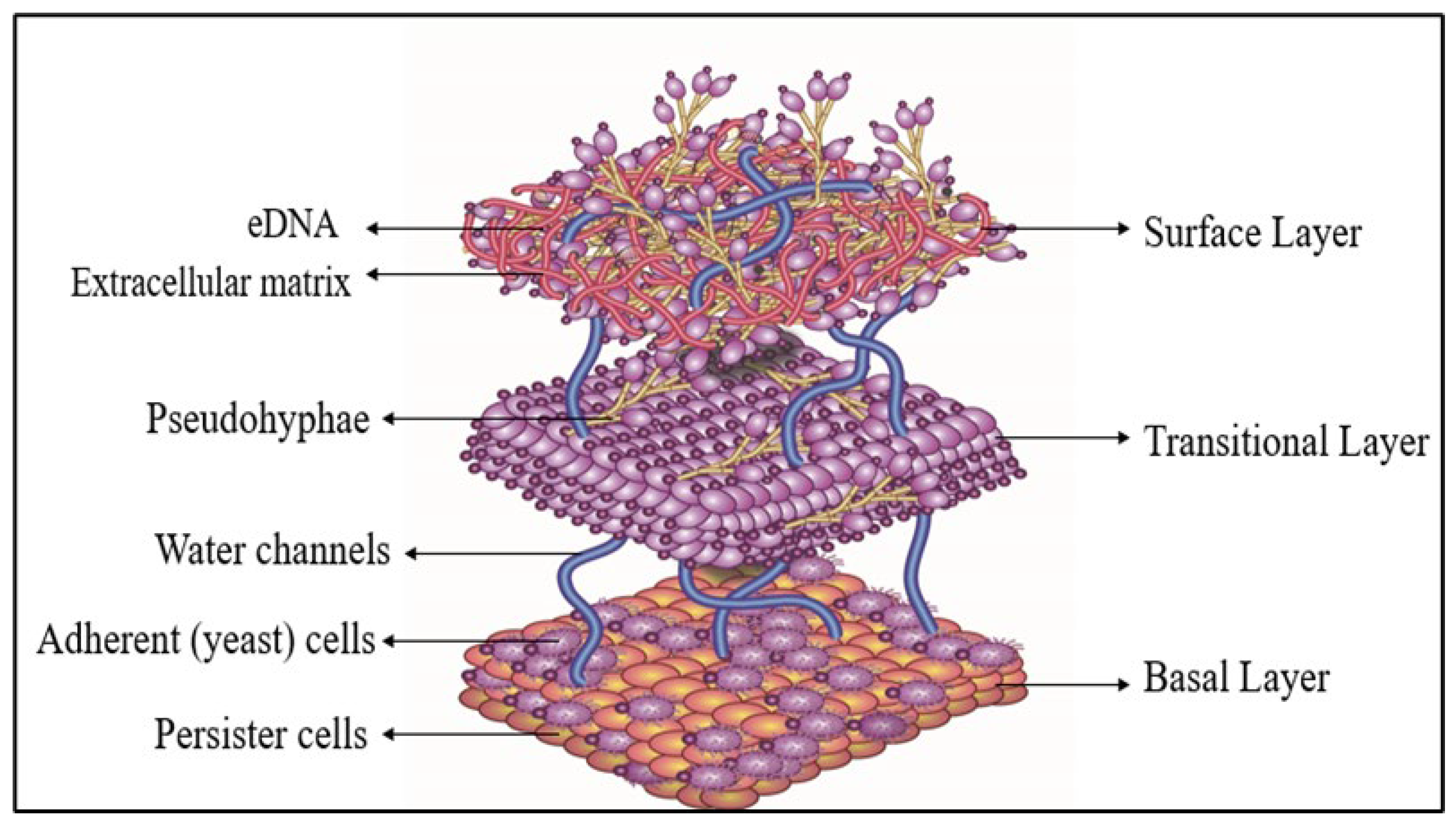
Figure 1. Three-dimensional architecture of C. tropicalis biofilm.
Materproc 29 00005 g001
Figure 2. Biofilm-mediated in vivo biofouling of prosthetic polymeric materials by C. tropicalis.
Materproc 29 00005 g002
Figure 3. Emerging antifungal strategies against C. tropicalis.
Materproc 29 00005 g003
Table 1. Primary experimental studies describing the stages of C. tropicalis biofilm formation.
Biofilm StageExperimental ApproachKey FindingsReferences
Initial adhesionIn vitro adhesion assays on silicone biomaterial surfaces under synthetic urine conditionsDemonstrated rapid adhesion of C. tropicalis to silicone surfaces, showing that surface properties and environmental conditions strongly influence early biofilm establishment[28]
In vitro adhesion assays on catheter materials using artificial urine flow modelShowed strong adhesion to and colonization of urinary catheter materials by C. tropicalis, mimicking device-associated infection conditions[29]
Adhesion and biofilm assays using clinical isolates on abiotic surfacesRevealed strain-dependent variability in adhesion and biofilm-forming capacity among C. tropicalis isolates[30]
Biofilm formation on silicone elastomer and polypropylene materialDemonstrated a strong correlation between biofilm formation and cell viability of C. tropicalis on silicone elastomer and polypropylene, highlighting the role of polymeric materials in supporting fungal persistence[4,31]
Microcolony formation (early biofilm)Scanning electron microscopy (SEM) of developing biofilms After initial attachment, yeast cells proliferated and formed localized clusters known as microcolonies. Early biofilms consisted of clustered yeast cells forming microcolonies with pseudohyphal elements[32]
In vitro biofilm growth models with microscopic observationDemonstrated proliferation of adherent cells and formation of structured microcolonies during early biofilm development[28]
Microscopic and biochemical analysis of early biofilm developmentShowed transition from yeast cells to pseudohyphal structures during early biofilm formation[29]
Extracellular matrix productionBiochemical characterization of extracellular matrixDuring biofilm development, C. tropicalis secreted extracellular matrix components including β-glucans, proteins, lipids, and extracellular DNA, contributing to structural stability[33]
Matrix composition analysis during biofilm growthDemonstrated that matrix polysaccharides play a major role in biofilm structural integrity and antifungal tolerance[34]
Microscopy and biochemical assays of ECM productionConfirmed accumulation of extracellular polymeric substances during biofilm development[28]
Biofilm maturationStructural analysis of mature biofilms using microscopyMature biofilms developed dense three-dimensional structures embedded within the extracellular matrix; nutrient and oxygen gradients developed within the biofilm, leading to metabolic heterogeneity[35]
Antifungal susceptibility assays on mature biofilmsMature biofilms showed significantly reduced susceptibility to antifungal agents[23]
Microscopy and metabolic activity analysis of mature biofilmsMature biofilms displayed metabolically heterogeneous cells within protective matrix layers[34]
Biofilm dispersionBiofilm dispersal assays and microscopy analysisIn the dispersion stage, cells were released from the mature biofilm and returned to the planktonic state, enabling colonization of new surfaces and contributing to the spread of device-associated infections[36,37]
Table 2. Clinical biofouling of polymeric prosthetic materials by C. tropicalis: devices, infections, and outcomes.
Polymeric MaterialMedical DeviceBiofouling-Associated InfectionGlobal Prevalence/StatisticsMortalityClinical ManagementRole of Drug-Resistant BiofoulingReferences
Polyurethane, Silicone ElastomerCentral venous catheters (CVCs)Catheter-related bloodstream infection (candidemia)Device-associated candidemia accounts for ~70–90% of ICU candidemia; C. tropicalis contributes ~15–40% in Asia and LMICs~20–60%Catheter removal + systemic antifungals (echinocandins/amphotericin B)Biofilm-embedded cells show high fluconazole tolerance; resistance leads to persistent candidemia until device removal[16]
PVC, Silicone ElastomerUrinary cathetersCatheter-associated urinary tract infection (CAUTI)Fungal CAUTIs account for ~20–30% of ICU UTIs; Candida spp. dominatesLow–moderate, but high morbidityCatheter replacement/removal + antifungals if invasiveBiofilms protect azole-resistant strains, enabling chronic colonization[28]
PMMA, Polyethylene, Polymer-coated MetalsProsthetic joints (hip, knee)Prosthetic joint infection (PJI)Fungal PJIs: ~1–2% of all PJIs; Candida spp. predominant~25–45% relapse riskSurgical debridement or prosthesis exchange + long-term antifungalsDrug-resistant biofilms necessitate surgical removal; medical therapy alone often fails [38]
Silicone Elastomer, PolyurethanePeritoneal dialysis (PD) cathetersFungal peritonitis~2–7% of PD-associated infectionsUp to ~30%Immediate catheter removal + antifungal therapyBiofilm-mediated resistance drives poor outcomes if catheter retained[39]
Polymer-coated MetalsProsthetic heart valves, cardiac devicesFungal endocarditisCandida causes ~2–10% of prosthetic valve endocarditis~25–40%Surgical valve replacement + prolonged antifungalsBiofilm-associated resistance contributes to high mortality[40]
Silicone Elastomer, Acrylic (PMMA)Dental and voice prosthesesLocal candidiasis, prosthesis failureVery common in long-term users; high recurrenceLow mortalityProsthesis cleaning/replacement + topical/systemic antifungalsResistant biofilms cause recurrent fouling and device failure[4,40]
Disclaimer/Publisher’s Note: The statements, opinions and data contained in all publications are solely those of the individual author(s) and contributor(s) and not of MDPI and/or the editor(s). MDPI and/or the editor(s) disclaim responsibility for any injury to people or property resulting from any ideas, methods, instructions or products referred to in the content.