Proceeding Paper

Structural, Optical, and Dielectric Behavior of MCr2O4 (M=Co, Cu, Ni) Spinel Chromites Prepared by Sol–Gel Route †

by
Pavithra Gurusamy
,
Anitha Gnanasekar
,
Geetha Deivasigamani
* and
Jose Luis Arias Mediano
MIT Campus, Anna University, Chennai 600 044, India
*
Author to whom correspondence should be addressed.
Presented at the 5th International Online Conference on Nanomaterials, 22–24 September 2025; Available online: https://sciforum.net/event/IOCN2025.
Mater. Proc. 2025, 25(1), 6; https://doi.org/10.3390/materproc2025025006
Published: 24 November 2025

Abstract

The influence of M site substitution in MCr2O4 nanoceramics on their properties is examined in this research. This study is an attempt to correlate the structural, morphological, and optical properties of M-site-modified chromites. The MCr2O4 nanoceramics-CuCr2O4, CoCr2O4, and NiCr2O4 were synthesized using a wet chemical sol–gel auto-combustion method, and all three samples were annealed for 4 h at 900 °C. X-ray diffraction analysis showed that the XRD patterns of CuCr2O4, CoCr2O4, and NiCr2O4 correspond to single-phase cubic crystal structures with the space group Fd-3m. Using the Scherrer equation, the crystallite sizes were found to be 9.86 nm, 6.73 nm, and 10.73 nm for CuCr2O4, CoCr2O4, and NiCr2O4, respectively. Other parameters, including crystal structure, micro-strain, lattice constant, unit cell volume, X-ray density, packing factor, and the stacking fault of the calcined powder samples, were determined from data acquired from the X-ray diffractometer. Energy dispersive X-ray spectroscopy (EDX) was employed to confirm the appropriate chromite elements in their expected stoichiometric proportions, removed from other impurities. The identification of the functional groups of the samples was performed using Fourier Transform Infrared Spectroscopy (FTIR). The absorption bands characteristic of tetrahedral and octahedral coordination compounds of the spinel structure are found between 450 and 750 cm−1 for all three samples in the spectrum. From the UV-absorption spectra, and using Tauc’s plot, the energy bandgap values for CuCr2O4, CoCr2O4, and NiCr2O4 were measured to be 1.66 eV, 1.82 eV, and 2.01 eV, respectively. The dielectric properties of the chromites were studied using an LCR meter. Frequency-dependent dielectric properties, including Dielectric constant and Tangent loss, were calculated. These findings suggest the feasibility of the use of these synthesized chromites for optical devices and other optoelectronic applications.

1. Introduction

Mixed spinel metal oxides have gained significant attention due to their wide-ranging applications in ceramics, semiconductors, sensors, and catalysis. Spinel structure has the general formula AB2O4, where both A and B are metal cations. In the oxygen framework, A ions occupy tetrahedral sites, whereas B ions reside in octahedral positions. A spinel unit cell contains 32 oxygen atoms and 24 metal ions, with 8 ions in tetrahedral (A) positions and 16 in octahedral (B) positions. Transition metal-based spinel oxides (MB2O4, where M2+ = Ni2+, Cu2+, Zn2+ and B3+ = Co3+, Mn3+, Fe3+, Cr3+, etc.) exhibit exceptional properties, making them promising for various technological uses [1]. In particular, chromium-based spinels (ACr2O4; A = Mg, Ni, Co, Fe, Zn, Cu, Mn, Cd, etc.) have attracted research interest for their remarkable electronic, magnetic, and optical characteristics, with potential applications in sensors, solid oxide fuel cells, and multiferroic devices due to their cubic spinel structure comprising tetrahedral (A2+) and octahedral (Cr3+) sites [2]. Among the spinel chromites, NiCr2O4, CoCr2O4, and CuCr2O4 have gained significant attention owing to their remarkable multiferroic nature. Materials with high dielectric constants and low dielectric losses are increasingly sought after for wireless communication and electronic applications. Various methods, including the sol–gel wet chemical method, hydrothermal treatment, co-precipitation technique, and conventional solid-state reaction method, have been employed for the preparation of chromites. Among the various synthesis techniques, the sol–gel auto-combustion method is preferred because of its cost efficiency, eco-friendly nature, and ability to control particle size and morphology while enhancing crystallinity. In this study, NiCr2O4, CoCr2O4, and CuCr2O4 materials were synthesized using the sol–gel auto-combustion method, and their structural, morphological, optical, and dielectric characteristics were investigated. The dielectric properties were analyzed using an impedance spectroscopic technique with varying frequency ranges at normal room temperature. Although individual studies on NiCr2O4, CoCr2O4, and CuCr2O4 exist, comparative investigations exploring their structural, optical, and dielectric behavior under identical synthesis conditions are still limited. Previous reports have primarily emphasized their magnetic, supercapacitor, or catalytic aspects [2,3,4], leaving the optical-dielectric behavior insufficiently addressed. In this work, all three chromites were synthesized via a uniform sol–gel route to ensure comparable microstructural features. The study provides new insights into how variations in the divalent cation influence lattice distortion, crystallite size, and band gap energy. This comparative approach offers a clearer understanding of cation-dependent behavior, which has been scarcely reported in the literature.

2. Experimental

2.1. Synthesis Procedure

In the present study, cobalt, copper, and nickel chromites were synthesized using a simple one-pot sol–gel auto-combustion method. Firstly, cobalt chromite was synthesized using raw materials, including cobalt nitrate hexahydrate (Co(NO3)2·6H2O) and chromium nitrate nonahydrate (Cr(NO3)3·9H2O), which were dissolved in distilled water according to their exact stoichiometric ratios. Citric acid was added to this metal nitrate solution in a 1:1 ratio. To ensure the thorough mixture of the above-described solution, a magnetic stirrer was employed. To maintain neutrality of the solution, pH adjustment was made using ammonia solution. After stirring at 1000 rpm for 3 h, the solution converts to a gel. This gel is then heated and transferred to a heating mantle, where the combustion reaction takes place. After combustion, cobalt chromite powders are synthesized. Similarly, copper chromite and nickel chromite powders were synthesized by replacing cobalt nitrate with copper nitrate and nickel nitrate, respectively, using the same synthesis procedure described above, and annealed at 900 °C for 4 h. For the dielectric study, the powder samples were mixed with 5% PVA binder and compressed into pellets using a 13 mm pelletizer under 3 tons of pressure at 900 °C for 3 h. The powders were then crushed using a mortar pestle to avoid agglomeration and were sent for characterization.

2.2. Characterization Techniques

The synthesized powders were characterized to study their crystal structure using an X-ray diffractometer (XRD: D8 Advance, Bruker AXS GmbH, Karlsruhe, Germany). The morphology and elemental composition were analyzed using a scanning electron microscope equipped with energy dispersive X-ray spectroscopy (SEM-EDX) (TESCAN VEGA3 XMU, TESCAN ORSAY HOLDING, a.s., Brno, Czech Republic). The functional groups of the chromites were examined using Fourier Transform Infrared Spectroscopy (FTIR, SHIMADZU Corporation, Kyoto, Japan). The optical bandgap was evaluated using a UV–Visible spectrophotometer (SHIMADZU UV-3600 PLUS, SHIMADZU Corporation, Kyoto, Japan). Dielectric measurements were performed using an LCR meter and impedance analyzer (Hioki IM3570, Hioki E.E. Corporation, Nagano, Japan).

3. Results and Discussion

3.1. XRD Analysis

The phase formation and crystal structure were analyzed using X-ray diffraction analysis. The XRD patterns observed in the 10° to 70° range are displayed in Figure 1. The diffraction peaks corresponding to 900 °C were obtained at 18.79°, 30.54°, 36.15°, 43.73°, 54.20°, 57.79°, and 63.52°. The XRD patterns of CuCr2O4, CoCr2O4, and NiCr2O4 matched with JCPDS card no. 04-0763, which confirmed the single-phase cubic crystal structure with space group Fd-3m. No other extra peaks were observed, confirming the single-phase formation.
The prominent XRD peaks with a considerable full width at half maximum (FWHM) confirm the formation of crystalline structures. It is well established that the reduction in particle size and the associated intrinsic strain resulting from this size confinement contribute to the broadening of XRD peaks. Therefore, the overall physical peak broadening mainly arises from two factors: size-related broadening and strain-induced effects [3]. Scherrer’s formula was utilized to calculate the average crystalline sizes of the chromites:
A v e r a g e   c r y s t a l l i t e   s i z e   ( D ) = K λ β c o s θ
where λ is the wavelength, K is a constant, β is the full width at half maximum value, and θ is the diffraction angle. The lattice parameter for the synthesized chromites was calculated using the formula given below:
L a t t i c e   p a r a m e t e r   a = h 2 + k 2 + l 2
Here, hkl corresponds to the Miller indices. Table 1 displays the structural parameters calculated using the XRD spectra.
Dislocation density (δ) was calculated using the relation
δ = 1/D2
where D is the crystallite size of the sample.
Stacking fault (SF) was estimated using the relation
S F = 2 π 2 45 3 t a n θ
Packing factor (P) was calculated using the following relation:
P = D d
where ‘D’ is the crystallite size and ‘d’ is the interplanar distance.

3.2. FTIR Analysis

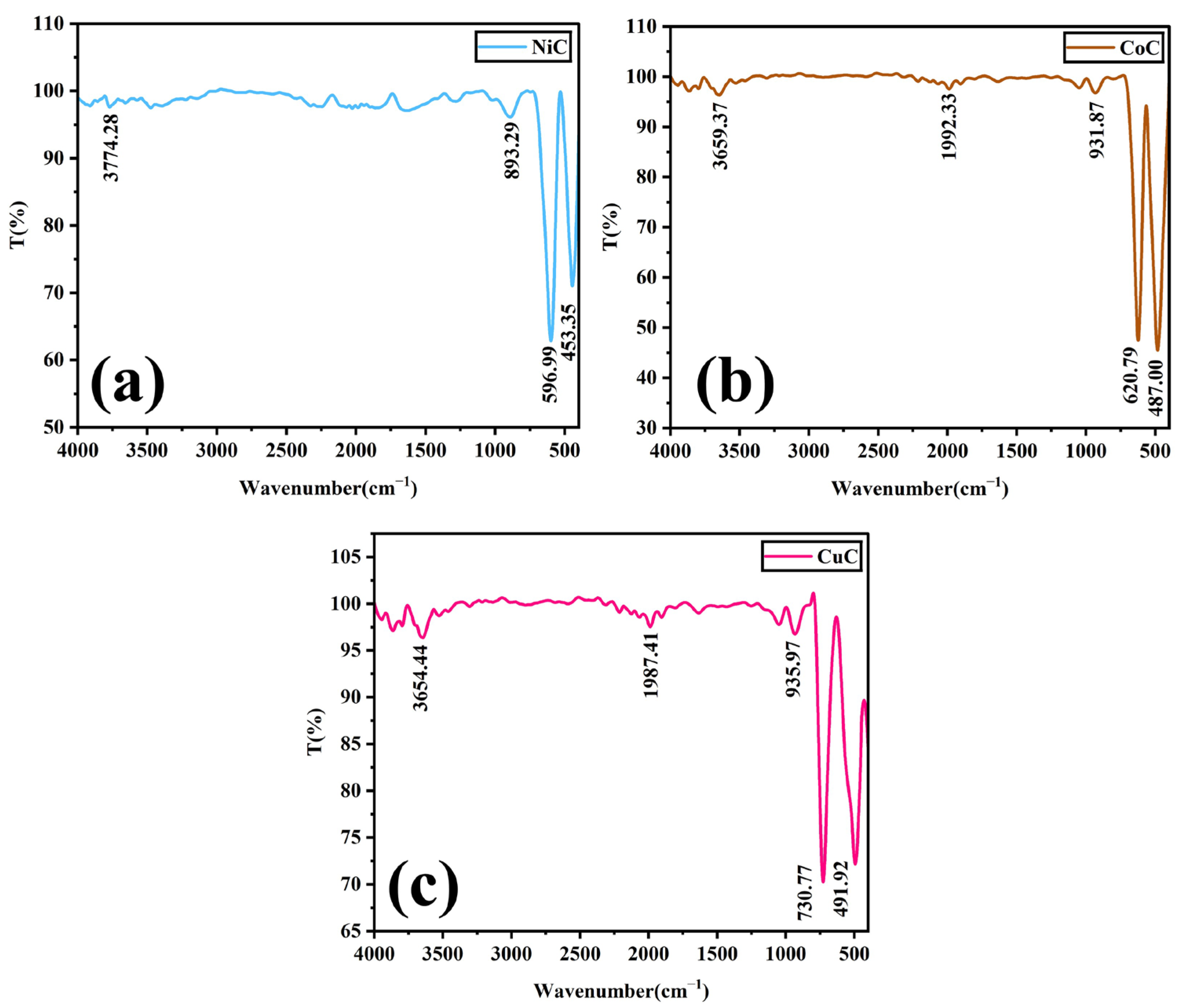
FTIR analysis was employed to determine the vibrational stretching frequencies associated with metal–oxygen bonds [5]. The FTIR spectra in Figure 2 display the percentage of transmittance against wavenumber. The nature of the A and B sites and the distribution of cations among them significantly influence the properties of spinel chromites [4]. In spinel chromites, the characteristic metal–oxygen stretching modes generally appear in the 400–600 cm−1 region [6]. Spinel structure is confirmed through two characteristic absorption bands observed at 590–730 cm−1 (high frequency, ν1) and 450–500 cm−1 (low frequency, ν2) in all three chromite samples, thereby detecting the Cr2O42− group of Cu–O and Cr–O, respectively [2]. The high-frequency band is attributed to the stretching vibrations of metal–oxygen bonds at the tetrahedral (A) sites, whereas the low-frequency band corresponds to the vibrations associated with the octahedral (B) sites. The presence of bands within 3600–3800 cm−1 in all three spectra is attributed to –OH stretching modes and deformation vibrations of water molecules [5]. The shift appearing around 890–940 cm−1 corresponds to the vibrational modes of amorphous CrO2 incorporated into the MCr2O4 (M = Co, Cu, Ni) spinel structure. The presence of bands near 1980–1990 cm−1 suggests the existence of residual reactant materials in the final nickel, cobalt, and copper chromite phase [7].

3.3. SEM Analysis

A scanning electron microscope was utilized to interpret the physical appearance and morphology of the chromite powders. Figure 3 displays the SEM images and atomic weight percentages of NiCr2O4, CoCr2O4, and CuCr2O4 samples. It is evident that the shape and size of the particles are random and agglomerated.
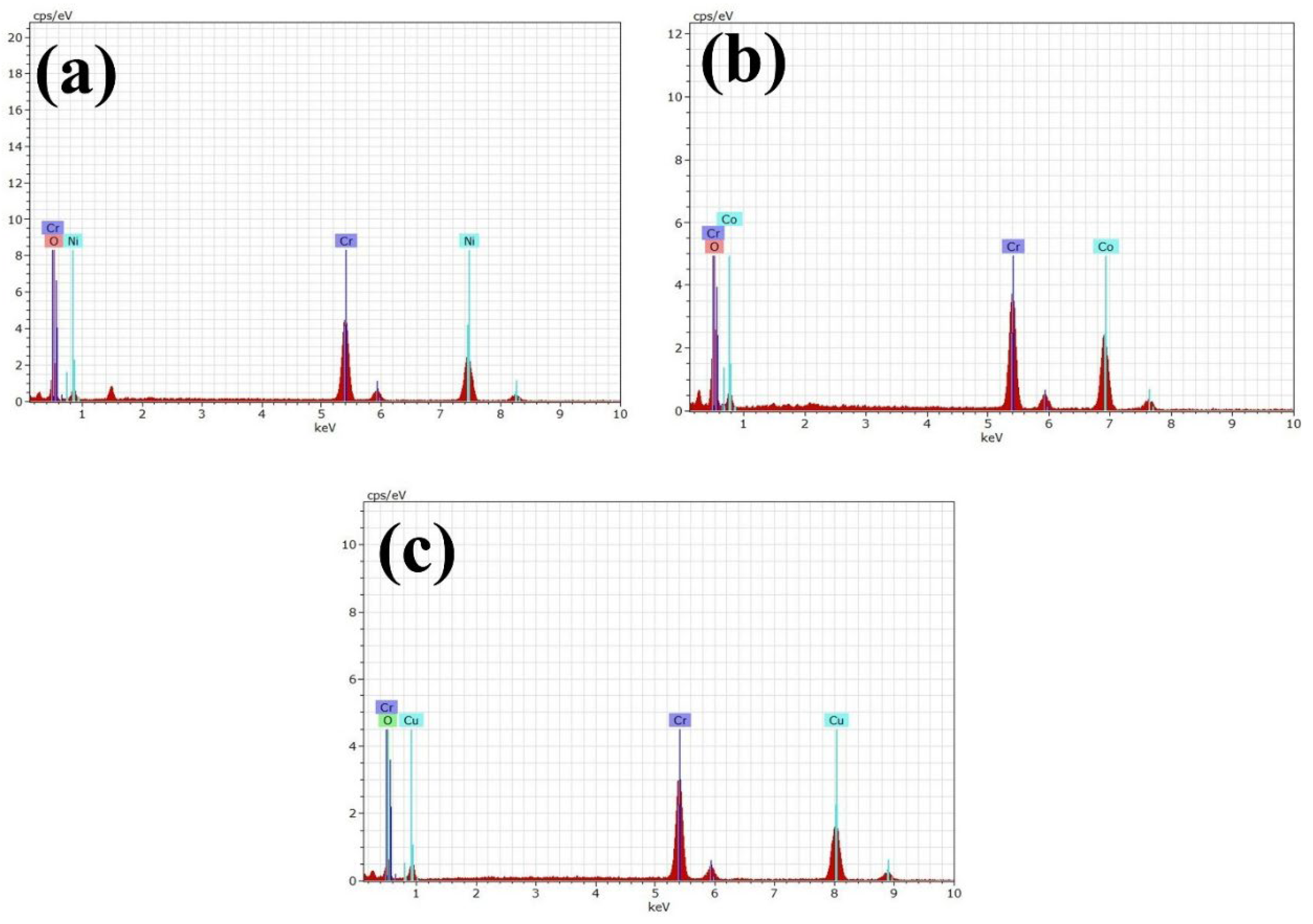
The observed porosity may be due to the gases emitted during the synthesis procedure. Since the mobility of grain boundaries plays a vital role in grain growth, the presence of chromium ions near the grain boundaries inhibits their movement, leading to a reduction in grain size with increasing chromium substitution. The release of gases during the combustion process leads to the formation of voids throughout the structure. The average grain sizes of CoCr2O4, CuCr2O4, and NiCr2O4 were found to be 1.23 μm, 3.12 μm, and 1.27 μm, respectively. The observed non-uniformity in grain size can be attributed to the presence of structural defects and impurity atoms within the nanomaterial. Figure 4 displays the EDX spectrum of the synthesized chromites. From the graph, we can observe that the synthesized materials include Ni, Cr, and O for NiCr2O4; Co, Cr, and O for cobalt chromite; and Cu, Cr, and O for copper chromite. The lack of any extra peaks in the EDS spectra confirms that the samples are free from impurities. The atomic percentages calculated from EDX are presented in Figure 3(2a–2c). The elemental composition obtained from EDS analysis closely matches the theoretical values.

3.4. Optical Analysis

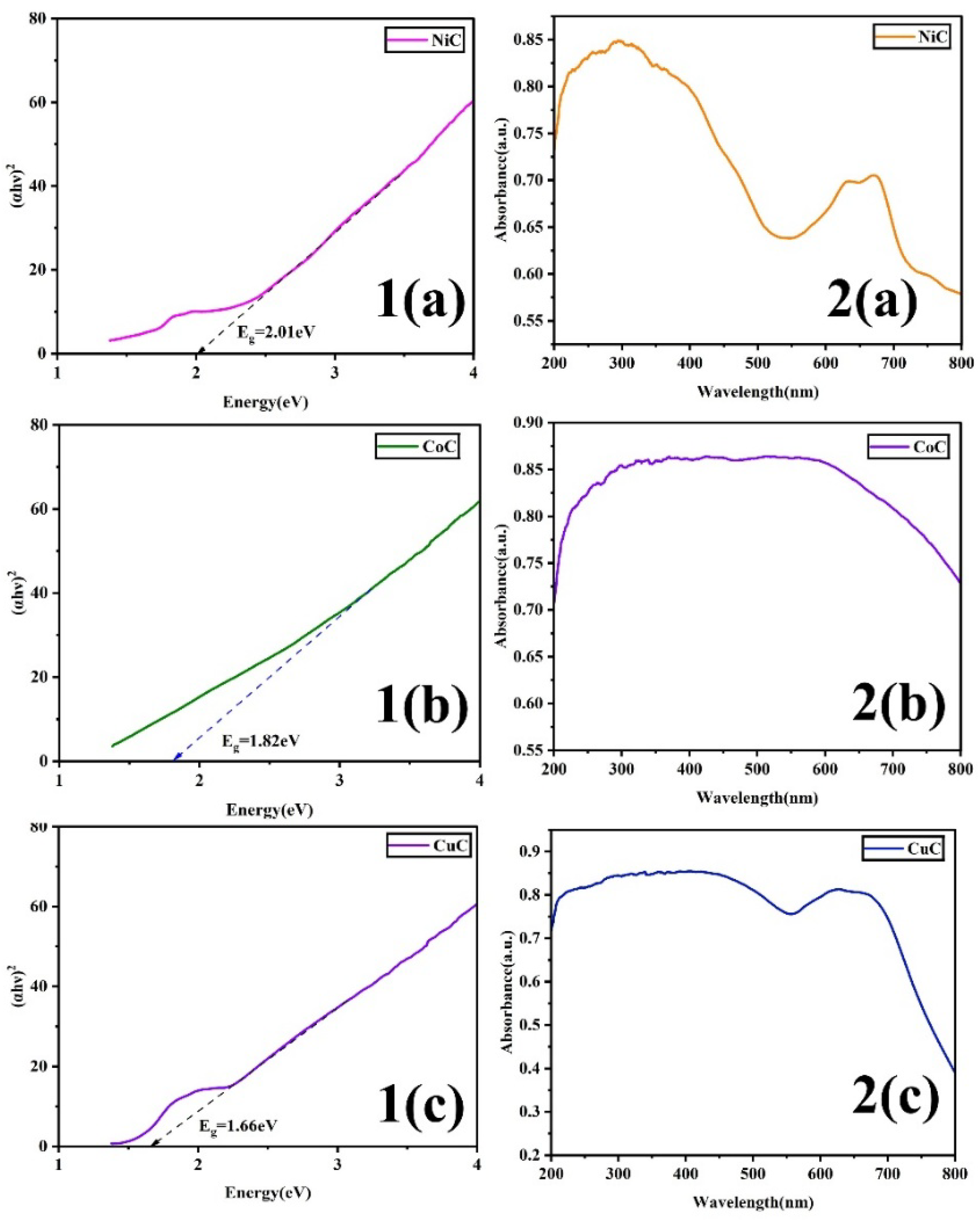
The UV-visible absorption spectrum reveals the band structure of the prepared chromites. Shown in Figure 5 are the Tauc plots and absorption spectra of NiCr2O4, CoCr2O4, and CuCr2O4 samples. The absorbance in the range of 200–800 nm is recorded at normal room temperature, exhibiting a remarkable optical response within this wavelength range. The band gaps of the corresponding chromites were extrapolated using Tauc’s equation, shown in Figure 5(1a–1c).
α h ν = A ( h ν E g ) n
Here, α denotes the absorption coefficient, hν indicates the incident photon energy, Eg denotes the bandgap of the material, and A denotes the absorption. In the case of a direct transition, n takes the value of 2, whereas, for an indirect transition, n equals 0.5 [8]. The band gap energy and crystallite size of the materials are strongly influenced by the calcination temperature. Therefore, annealing at 900 °C resulted in an evident optical response, consistent with this behavior.

3.5. Dielectric Property Studies

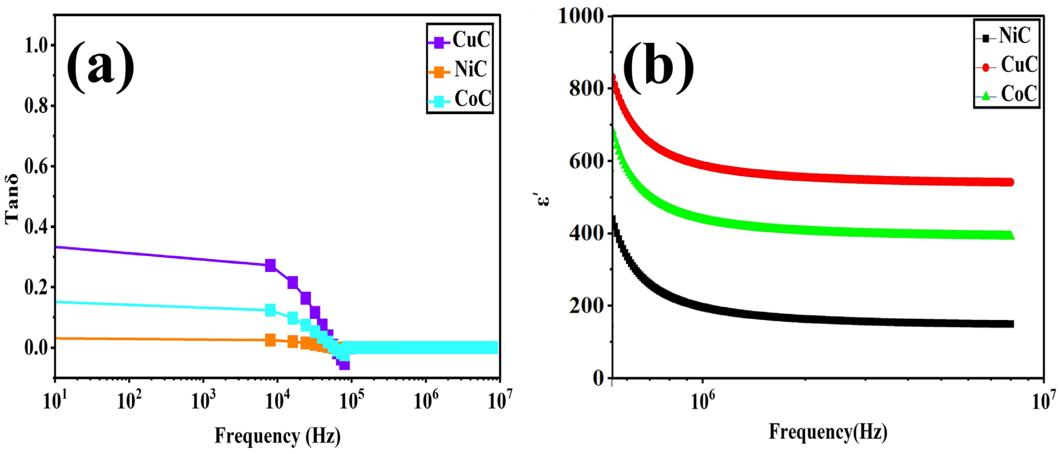
The dielectric properties of the NiCr2O4, CoCr2O4, and CuCr2O4 samples were studied using an LCR meter. Shown in Figure 6a is the dielectric constant, and Figure 6b shows the tangent loss values of the prepared chromite samples, recorded at room temperature. We can observe that the value of the dielectric constant decreases with increasing frequency. The change in dielectric values is mainly attributed to the polarization mechanism, which varies with frequency [6]. The dielectric behavior is affected by several key parameters such as dopant concentration, annealing temperature, synthesis technique, morphological consistency, porosity, grain size, and density. The observed enhanced dielectric constant values can be attributed to factors like interfacial polarization, oxygen vacancies, and grain boundary defects [9]. The dielectric constant values of NiCr2O4, CoCr2O4, and CuCr2O4 were found to be 421, 637, and 823. Values of tanδ were found to be 0.32, 0.16, and 0.02 for CuCr2O4, CoCr2O4, and NiCr2O4, respectively.
At higher frequencies, dipoles are unable to reorient quickly enough and consequently lag behind the applied electric field, leading to the absence of orientation polarization. As a result, only interfacial or space charge polarization occurs, causing a reduction in the overall dielectric constant [9]. In chromites, the electron exchange between Cr2+↔Cr3+ plays a significant role at lower frequencies. However, with increasing frequency, this polarization gradually diminishes until it reaches a minimum, steady value. The dielectric loss tangent is influenced by factors such as stoichiometry, structural uniformity, and the presence of Cr3+ ions. Additionally, the conduction mechanism also contributes to variations in the loss tangent [10]. As the frequency increases, we can note that the dielectric loss decreases. In the low-frequency region, the presence of larger grains, indicated by sharp diffraction peaks with narrow FWHM, promotes charge accumulation at the grain boundaries, leading to an increased dielectric constant. However, as the frequency rises, the accumulated charges can no longer follow the alternating field, causing a deviation from the initial trend and a subsequent reduction in the tangent loss [10]. The obtained dielectric results highlight chromites as fascinating materials with potential use in microwave frequency circuits and other dielectric applications.

4. Conclusions

This work successfully demonstrated the influence of M-site substitution on the structural, morphological, and optical properties of MCr2O4 (M = Cu, Co, Ni) nanoceramics synthesized via the sol–gel auto-combustion method. From the XRD analysis, the crystallite size was found to be in the range of 6.73–10.73 nm for all three chromites, indicating well-crystallized ceramic formation. The EDX spectra confirmed the stoichiometric composition and purity of the synthesized compounds, while FTIR results evidenced the presence of metal–oxygen vibrational modes corresponding to tetrahedral and octahedral coordination, validating the spinel framework. The optical studies indicated a systematic variation in energy bandgap values—ranging from 1.66 eV to 2.01 eV—for the prepared chromites, demonstrating that the substitution at the M-site significantly tunes the optical properties. Dielectric analysis further revealed strong frequency-dependent behavior, suggesting potential for the use of these materials in electronic and optoelectronic applications due to their higher dielectric constant value and low tangent loss. Overall, the findings establish that M-site engineering in MCr2O4 nanoceramics effectively modifies their microstructural, optical, and dielectric characteristics, making them promising candidates for applications in optical devices, sensors, and dielectric components in energy and communication technologies.

Author Contributions

Conceptualization, Methodology, Software, and Validation, P.G., A.G., and G.D.; Formal Analysis and Investigation, P.G. and G.D.; Writing—Original Draft Preparation, Writing—Review and Editing, P.G. and A.G.; Visualization, G.D.; Supervision, G.D. All authors have read and agreed to the published version of the manuscript.

Funding

This research received no external funding.

Institutional Review Board Statement

Not applicable.

Informed Consent Statement

Not applicable.

Data Availability Statement

Data will be made available upon request.

Acknowledgments

We thank J. Ramajothi, Department of Applied Sciences and Humanities, MIT Campus, Anna University, Chennai, for helping in the dielectric characterization facility.

Conflicts of Interest

The authors declare no conflicts of interest.

References

  1. Anitha, G.; Pavithra, G.; Geetha, D. Tailoring ZnAl2O4 properties through divalent metal ions (Cu2+, Mg2+, & Mn2+) incorporation: A study on dielectric and photocatalytic enhancement. J. Mater. Sci. Mater. Electron. 2025, 36, 1–27. [Google Scholar] [CrossRef]
  2. Anandha Kumar, S.; Shahanas, T.; Harichandran, G. Synthesis and fabrication of tetragonal copper chromite nanospheres for usage as the cathode material in asymmetric supercapacitors. J. Energy Storage 2025, 106, 114839. [Google Scholar] [CrossRef]
  3. Mamidipalli, N.R.; Tiyyagura, P.; Punna Rao, S.; Kothamasu, S.B.; Pothu, R.; Boddula, R.; Al-Qahtani, N. Crystallite Size Effects on Electrical Properties of Nickel Chromite (NiCr2O4) Spinel Ceramics: A Study of Structural, Magnetic, and Dielectric Transitions. ChemEngineering 2024, 8, 100. [Google Scholar] [CrossRef]
  4. Ajith, A.; Wani, T.A.; Suresh, G.; Saravanan, N.; Masrour, R. Sol–Gel Synthesis and Characterizations of Spinel Nickel Chromite (NiCr2O4) Nanoparticles to Study their Photocatalytic, Antibacterial and Anticancer Activities. Nano 2024, 19, 2450050. [Google Scholar] [CrossRef]
  5. Kumar, K.V.; Bhavani, S.D. Influence of Calcination Temperature on Physicaland Optical Properties of Nickel Chromite Nanoparticles. Sci. Sinter. 2022, 54, 457–468. [Google Scholar] [CrossRef]
  6. Choudapur, V.H.; Alrashidi, K.A.; Sajjan, A.M.; Ayachit, N.H.; Adil, S.F.; Fernandes, B.J.; Angadi, V.J. Effect of Rare Earth (La3+) on the Structural, Microstructural, and Transport Properties of CoCr2O4 for Industrial Applications. Int. J. Self-Propagating High-Temp. Synth. 2024, 33, 258–265. [Google Scholar] [CrossRef]
  7. Bisht, N.; Rao, S.S.; Deshawal, S.; Khanna, P.K. Novel microwave synthesis of copper chromite nanoparticles. Inorg. Chem. Commun. 2021, 134, 109072. [Google Scholar] [CrossRef]
  8. Gurusamy, P.; Gnanasekar, A.; Deivasigamani, G. Investigation of Structural, Morphological, Optical, and Dielectric Properties of Magnesium Chromite (MgCr2O4) Spinel Oxide. Eng. Proc. 2025, 87, 109. [Google Scholar] [CrossRef]
  9. Priya, A.S.; Geetha, D.; Madhavan, J. Synthesis and Structural, Dielectric and Photocatalytic Properties of (Ti, La)-co-Doped Calcium Ferrite Ceramic Powders. Arab. J. Sci. Eng. 2022, 47, 7657–7667. [Google Scholar] [CrossRef]
  10. Garg, T.; Parmar, L.K.; Koser, A.A.; Kaurav, N.; Yadav, A. Impact of Zn-Ion Doping on the Structural and Optical Properties of Cobalt Chromite System. J. Mol. Eng. Mater. 2025, 13, 2–11. [Google Scholar] [CrossRef]
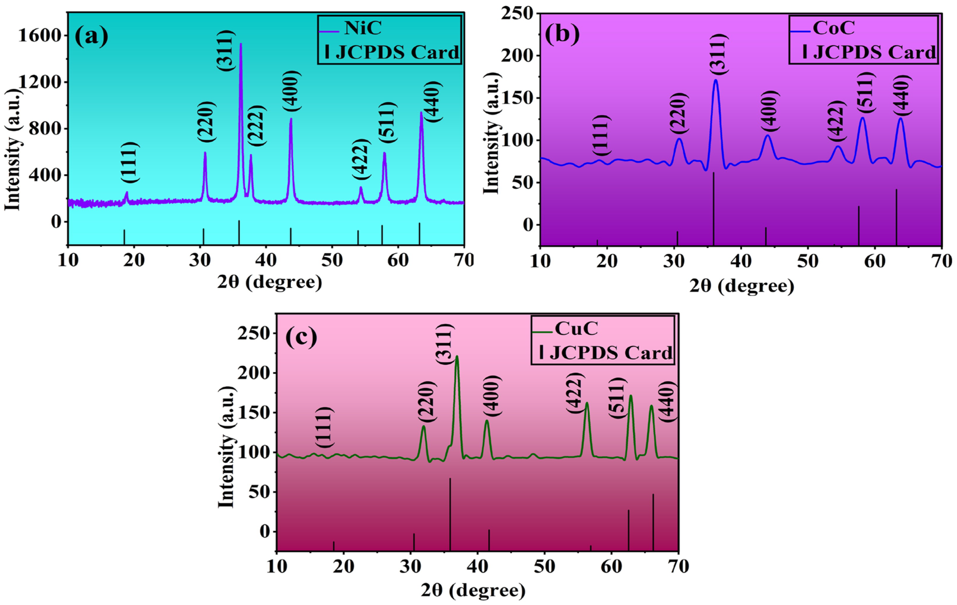
Figure 1. X-ray diffraction patterns of (a) NiCr2O4, (b) CoCr2O4, and (c) CuCr2O4.
Materproc 25 00006 g001
Figure 2. FTIR Spectra of (a) NiCr2O4, (b) CoCr2O4, and (c) CuCr2O4.
Materproc 25 00006 g002
Figure 3. (1a1c) SEM images and (2a2c) atomic weight percentages of NiCr2O4, CoCr2O4, and CuCr2O4.
Materproc 25 00006 g003
Figure 4. EDX images of (a) NiCr2O4, (b) CoCr2O4, and (c) CuCr2O4.
Materproc 25 00006 g004
Figure 5. (1a1c) Tauc plots and (2a2c) absorption spectra of NiCr2O4, CoCr2O4, and CuCr2O4.
Materproc 25 00006 g005
Figure 6. (a) Dielectric constant and (b) tangent loss of NiCr2O4, CoCr2O4, and CuCr2O4.
Materproc 25 00006 g006
Table 1. Structural parameters of NiCr2O4, CoCr2O4, and CuCr2O4 obtained from XRD.
ParametersCoCCuCNiC
Crystallite size (D)nm6.739.8610.73
Micro Strain (ε)2.591.831.49
Interplanar distance (d) Å2.021.992.14
Lattice constant Å8.237.658.25
Volume of the unit cell (a3) Å558.15448.44562.73
Packing factor P3.214.954.99
Density Dx (g/cm3)3.013.892.98
Specific surface area S (cm2/g)2.951.561.87
Stacking fault SF0.380.380.40
Dislocation density (δ) (10−3/m2)0.0220.0100.008
Disclaimer/Publisher’s Note: The statements, opinions and data contained in all publications are solely those of the individual author(s) and contributor(s) and not of MDPI and/or the editor(s). MDPI and/or the editor(s) disclaim responsibility for any injury to people or property resulting from any ideas, methods, instructions or products referred to in the content.