Proceeding Paper

Synthesis and Characterization of ZnSnO3/PVP Electrospun Composite Nanofibers †

by
R. Indhumathi
and
A. Sathiya Priya
*
Department of Physics, Sri Sai Ram Engineering College, Chennai 600 044, Tamil Nadu, India
*
Author to whom correspondence should be addressed.
Presented at the 5th International Online Conference on Nanomaterials, 22–24 September 2025; Available online: https://sciforum.net/event/IOCN2025.
Mater. Proc. 2025, 25(1), 5; https://doi.org/10.3390/materproc2025025005
Published: 21 November 2025

Abstract

Zinc stannate (ZnSnO3), a lead-free perovskite oxide, possesses a wide band gap along with ferroelectric and piezoelectric characteristics, but its practical use is limited by nanoparticle agglomeration and poor mechanical stability. In this study, ZnSnO3 nanoparticles were synthesized via a chemical precipitation method and incorporated into electrospun polyvinylpyrrolidone (PVP) nanofibers to overcome these drawbacks. XRD confirmed the formation of orthorhombic perovskite ZnSnO3, while FTIR verified successful embedding into the PVP matrix without chemical degradation. UV–Vis analysis revealed a slight blue shift in the absorption edge, with the optical band gap widening from 3.61 eV (ZnSnO3) to 3.73 eV (ZnSnO3/PVP), accompanied by enhanced visible light absorption. SEM images showed that agglomerated ZnSnO3 nanoparticles were transformed into smooth, bead-free nanofibers with diameters ranging from 0.22 to 1.80 μm. The synergy between ZnSnO3 crystallinity and PVP flexibility imparts structural integrity, tunable optical behavior, and mechanical robustness, making the hybrid nanofibers promising candidates for photocatalysis, flexible optoelectronics, and energy-harvesting applications.

1. Introduction

Perovskite oxides with the general formula ABO3 have emerged as an important class of functional materials, owing to their wide range of electronic, optical, and energy-related properties. Among them, zinc stannate (ZnSnO3), a ternary perovskite oxide, has attracted considerable attention because of its wide band gap, high dielectric constant, ferroelectric and piezoelectric characteristics, and environmental stability [1,2,3,4]. These intrinsic features make ZnSnO3 highly relevant for applications in photocatalysis, gas sensing, energy storage, and flexible optoelectronic devices [5,6,7]. Furthermore, its non-toxic nature and structural tunability offer an additional advantage over conventional lead-based perovskites. Despite these promising attributes, the practical deployment of ZnSnO3 nanoparticles is often restricted by issues such as particle agglomeration, poor mechanical strength, and limited dispersion when integrated into device architectures. To overcome these limitations, embedding inorganic nanoparticles within a polymer matrix has proven to be an effective strategy. Such hybrid systems combine the functional performance of the nanoparticles with the flexibility and processability of polymers, making them attractive for next-generation flexible and wearable technologies [8,9,10]. Polyvinylpyrrolidone (PVP) is a widely used polymer matrix due to its high solubility, excellent film-forming ability, mechanical robustness, and biocompatibility. Importantly, PVP can be readily processed into continuous nanofibers through electrospinning, a simple and scalable technique that produces fibrous networks with high surface-to-volume ratios and interconnected porous structures [11,12,13]. Incorporating ZnSnO3 nanoparticles into PVP nanofibers not only minimizes nanoparticle agglomeration but also enhances interfacial interactions, leading to improved optical absorption and charge transport properties. Recent studies have demonstrated that oxide–polymer nanocomposites fabricated via electrospinning hold great promise for applications in photocatalysis, photovoltaics, and flexible energy-harvesting devices [14,15]. Electrospun ZnSnO3/polymer nanofibers have attracted significant attention for energy harvesting, sensing, and damping applications due to their high surface area, tunable morphology, and multifunctional properties. However, reports on ZnSnO3/PVP electrospun nanofibers remain scarce, leaving a gap in understanding the structural stability, optical modulation, and morphology of these composites. Kavarthapu et al. [16] fabricated ZnSnO3/PVDF-HFP nanofibrous composite films via a two-step process comprising the hydrothermal synthesis of ZnSnO3 nanoparticles and subsequent electrospinning of the composite solution. The incorporation of ZnSnO3 nanoparticles into the PVDF-HFP matrix enhanced dielectric properties, surface charge density, and triboelectric output performance of the films. Zhang et al. [17] fabricated a (ZnSnO3/PVDF)@PPy/epoxy composite via electrospinning, where the ZnSnO3/PVDF nanofibers and PPy coating significantly enhanced damping and mechanical properties. Chen et al. [18] synthesized Au-functionalized In-doped ZnSnO3 nanofibers via electrospinning followed by annealing, where the incorporation of Au nanoparticles enhanced acetone gas-sensing performance by providing catalytic active sites, Schottky barrier formation, and increased surface area. Haq et al. [19] fabricated NiO/ZnSnO3 microsphere-decorated nanofibers via electrospinning ZnSnO3 followed by the hydrothermal growth of NiO, where the resulting p–n heterostructure exhibited enhanced ethanol-sensing performance with high sensitivity, selectivity, and fast response/recovery times. While ZnSnO3 has been combined with various polymers such as PVDF, PVDF-HFP, and PMMA for energy-harvesting and sensing applications, reports on its integration with PVP via electrospinning are not available to date. In this study, we present for the first time the synthesis of ZnSnO3/PVP composite nanofibers through electrospinning, providing insight into their structural stability, optical modulation, and morphology. This approach offers a simple and scalable route to flexible ZnSnO3-based materials for energy and optoelectronic applications.
In this study, ZnSnO3 nanoparticles were synthesized by a simple chemical precipitation method and incorporated into PVP nanofibers via electrospinning. A comprehensive investigation of their structural, functional, optical, and morphological properties was carried out using XRD, FTIR, UV–Vis spectroscopy, and FESEM. The study provides fundamental insights into the stabilization of ZnSnO3 within a polymeric framework and demonstrates the potential of ZnSnO3/PVP nanofibers as multifunctional materials for flexible electronics and energy-harvesting applications.

2. Experimental Methods

2.1. Materials

Zinc chloride (ZnCl2) and tin (IV) chloride pentahydrate (SnCl4⋅5H2O) were used as precursor materials for the synthesis of ZnSnO3 nanoparticles. Polyvinylpyrrolidone (PVP, Mw = 100,000) was purchased from Sigma- Aldrich, (Bengaluru, India). The solvents N,N-dimethylformamide (DMF) and acetonitrile (ACN) were obtained from commercial suppliers. All chemicals and reagents were used as received without further purification.

2.2. Synthesis of ZnSnO3 Nanoparticles

ZnSnO3 nanoparticles were prepared by a chemical precipitation method. Equimolar aqueous solutions of ZnCl2 and SnCl4·5H2O (1:1 molar ratio) were mixed under magnetic stirring, and a NaOH solution was added dropwise until complete precipitation occurred. The resulting white precipitate was filtered, thoroughly washed with double-distilled water to remove residual chloride ions, and dried at 80 °C for 6 h. The dried powder was calcined in air at 500 °C for 3 h using a heating rate of 5 °C/min, yielding crystalline ZnSnO3 nanoparticles with improved phase stability.

2.3. Preparation of ZnSnO3/PVP Electrospun Nanofibers

For the fabrication of composite nanofibers, polyvinylpyrrolidone (PVP, Mw ≈ 100,000) was dissolved in a solvent mixture consisting of N,N-dimethylformamide (DMF) and acetonitrile (ACN) mixed in a 7:3 volume ratio. The polymer concentration was maintained at 21 wt% with continuous stirring until a clear solution was obtained. Separately, calcined ZnSnO3 nanoparticles were dispersed in the same DMF/ACN mixture at a loading of 5 wt% and ultrasonicated for 15 min to minimize agglomeration. The nanoparticle dispersion was then combined with the PVP solution under vigorous stirring to form a homogeneous ZnSnO3/PVP precursor solution.

2.4. Electrospinning of Composite Nanofibers

The precursor solution was transferred into a syringe equipped with a metallic needle and electrospun under optimized conditions, involving an applied voltage of 11 kV, tip-to-collector distance of 15 cm, and solution feed rate of 0.5 mL/h. The collector consisted of a flat aluminum foil substrate attached to a grounded metallic plate. Under these optimized parameters, the polymer jet was continuously ejected from the needle tip and elongated into ultrafine fibers by the electrostatic field, resulting in the deposition of uniform ZnSnO3/PVP nanofibers onto the aluminum foil. After electrospinning, the as-spun nanofibers were carefully peeled off from the aluminum foil and dried in a vacuum oven at 60 °C for 5 h to remove residual solvents and stabilize the fibrous structure. No post-annealing was carried out to preserve the polymeric phase and maintain the ZnSnO3/PVP composite nature of the fibers.

2.5. Characterization Techniques

The crystal structure of ZnSnO3 nanoparticles and ZnSnO3/PVP nanofibers was analyzed by XRD (PANalytical X’Pert, Malvern Panalytical, Almelo, The Netherlands; CuKα, λ = 1.5406 Å, 2θ = 10–70°). Functional groups were identified by FTIR (IRAffinity-1, Shimadzu, Kyoto, Japan; Nicolet iS50, Thermo Fisher Scientific, Waltham, MA, USA) in the range 400–4000 cm−1, and optical properties were measured by UV-Vis spectroscopy (PerkinElmer Lambda 25, PerkinElmer, Waltham, MA, USA) to determine band gap and Urbach energy. Surface morphology was examined using (FESEM TESCAN VEGA3, Tescan Brno, S.R.O., Brno, Czech Republic), and the particle size was evaluated using ImageJ version 1.54 (Windows, Java 8 build) software.

3. Results and Discussion

3.1. Structural Analysis

X-ray diffraction (XRD) analysis was employed to investigate the crystalline structure of the synthesized ZnSnO3 nanoparticles and ZnSnO3/PVP composite nanofibers, as shown in Figure 1. The diffraction pattern of calcined ZnSnO3 exhibits sharp and intense peaks at 2θ values corresponding to the (012), (110), (015), (116), (018), (214), (208), (217), and (036) planes, confirming the formation of a well-crystallized orthorhombic perovskite structure of ZnSnO3 (JCPDS card no. 028-1486) [20,21,22]. The high intensity and narrow width of the peaks indicate good crystallinity and phase purity of the nanoparticles obtained at 500 °C.
The average crystallite size of the synthesized samples was estimated using the Debye–Scherrer equation:
D =   K λ β cos θ
where D is the crystallite size (nm), K is the shape factor (typically 0.89), λ is the wavelength of the incident X-ray radiation (CuKα = 1.5406 Å), β is the full width at half maximum (FWHM) of the diffraction peak (in radians), and θ is the Bragg diffraction angle. The estimated crystallite size of ZnSnO3 was found to be approximately 42.5 nm.
In contrast, the ZnSnO3/PVP composite nanofibers show comparatively broad and less intense diffraction peaks. This reduction in intensity and peak broadening can be attributed to the amorphous nature of the PVP polymer matrix, which partially suppresses the diffraction signal of ZnSnO3. Nonetheless, the characteristic diffraction peaks of ZnSnO3 are still discernible in the composite, signifying that the crystalline phase of the nanoparticles is retained even after incorporation into the polymer matrix. The absence of impurity peaks further confirms the phase purity and successful embedding of ZnSnO3 nanoparticles within the PVP network. However, a few additional sharp reflections observed at 2θ ≈ 38.3°, 44.6°, and 64.9° in the ZnSnO3/PVP composite pattern are attributed to the aluminum foil used as the collector substrate during electrospinning. The peaks at 38.63°, 44.75°, and 65.14° correspond to the (111), (200), and (220) planes of aluminum (JCPDS card no. 85-1327) and are not related to the ZnSnO3 phase. Similar aluminum substrate peaks have been reported for ZnO films grown on aluminum foil [23]. The presence of these reflections thus arises from the substrate contribution rather than from any secondary phase of ZnSnO3. This observation is consistent with previous reports on polymer–nanoparticle composites, where the amorphous polymer matrix typically masks the nanoparticle diffraction but does not eliminate it entirely, allowing for partial retention of the crystalline signature of preformed nanoparticles in as-spun fibers [24,25].

3.2. Infrared Spectra Analysis

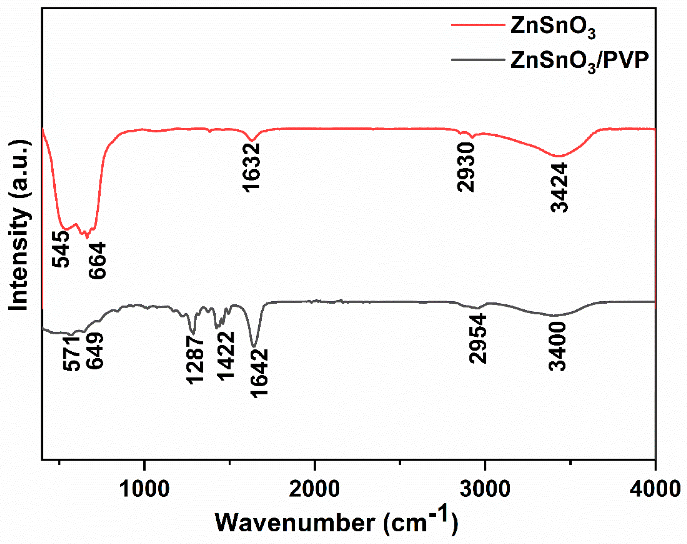
FTIR analysis was performed to identify the functional groups and confirm the formation of ZnSnO3 and ZnSnO3/PVP composites, as shown in Figure 2. The spectrum of ZnSnO3 nanoparticles exhibits characteristic absorption bands at 545, 571, 649, and 664 cm−1, which correspond to the stretching vibrations of Zn–O and Sn–O bonds, confirming the formation of the perovskite ZnSnO3 phase [26]. A weak band at 1632 cm−1 is assigned to the bending vibration of adsorbed O–H molecules [27], while the broad peak around 3424 cm−1 arises from O–H stretching vibrations due to surface hydroxyl groups and physisorbed water [28]. In the case of ZnSnO3/PVP nanofibers, additional peaks are observed at 1287, 1422, and 1642 cm−1, corresponding to C–N stretching, CH2 bending, and C=O stretching vibrations of the PVP backbone [29]. The absorption bands near 2954 and 3400 cm−1 are attributed to C–H stretching and N–H/O–H stretching vibrations, respectively, confirming the presence of the polymer matrix [30,31]. Importantly, the persistence of Zn–O and Sn–O related peaks in the composite spectrum indicates the successful incorporation of ZnSnO3 nanoparticles into the PVP matrix without altering their structural identity.

3.3. Optical Properties

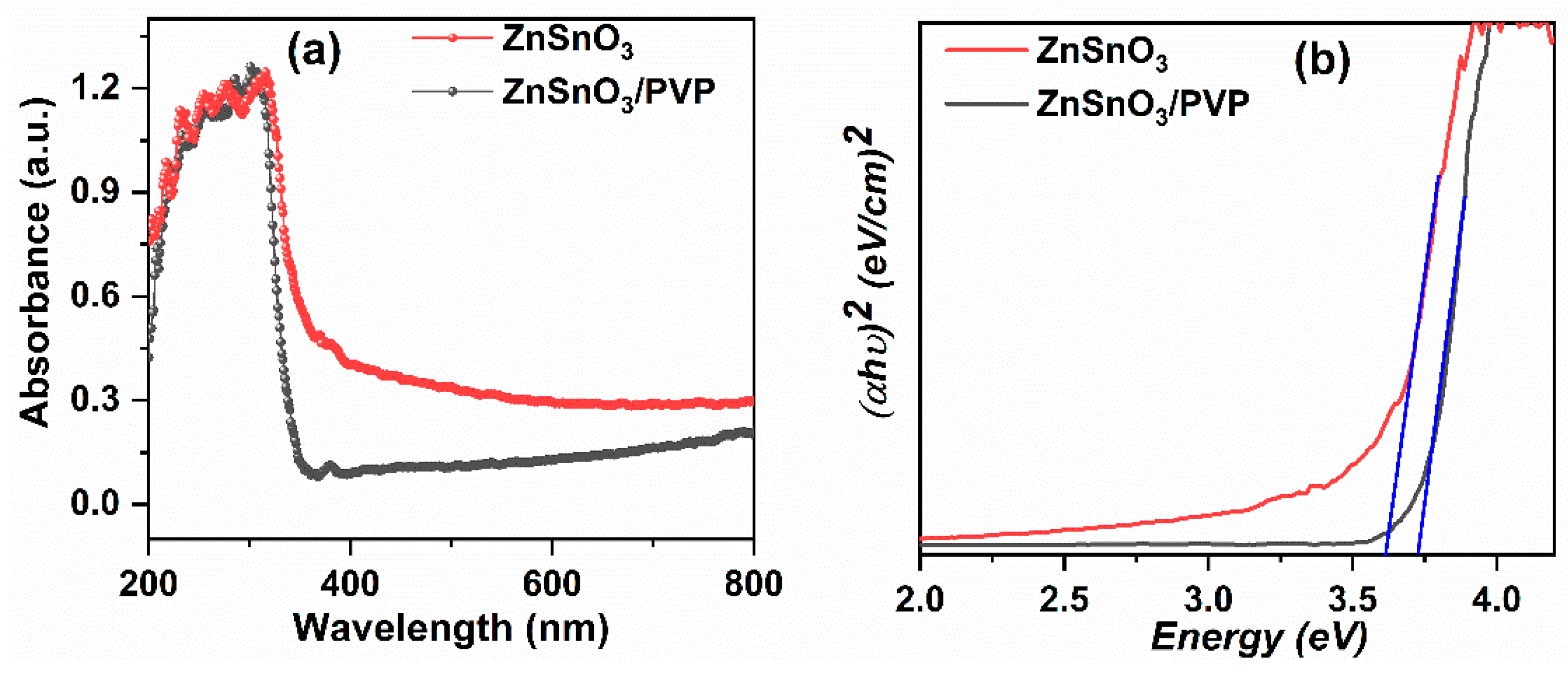
Figure 3a shows the UV–Vis absorbance spectra of ZnSnO3 and ZnSnO3/PVP nanofibers in the wavelength range of 200–800 nm. Both samples exhibit strong absorbance in the UV region (200–380 nm), corresponding to fundamental electronic transitions. The ZnSnO3 nanoparticles display a sharp absorption edge at ~315 nm [32], while the ZnSnO3/PVP nanofibers show a slight blue shift in the edge to ~301 nm, which can be attributed to the effect of the polymer matrix. Additionally, the ZnSnO3/PVP sample exhibits a lower overall absorbance intensity due to the presence of PVP [33]. The observed blue shift indicates a marginal increase in the band gap of the composite compared to pure ZnSnO3, suggesting a slight widening of the band gap that may influence the optical absorption properties. Furthermore, the more pronounced absorbance tail in the visible region (400–800 nm) for the ZnSnO3/PVP nanofibers indicates the presence of localized defect states or enhanced interfacial charge transfer arising from PVP incorporation, which could be advantageous for photocatalytic and photovoltaic applications. Figure 3b displays the Tauc plots ((αhν)2 versus photon energy (hν)) for pristine ZnSnO3 and ZnSnO3/PVP nanocomposites, used to determine their optical band gap energies. Both samples exhibit a clear absorption edge in the UV region, indicative of their semiconducting nature. The linear extrapolation of the plots to the energy axis suggests that pristine ZnSnO3 has a direct band gap of approximately 3.61 eV, whereas the ZnSnO3/PVP composite shows a slightly higher band gap of ~3.73 eV. This slight blue shift can be attributed to the influence of the PVP polymer matrix, which may induce quantum confinement effects or modify the local electronic environment, resulting in a marginal widening of the band gap. Previous reports have demonstrated comparable Tauc plot analyses, indicating similar optical band gap values [34,35]. Such band gap modulation can enhance the optical absorption properties and is advantageous for photocatalytic and optoelectronic applications.

3.4. Morphology Analysis

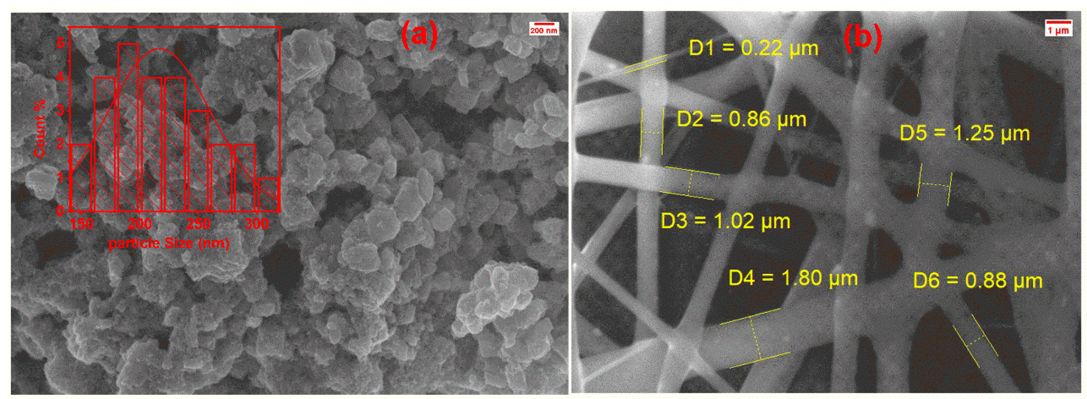
The SEM micrograph of pure ZnSnO3 nanoparticles, shown in Figure 4a, reveals a highly agglomerated structure consisting of nearly cubic-like grains with nanometer-scale dimensions [1,36]. The average grain size obtained for ZnSnO3 is 220 nm. The particles exhibit slight faceting and tend to form dense clusters, which can be attributed to strong interparticle interactions and the high surface energy of the nanoparticles. Such aggregation leads to a porous texture that is favorable for enhanced surface activity in photocatalytic and gas-sensing applications. In contrast, the ZnSnO3/PVP composite, presented in Figure 4b, displays a continuous fibrous network formed by electrospinning. The fibers are smooth and uniform without the presence of beads, confirming the optimized polymer–nanoparticle blending and spinning conditions. The measured fiber diameters range from 0.22 to 1.80 μm, reflecting a relatively broad size distribution [17,37]. The incorporation of ZnSnO3 nanoparticles within the polymeric matrix ensures structural integrity while maintaining the crystalline phase of ZnSnO3, as also confirmed by XRD. The interconnected fibrous morphology is expected to provide high mechanical flexibility along with enhanced interfacial charge transfer pathways, making the ZnSnO3/PVP composite suitable for optoelectronic and photocatalytic applications.

4. Conclusions

ZnSnO3 nanoparticles with an orthorhombic perovskite structure were successfully synthesized and embedded into electrospun PVP nanofibers. XRD and FTIR analyses confirmed that the crystalline phase of ZnSnO3 was preserved within the polymer matrix without impurity formation. UV–Vis measurements revealed a slight blue shift in the absorption edge, corresponding to a band gap increase from 3.61 eV to 3.73 eV, along with enhanced absorption in the visible region due to polymer–nanoparticle interactions. SEM observations showed that while pristine ZnSnO3 nanoparticles tend to agglomerate, their incorporation into PVP produced uniform, bead-free nanofibers with well-dispersed nanoparticles. The resulting composite combines the structural stability of the polymer with the optical activity of ZnSnO3, offering mechanical flexibility and improved interfacial charge transfer. These features make ZnSnO3/PVP nanofibers attractive candidates for applications in photocatalysis, flexible optoelectronics, and energy-harvesting devices.

Author Contributions

R.I.: writing—original draft, methodology, investigation, formal analysis, data curation. A.S.P.: writing—review and editing, supervision, conceptualization. All authors have read and agreed to the published version of the manuscript.

Funding

This research received no external funding.

Institutional Review Board Statement

Not applicable.

Informed Consent Statement

Not applicable.

Data Availability Statement

The raw data supporting the conclusions of this article will be made available by the authors on request.

Conflicts of Interest

The authors declare no conflict of interest.

References

  1. Ibrahim, D.M.; Gaber, A.A.; Reda, A.E.; Abdel Aziz, D.A.; Ajiba, N.A. Structural, optical, and dielectric properties of sol-gel derived perovskite ZnSnO3 nanomaterials. J. Sol-Gel Sci. Technol. 2024, 112, 703–714. [Google Scholar] [CrossRef]
  2. Mukherjee, D.; Datta, A.; Kons, C.; Hordagoda, M.; Witanachchi, S.; Mukherjee, P. Intrinsic anomalous ferroelectricity in vertically aligned LiNbO3-type ZnSnO3 hybrid nanoparticle-nanowire arrays. Appl. Phys. Lett. 2014, 105, 212903. [Google Scholar] [CrossRef]
  3. Anith, A.; Ponnusamy, V. Optical and electrochemical studies on single-phase ZnSnO3 nanostructures—A photosensitive approach. Surf. Interfaces 2024, 51, 104747. [Google Scholar]
  4. Rovisco, A.; dos Santos, A.; Cramer, T.; Martins, J.; Branquinho, R.; Aguas, H.; Fraboni, B.; Fortunato, E.; Martins, R.; Igreja, R.; et al. Piezoelectricity Enhancement of Nanogenerators Based on PDMS and ZnSnO3 Nanowires through Microstructuration. ACS Appl. Mater. Interfaces 2020, 12, 18421–18430. [Google Scholar] [CrossRef]
  5. George, S.D.B.; Nagarajan, K.; Ali, A.A.; Madamala, S.; Subramanian, D.; Kuppamuthu, S.; Aruckiasamy, J.J. Morphology-optimized ZnSnO3 nanopentagons as efficient electron transport layers for high-efficient perovskite solar cells. J. Solid. State Chem. 2025, 347, 125322. [Google Scholar] [CrossRef]
  6. Sim, C.K.; Majid, S.R.; Mahmood, N.Z. ZnSnO3/mesoporous biocarbon composite towards sustainable electrode material for energy storage device. Microchem. J. 2021, 164, 105968. [Google Scholar] [CrossRef]
  7. Dong, S.; Cui, L.; Zhang, W.; Xia, L.; Zhou, S.; Russell, C.K.; Fan, M.; Feng, J.; Sun, J. Double-shelled ZnSnO3 hollow cubes for efficient photocatalytic degradation of antibiotic wastewater. Chem. Eng. J. 2020, 384, 123279. [Google Scholar] [CrossRef]
  8. Choi, K.H.; Siddiqui, G.U.; Yang, B.-s.; Mustafa, M. Synthesis of ZnSnO3 nanocubes and thin film fabrication of (ZnSnO3/PMMA) composite through electrospray deposition. J. Mater. Sci. Mater. Electron. 2015, 26, 5690–5696. [Google Scholar] [CrossRef]
  9. Siddiqui, G.U.; Rehman, M.M.; Choi, K.H. Enhanced resistive switching in all-printed, hybrid and flexible memory device based on perovskite ZnSnO3 via PVOH polymer. Polymer 2016, 100, 102–110. [Google Scholar] [CrossRef]
  10. Prinscilla, J.; Vijayaraghavan, G.V.; Vilvanathaprabu, A.; Suriakarthick, R. Electrical and ferroelectric properties of novel PVDF/ZnSnO3 polymer nanocomposites for flexible energy storage application. J. Alloys Compd. 2025, 1037, 182303. [Google Scholar] [CrossRef]
  11. Teodorescu, M.; Bercea, M. Poly(vinylpyrrolidone)—A Versatile Polymer for Biomedical and Beyond Medical Applications. Polym. Plast. Technol. Eng. 2015, 54, 923–943. [Google Scholar] [CrossRef]
  12. Xue, J.; Wu, T.; Dai, Y.; Xia, Y. Electrospinning and Electrospun Nanofibers: Methods, Materials, and Applications. Chem. Rev. 2019, 119, 5298–5415. [Google Scholar] [CrossRef]
  13. Wang, P.; Lv, H.; Cao, X.; Liu, Y.; Yu, D.-G. Recent Progress of the Preparation and Application of Electrospun Porous Nanofibers. Polymers 2023, 15, 921. [Google Scholar] [CrossRef]
  14. Kim, O.S.; Kwon, J.B.; Kim, S.W.; Xu, B.; Seo, K.H.; Park, C.E.; Do, W.J.; Bae, J.H.; Kang, S.W. Effect of PVP-Capped ZnO Nanoparticles with Enhanced Charge Transport on the Performance of P3HT/PCBM Polymer Solar Cells. Polymers 2019, 11, 1818. [Google Scholar] [CrossRef]
  15. Geetha, K.; Sivasangari, D.; Kim, H.-S.; Murugadass, G.; Kathalingam, A. Electrospun nanofibrous ZnO/PVA/PVP composite films for efficient antimicrobial face masks. Ceram. Int. 2022, 48, 29197–29294. [Google Scholar] [CrossRef]
  16. Kavarthapu, V.S.; Graham, S.A.; Manchi, P.; Paranjape, M.V.; Yu, J.S. Electrospun ZnSnO3/PVDF-HFP Nanofibrous Triboelectric Films for Efficient Mechanical Energy Harvesting. Adv. Fiber Mater. 2023, 5, 1685–1698. [Google Scholar] [CrossRef]
  17. Zhang, C.; Li, H.; Zhuo, Z.; Dugnani, R.; Xue, W.; Zhou, Y.; Chen, Y.; Liu, H. Characterization of the damping and mechanical properties of a novel (ZnSnO3/PVDF)@PPy nanofibers/EP composite. RSC Adv. 2017, 7, 37130. [Google Scholar] [CrossRef]
  18. Chen, Q.; Wang, Y.; Wang, M.; Ma, S.; Wang, P.; Zhang, G.; Chen, W.; Jiao, H.; Liu, L.; Xu, X. Enhanced acetone sensor based on Au functionalized In-doped ZnSnO3 nanofibers synthesized by electrospinning method. J. Colloid. Interface Sci. 2019, 543, 285–299. [Google Scholar] [CrossRef]
  19. ul Haq, M.; Zhang, Z.; Chen, X.; Rahman, N.; Khan, S.; Khatoon, R.; Hassan, S.S.; Ye, Z.; Zhu, L. A two-step synthesis of microsphere-decorated fibers based on NiO/ZnSnO3 composites towards superior ethanol sensitivity performance. J. Alloys Compd. 2019, 777, 73–83. [Google Scholar] [CrossRef]
  20. Kovacheva, D.; Petrov, K. Preparation of crystalline ZnSnO from Li SnO by low-temperature ion exchange. Solid. State Ionics 1998, 109, 327–332. [Google Scholar] [CrossRef]
  21. Shujah, T.; Ikram, M.; Butt, A.R.; Hussain, S.G.; Shahzad, M.K.; Zafar, Q.; Ali, S. Growth of Zinc Oxide and Zinc Stannate Nanostructured Thin Films for Carbon Monoxide Sensing Application. Nanosci. Nanotechnol. Lett. 2019, 11, 1050–1059. [Google Scholar] [CrossRef]
  22. Klobes, B. Comment on Modification of Gas Sensitivity in ZnSnO3 Film from Carrier Behaviors: Derivation, Concentration, and Depletion: Rather SnO2 than ZnSnO3. J. Phys. Chem. C 2024, 128, 16824–16825. [Google Scholar] [CrossRef]
  23. Gaddam, V.; Kumar, R.R.; Parmar, M.; Yaddanapudi, G.R.K.; Nayak, M.M.; Rajanna, K. Morphology controlled synthesis of Al doped ZnO nanosheets on Al alloy substrate by low-temperature solution growth method. RSC Adv. 2015, 5, 13519–13524. [Google Scholar] [CrossRef]
  24. Atisme, T.B.; Yu, C.-Y.; Tseng, E.N.; Chen, Y.-C.; Hsu, P.-K.; Chen, S.Y. Interface Interactions in Conjugated Polymer Composite with Metal Oxide Nanoparticles. Nanomaterials 2019, 9, 1534. [Google Scholar] [CrossRef]
  25. Brahmi, C.; Benltifa, M.; Vaulot, C.; Michelin, L.; Dumur, F.; Airoudj, A.; Morlet-Savary, F.; Reveau, B.; Bousselmi, L.; Lalevée, J. New hybrid perovskites/polymer composites for the photodegradation of organic dyes. Eur. Polym. J. 2021, 157, 110641. [Google Scholar] [CrossRef]
  26. Indhumathi, R.; Priya, A.S.; Aepuru, R. Effect of calcination temperature on the structure, optical and dielectric properties of ZnSnO3 ceramics. Ceram. Int. 2025, 51, 55030–55055. [Google Scholar] [CrossRef]
  27. Sá, B.S.; Zito, C.A.; Perfecto, T.M.; Volanti, D.P. Porous ZnSnO3 nanocubes as a triethylamine sensor. Sens. Actuators B 2021, 338, 129869. [Google Scholar] [CrossRef]
  28. Bora, T.; Al-Hinai, M.H.; Al-Hinai, A.T.; Dutta, J. Phase Transformation of Metastable ZnSnO3 Upon Thermal Decomposition by In-Situ Temperature-Dependent Raman Spectroscopy. J. Am. Ceram. Soc. 2015, 98, 4044–4049. [Google Scholar] [CrossRef]
  29. Deniz, A.E.; Vural, H.A.; Ortaç, B.; Uyar, T. Gold nanoparticle/polymer nanofibrous composites by laser ablation and electrospinning. Mater. Lett. 2011, 65, 2941–2943. [Google Scholar] [CrossRef]
  30. Baganizi, D.R.; Nyairo, E.; Duncan, S.A.; Singh, S.R.; Vida, A. Interleukin-10 Conjugation to Carboxylated PVP-Coated Silver Nanoparticles for Improved Stability and Therapeutic Efficacy. Nanomaterials 2017, 7, 165. [Google Scholar] [CrossRef]
  31. Kallum, M.K.; Stefanos, M.; Lakshminarayana, P.; Sara, E.S. Polyvinylpyrrolidone (PVP) in nanoparticle synthesis. Dalton Trans. 2015, 44, 17883–17905. [Google Scholar] [CrossRef]
  32. Paul, S.; Basak, S.; Ali, W. Zinc Stannate Nanostructure: Is It a New Class of Material for Multifunctional Cotton Textiles? ACS Omega 2019, 4, 21827–21838. [Google Scholar] [CrossRef]
  33. Soltani, N.; Saion, E.; Erfani, M.; Razaee, K.; Bahmanrokh, G.; Drummen, G.P.C.; Bahrami, A.; Hussein, M.Z. Influence of the Polyvinyl Pyrrolidone Concentration on Particle Size and Dispersion of ZnS Nanoparticles Synthesized by Microwave Irradiation. Int. J. Mol. Sci. 2012, 13, 12412–12427. [Google Scholar] [CrossRef]
  34. Saafi, I.; Dridi, R.; Mimouni, R.; Amlouk, A.; Yumak, A.; Boubaker, K.; Petkova, P.; Amlouk, M. Microstructural and optical properties of SnO2-ZnSnO3 ceramics. Ceram. Int. 2016, 42, 6273–6381. [Google Scholar] [CrossRef]
  35. Soltani, N.; Dehzangi, A.; Kharazmi, A.; Saion, E.; Yunus, W.M.M.; Majlis, B.Y.; Zare, M.R.; Gharibshahi, E.; Khalilzadeh, N. Structural, Optical and Electrical Properties of ZnS nanoparticles affecting by organic coating. Chalcogenide Lett. 2014, 11, 79–90. [Google Scholar]
  36. Saeidi, M.; Abrari, M.; Ahmadi, M. Fabrication of dye-sensitized solar cell based on mixed tin and zinc oxide nanoparticles. Appl. Phys. A 2019, 125, 409. [Google Scholar] [CrossRef]
  37. Turky, A.O.; Barhoum, A.; MohamedRashad, M.; Bechlany, M. Enhanced the structure and optical properties for ZnO/PVP noanofibres fabricated via electrospinning technique. J. Mater. Sci. Mater. Electron. 2017, 28, 17526–17532. [Google Scholar] [CrossRef]
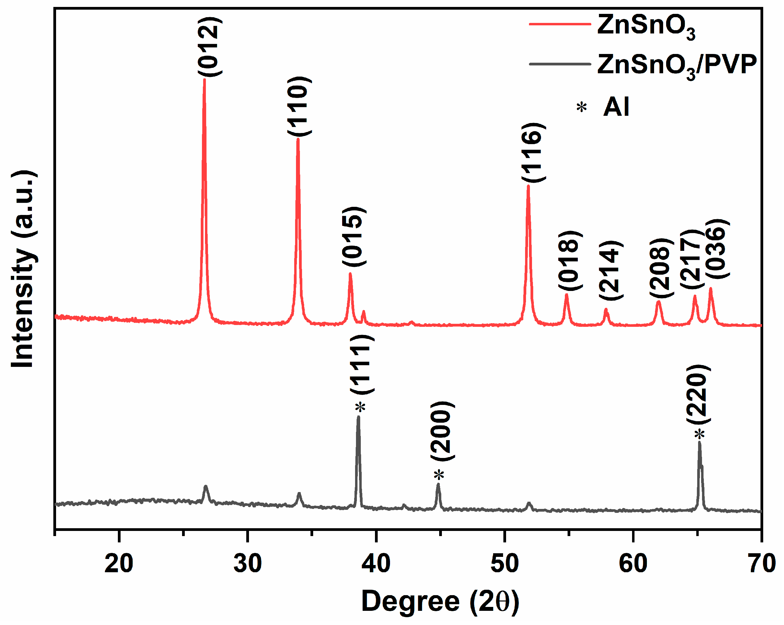
Figure 1. XRD patterns of the prepared ZnSnO3 nanoparticles and ZnSnO3/PVP composite nanofibers.
Materproc 25 00005 g001
Figure 2. FT−IR spectra of the prepared ZnSnO3 nanoparticles and ZnSnO3/PVP composite nanofibers.
Materproc 25 00005 g002
Figure 3. (a) UV–Vis absorbance spectra and (b) Tauc plots of ZnSnO3 and ZnSnO3/PVP.
Materproc 25 00005 g003
Figure 4. SEM images of (a) ZnSnO3 nanoparticles with particle size distribution in the inset and (b) ZnSnO3/PVP electrospun nanofibers.
Materproc 25 00005 g004
Disclaimer/Publisher’s Note: The statements, opinions and data contained in all publications are solely those of the individual author(s) and contributor(s) and not of MDPI and/or the editor(s). MDPI and/or the editor(s) disclaim responsibility for any injury to people or property resulting from any ideas, methods, instructions or products referred to in the content.