Proceeding Paper

A Comparative Study of Titanium-Based Coatings Prepared by Magnetron Sputtering for Biomedical Applications †

1
Division Milieux Ionisés, Centre de Développement des Technologies Avancées CDT, Cité du 20 Août 1956, Baba Hassen, BP. n 17, Alger 16000, Algeria
2
Laboratoire de Sciences et Génie des Matériaux, Département de Métallurgie, École Nationale Polytechnique, 10 Avenue Pasteur, BP. 182, El-Harrach, Alger 16000, Algeria
*
Author to whom correspondence should be addressed.
Presented at the 1st International Online Conference on Bioengineering, 16–18 October 2024; https://sciforum.net/event/IOCBE2024.
Eng. Proc. 2024, 81(1), 9; https://doi.org/10.3390/engproc2024081009
Published: 27 February 2025
(This article belongs to the Proceedings of The 1st International Online Conference on Bioengineering)

Abstract

:
This study investigates the effects of substrate bias voltage on the properties of titanium nitride (TiN) and titanium oxynitride (TiON) thin films deposited via High-Power Impulse Magnetron Sputtering (HiPIMS). The structure and morphology of the obtained coatings were characterized using X-ray diffraction (XRD), scanning electron microscopy (SEM), and atomic force microscopy (AFM). TiN coatings exhibited hydrophilic behavior, while TiON coatings demonstrated hydrophobic characteristics. Electrochemical corrosion testing in Hank’s solution revealed superior corrosion resistance for TiON films deposited at −100 V, indicating their potential for biomedical applications. The observed differences in wettability and corrosion resistance are attributed to the influence of substrate bias voltage on the films’ microstructure and surface chemistry.

1. Introduction

316L stainless steel (SS) has long been a popular choice for medical metallic implants due to its favorable mechanical characteristics and comparatively low fabrication costs, making it a preferred alternative to cobalt-based materials. Its excellent corrosion resistance, strength, and toughness make it an ideal candidate for biomedical applications, facilitating its extensive use in the field of medical implants. However, this material needs to have a high wear resistance and a combination of a low Young’s modulus and a high strength that is comparable to human cortical bone. These properties are all necessary for this biomaterial implant to last longer and to avoid revision surgery [1]. The implant should also prevent cytotoxicity and the formation of bacterial biofilms while integrating well with the surrounding bone. Cytotoxic elements such as chromium (Cr), nickel (Ni), cobalt (Co), and aluminum (Al) are commonly found in metallic implants (316L; CoCr, and Ti6Al4V, etc.) [2]. Coating metallic medical implants with ceramic materials is an effective way to enhance the surface properties of 316L stainless steel (SS) [3,4]. Titanium nitride (TiN) and titanium oxynitride (TiON) are considered advanced ceramics due to their exceptional properties. TiON, in particular, is a versatile alternative that combines the characteristics of metal oxides and those of nitrides [5,6]. These coatings have been extensively researched to enhance the durability and lifespan of surgical implants and prostheses by providing resistance to wear and corrosion. Several methods are used for medical surface treatments, including chemical techniques like anodization [7] and physical technics such as sputtering [8], High-Power Impulse Magnetron Sputtering (HIPIMS) is a recently developed physical vapor deposition (PVD) technique used to deposit high-quality thin coatings. This method generates a highly dense plasma with a greater concentration of metal ions compared to neutral particles, which sets it apart from conventional sputtering techniques (like Radio Frequency and Direct Current magnetron sputtering) [9]. During this HIPIMS process, the electron density reaches levels between 1018 and 1019 m−3, comparable to the mean free path of electron impact ionization, which is typically around 1 cm or less [10].
Among the in vitro tests conducted to evaluate medical implants, the control of surface corrosion resistance in various simulated body fluid (SBF) solutions, such as Ringer’s and Hank’s solutions, is a crucial assessment. The corrosion resistance assessment in different SBF solutions allows for the examination of the implant material’s stability and durability in environments that closely replicate those found in the human body. Additionally, the evaluation of wettability and surface roughness helps predict the implant’s interactions with the surrounding biological fluids and tissues, which can directly impact the formation of biofilms and the overall integration of the implant within the body [11].
Our study aims to study titanium nitride and titanium oxynitride thin coatings deposited on 316L stainless steel (SS) using High-Power Impulse Magnetron Sputtering (HIPIMS). We explore the effects of applying a negative substrate bias voltage of –100 volts, comparing conditions with and without this bias. Our primary focus is to determine how substrate bias voltage affects the surface roughness, wettability, and corrosion properties of the coated samples.

2. Materials and Methods

2.1. Deposition Protocol

The coatings were deposited using a High-Power Impulse Magnetron Sputtering (HIPIMS) system. A high-purity titanium nitride (TiN) target with a diameter of 76.2 mm, thickness of 6.35 mm, and purity of 99.9% was used. The substrate–target distance was set to 30 mm. For more detailed information about the sputtering system, please refer to our previous work [12]. Prior to each experiment, a turbomolecular pump was used to evacuate the vacuum chamber to achieve a base pressure of 1.33 × 10⁻³ Pa. During the deposition, argon (99.9997% purity) and oxygen (99.9995% purity) gases were introduced into the deposition chamber via two mass flow controllers. The Ar and O2 flow rates were fixed at 19 sccm and 6.4 sccm, respectively. The total work pressure was fixed at 1.33 Pa. A negative DC bias voltage (0 V and −100 V) was applied to the substrates during a 30 min period. The HiPIMS power supply delivered a sputtering power of 100 W and a voltage of 400 V to the target. Single-pole pulses with a length of 25 μs and a frequency of 600 Hz were supplied by a HiPSTER 1 pulse unit (Ionautics AB) powered by a DCPSU power supply. Before deposition, the substrates (316L SS and Si) were ultrasonically cleaned with an acetone and ethanol solution for 10 min and then dried in a drying oven at 70 °C.

2.2. Characterization Methods

The structures of the coatings were analyzed using X-ray diffraction (XRD, Bruker AXS D8 Advance) and scanning electron microscopy (SEM, JEOL JSM 6360LV, Japan). Atomic force microscopy (AFM) was conducted in tapping mode over 3 μm × 3 μm areas. The wettability behavior of the deposited coatings was investigated using an OCA 15 (Plus Data Physics Instruments, GmbH Germany) with sessile drop method, where a 5 μml water drop was placed on the surface. The electrochemical tests were carried out using a PARSTAT 4000 (AMETEK, USA), which consists of a galvanostat potentiostat connected to a microcomputer. The results were analyzed using Versa Studio (2.66.2) software. Electrochemical behavior was evaluated in a physiologically simulated solution, with the open-circuit potential recorded over 12 h of immersion in Hank’s solution, the composition of which is detailed in Table 1 [13].

3. Results and Discussion
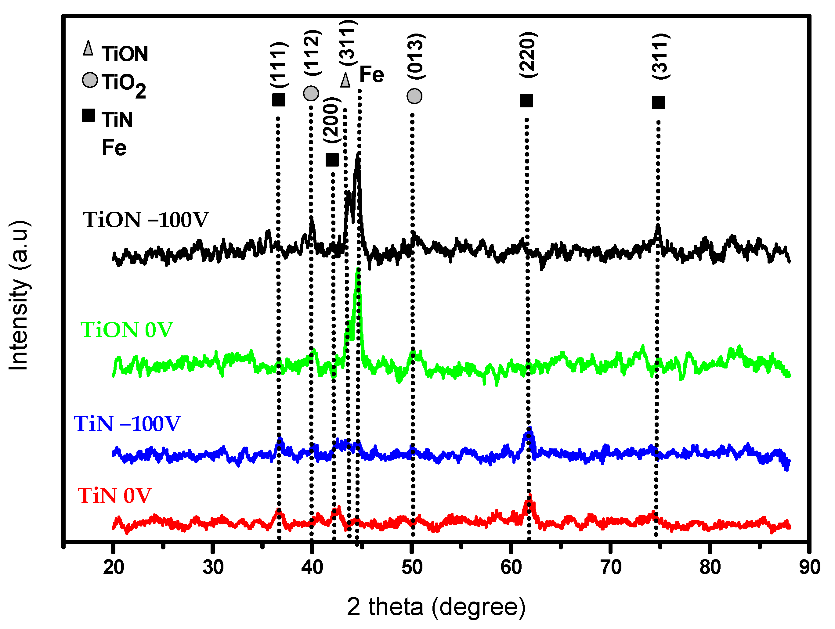
3.1. Structure

The X-ray diffraction (XRD) analysis was conducted to investigate the crystallographic structure of the coatings and to measure the size of the crystallites. Figure 1 shows the diffractograms recorded for the deposited TiN and TiON coatings, which clearly demonstrate the crystallinity of the films. Both TiN and Ti2N phases coexisted in the two titanium nitride samples, with a clear dominance of the TiN phase exhibiting a preferred orientation along the (220) plane. Substrate bias voltage led to an increase in the intensity of the peaks, and therefore to greater crystallization. The introduction of oxygen as a reactive gas for the deposition of TiON resulted in the formation of oxidized thin films, including TiON and TiO2. In the case of the TiON coatings, the preferred orientation was found to be (311) under both biased and unbiased conditions [14].
The effect of negative bias voltage on crystallite size values is presented in Table 1. The crystallite size D is determined using the Scherer formula (Equation (1)):
                              D = K λ β h k l cos θ                              
where (D) is the crystallite size, (K) is the shape factor (0.9), and (βhkl) is the full width at half maximum (FWHM) of the peak, while λ is the wavelength of CuKα radiation and ϴ is Bragg’s angle.
Figure 2 presents SEM cross-sectional images of TiN and TiON coatings deposited on a silicon substrate at 0 V and −100 V bias, from which the coating thickness (H) is measured. Table 2 summarizes the coating’s structural parameters based on the substrate bias voltage—the electrical potential applied during the deposition process. Specifically, the coating thickness (H, in nm) was determined via SEM, the grain size (D) was estimated from the broadening of XRD peaks, and the average surface roughness (Ra) was measured using AFM.
Substrate bias voltage had no significant effect on the thickness and grain size of the coatings. All coatings displayed smooth surfaces with low surface roughness. In fact, the nanoscale values (1.4–7.4 nm) of surface roughness indicate very smooth morphologies, which are probably due to the maintenance of the preferred growth orientation [13].

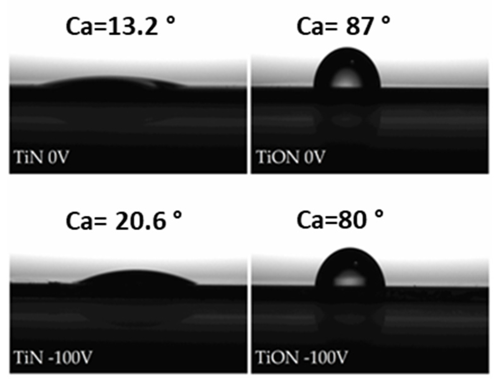
3.2. Surface Wettability
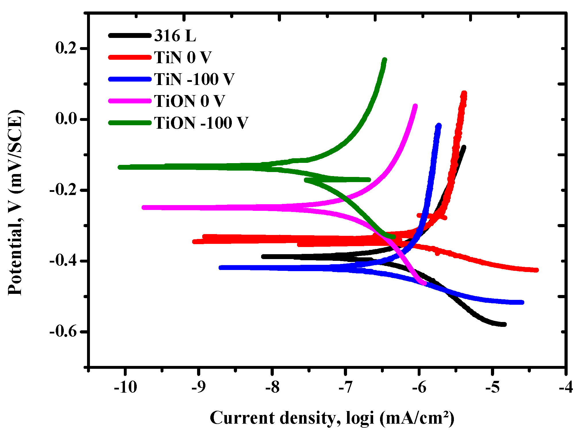
The contact angle reflects the wettability of a liquid on a solid surface. The shape of a droplet on a surface depends on the interplay between the liquid’s surface tension and the inherent properties of the surface. Surface tension causes the droplet’s interface with the surrounding air to curve. A higher contact angle indicates lower wettability and a more hydrophobic surface [15]. Figure 3 shows the wettability characteristics of titanium nitride (TiN) and titanium oxynitride (TiON) coatings for two substrate bias voltages (0 V and −100 V), with corresponding contact angles (Ca) indicating their hydrophilic or hydrophobic nature. The TiN coating at 0 V exhibits a very low contact angle of 13.2°, demonstrating excellent hydrophilicity and promoting significant droplet spreading. In contrast, the TiON coating at 0 V shows a high contact angle of 87°, indicative of hydrophobic behavior and limited wettability. Interestingly, while the TiN coating at −100 V maintains good wettability with a slightly increased contact angle of 20.6°, the TiON coating shows a modest improvement in wettability with a contact angle of 80°. These findings are similar to those of Koerner et al. [16]. The high contact angle (Ca) observed for TiON coatings indicates their hydrophobic nature attributed to the presence of oxygen [17,18].

3.3. Corrosion Test

Corrosion testing uses potentiodynamic polarization measurements. The potential is presented as a function of current density (I) at each measured point, providing the polarization curve (Tafel plots). Figure 4 shows the polarization curves for coated and uncoated 316L (SS). Extrapolating the Tafel regions of the respective TiN and TiON curves gave the corrosion potential (Ecorr), corrosion current density (Icorr), and anodic/cathodic Tafel constants. The Icorr values for TiN (0 V and −100 V) were significantly lower than those for 316L steel. TiON coatings demonstrated even greater corrosion resistance, significantly outperforming uncoated steel. This improvement is attributed to the formation of highly stable oxidized phases, resulting in overall passivation of the thin films.
The parameters extracted from the electrochemical tests, including corrosion voltage and current, polarization resistance, and protection efficiency, are presented in Table 3. The Icorr values for TiN (0 V and −100 V) are significantly lower than those for 316L steel (114.012 nA/cm2). TiON coatings exhibit even better corrosion resistance, far superior to that of uncoated steel. This improvement is attributed to the formation of highly thermodynamically stable oxidized phases (TiO2, TiO, and TiON), which lead to overall passivation of the thin films. A high corrosion rate was observed in the TiN and TiON coatings, where 0.0017 mm/year was measured for TiON with a negative bias voltage (−100 V), and the corrosion rate increased considerably in the experiments with lower negative bias voltage. Resistance was increased in TiON (−100 V) compared to TiN (−100 V), which is because of the formation of fine grain structures and highdensity coatings. The significant enhancements in electrochemical parameters are also due to the application of polarization, which results in a more compact structure and reduces the number of defects [19].
A range of inorganic ions, including sodium, potassium, calcium, magnesium, chloride, phosphate, and bicarbonate, are commonly present in the aqueous solution known as Hank’s solution. The following significant chemical processes could result in the electrochemical treatment’s conversion of TiON to TiO2 in the presence of this aqueous solution:
Oxidation of TiN:       TiN + O2 → TiO2 (rutile) + ½ N2
The TiN phase may be oxidized by the oxygen contained in Hank’s solution, changing it into the more stable rutile TiO2 phase [20].
Hydrolysis of TiN:       TiN + H2O → TiO2 (anatase) + ½ N2 + 2 H2

4. Conclusions

In this study, titanium nitride (TiN) and titanium oxynitride (TiON) coatings were deposited on 316L stainless steel substrates using High-Power Impulse Magnetron Sputtering. The study focused on how a negative substrate bias voltage influences the coatings’ structure, surface morphology, wettability, and electrochemical behavior.
All coatings displayed exceptionally smooth surfaces, with the TiON coating at 0 V showing the lowest roughness (1.4 nm). The substrate bias voltage did not noticeably affect either the thickness or the grain size. TiN coatings measured about 500 nm in thickness, while TiON coatings were around 100 nm, and all coatings had a similar grain size of about 20 nm. However, applying a negative bias voltage significantly enhanced corrosion resistance in both materials, with TiON at −100 V showing the lowest corrosion current density (29.15 nA/cm2). The TiN coating at 0 V demonstrated superior wettability, with a contact angle of only 13.2°, compared to the TiON coating, which exhibited poor wettability with a contact angle of approximately 80°. Overall, TiN coatings, with their excellent wettability, appear to be more suitable for surgical instruments, while TiON coatings, demonstrating improved corrosion resistance, hold promise for medical implant applications.

Author Contributions

Methodology, N.S.; investigation, F.L., G.C. and Y.A.; writing—original draft preparation, F.L., N.M., M.A. and A.H.; writing—review and editing, N.S.; supervision, N.S. and F.L. All authors have read and agreed to the published version of the manuscript.

Funding

The ministry of higher education and scientific research and The Directorate General for Scientific Research and Technological Development (DGRSDT) of Algeria.

Institutional Review Board Statement

Not applicable.

Informed Consent Statement

Not applicable.

Data Availability Statement

Dataset available on request from the authors.

Conflicts of Interest

The authors declare no conflicts of interest.

References

  1. Jimenez-Marcos, C.; Mirza-Rosca, J.C.; Baltatu, M.S.; Vizureanu, P. Experimental research on new developed titanium alloys for biomedical applications. Bioengineering 2022, 9, 686. [Google Scholar] [CrossRef] [PubMed]
  2. Quinn, J.; McFadden, R.; Chan, C.-W.; Carson, L. Titanium for orthopedic applications: An overview of surface modification to improve biocompatibility and prevent bacterial biofilm formation. iScience 2020, 23, 5–6. [Google Scholar] [CrossRef] [PubMed]
  3. Bekmurzayeva, A.; Duncanson, W.J.; Azevedo, H.S.; Kanayeva, D. Surface modification of stainless steel for biomedical applications: Revisiting a century-old material. Mater. Sci. Eng. C 2018, 93, 1073–1089. [Google Scholar] [CrossRef] [PubMed]
  4. Mejía, C.P.; Vanegas, H.S.; Olaya, J.J. Electrochemical and optical behavior of ZrN-Ag coatings deposited by means of DC reactive magnetron sputtering technique. Coatings 2022, 12, 754. [Google Scholar] [CrossRef]
  5. Rajan, S.T.; Arockiarajan, A. Thin film metallic glasses for bioimplants and surgical tools: A review. J. Alloys Compd. 2021, 876, 159939. [Google Scholar] [CrossRef]
  6. Amirtharaj Mosas, K.K.; Chandrasekar, A.R.; Dasan, A.; Pakseresht, A.; Galusek, D. Recent advancements in materials and coatings for biomedical implants. Gels 2022, 8, 323. [Google Scholar] [CrossRef] [PubMed]
  7. Atmani, D.; Saoula, N.; Chouchane, K.; Lalmi, M.; Abdi, A.; Olaru, P. Study of Mg-Rich Sulfato-Calcium Hydroxyapatite Coating on Anodized Ti6Al4V-Alloy for Biomedical Applications. J. Mater. Eng. Perform. 2024, 1–15. [Google Scholar] [CrossRef]
  8. Madaoui, N.; Saoula, N.; Kheyar, K.; Nezar, S.; Tadjine, R.; Hammouche, A.; Belhousse, S. The effect of substrate bias voltage on the electrochemical corrosion behaviors of thin film deposited on stainless steel by r. f magnetron sputtering. Prot. Met. Phys. Chem. Surf. 2017, 53, 527–533. [Google Scholar] [CrossRef]
  9. Jiang, Q.; Ma, D.; Li, Y.; Chen, C. The Deposition and Properties of Titanium Films Prepared by High Power Pulsed Magnetron Sputtering. Materials 2023, 16, 7294. [Google Scholar] [CrossRef] [PubMed]
  10. Yan, Y.; Lizhen, Y.; Zhongwei, L.; Qiang, C. High power impulse magnetron sputtering and its applications. Plasma Sci. Technol. 2018, 20, 065501. [Google Scholar] [CrossRef]
  11. Yang, K.; Shi, J.; Wang, L.; Chen, Y.; Liang, C.; Yang, L.; Wang, L.-N. Bacterial anti-adhesion surface design: Surface patterning, roughness and wettability: A review. J. Mater. Sci. Technol. 2022, 99, 82–100. [Google Scholar] [CrossRef]
  12. Zinai, N.; Bouzidi, A.; Saoula, N.; Miloua, R.; Medles, M.; Filali, W.; Garoudja, E.; Azibi, M.; Connelly, P.; Nakrela, A. Tailoring the structural and optical properties of HiPIMS TiO2 thin films for photovoltaic applications. Opt. Mater. 2022, 131, 112590. [Google Scholar] [CrossRef]
  13. Azibi, M.; Saoula, N.; Madaoui, N.; Aknouche, H. Effects of nitrogen content on the structural, mechanical, and corrosion properties of ZrN thin films grown on AISI 316L by radiofrequency magnetron sputtering. Cryst. Res. Technol. 2021, 56, 2100096. [Google Scholar] [CrossRef]
  14. Hunt, J.; Soletta, I.; Battezzati, L.; Cowlam, N.; Cocco, G. On the influence of gaseous impurities in the amorphization reaction of some titanium-based alloys. J. Alloys Compd. 1993, 194, 311–317. [Google Scholar] [CrossRef]
  15. Čolović, B.; Kisić, D.; Jokanović, B.; Rakočević, Z.; Nasov, I.; Petkoska, A.T.; Jokanović, V. Wetting properties of titanium oxides, oxynitrides and nitrides obtained by DC and pulsed magnetron sputtering and cathodic arc evaporation. Mater. Sci.-Pol. 2019, 37, 173–181. [Google Scholar] [CrossRef]
  16. Koerner, R.; Butterworth, L.; Mayer, I.; Dasbach, R.; Busscher, H. Bacterial adhesion to titanium-oxy-nitride (TiNOX) coatings with different resistivities: A novel approach for the development of biomaterials. Biomaterials 2002, 23, 2835–2840. [Google Scholar] [CrossRef] [PubMed]
  17. Estrada-Martínez, J.; Reyes-Gasga, J.; García-García, R.; Vargas-Becerril, N.; Zapata-Torres, M.G.; Gallardo-Rivas, N.V.; Mendoza-Martínez, A.M.; Paramo-García, U. Wettability modification of the AISI 304 and 316 stainless steel and glass surfaces by titanium oxide and titanium nitride coating. Surf. Coat. Technol. 2017, 330, 61–70. [Google Scholar] [CrossRef]
  18. Mrabet, C.; Mahdhi, N.; Boukhachem, A.; Amlouk, M.; Manoubi, T. Effects of surface oxygen vacancies content on wettability of zinc oxide nanorods doped with lanthanum. J. Alloys Compd. 2016, 688, 122–132. [Google Scholar] [CrossRef]
  19. Azibi, M.; Madaoui, N.; Saoula, N.; Idir, B.; Sam, S.; Khelladi, M.R. Tailoring Zr-N Thin Film Characteristics on Ti-6Al-4 V Alloy: Effect of Nitrogen and Bias Voltage. Braz. J. Phys. 2024, 54, 90. [Google Scholar] [CrossRef]
  20. Piallat, F.; Gassilloud, R.; Caubet, P.; Vallée, C. Investigation of TiN thin film oxidation depending on the substrate temperature at vacuum break. J. Vac. Sci. Technol. A 2016, 34, 051508. [Google Scholar] [CrossRef]
Figure 1. XRD patterns of the deposited coatings.
Engproc 81 00009 g001
Figure 2. SEM cross-sectional views of the TiN and TiON coatings deposited on Si substrate.
Engproc 81 00009 g002
Figure 3. Water contact angle (Ca) of TiN and TiOn coatings.
Engproc 81 00009 g003
Figure 4. Potentiodynamic polarization curves for the TiN, TiON coatings and uncoated 316L stainless steel alloy in Hank’s solution at 37 °C.
Engproc 81 00009 g004
Table 1. Chemical composition of Hank’s solution.
CompoundsQuantity g/L
NaCl8
KCl0.4
CaCl20.18
KH2PO40.06
NaHCO30.35
Na2HPO40.0475
MgCl2·6H2O0.10
MgSO4·7H2O0.10
C6H12O61
Table 2. Thickness H, grain size (D) and average roughness of the deposited TiN and TiON coatings deposited at 0 and −100 V.
Bias Substrate Voltage (V)Thickness H (nm)Grain Size (nm)Roughness Ra (nm)
TiN 0500167.4
TiN −100450121.5
TiON 0<100161.4
TiON −100<100201.5
Table 3. The corrosion parameters measured for the TiN, TiON coatings in a physiological medium (Hank’s solution at 37 °C).
SamplesEcorr
(mV vs. SCE)
Icorr (nA/cm2)Rp (KΩ.cm2)Cr (mm/an)Pe
(%)
316L SS−380.435114.012274.290.0043--
TiN 0−335.065760.67130.1960.03470
TiN −100−417.186461.74480.9580.0130
TiON 0−248.73885.271335.4260.003625
TiON −100−139.16829.156708.3780.001774
Disclaimer/Publisher’s Note: The statements, opinions and data contained in all publications are solely those of the individual author(s) and contributor(s) and not of MDPI and/or the editor(s). MDPI and/or the editor(s) disclaim responsibility for any injury to people or property resulting from any ideas, methods, instructions or products referred to in the content.

