1. Introduction
Non-destructive testing (NDT) is a significant methodology both for composite material maintenance and cultural heritage conservation. Infrared thermography (IRT) is a well-known NDT technique, and it has the advantages of a fast inspection rate, the absence of contact, excellent spatial resolution and acquisition rate [
1]. The heat transfer process through materials, which is the basis of IRT, determines its sensitivity for the detection of both surface and sub-surface defects. Terahertz time-domain spectroscopy (THz-TDS) is a non-invasive, high-resolution imaging technique which can discriminate between materials effectively [
2]. However, complex material composition and geometry suppress the signal-to-noise ratio (SNR) of imaging on the internal defects using a single imaging modality.
To tackle this problem and provide high-contrast imaging results, a non-invasive imaging technique is proposed here for non-destructive inspection on both cultural heritage objects and natural fiber composites. The proposed technique combines the surface information provided by IRT and the internal structure retrieved with THz-TDS using an unsupervised deep residual fusion network. Experiments show that the fusion results provide more material information than a single modality. In addition, 3D imaging has been achieved using the fusion results on natural fiber composites.
2. Specimens
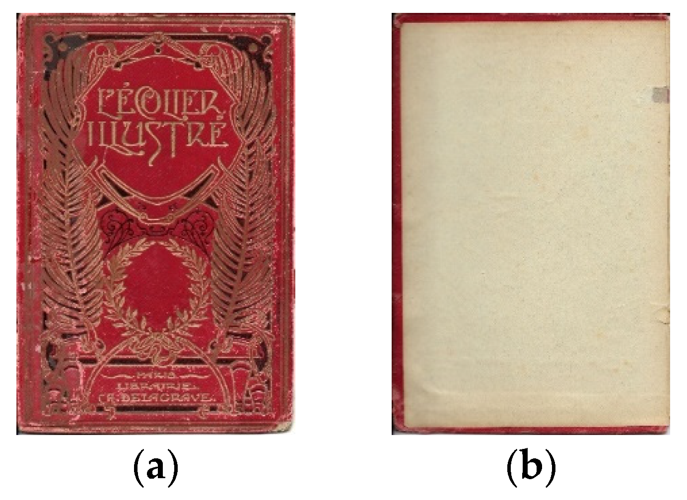
As a representative sample of cultural heritage, an old book cover was investigated in this study. Photographs of the book cover of a 19th century book is shown in
Figure 1. The book, entitled The Little Illustrated Student, was published in Paris, France.
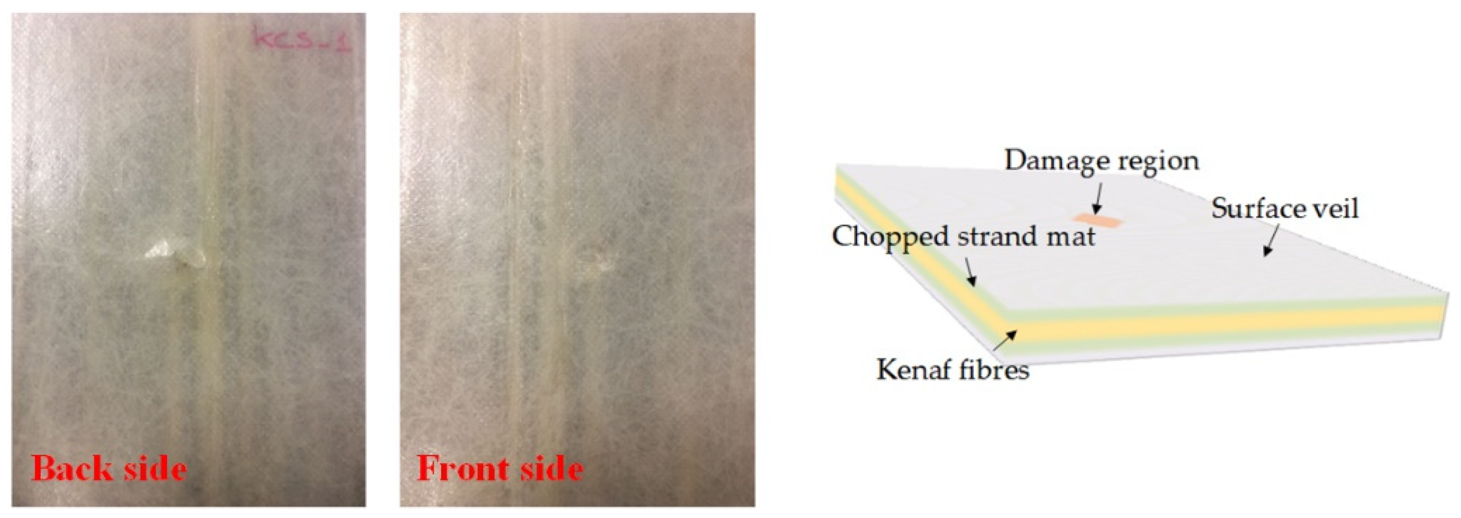
Additionally, a natural fiber composite, mainly constituted by kenaf, was inspected using the proposed approach. The structure of the kenaf fiber composite is illustrated in
Figure 2. In particular, the composite was realized with a hybrid method using both kenaf and glass fibers. A sandwich structure was realized at the end of the fabrication process. The core was fabricated with continuous kenaf/glass hybrid fiber yarns, whereas the two skins were made of a chopped strand matrix with a surface veil. The composite was later impacted in order to provoke damage partially visible to the naked eye.
3. Infrared-Terahertz Fusion
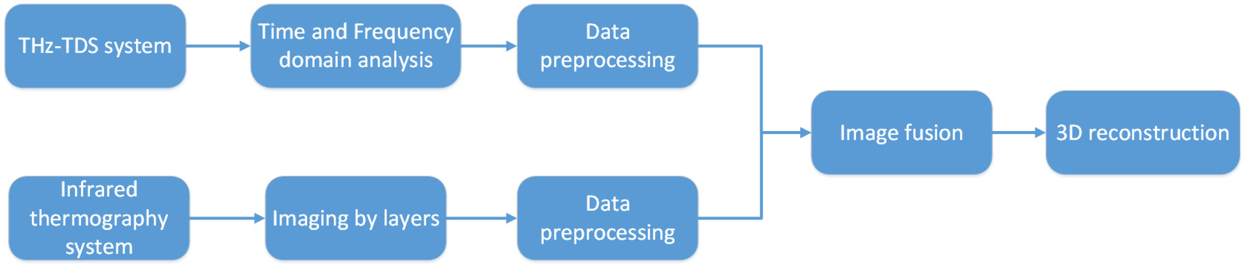
The proposed infrared-terahertz fusion approach is illustrated in
Figure 3. Raw data collected from THz-TDS system was analyzed both in the time domain and frequency domain to obtain images based on different time domain representations and spectral characteristics of the materials.
The obtained thermal sequence from the IRT technique was also analyzed to image the different depths. Then, the analytical results from THz-TDS and IRT were pre-processed in order to confirm that the images from different techniques were in strict geometrical alignment and were intensity-matched. An unsupervised deep residual fusion network was developed to fuse the images from the different techniques (i.e., THz and IRT), and then the fused results were reconstructed in a 3D form to show the through-depth imaging results of the materials.
4. Results
Figure 4c shows the fused results on the book cover. The IRT imaging provided the texture details of it, along with the slight detachment, shown both at the upper and at the bottom right corner. However, the heat penetration depth limited the performance in inspecting internal defects. The THz imaging performed in transmission mode (
Figure 4b) provided the internal defects and detachment clearly. The fusion imaging shown in
Figure 4c combined both surface details (
Figure 4a) and the internal defect information (
Figure 4b). Moreover, the contrast of different materials was also improved.
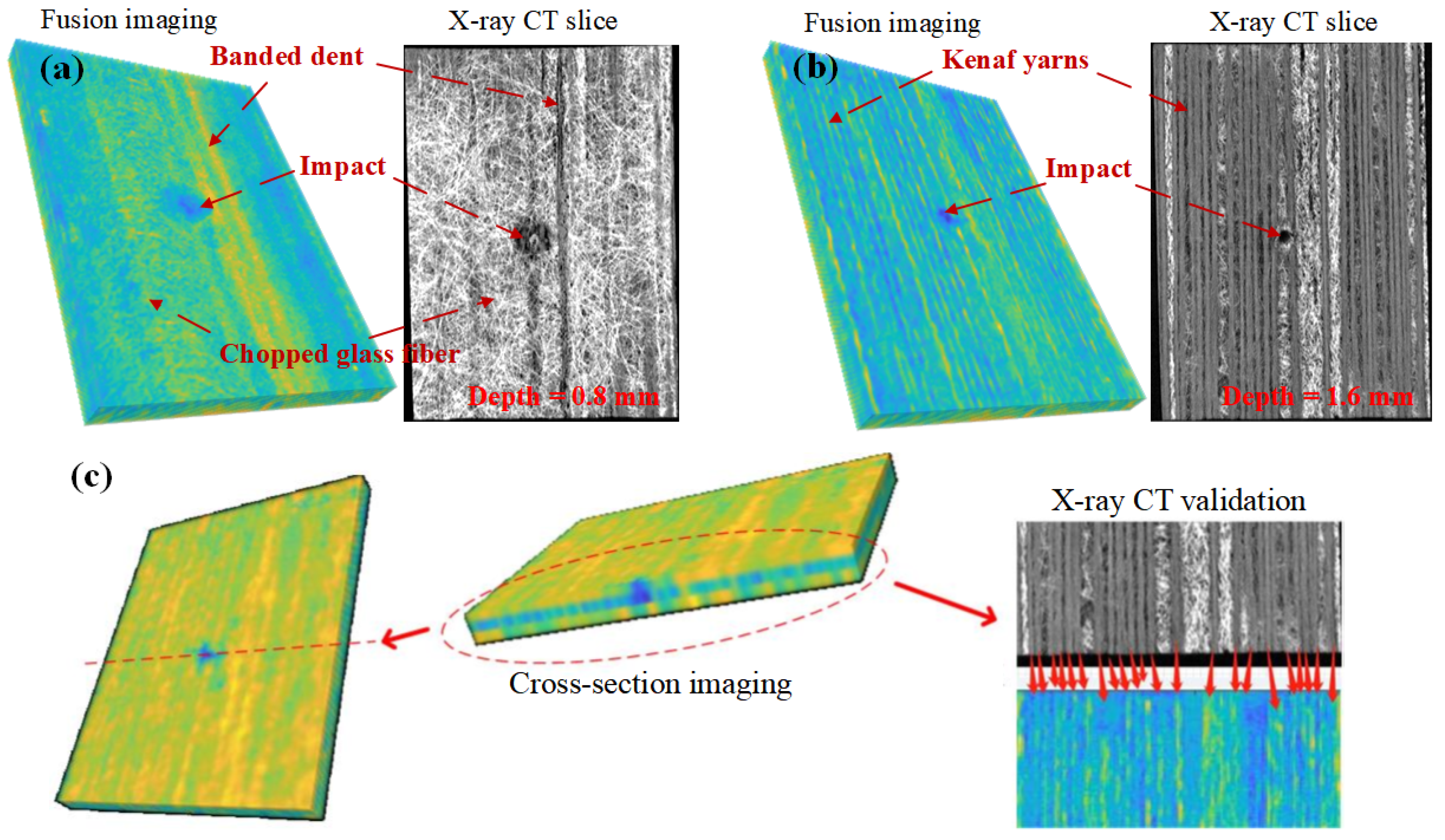
The 3D reconstruction results of the kenaf fiber composite are shown in
Figure 5. In the second row of the figure, the images observed from different views of the 3D reconstruction model are reported.
Figure 5a represents a cut on the chopped strand mat layer, compared with the X-ray computed tomography (CT) slice at the depth of 0.8 mm from the back panel. The low-velocity impact-induced damage and banded dents observed in IRT imaging are merged with the glass fiber content distribution viewed in THz imaging. All these patterns are presented in the fusion imaging results and can be mapped by the referred X-ray CT slice.
Figure 5b represents a cut on the kenaf yarn layer, which refers to the CT slice obtained at the depth of 1.6 mm from the back panel. It is worth mentioning that, as an internal layer, the kenaf yarns are observed clearly with the impact-induced damage in the 3D fused result.
Figure 5c shows the cross-section imaging cut at the position of 71.5 and 88.5 mm from the upper edge of the sample. It is interesting that the impact-induced damage appears at the position of 71.5 mm from upper edge. Therefore, the morphology of the impact can be observed in this cross-section imaging.
Figure 5d presents the horizontal section of the kenaf fiber yarn layer. As an internal layer in this unusual 3D structure, the kenaf yarn layer was chosen for the detailed comparison with the CT slice. The kenaf yarns present both on the cropped horizontal section of 3D fusion imaging and the cropped CT slice are matched by means of a red arrow. A total of 26 kenaf yarns can be mapped into the X-ray CT inspection result, one-by-one.
5. Conclusions
In this study, a non-invasive imaging technique is proposed for non-destructive inspection of both cultural heritage objects and natural fiber composites. The proposed technique combines the surface information using IRT and the internal ones obtained via THz-TDS; an unsupervised deep residual fusion network is here developed for the first time. The results for an old book cover combine the defect information of two modalities and enhance the contrast in imaging. The results for a natural fiber composite using 3D imaging offer a better way to understand the characteristics of the composite itself. Comparison between the 3D fusion imaging results and X-ray CT inspection proves that the proposed fusion imaging technique has a promising performance in the imaging of different layers in such a complex composite structure.


