Proceeding Paper

Photocatalytic Degradation of Malathion Using Hydroxyapatite Derived from Chanos chanos and Pangasius dory Bones †

by
Allen S. Credo
,
Mckenneth G. Pascual
,
Mark Jerome C. Villagracia
,
Alden D. Villaruz
,
Erison C. Roque
,
Edgar Clyde R. Lopez
* and
Rugi Vicente C. Rubi
*
Chemical Engineering Department, Adamson University, 900 San Marcelino St., Ermita, 1000 Manila, Philippines
*
Authors to whom correspondence should be addressed.
Presented at the 2nd International Electronic Conference on Processes: Process Engineering—Current State and Future Trends (ECP 2023), 17–31 May 2023; Available online: https://ecp2023.sciforum.net/.
Eng. Proc. 2023, 37(1), 7; https://doi.org/10.3390/ECP2023-14618
Published: 17 May 2023

Abstract

:
Farmers widely use malathion, even in households, and significant amounts seep through groundwater and effluent wastewater. It is toxic to animal and human life. Hence, its removal from wastewater is necessary. Here, we report the applicability of hydroxyapatite as a catalyst in the UV-light-assisted degradation of malathion. The hydroxyapatite was synthesized via calcination from milkfish (MF1000) and cream dory (CD1000) bones. FTIR and PXRD results proved the successful synthesis of hydroxyapatite from the fish bones. SEM images revealed that the synthesized hydroxyapatite varies in size from 19 to 52 nm with a pseudo-spherical morphology. Degradation efficiency increases when catalyst dosage or irradiation time are increased. Degradation efficiencies range from 8.18% to 67.80% using MF1000 and from 20.50% to 67.90% using CD1000. Malathion obeys first-order kinetics with a kinetic constant up to 7.0289 × 10⁻³ min−1 for 0.6 g catalyst loading. Meanwhile, malathion obeys second-order kinetics with a kinetic constant up to 1.1946 × 10⁻³ L min−1 mg−1 for 0.6 g loading. Across all catalyst loadings, CD1000 has faster degradation kinetics compared to MF1000. The results of this study validate that the calcined fish bones are effective in removing malathion in an aqueous solution, which significantly lessens the detrimental effects of pesticides in groundwater and wastewater.

1. Introduction

The consumption of agrochemicals in the Philippines is relatively large due to the massive farming activities in the country. Agrochemicals such as fertilizers and pesticides are used to boost production, ensure productivity, and prevent crop damage. Despite their benefits in elongating the lives of crops through the prevention of pests, pesticides are poisonous to insects and humans. Since these chemicals have been occurring for decades, consequences emerge, such as contamination of water sources and indirect harm to human health [1]. Fungicides such as dithane, mancozeb, and funguran; insecticides such as cypermethrin and malathion; and weedicides were found to be the pesticides that are most consumed by Filipino farmers [1,2]. Brought about by the large consumption of the said pesticides, some of these have been found in bodies of water. Specifically, cyhalothrin, cypermethrin, and deltamethrin were detected on streams and rivers in the islands of Leyte [3]. Organochlorine pesticides such as dieldrin, endosulfan II, α-hexachlorocyclohexane, and heptachlor are detected in the surface water and groundwater in Pampanga [4]. Additionally, catchments along Laguna Lake have also been proven to be positive in terms of pesticide contamination due to agricultural activities performed around the area [5]. Presently, pesticide contamination in bodies of water and wastewater is of high concern, as current wastewater treatment methods are somehow inefficient in removing such hazardous chemicals.
One solution to prevent further damages brought about by pesticide contamination of water streams is the treatment of such wastewater prior to final disposal, which leads to a high demand for technologies for the removal of pesticides from agricultural run-off and wastewater [6]. Among these treatments are electrochemical methods [7], adsorption [8], phytoremediation [9], and photocatalysis [10]. Among these, photocatalysis remains the top choice of researchers due to its simplicity in completely degrading the pollutants. In photocatalysis, the system is irradiated by light to induce electron and hole formation on the catalyst’s surface. These electron holes would then function as mediators of the redox reaction. The end product would then react with dissolved contaminants in the water, following the degradation or separation of the contaminant from the water [11]. A wide range of photocatalysts have been investigated for pesticide degradation, such as TiO2 [12], ZnO [13], and other semiconductors [14], among others.
Recently, the use of greener photocatalysts such as hydroxyapatite (HAp, Ca10(PO4)(OH)2) for photocatalysis has been studied to replace conventional semiconducting photocatalysts [15]. Hydroxyapatite has been proven to degrade via photocatalysis, dyes [16], pharmaceuticals [17], and pesticides [18] from wastewater and aqueous solutions.
Here we report the synthesis of HAp from the fish bones of milkfish, Chanos chanos, and cream dory, Pangasius dory, via calcination. The fish bone-derived HAp was then used for the photocatalytic degradation of malathion in aqueous solutions. We investigated the effect of catalyst loading, UV light irradiation time, and HAp type on the degradation of malathion.

2. Materials and Methods

2.1. Materials

The milkfish bones were obtained from Limay, Bataan. The cream dory bones were obtained from Libertad Public Market. 100 mg of Malathion Pestanal (C10H9O6PS2, 98%, Belman Laboratories) was diluted with 1.0 L of deionized water to produce the simulated malathion-contaminated water.

2.2. Synthesis and Characterization of Hydroxyapatite

The fish bones of the milkfish and cream dory were cleaned separately in boiling water to remove any impurities such as meat, fats, scales, and any other contaminants. The fish bones were then subjected to convection air drying at 40 °C. Furthermore, the fish bones were cut into 20–30 mm sections. The fish bones were subjected to calcination (EYELA TMF-2200) at 1000 °C for 2 h. The milkfish and cream dory bones were calcined separately. The HAp derived from milkfish bones was named MF1000, and the HAp derived from cream dory bones was named CD1000.
The structure of the fish bone-derived HAp was characterized via powder X-ray diffraction (PXRD, Shimadzu Maxima-X XRD-7000, Korea; 40 kV; 30 mA) with CuKα radiation. The morphology of the fish bones derived from hydroxyapatite was evaluated via scanning electron microscopy with energy dispersive X-ray analysis (SEM-EDX, JEOL 5300, Japan). The functional groups on the synthesized powders were determined using Fourier transform infrared spectroscopy (FTIR, Perkin Elmer Spectrum Two L60000A, USA).

2.3. Photocatalytic Degradation of Malathion

A simulated aqueous malathion solution was prepared by dissolving 100 mg of malathion in 1.0 L of deionized water, resulting in an initial concentration of 100 ppm. A homemade photocatalytic reactor box was made for the photocatalytic degradation, and a UV lamp (λmax = 254 nm; Sankyo Denki, Hiratsuka, Japan) was installed in the photoreactor box. The lamp was positioned horizontally and 10 cm above the surface of the solution. The box’s interior was covered with aluminum foil to prevent outside light from passing into the box. 0.2 g of the hydroxyapatite catalyst was added to the solution before UV irradiation. A magnetic stirrer was also installed on the reactor to maintain adsorption equilibrium between the hydroxyapatite catalyst and the Malathion solution. The solution was kept at room temperature. The solution was irradiated for 30, 60, 90, 120, and 150 min. Following this, the malathion absorbance of the filtrate was determined using a UV-Vis spectrophotometer (Lambda 25 L60000B UV-Visible Perkin Elmer, 190–1100 nm) measured at 210 nm. The malathion’s wavelength was adopted from Sharma (2016). The degradation efficiency was expressed in terms of the initial and final concentrations and was estimated using the equation below:
%   d e g r a d a t i o n = C 0 C C 0 × 100 ,
where C 0 is the concentration at 0 min, and C is the variable concentration (concentration at the time of sample withdrawal). The same process was repeated using 0.4 and 0.6 g of hydroxyapatite to determine the effect of the catalyst dosage on the degradation efficiency. The hydroxyapatite from the milkfish bones and cream dory bones was used separately as a photocatalyst.

3. Results

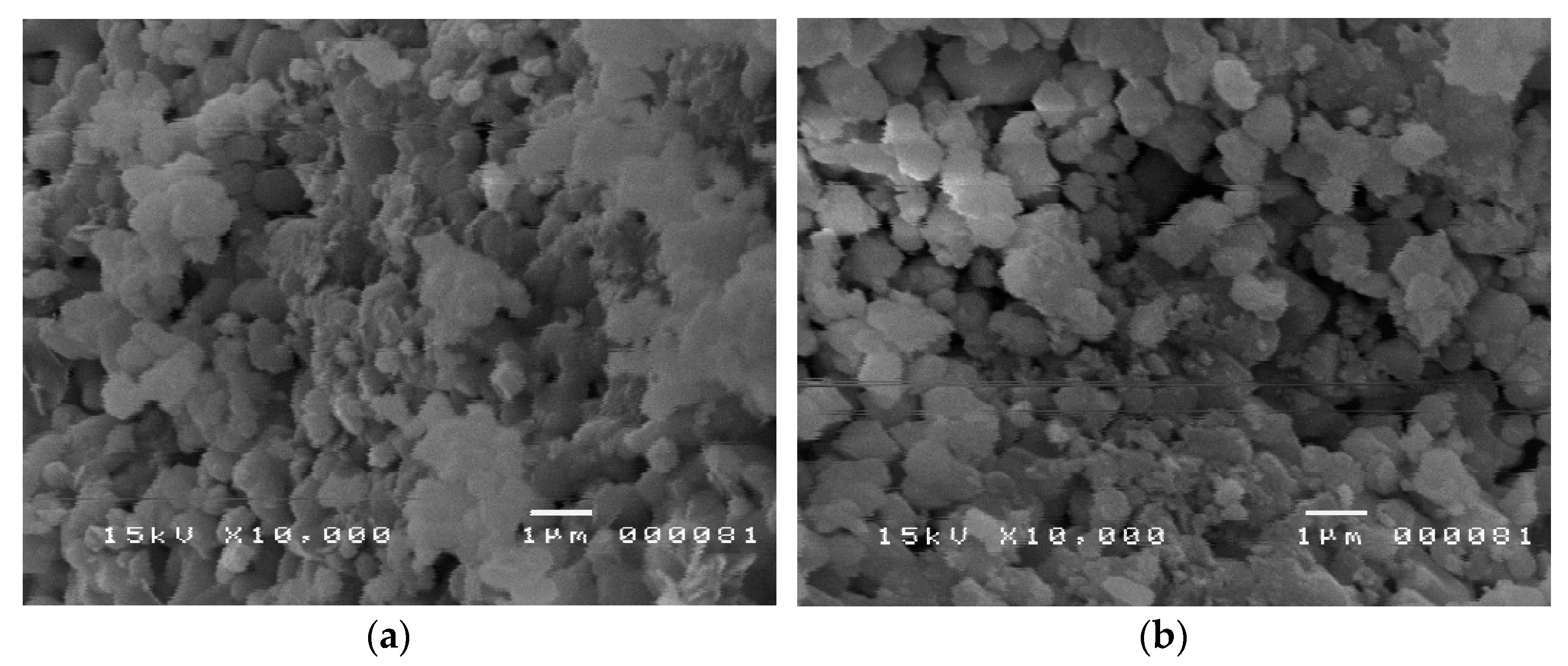
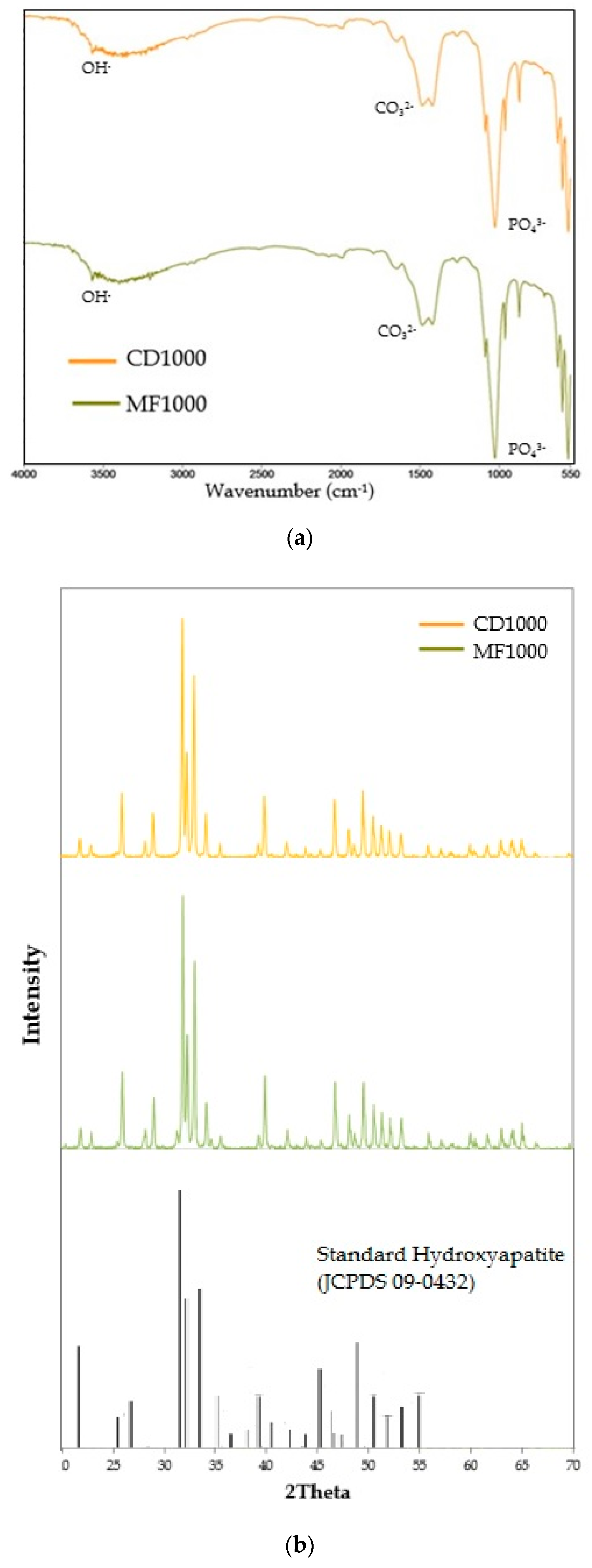
3.1. Characterization of Hydroxyapatite

The morphology of the hydroxyapatite was analyzed using a Scanning Electron Microscope. Both samples derived from the two fish appear to be pseudospherical. Figure 1a shows the SEM image for the cream dory-derived hydroxyapatite, with particle sizes ranging from 19 nm to 43 nm. The milkfish-derived hydroxyapatite’s SEM image is shown in Figure 1b, with particle sizes ranging from 26 to 52 nm. The HAp particles appear clustered because of the moisture content, which causes the powder particles to stick together. Nevertheless, the composition of the CD1000 and MF1000 is almost identical.
The FTIR spectra of CD1000 and MF1000, shown in Figure 2a, prove that numerous functional groups are present in the HAp samples. The peak around 3500 cm−1 may be attributed to -OH vibrations from the moisture adhering to the powders. Peaks at around 1430 cm−1 and 1520 cm−1 corresponding to C=O stretching could be linked to CO2 adsorbed on the sample surface during testing. The peaks at about 550 and 600 cm−1 are more likely due to the vibrational modes of the phosphate group in the apatite.
Figure 2b shows the PXRD results of CD1000 and MF1000, which were compared to a standard hydroxyapatite reference. CD1000 and MF1000 have similar peaks, indicating similar crystal structures in the HAp powders. The major peaks for the milkfish-derived catalyst were at 31.97°, 33.10°, and 32.38°, while the cream dory-derived catalyst had 31.91°, 33.04°, and 32.32° as its peaks. All peaks observed show that a typical hydroxyapatite was produced from both fish bones compared to a standard reference (JCPDS: 09-0432).

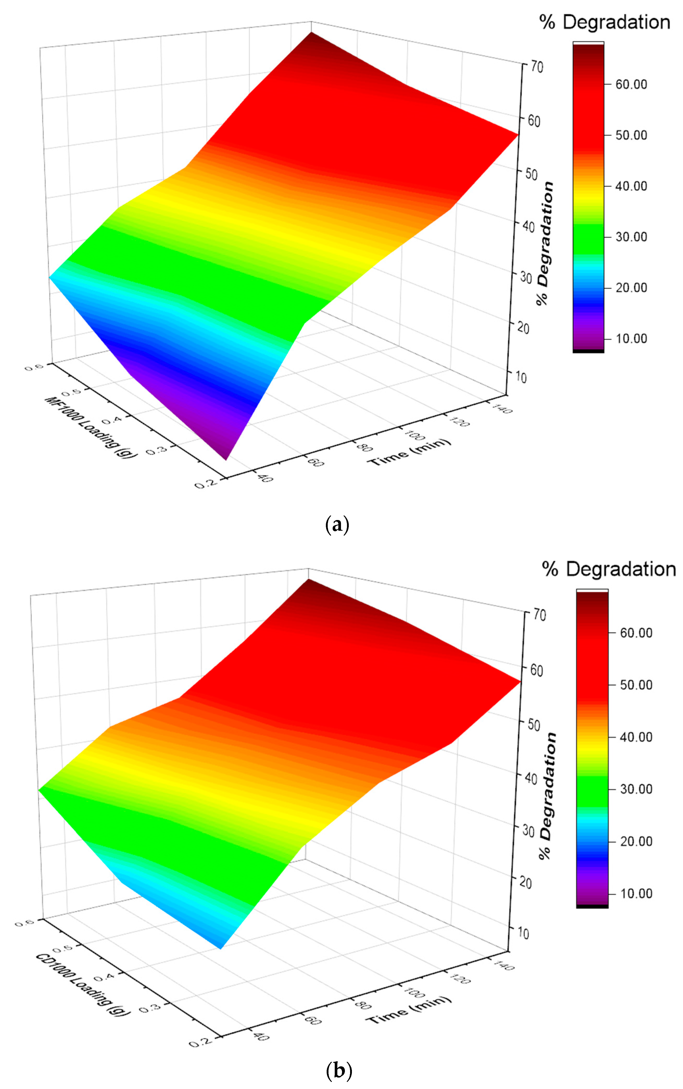
3.2. Photocatalytic Degradation of Malathion

As shown in Figure 3, the degradation efficiency of malathion increases with an increase in catalyst dosage and UV exposure time. The highest degradation was reached after 150 min of exposure to UV light with 0.6 g of the catalyst. The highest degradation efficiency obtained for the milkfish-derived catalyst was 67.80%, while the cream-dory-derived catalyst resulted in 67.99% degradation. Both were obtained with 0.6 g of catalyst and UV light exposure for 150 min. These results may be due to kinetic effects since the catalyst and the pollutant have a longer time to collide and react with each other at longer UV exposure times (Nuengmatcha, 2016). Moreover, increased catalyst loading means more active sites for malathion to react with during its degradation, hence the higher degradation efficiencies.
A three-way ANOVA (Table 1) was used to determine the significant effect of varying the parameters (catalyst loading, UV exposure time, and catalyst type) on the photocatalytic degradation of malathion. The linear effects of catalyst loading, UV exposure time, and catalyst type significantly affect malathion’s degradation efficiency. Moreover, it was found that the two-factor interaction between UV exposure time and catalyst loading and between UV exposure time and HAp type also significantly affects the degradation of malathion. Meanwhile, the two-factor interaction between catalyst loading and HAp type has an insignificant impact on malathion degradation.

3.3. Degradation Kinetics

We fitted the experimental data to first- and second-order kinetics (Table 2). When MF1000 is used as a photocatalyst, malathion obeys first-order kinetics with a kinetic constant ranging from 5.2503 × 10⁻³ min−1 for 0.2 g catalyst loading to 7.0289 × 10⁻³ min−1 for 0.6 g catalyst loading. Meanwhile, malathion obeys second-order kinetics, with a kinetic constant ranging from 0.8520 × 10⁻³ L min−1 mg−1 for 0.2 g loading to 1.1946 × 10⁻³ L min−1 mg−1 for 0.6 g loading. Across all catalyst loadings, CD1000 has faster degradation kinetics compared to MF1000. More studies are needed to elucidate the mechanism of malathion degradation in water using the synthesized HAp.

4. Conclusions

Hydroxyapatite (HAp) was successfully synthesized via calcination. The HAp produced has a particle size ranging from 19 nm to 52 nm with a pseudo-spherical morphology. PXRD and FTIR analysis confirmed the successful synthesis of HAp. A three-way ANOVA showed that catalyst loading, UV exposure time, and HAp type are significant factors in the photocatalytic degradation efficiency of malathion in water. Moreover, the two-factor interaction between UV exposure time and catalyst loading and between UV exposure time and HAp type affects the degradation efficiency. A kinetic study shows that the degradation mechanism of malathion is best fit in a first-order reaction when using milkfish-derived HAp (MF1000) and in a second-order reaction when using cream-dory-derived HAp (CD1000). Overall, CD1000 yielded faster degradation kinetics compared to MF1000.

Author Contributions

Conceptualization, R.V.C.R.; methodology, A.S.C., M.G.P., M.J.C.V., A.D.V. and R.V.C.R.; software, E.C.R.L.; validation, E.C.R.L. and R.V.C.R.; formal analysis, A.S.C., M.G.P., M.J.C.V., A.D.V., E.C.R.L. and R.V.C.R.; investigation, A.S.C., M.G.P., M.J.C.V., A.D.V., E.C.R.L. and R.V.C.R.; resources, E.C.R. and R.V.C.R.; data curation, A.S.C., M.G.P., M.J.C.V., A.D.V., E.C.R.L. and R.V.C.R.; writing—original draft preparation, A.S.C., M.G.P., M.J.C.V., and A.D.V.; writing—review and editing, E.C.R.L., E.C.R. and R.V.C.R.; visualization, E.C.R.L.; supervision, R.V.C.R.; project administration, E.C.R. and R.V.C.R.; funding acquisition, E.C.R. and R.V.C.R. All authors have read and agreed to the published version of the manuscript.

Funding

This research received no external funding.

Institutional Review Board Statement

Not applicable.

Informed Consent Statement

Not applicable.

Data Availability Statement

Not applicable.

Conflicts of Interest

The authors declare no conflict of interest.

References

  1. Lu, J.L.; Cosca, K.Z.; del Mundo, J. Trends of Pesticide Exposure and Related Cases in the Philippines. J. Rural Med. 2010, 5, 153–164. [Google Scholar] [CrossRef] [PubMed]
  2. Varca, L.M. Pesticide residues in surface waters of Pagsanjan-Lumban catchment of Laguna de Bay, Philippines. Agric. Water Manag. 2012, 106, 35–41. [Google Scholar] [CrossRef]
  3. Elfman, L.; Tooke, N.E.; Patring, J.D. Detection of pesticides used in rice cultivation in streams on the island of Leyte in the Philippines. Agric. Water Manag. 2011, 101, 81–87. [Google Scholar] [CrossRef]
  4. Navarrete, I.A.; Tee, K.A.M.; Unson, J.R.S.; Hallare, A.V. Organochlorine pesticide residues in surface water and groundwater along Pampanga River, Philippines. Environ. Monit. Assess. 2018, 190, 289. [Google Scholar] [CrossRef] [PubMed]
  5. Bajet, C.; Kumar, A.; Calingacion, M.; Narvacan, T. Toxicological assessment of pesticides used in the Pagsanjan-Lumban catchment to selected non-target aquatic organisms in Laguna Lake, Philippines. Agric. Water Manag. 2012, 106, 42–49. [Google Scholar] [CrossRef]
  6. Saleh, I.A.; Zouari, N.; Al-Ghouti, M.A. Removal of pesticides from water and wastewater: Chemical, physical and biological treatment approaches. Environ. Technol. Innov. 2020, 19, 101026. [Google Scholar] [CrossRef]
  7. Trellu, C.; Vargas, H.O.; Mousset, E.; Oturan, N.; Oturan, M.A. Electrochemical technologies for the treatment of pesticides. Curr. Opin. Electrochem. 2021, 26, 100677. [Google Scholar] [CrossRef]
  8. Ho, S. Low-Cost Adsorbents for the Removal of Phenol/Phenolics, Pesticides, and Dyes from Wastewater Systems: A Review. Water 2022, 14, 3203. [Google Scholar] [CrossRef]
  9. Karthikeyan, R.; Davis, L.C.; Erickson, L.E.; Al-Khatib, K.; Kulakow, P.; Barnes, P.L.; Hutchinson, S.; Nurzhanova, A.A. Potential for Plant-Based Remediation of Pesticide-Contaminated Soil and Water using Nontarget Plants such as Trees, Shrubs, and Grasses. Crit. Rev. Plant Sci. 2004, 23, 91–101. [Google Scholar] [CrossRef]
  10. Bruckmann, F.S.; Schnorr, C.; Oviedo, L.R.; Knani, S.; Silva, L.F.O.; Silva, W.L.; Dotto, G.L.; Rhoden, C.R.B. Adsorption and Photocatalytic Degradation of Pesticides into Nanocomposites: A Review. Molecules 2022, 27, 6261. [Google Scholar] [CrossRef] [PubMed]
  11. Zeshan, M.; Bhatti, I.A.; Mohsin, M.; Iqbal, M.; Amjed, N.; Nisar, J.; AlMasoud, N.; Alomar, T.S. Remediation of pesticides using TiO2 based photocatalytic strategies: A review. Chemosphere 2022, 300, 134525. [Google Scholar] [CrossRef] [PubMed]
  12. Andronic, L.; Lelis, M.; Enesca, A.; Karazhanov, S. Photocatalytic activity of defective black-titanium oxide photocatalysts towards pesticide degradation under UV/VIS irradiation. Surf. Interfaces 2022, 32, 102123. [Google Scholar] [CrossRef]
  13. Khan, S.H.; Pathak, B. Zinc oxide based photocatalytic degradation of persistent pesticides: A comprehensive review. Environ. Nanotechnol. Monit. Manag. 2020, 13, 100290. [Google Scholar] [CrossRef]
  14. Vaya, D.; Surolia, P.K. Semiconductor based photocatalytic degradation of pesticides: An overview. Environ. Technol. Innov. 2020, 20, 101128. [Google Scholar] [CrossRef]
  15. Rocha, R.L.P.; Honorio, L.M.C.; Bezerra, R.D.d.S.; Trigueiro, P.; Duarte, T.M.; Fonseca, M.G.; Silva-Filho, E.C.; Osajima, J.A. Light-Activated Hydroxyapatite Photocatalysts: New Environmentally-Friendly Materials to Mitigate Pollutants. Minerals 2022, 12, 525. [Google Scholar] [CrossRef]
  16. Mariappan, A.; Pandi, P.; Rajeswarapalanichamy, R.; Neyvasagam, K.; Sureshkumar, S.; Gatasheh, M.K.; Hatamleh, A.A. Bandgap and visible-light-induced photocatalytic performance and dye degradation of silver doped HAp/TiO2 nanocomposite by sol-gel method and its antimicrobial activity. Environ. Res. 2022, 211, 113079. [Google Scholar] [CrossRef] [PubMed]
  17. Zhang, Y.; Wang, L.; Lu, L.; Liu, M.; Yuan, Z.; Yang, L.; Liu, C.; Huang, S.; Rao, Y. Highly efficient decontamination of tetracycline and pathogen by a natural product-derived Emodin/HAp photocatalyst. Chemosphere 2022, 305, 135401. [Google Scholar] [CrossRef] [PubMed]
  18. Rubi, R.V.; Roque, E.; Rosa, F.D.; Estoque, R.M.; Olvido, G.; Perey, P.J.; Teresa, J.S.; Tesalona, M.A. Photocatalytic degradation of Atrazine herbicide using nano-Hydroxyapatite from Cow Bone synthesized via Simulated Body Fluid. IOP Conf. Ser. Mater. Sci. Eng. 2020, 778, 012013. [Google Scholar] [CrossRef]
Figure 1. Surface morphology of (a) cream dory-derived hydroxyapatite and (b) milkfish-derived hydroxyapatite.
Engproc 37 00007 g001
Figure 2. (a) FTIR spectra; (b) X-ray diffraction profiles of cream dory-derived hydroxyapatite (CD1000) and milkfish-derived hydroxyapatite (MF1000).
Engproc 37 00007 g002
Figure 3. 3D plots for the photocatalytic degradation of malathion under UV light using (a) MF1000 and (b) CD1000 as photocatalysts.
Engproc 37 00007 g003
Table 1. Effect of catalyst loading, UV exposure time, and catalyst type on the photocatalytic degradation of malathion.
DFSum of SquaresMean SquareF Valuep Value
Model216946.0871330.7661392.68027.4364 × 10−10
Time **46233.64921558.41231850.12236.7926 × 10−12
Loading **2382.2993191.1496226.92989.0016 × 10−8
HAp Type **1168.1328168.1328199.60456.1246 × 10−7
Time x loading**876.30089.537611.32291.2400 × 10−3
Time x HAp Type **483.002920.750724.63491.4941 × 10−4
Loading x HAp Type22.70221.35111.60402.5956 × 10−1
** significant effect.
Table 2. Degradation Kinetics of Malathion.
MF1000CD1000
0.2 g0.4 g0.6 g0.2 g0.4 g0.6 g
First-Order Kinetics
k1 (×10−3), min−15.25036.07387.02896.06896.92247.7919
Std. Error (×10−3)0.31200.21100.37270.34150.36190.7419
Reduced Χ20.14040.05360.13590.14040.13110.4584
SSE0.56150.21430.54360.56140.52451.8336
R20.95770.98450.95470.92790.94480.7586
Second-Order Kinetics
k2 (×10−3), L min−1 mg−10.69200.83251.01740.85201.00681.1946
Std. Error (×10−3)0.07570.07900.09630.02480.04390.1044
Reduced Χ20.28990.23160.23920.02200.05070.2057
SSE1.15980.92620.95680.08800.20300.8229
R20.91270.93310.92020.98870.97860.8917
Disclaimer/Publisher’s Note: The statements, opinions and data contained in all publications are solely those of the individual author(s) and contributor(s) and not of MDPI and/or the editor(s). MDPI and/or the editor(s) disclaim responsibility for any injury to people or property resulting from any ideas, methods, instructions or products referred to in the content.

