Proceeding Paper

Biocompatible and Flexible Transparent Electrodes for Skin-Inspired Sensing †

by
Raquel L. Pereira
1,2,3 and
Gabriela V. Martins
1,2,3,*
1
BioMark@ISEP, School of Engineering, Polytechnique School of Porto, 4200-072 Porto, Portugal
2
Centre of Biological Engineering (CEB), University of Minho, 4710-057 Braga, Portugal
3
LABBELS—Associate Laboratory, 4710-057 Braga, Portugal
*
Author to whom correspondence should be addressed.
Presented at the 3rd International Electronic Conference on Biosensors, 8–21 May 2023; Available online: https://iecb2023.sciforum.net/.
Eng. Proc. 2023, 35(1), 26; https://doi.org/10.3390/IECB2023-14588
Published: 9 June 2023
(This article belongs to the Proceedings of The 3rd International Electronic Conference on Biosensors)

Abstract

:
In recent years, flexible electronics have experienced a massive growth as a response to the high demand for new skin-patch sensor devices targeted at personal health-monitoring. In this context, the incorporation of biopolymers into the backbone of these soft systems brings new opportunities in terms of biocompatibility and sustainability performance. However, the suitable integration of a conductive patterned material is still a challenge, in order to achieve good adhesion and high transparency. Thus, silver nanowires (AgNWs) constitute promising candidates for the fabrication of flexible transparent conductive films. Herein, a chitosan membrane doped with a plasticizer element was made conductive, through a one-step process, by using an optimized ratio of chitosan–lactic acid–AgNWs (Chi-LaA-AgNWs) dispersion. This formulation was applied using screen-printing, and the influence of the polymer ratio, cure temperature, and number of layers applied with the AgNW-based ink was investigated. Compared with conventional water-based AgNW dispersions, the here-proposed chitosan-doped ink enabled the fabrication of transparent electrode platforms holding good stability, homogeneity, and electrical features.

1. Introduction

Electronics are currently evolving to allow a deeper connection with the human body, particularly with the skin. The new field of “skin-inspired electronics” arose from the need to develop new (electrochemical) platforms that combine a high electrical performance with skin-like features, such as flexibility, stretchability, and breathability [1]. Typically, these platforms are made up of a polymeric backbone, and an epidermal electrode, which should possess not only good conductivity features, but also high transparency and facile patterning [2,3].
Chitosan is a linear polysaccharide composed of β-(1-4)-linked D-glucosamine and N-acetyl-D-glucosamine [4]. It is the second-largest biological resource on earth [4,5] and a low-cost material for the production of biopolymers [5]. Chitosan has long been employed in wound-dressing fabrication, due to its excellent biocompatibility, biodegradability, physiological activity, extracellular-matrix resemblance, cell-growth support, flexibility, sweat permeability, and film-forming properties [3,4,5]. Nevertheless, there are some studies that suggest that pure chitosan films can have a slight inhibitory effect on cell adhesion and proliferation [4]. Chitosan has also been used as a substrate for electrode fabrication thanks to its high transparency, good formability on the nanoscale, strong adhesion to substrates, and good mechanical strength [3,4,6]. Moreover, chitosan films do not significantly change size under aqueous conditions, and they have functional groups such as -OH and -NH2, which allow a variety of surface modifications [4], favoring their use as a substrate for electronic skins [3].
Recently, metallic nanofiber networks, such as silver nanowires (AgNW), have attracted great attention in the field of transparent conductive films, due to their high transparency, excellent biocompatibility, and favorable water–air permeability [3,6,7]. Nonetheless, in some cases, printed AgNW networks possess poor stability, and unsuitable adhesion and surface roughness [6,7]. Thus, conjugating the AgNW networks into composites is crucial to improve their usability as building blocks for the fabrication of transparent electrode platforms. Recently, Wang et al., 2020, demonstrated how the incorporation of a chitosan–lactic acid (Chi-LaA) mixture into an aqueous AgNW solution could significantly improve the adhesion of the AgNWs to the chitosan substrate, which resulted in the production of stable conductive films coated on glass substrates [6].
In the literature, to the best of our knowledge, very few articles have reported the use of AgNW ink-based electrochemical sensors for wearable sensing applications [8,9]. This work reports the development of a Chi-LaA-AgNWs dispersion, which can be screen-printed onto chitosan membranes, to yield transparent electrode platforms that surpass the stability and electrical features of commercial water-based AgNW dispersions. Moreover, this work explores how the membrane composition, the cure parameters, and the Chi:AgNW mass ratio in the dispersions impact the electrochemical performance and mechanical stability of the resulting electrodes.

2. Materials and Methods

2.1. Reagents and Solutions

The chemicals used in the experiments were all of analytical grade. Solutions were prepared with ultrapure water (obtained from a Milli-Q water-purification system). Potassium hexacyanoferrate II-3-hydrate (K4[Fe(CN)6]) and potassium hexacyanoferrate III (K3[Fe(CN)6]) were obtained from Riedel de Haën; potassium chloride (KCl) from Normapur; acetic acid glacial 100% (CH3COOH) from Carlo Erba; glycerine (C3H8O3) from Pronalab; lactic acid (LaA, C3H6O3) from Fluka; chitosan (Chi, molecular weight: 100,000–300,000) and 0.5% (w/w) silver nanowire (AgNW, diam. × L 120–150 nm × 20–50 μm) suspension in isopropyl alcohol (IPA) from Sigma Aldrich; AgNW aqueous ink from Dycotec Materials.

2.2. Apparatus

The electrochemical data was obtained using the PGSTAT302N (Potencial Zero, Lisboa, Portugal), a potentiostat/galvanostat device from Metrohm Autolab, controlled by the Nova 2.1 software. The electrodes were connected to the potentiostat via a box connector from BioTid Electronical.

2.3. Fabrication of the Transparent Electrode Platforms

2.3.1. Preparation of the Chitosan Membranes

To form flexible and transparent membranes, a 2% (w/v) chitosan solution in 1% (v/v) acetic acid, doped with 1% or 2% (v/v) glycerine, was cast on petri dishes, placed under vacuum for 30 min, and left to dry overnight, at room temperature, inside a fume hood.

2.3.2. Preparation of the Chi-LaA-AgNW Dispersions

The preparation of the Chi-LaA-AgNW dispersion was adapted from Wang et al., 2020 [6]. Firstly, a solution of 2% (w/v) chitosan in 51% (v/v) lactic acid was prepared. Then, this solution was added to the 0.5% (w/w) AgNW dispersion in IPA to obtain a Chi-LaA-AgNW dispersion, where the Chi:AgNW mass ratio was 1:1. Alternatively, a solution of 4% (w/w) chitosan in 51% (v/v) lactic acid was used to obtain a Chi-LaA-AgNW dispersion, where the Chi:AgNW mass ratio was 2:1.

2.3.3. Screen-Printing of the Chitosan Membranes

After completely drying, a 3-electrode system was manually screen-printed onto the surface of the chitosan membranes, which was intended to be like conventional commercial screen-printed electrodes composed of a working-, a counter-, and a pseudo-reference-electrode. For that, an adhesive counter mask was placed on the membrane’s surface, and the ink was screen-printed using a microscope slide. Four types of ink were tested: the AgNW aqueous ink from Dycotec Materials, the unmodified 0.5% (w/w) AgNW suspension in IPA, and the two Chi-LaA-AgNW dispersions prepared in Section 2.3.2. For the Dycotec ink, only one layer of ink was applied to the surface of the membranes, which was cured for 10 min, at 60 °C. After mask removal, a final cure was performed (for 10 min, at 140 °C) to minimize ink removal upon washing. For the AgNW dispersions, the number of ink layers, the cure temperature, and the cure time of each layer were optimized. The number of layers was varied between one and four, the cure temperature between 75 °C and 140 °C, and the cure time between 15 min and 2 h. No final cure was performed in these cases. Finally, all produced electrodes were stored under vacuum, until their electrochemical properties were assayed.

2.4. Electrochemical Assays

The electrochemical properties of the sensors were determined using a solution of 5 mM of K4[Fe(CN)6] and 5 mM of K3[Fe(CN)6] in KCl 0.1 M. Cyclic voltammetry (CV) assays were performed through five consecutive cycles, ranging from −0.5 to +0.8 V, with a scan rate of 50 mV/s.

3. Results and Discussion

3.1. Fabrication of the Transparent Electrode Platforms

Initially, the fabrication process of the polymeric membranes was only performed with single chitosan, but the outcome was rigid and plastic substrates, in particular after experiencing elevated cure temperatures. Thus, in order to achieve more flexible and skin-like membranes, a natural plasticizer as glycerine was incorporated into the backbone of the polymeric matrix [3,10]. The two types of membranes (doped with either 1% or 2% of glycerine) were found to be indistinguishable to the naked eye, presenting themselves as homogeneous yellowish films. However, the texture of the membranes containing 2% of glycerine was rubberier, more elastic, and stickier than the texture of the membranes containing 1% of glycerine. In addition, the membranes doped with 2% of glycerine also appeared to have a higher amount of oil residue on their surface.
Table 1 contains a summary of all the conditions tested, as well as representative examples of the electrodes produced by said conditions. The first parameter to be optimized was cure temperature. All cure temperatures above 75 °C resulted in the melting of the masks and, therefore, in the inability to produce electrodes. The second parameter to be optimized was the mass ratio of Chi:AgNW in the dispersions. From Table 1, it is obvious that the electrodes prepared with the 0.5% (w/w) AgNW suspension are less homogeneous than the ones prepared with the Chi-LaA-AgNW dispersions, which constitutes strong evidence that the presence of chitosan in the conductive ink greatly improves its adhesion properties. Likewise, it is also clear that the electrodes prepared with the dispersion where the Chi:AgNW mass ratio is 2:1 are far too opaque to be considered transparent electrode platforms. Thus, this last type of electrode will be excluded from the electrochemical assays. Regarding the influence of the percentage of glycerine in the membranes, the cure time, and the number of ink layers applied on the resulting electrodes, no significant visual differences can be gathered from the analysis of Table 1. However, an exception should be made for the fact that the electrodes prepared with only one layer of ink appear to be more sheer and less uniform than their counterparts with various ink layers. To obtain more information about the effect of these parameters on the electrodes, their electrochemical properties were analyzed through CV assays.

3.2. Electrochemical Assays

To further explore the effects of the amount of glycerine in the chitosan substrates, the number of ink layers, and the cure time, on the produced electrodes, the electrodes were analyzed through CV. Starting with the effect of the number of ink layers, the electrochemical properties of membranes doped with 2% of glycerine and screen-printed with one to four layers of a Chi-LaA-AgNW dispersion (where the Chi:AgNW mass ratio is 1:1) were measured. Each ink layer was cured for 30 min, at 75 °C, and the results of this assay are summarized in Figure 1. As expected, the obtained cyclic voltammograms displayed a couple of well-defined redox peaks, as a result of the reversible electron transfer of the redox pair [Fe(CN)6]3−/4−. Looking at Figure 1, it is clear that screen-printing the membranes with only one ink layer resulted in lower redox peak currents, which can be justified by an insufficient amount of conductive material. When using two and three ink layers, the current of the redox peaks was greatly enhanced, which does not happen with the four-layer condition; thus, for the remainder of the studies, only two and three ink layers were considered.
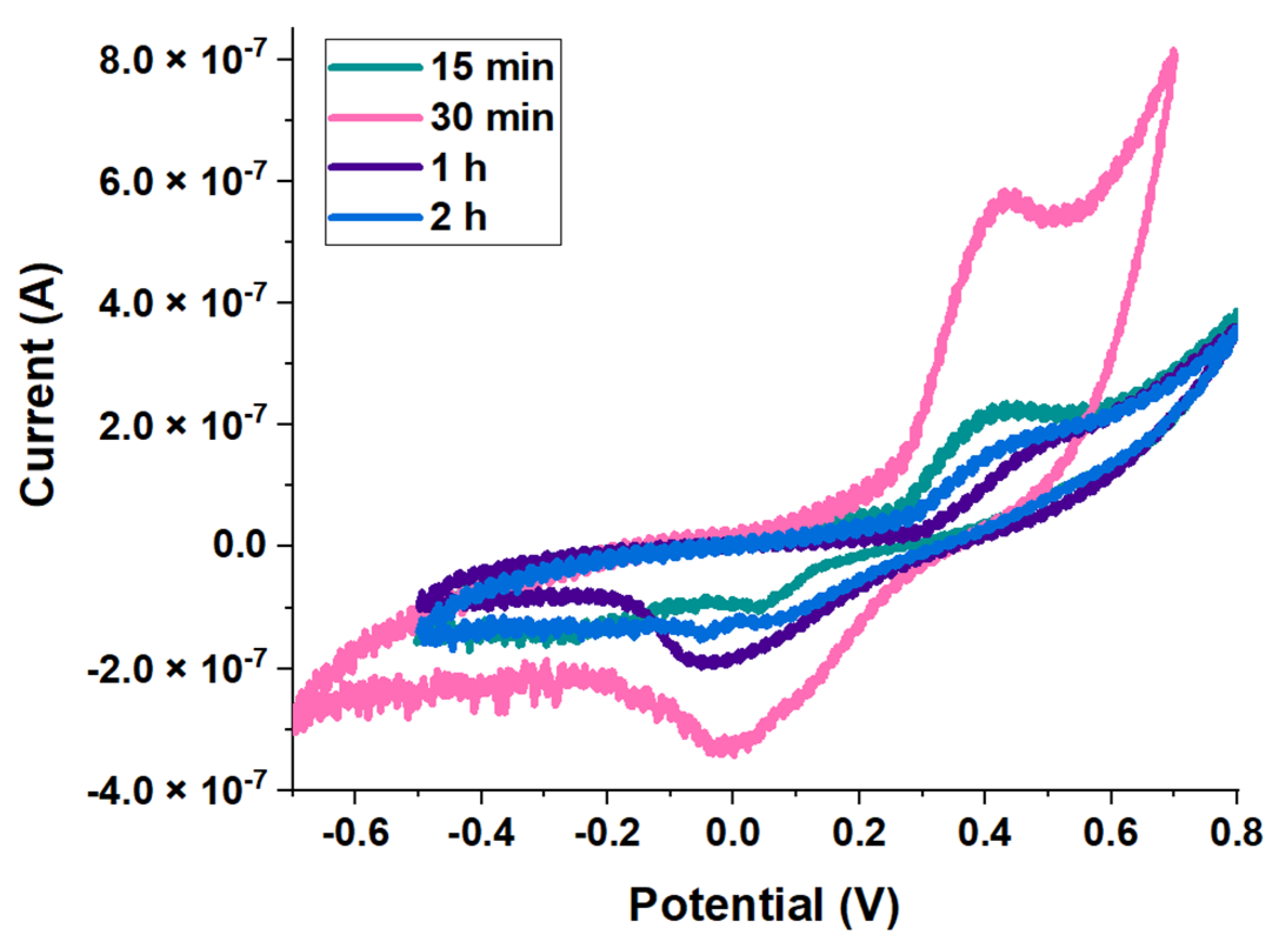
Moving onto the effects of the cure time on the electrodes’ performance, the electrochemical properties of membranes doped with 2% of glycerine and screen-printed with two layers of the Chi-LaA-AgNW dispersion (Chi:AgNW, mass ratio 1:1) were measured. Each ink layer was cured for either 15 min, 30 min, 1 h, or 2 h (at 75 °C), and the resulting cyclic voltammograms of the electrodes are presented in Figure 2. The obtained results show that the performance of the electrodes cured for 30 min clearly stands out from the others, achieving more pronounced redox peaks, and allowing, overall, higher current values—the oxidation peak current values of the electrodes cured for 30 min were around +0.8 μA, while the others were no higher than +0.35 μA. Therefore, 30 min was the cure time selected to move forward with.
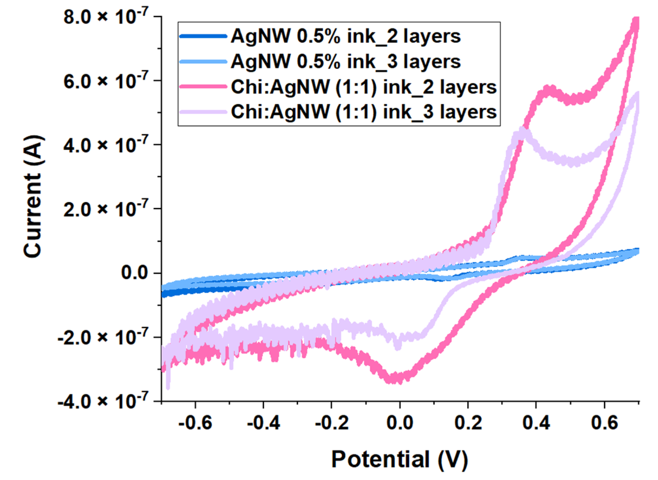
To clarify which conductive dispersion had the best electrochemical performance, membranes doped with 2% of glycerine were screen-printed with 2/3 layers of either the Chi-LaA-AgNW dispersion where the Chi:AgNW mass ratio was 1:1, or the unmodified 0.5% (w/w) AgNW suspension in IPA. Each ink layer was cured for 30 min, at 75 °C, and the electrochemical properties were assessed through CV, as shown in Figure 3. From Figure 3, it is obvious that the Chi-LaA-AgNW dispersion produces electrodes with a much superior electrochemical performance to the unmodified 0.5% (w/w) AgNW suspension, allowing the flow of currents around eight times higher, independently of the number of ink layers applied. This observation is in line with the images in Table 1, which show that the electrodes produced with the Chi-LaA-AgNW dispersion are more uniform than those produced with the unmodified 0.5% (w/w) AgNW suspension.
To understand the effect of the percentage of glycerine used to produce the membranes on the electrochemical performance of the electrodes, the two types of membranes (doped with either 1% or 2% of glycerine) were screen-printed with 2/3 layers of the Chi-LaA-AgNW dispersion (where the Chi:AgNW mass ratio is 1:1) and each ink layer was cured for 30 min, at 75 °C. Lastly, the electrochemical properties were investigated, and the results were summarized in Figure 4. The obtained voltammograms show that the current values of the electrodes prepared using membranes doped with 1% of glycerine were higher, and the anodic peaks were more pronounced, than that of the electrodes prepared using membranes doped with 2% of glycerine. This suggests that the electrochemical performance of the sensors is favored by the use of 1% of glycerine rather than 2% of glycerine, which can be explained by the higher amount of oil residue on the surface of the membranes doped with 2% of glycerine, which may hinder the adhesion of the conductive patterned component of the electrodes to their substrate.
Finally, the best-performing electrodes produced using the innovative Chi-LaA-AgNW dispersion were compared with the electrodes produced using the commercially available aqueous-based Dycotec Materials ink. After evaluating the above-mentioned results, the electrodes selected were the ones produced by screen-printing membranes doped with 1% of glycerine with two layers of the Chi-LaA-AgNW dispersion (where the Chi:AgNW mass ratio is 1:1), and cured for 30 min, at 75 °C. Besides the poor electrochemical properties, it is important to note that the aqueous-based AgNW ink was partially removed from the surface of the chitosan membranes as the electrochemical assays were being performed, which constitutes a further disadvantage of this type of ink.

4. Conclusions

Overall, the work herein presented showed the successful production of transparent, flexible electrode platforms based on chitosan membranes, doped with glycerine as a natural plasticizer, and screen-printed with a Chi-LaA-AgNW ink. This innovative dispersion outperformed commercially available AgNW preparations, both in terms of stability and electrical features. Moreover, these platforms demonstrated suitable electrochemical properties for application in biosensors, which opens up new possibilities in the realm of skin-patch monitoring devices.

Author Contributions

Conceptualization, resources, supervision, funding acquisition, project administration and writing—review and editing, G.V.M.; validation, formal analysis, investigation, data curation and writing—original draft preparation, R.L.P.; methodology and visualization, G.V.M. and R.L.P. All authors have read and agreed to the published version of the manuscript.

Funding

This research was funded by the SmartBioPatch project (Self-indicated biosensors integrated into flexible membranes of natural origin for application in chronic wounds), with the reference EXPL/EMD-EMD/1095/2021, financed by national funds through the Foundation for Science and Technology (FCT).

Institutional Review Board Statement

Not applicable.

Informed Consent Statement

Not applicable.

Data Availability Statement

Data sharing not applicable.

Conflicts of Interest

The authors declare no conflict of interest.

References

  1. Fu, F.; Wang, J.; Zeng, H.; Yu, J. Functional Conductive Hydrogels for Bioelectronics. ACS Mater. Lett. 2020, 2, 1287–1301. [Google Scholar] [CrossRef]
  2. Wang, S.; Oh, J.Y.; Xu, J.; Tran, H.; Bao, Z. Skin-Inspired Electronics: An Emerging Paradigm. Acc. Chem. Res. 2018, 51, 1033–1045. [Google Scholar] [CrossRef]
  3. Peng, X.; Dong, K.; Zhang, Y.; Wang, L.; Wei, C.; Lv, T.; Wang, Z.L.; Wu, Z. Sweat-Permeable, Biodegradable, Transparent and Self-Powered Chitosan-Based Electronic Skin with Ultrathin Elastic Gold Nanofibers. Adv. Funct. Mater. 2022, 32, 2112241. [Google Scholar] [CrossRef]
  4. Lin, Y.-H.; Kang, P.-L.; Xin, W.; Yen, C.-S.; Hwang, L.-C.; Chen, C.-J.; Liu, J.-T.; Chang, S.J. Preparation and Evaluation of Chitosan Biocompatible Electronic Skin. Comput. Ind. 2018, 100, 1–6. [Google Scholar] [CrossRef]
  5. Lü, X.; Zhang, H.; Huang, Y.; Zhang, Y. A Proteomics Study to Explore the Role of Adsorbed Serum Proteins for PC12 Cell Adhesion and Growth on Chitosan and Collagen/Chitosan Surfaces. Regen. Biomater. 2018, 5, 261–273. [Google Scholar] [CrossRef] [PubMed]
  6. Wang, K.; Jin, Y.; Qian, B.; Wang, J.; Xiao, F. One-Step Aqueous Fabrication of a Silver Nanowire Composite Transparent Conductive Film with High Uniformity and Stability. J. Mater. Chem. C 2020, 8, 4372–4384. [Google Scholar] [CrossRef]
  7. Ding, S.; Zhang, S.; Yin, T.; Zhang, H.; Wang, C.; Wang, Y.; Li, Q.; Zhou, N.; Su, F.; Jiang, Z.; et al. Room-Temperature Nanojoining of Silver Nanowires by Graphene Oxide for Highly Conductive Flexible Transparent Electrodes. Nanotechnology 2022, 34, 045201. [Google Scholar] [CrossRef] [PubMed]
  8. Min, K.; Umar, M.; Seo, H.; Yim, J.H.; Kam, D.G.; Jeon, H.; Lee, S.; Kim, S. Biocompatible, Optically Transparent, Patterned, and Flexible Electrodes and Radio-Frequency Antennas Prepared from Silk Protein and Silver Nanowire Networks. RSC Adv. 2016, 7, 574–580. [Google Scholar] [CrossRef]
  9. Chen, Y.; Carmichael, R.S.; Carmichael, T.B. Patterned, Flexible, and Stretchable Silver Nanowire/Polymer Composite Films as Transparent Conductive Electrodes. ACS Appl. Mater. Interfaces 2019, 11, 31210–31219. [Google Scholar] [CrossRef] [PubMed]
  10. Chiaoprakobkij, N.; Suwanmajo, T.; Sanchavanakit, N.; Phisalaphong, M. Curcumin-Loaded Bacterial Cellulose/Alginate/Gelatin as A Multifunctional Biopolymer Composite Film. Molecules 2020, 25, 3800. [Google Scholar] [CrossRef] [PubMed]
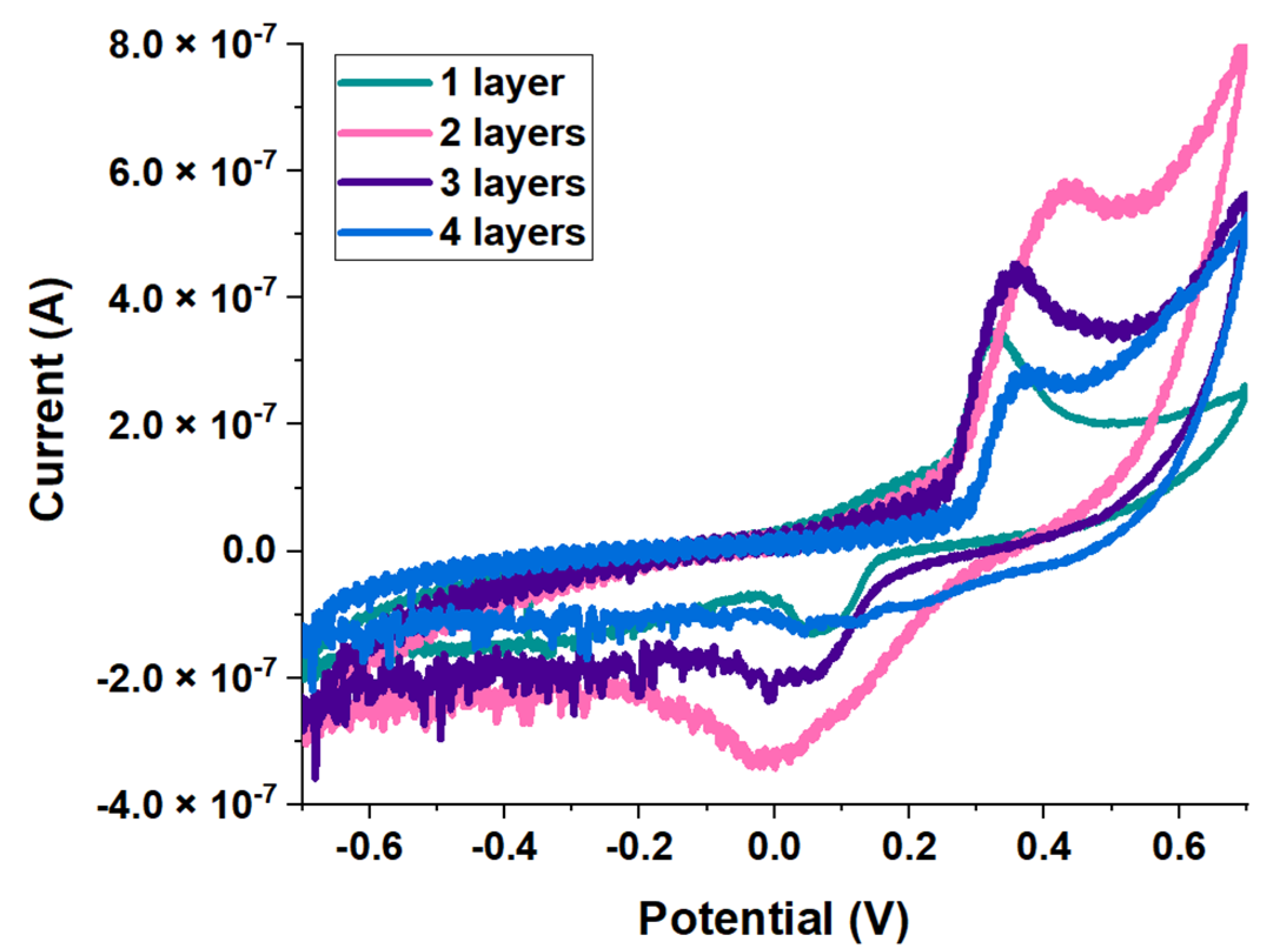
Figure 1. Effect of the number of ink layers on the electrochemical performance of the sensors. CV analysis performed on membranes doped with 2% of glycerine and screen-printed with one to four layers of a Chi-LaA-AgNW dispersion, where the Chi:AgNW mass ratio is 1:1. Ink layers cured for 30 min, at 75 °C.
Engproc 35 00026 g001
Figure 2. Effect of the cure time on the electrochemical performance of the sensors. CV analysis performed on membranes doped with 2% of glycerine and screen-printed with a Chi-LaA-AgNW dispersion, where the Chi:AgNW mass ratio is 1:1. Ink layers cured for 15 min, 30 min, 1 h, or 2 h, at 75 °C.
Engproc 35 00026 g002
Figure 3. Effect of the Chi:AgNW mass ratio of the ink on the electrochemical performance of the sensors. CV analysis performed on membranes doped with 2% of glycerine and screen-printed with 2/3 layers of the Chi-LaA-AgNW dispersion where the Chi:AgNW mass ratio is 1:1 or the unmodified 0.5% (w/w) AgNW suspension. Ink layers cured for 30 min, at 75 °C.
Engproc 35 00026 g003
Figure 4. Effect of the percentage of glycerine in the membranes on the electrochemical performance of the sensors. CV analysis performed on membranes doped with either 1% or 2% of glycerine and screen-printed with 2/3 layers of the Chi-LaA-AgNW dispersion (where the Chi:AgNW mass ratio is 1:1). Ink layers cured for 30 min, at 75 °C.
Engproc 35 00026 g004
Table 1. Summary of conditions tested, and images of the resulting electrodes.
Ink Used[Glycerine] (v/v)Cure TemperatureCure TimeNumber of Ink LayersResulting Electrodes
AgNW aqueous ink1%60 °C 110 min1Engproc 35 00026 i001
2%Engproc 35 00026 i002
AgNW 0.5% (w/w) suspension2%75 °C30 min2Engproc 35 00026 i003
3Engproc 35 00026 i004
Chi-LaA-AgNW dispersion, Chi:AgNW (1:1)1%75 °C 230 min1Engproc 35 00026 i005
2Engproc 35 00026 i006
3Engproc 35 00026 i007
2%15 min2Engproc 35 00026 i008
30 min1Engproc 35 00026 i009
2Engproc 35 00026 i010
3Engproc 35 00026 i011
4Engproc 35 00026 i012
1 h2Engproc 35 00026 i013
3Engproc 35 00026 i014
2 h2Engproc 35 00026 i015
Chi-LaA-AgNW dispersion, Chi:AgNW (2:1)2%75 °C30 min2Engproc 35 00026 i016
3Engproc 35 00026 i017
1 h2Engproc 35 00026 i018
3Engproc 35 00026 i019
1 A final cure (without the masks) was performed for 10 min, at 140 °C. 2 Every temperature tested above 75 °C resulted in the melting of the masks.
Disclaimer/Publisher’s Note: The statements, opinions and data contained in all publications are solely those of the individual author(s) and contributor(s) and not of MDPI and/or the editor(s). MDPI and/or the editor(s) disclaim responsibility for any injury to people or property resulting from any ideas, methods, instructions or products referred to in the content.