1. Introduction
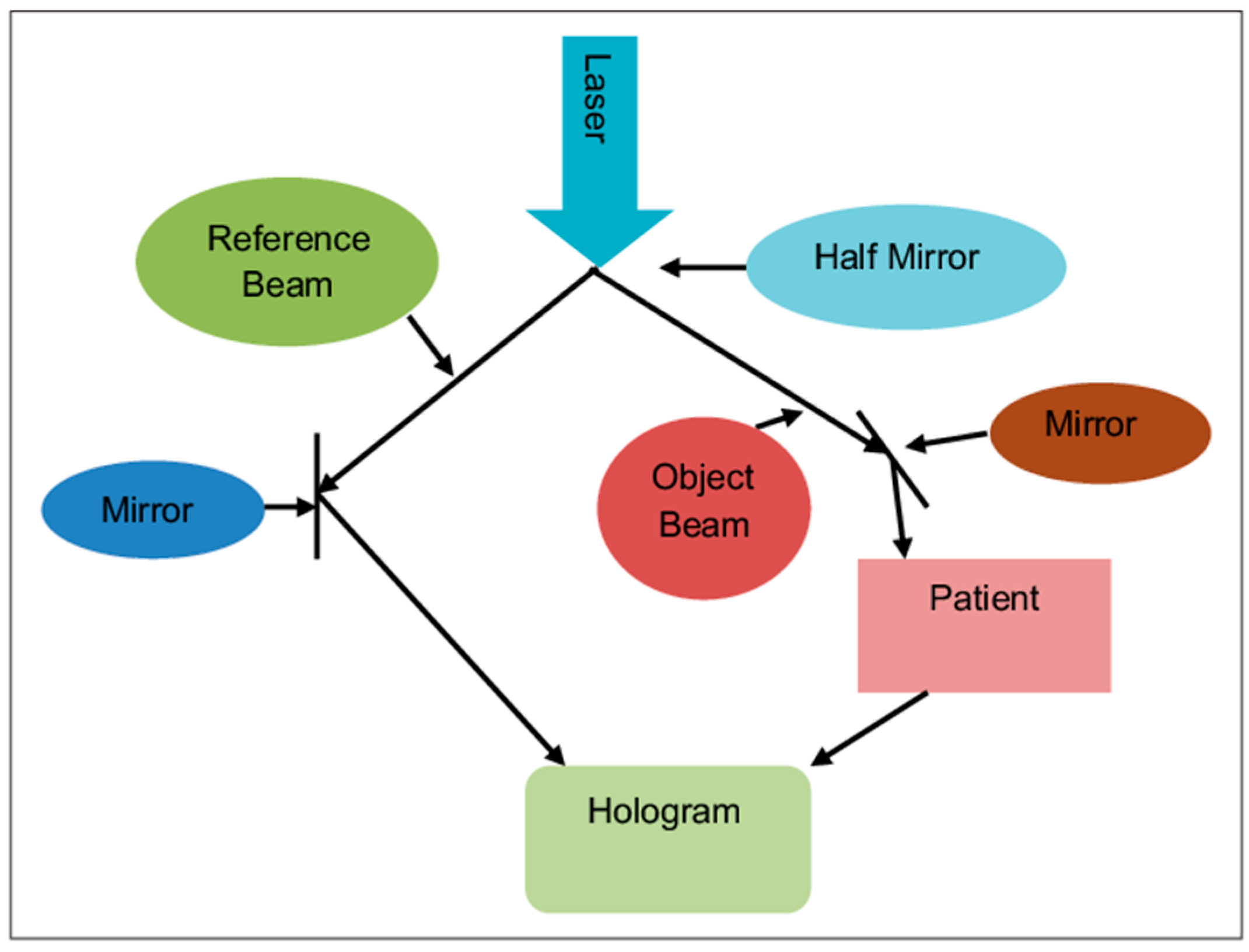
With the advent of newer technology and evolution of artificial intelligence, we can improve our expertise and decrease the errors in various medical fields. A hologram provides a non-contact three-dimensional (3D) image that can be seen with the naked eye. These 3D images provide details of the human anatomy and activity of an internal organ of the body in high resolution. Holography is a two-step process. In the first step, it records a hologram, in which a radiographic image is converted into a photographic record. The second step is to convert a hologram into a virtual image.
Figure 1 [
1].
This provides better hands-on experience for medical and paramedical personnel. It can be employed in various fields of medicine, ranging from laboratory investigations to complex surgical procedures.
A 3D hologram is a highly efficient simulation technique, which could be effectively used for teaching and training for students in various aspects of the medical field at different levels.
Most residents specialize in a surgical field (especially in orthopedics) in a mid-level tertiary care center, peripheral center and post-graduate roles in corporate hospitals where the access to an anatomy dissection hall to learn in-depth anatomy would be difficult, which is required for better a understanding for the students while performing surgeries; to learn the techniques quickly, holograms will make it significantly easier for the students to achieve this. To err is human, but keeping our ethics in mind, it is permissible and acceptable in a simulated environment rather than on actual patients. Creating a simulated environment to learn reduces the damage to actual patients.
In this study, we focus on how holograms help in training orthopedic residents in various surgical techniques and critical situations to handle patients.
2. Methods
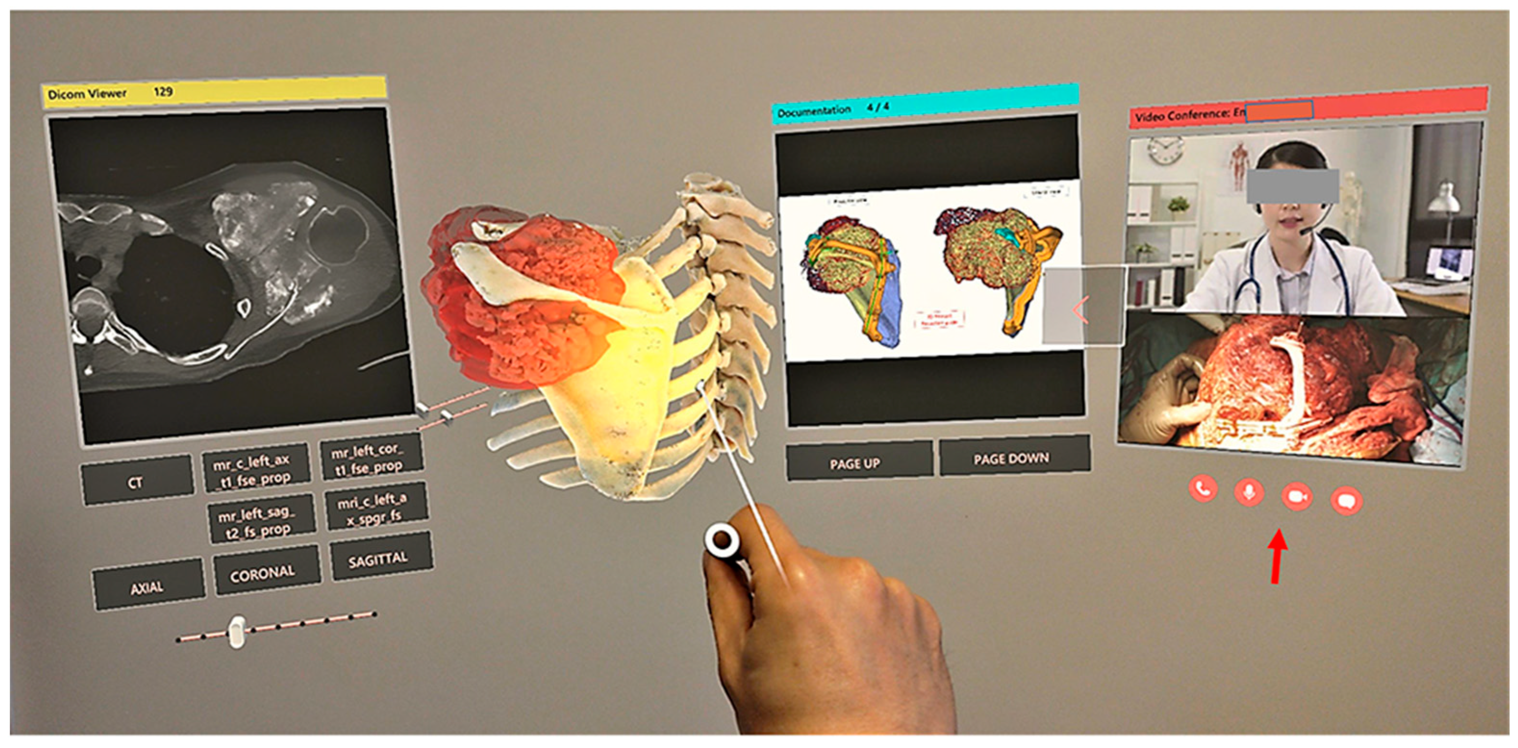
Due to constraints in the time period during residency in orthopedics, we are not able to gain adequate hands-on experience for most of the surgical procedures. The reasons are mainly due to the confidence of the consultants toward the resident, as we need to operate on actual patients and there should be minimal chances for mistakes. The role of the hologram is highlighted at this juncture. Using Microsoft’s HoloLens glasses [
2] in combination with the mixed reality system, it can project 3D holograms in real time under different clinical scenarios.
Figure 2 shows that the process of obtaining surgical skills comes with practice, which, in turn, boosts the confidence level of the resident and the consultant, which is knowledge that cannot be gained from books alone. Hereby, holograms will provide different clinical, surgical and even emergency scenarios under simulation.
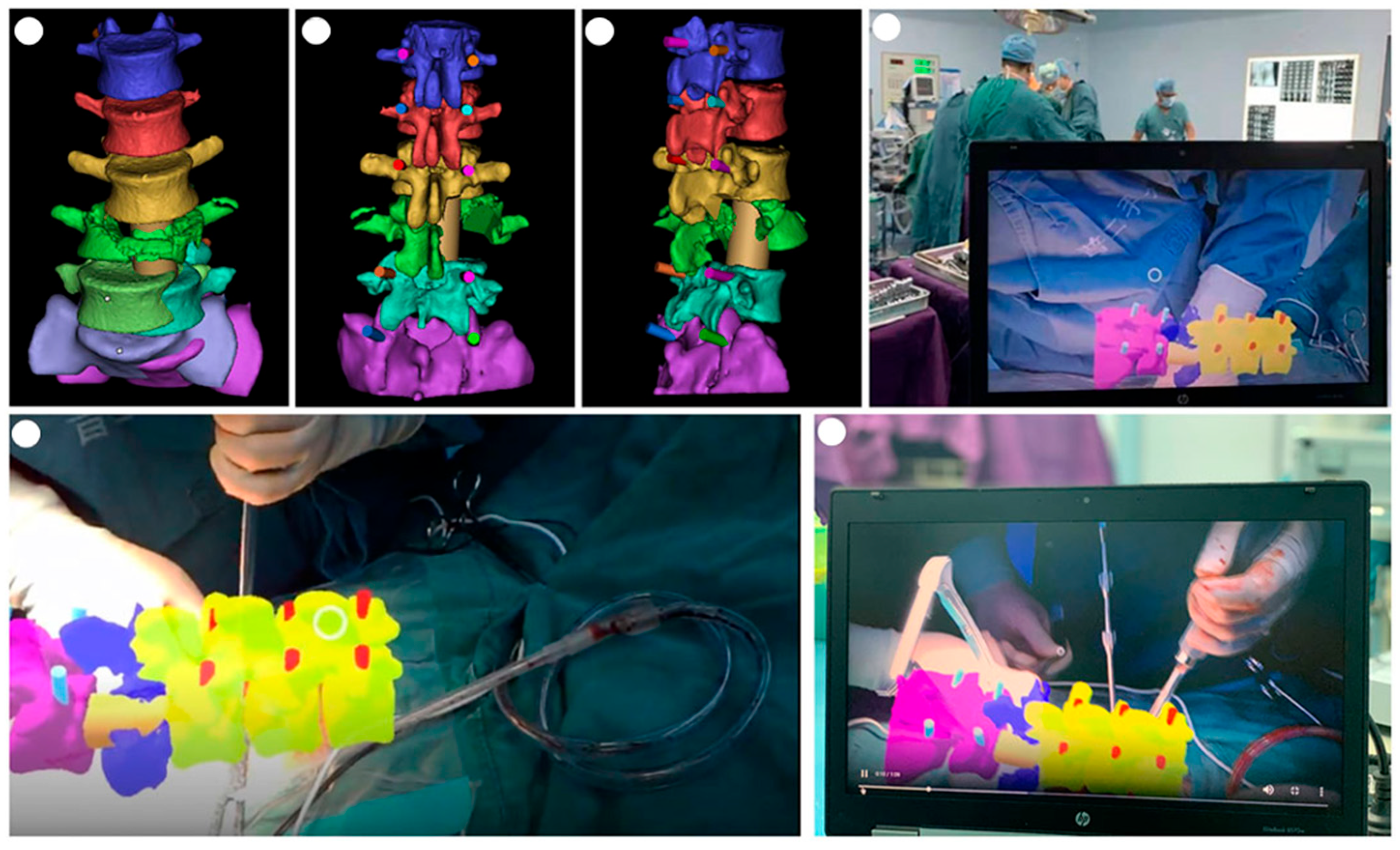
The average total residency period for orthopedic surgeons is 2 or 3 years in India, which is less when compared to the courses abroad. The students have inadequate operating experience on actual patients, which may lead to a lack of confidence during the practice period. Using holograms, residents can have a real-time experience, which mimics a patient presenting with polytrauma along with an appropriate feedback system, which enables proper learning of the adequate resuscitative measures to help save the patient. This helps students deal with actual patients with a better level of confidence, and to minimize human errors. In India, due to the non-uniformity of exposure of orthopedic residents to various kinds of patients and surgical procedures, holograms help in bringing the learning exposure on a par. They can help the consultants sharpen their fine skills such as soft tissue procedures (tendon suturing, nerve grafting, etc.) with the appropriate simulation. Holograms can simulate a scenario and help in research that will be beneficial for patients. They help the consultant to superimpose the hologram image of the specific patients during surgery, such as spine in
Figure 3, orthopedic oncology [
3], etc., to predict the actual plane and degree of the implant and screw placement, and for proper resection of the tumor-free margin.
3. Results
This technology of using holograms that simulate various situations for surgeons and physicians may be helpful for better training residents. If this idea of using holograms for residents is beneficial, it can be extended even to undergraduates to develop knowledge about the basics in every specialty, which makes it easy to learn more things very early when they enter residency. It was found that surgeons who have started using holograms for a patient-tailored pre-op and intra-op planning had better outcomes. Hence, it will be more beneficial if this technology is extended for academic purposes.
4. Limitations
Considering extended reality will not be a better option for residents in certain scenarios, as it will give tactile feedback such as performing surgeries on actual patients, but it will enable them to learn better about anatomy and dissection, which is of the utmost importance for surgeons in their learning period.
In 1979, Piwernetz, et al. [
4] published an idea of “Holography in orthopaedics” as a part of the “springer series in optical sciences” book, but it took many years to lead to a good knowledge base about this and create a high-resolution hologram for medical professionals, making this technology a difficult one to understand.
5. Conclusions
Holograms will change the learning technique of students by providing virtual imaging for better understanding the subject. Simulation of some basic surgeries will help residents to learn faster and decrease the incidence of errors in real-time surgeries in future. Holograms can change future methods of learning. However, there are certain limitations concerning hologram use for academic purposes, which includes the cost of manufacturing and maintenance and the source of funds to produce them. Hence, the feasibility of the advanced technology for residents is still debatable, for which monetary benefits are required from the government to every teaching institute.


