
Artificial Intelligence-Powered Breast Cancer Prognosis: Optimizing Deep Learning with Image Normalization †

Abstract
1. Introduction
2. Literature Review
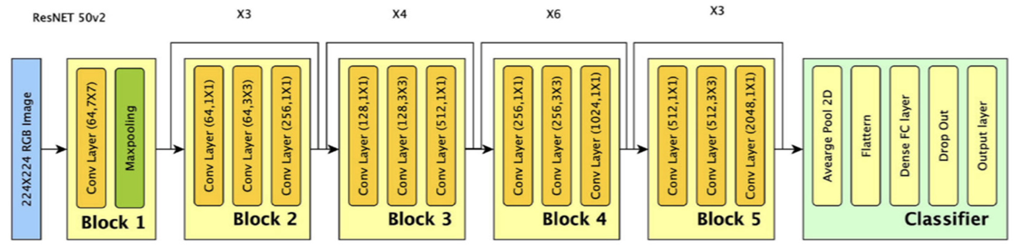
2.1. ResNet-50
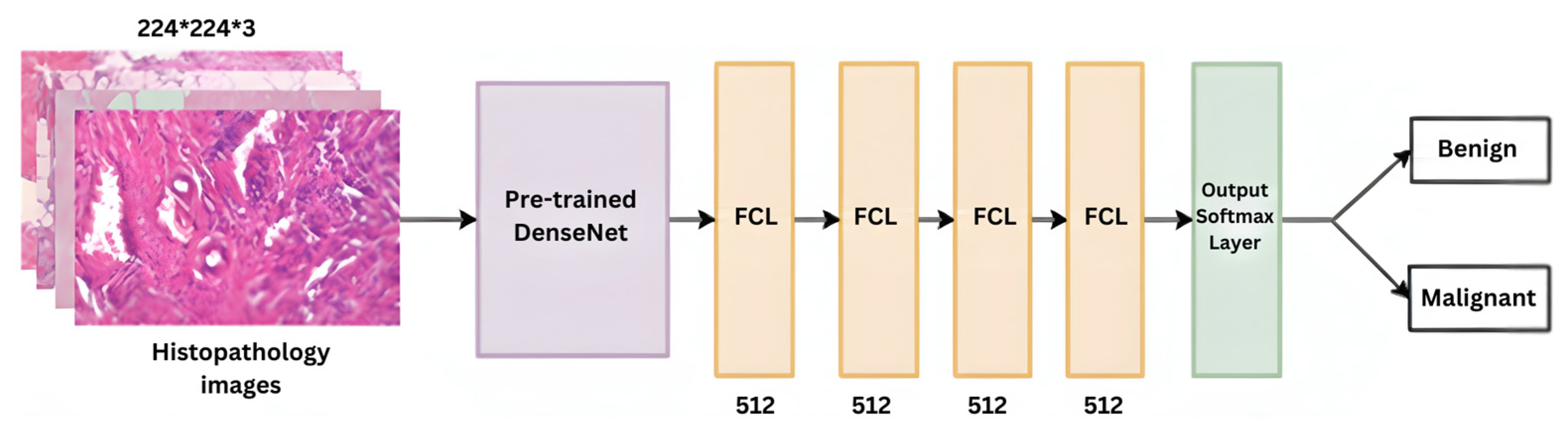
2.2. DenseNet
- Efficient feature reuse: The dense connections ensure that features learned earlier in the network are reused throughout the model, making it highly efficient at extracting important patterns in histopathological images [19].
- Fewer parameters: Despite its deep architecture, DenseNet uses fewer parameters than ResNet-50, making it memory-efficient and better suited for balancing accuracy and resource constraints [15].
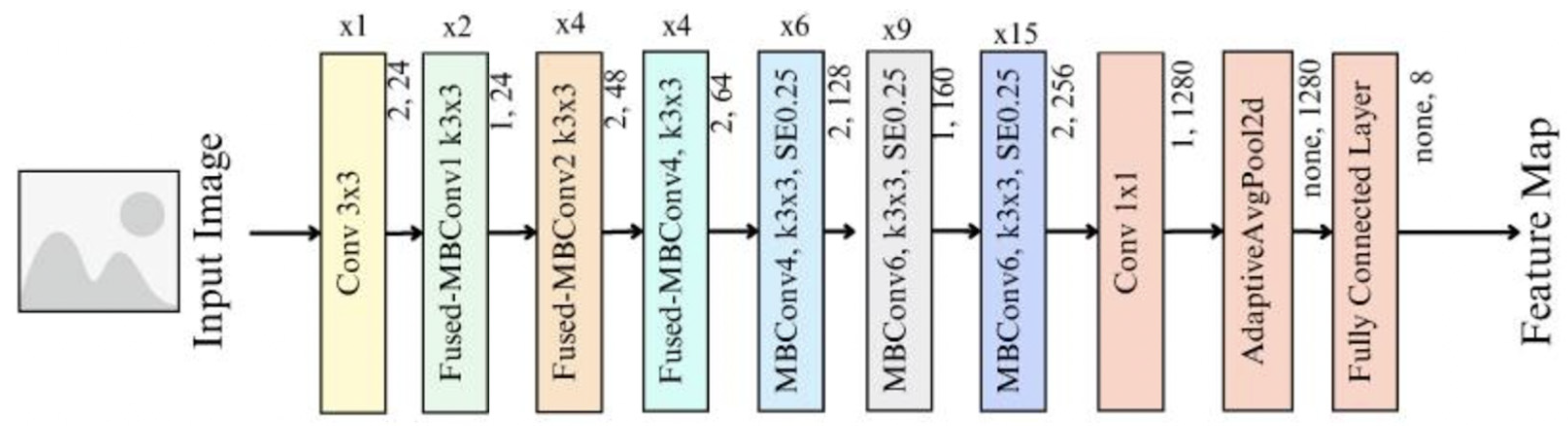
2.3. EfficientNet
- Balanced performance and efficiency: EfficientNet is designed to deliver high accuracy while keeping computational requirements low, making it ideal for processing large histopathological datasets [16].
- Scalability: EfficientNet offers scalable models such as EfficientNet-B0 for quick development and EfficientNet-B7 for higher accuracy, providing flexibility in handling different computational resources during the project [21].
3. Methodology
3.1. Dataset
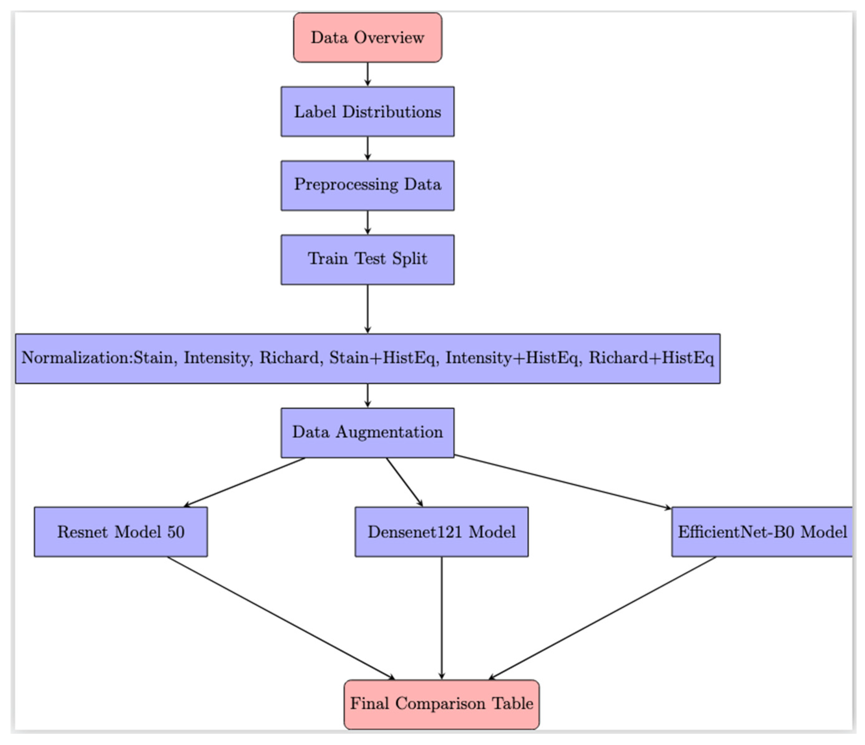
3.2. Data Preprocessing
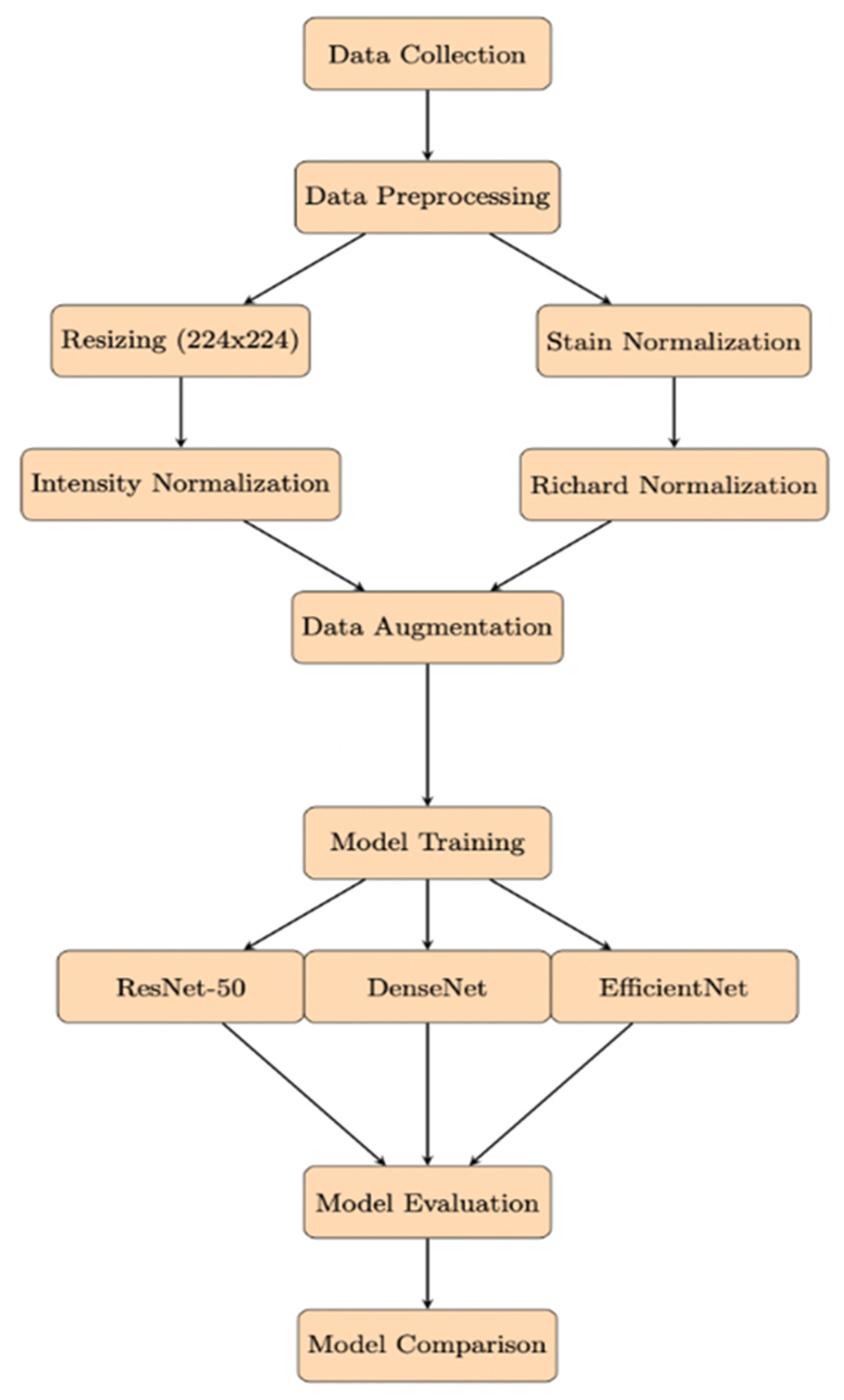
- Stain normalization: Variability in staining protocols and equipment introduces color inconsistencies that obscure tissue morphology. The Macenko algorithm is used to standardize the color space by aligning images to a reference stain profile, ensuring consistent input and improving model generalization.
- Intensity normalization: Pixel values are normalized to a uniform range (e.g., [0, 1] or [−1, 1]) to compensate for differences in brightness and contrast caused by imaging conditions. By dividing raw pixel values by 255, intensity normalization stabilizes training, reduces the risk of gradient instability, and improves data manageability for deep learning models.
- Richard normalization: Contrast enhancement methods such as histogram equalization and contrast-limited adaptive histogram equalization (CLAHE) are employed to amplify critical features in histopathological images. These techniques redistribute pixel intensities to highlight malignant regions while preserving structural integrity, thereby facilitating more accurate differentiation between healthy and cancerous tissues.
3.3. HE
- Enhance image contrast: In medical imaging, such as histopathological images of breast tissue, the contrast between cancerous and non-cancerous is not always sharp. Applying histogram equalization helps enhance these contrasts, making cancerous regions more detectable.
- Improve feature extraction: By improving the contrast, the models better detect patterns and structures, which are critical for distinguishing between IDC-positive (+) and IDC-negative (−) regions in tissue samples.
3.4. Data Augmentation
- Random sampling (IDC(−) = 1000, IDC(+) = 1200)
- Random brightness (0.5 px~2.0 px)
- Random shear (x: 0.0~0.2, y: 0.0~0.2)
- Class weights in the loss function
- Oversampling of the minority class
- Undersampling the majority class
- Combination of oversampling and undersampling
4. Result and Discussion
4.1. Effectiveness of HE
4.2. Stain Normalization
4.3. Intensity Normalization
4.4. Richard Normalization
5. Conclusions
Author Contributions
Funding
Institutional Review Board Statement
Informed Consent Statement
Data Availability Statement
Acknowledgments
Conflicts of Interest
References
- Zhang, Y.; Xia, K.; Li, C.; Wei, B.; Zhang, B. Review of Breast Cancer Pathologigcal Image Processing. BioMed Res. Int. 2021, 2021, 1994764. [Google Scholar] [CrossRef] [Scilit] [PubMed]
- Gurcan, M.; Boucheron, L.; Can, A.; Madabhushi, A.; Rajpoot, N.; Yener, B. Histopathological Image Analysis: A review. IEEE Rev. Biomed. Eng. 2009, 2, 147–171. [Google Scholar] [CrossRef] [Scilit] [PubMed]
- Janowczyk, A.; Madabhushi, A. Deep learning for digital pathology image analysis: A comprehensive tutorial with selected use cases. J. Pathol. Inform. 2016, 7, 29. [Google Scholar] [CrossRef] [Scilit] [PubMed]
- Aldakhil, L.; Alhasson, H.; Alharbi, S. Attention-Based Deep Learning Approach for Breast Cancer Histopathological Image Multi-Classification. Diagnostics 2024, 14, 1402. [Google Scholar] [CrossRef] [Scilit] [PubMed]
- Cruz-Roa, A.; Gilmore, H.; Basavanhally, A.; Feldman, M.; Ganesan, S.; Shih, N.N.; Tomaszewski, J.; González, F.A.; Madabhushi, A. Accurate and reproducible invasive breast cancer detection in whole-slide images: A Deep Learning approach for quantifying tumor extent. Sci. Rep. 2017, 7, 46450. [Google Scholar] [CrossRef] [Scilit] [PubMed]
- Angayarkanni, S.P. Hybrid Convolution Neural Network in Classification of Cancer in Histopathology Images. J. Digit. Imaging 2022, 35, 248–257. [Google Scholar] [CrossRef] [Scilit] [PubMed]
- Potsangbam, J.; Devi, S.S. Classification of Breast Cancer Histopathological Images Using Transfer Learning with DenseNet121. Procedia Comput. Sci. 2024, 235, 1990–1997. [Google Scholar] [CrossRef] [Scilit]
- Hossain, M.S.; Armstrong, L.J.; Cook, D.M.; Zaenker, P. Application of histopathology image analysis using deep learning networks. Hum.-Centric Intell. Syst. 2024, 4, 417–436. [Google Scholar] [CrossRef] [Scilit]
- Madani, M.; Behzadi, M.M.; Nabavi, S. The role of Deep Learning in Advancing breast cancer detection using different imaging modalities: A Systematic review. Cancers 2022, 14, 5334. [Google Scholar] [CrossRef] [Scilit] [PubMed]
- Belsare, A. Histopathological image analysis using image Processing techniques: An Overview. Signal Image Process. Int. J. 2012, 3, 23–36. [Google Scholar] [CrossRef] [Scilit]
- Hoque, M.Z.; Keskinarkaus, A.; Nyberg, P.; Seppänen, T. Stain normalization methods for histopathology image analysis: A comprehensive review and experimental comparison. Inf. Fusion 2023, 102, 101997. [Google Scholar] [CrossRef] [Scilit]
- JEOL Ltd. Richardson-Lucy Method|Glossary. Available online: https://www.jeol.com/words/emterms/20121023.105457.php#gsc.tab=0 (accessed on 1 September 2024).
- Tan, M.; Le, Q.V. EfficientNet: Rethinking model scaling for convolutional neural networks. arXiv 2019, arXiv:1905.11946. [Google Scholar] [CrossRef] [Scilit]
- Komura, D.; Ishikawa, S. Machine learning methods for histopathological image analysis. Comput. Struct. Biotechnol. J. 2018, 16, 34–42. [Google Scholar] [CrossRef] [Scilit] [PubMed]
- Potrimba, P. What is ResNet-50? Roboflow Blog, 15 April 2024. Available online: https://blog.roboflow.com/what-is-resnet-50/ (accessed on 1 September 2014).
- Li, Z.; Zhang, X.; Müller, H.; Zhang, S. Large-scale retrieval for medical image analytics: A comprehensive review. Med. Image Anal. 2017, 43, 66–84. [Google Scholar] [CrossRef] [Scilit] [PubMed]
- Wei, J. Classification of Histopathology Images with Deep Learning: A Practical Guide. Medium, 7 December 2021. Available online: https://medium.com/health-data-science/classification-of-histopathology-images-with-deep-learning-a-practical-guide-2e3ffd6d59c5 (accessed on 1 September 2014).
- Das, S. Implementing DenseNet-121 in PyTorch: A Step-by-Step Guide. Medium, 19 March 2023. Available online: https://medium.com/deepkapha-notes/implementing-densenet-121-in-pytorch-a-step-by-step-guide-c0c2625c2a60 (accessed on 1 September 2014).
- Amin, H.; Darwish, A.; Hassanien, A.E.; Soliman, M. End-to-End Deep learning model for corn leaf disease classification. IEEE Access 2022, 10, 31103–31115. [Google Scholar] [CrossRef] [Scilit]
- Keras Documentation: EfficientNet B0 to B7. Available online: https://keras.io/api/applications/efficientnet/ (accessed on 1 September 2014).
- Allunia. Breast Cancer. Kaggle, 22 June 2021. Available online: https://www.kaggle.com/code/allunia/breast-cancer (accessed on 1 September 2014).
- Garcea, F.; Serra, A.; Lamberti, F.; Morra, L. Data augmentation for medical imaging: A systematic literature review. Comput. Biol. Med. 2022, 152, 106391. [Google Scholar] [CrossRef] [Scilit] [PubMed]



| Model | Purpose | Implementation | Advantages |
|---|---|---|---|
| ResNet-50 | Chosen for its ability to handle deep neural networks with skip connections that mitigate the vanishing gradient problem, enabling it to learn complex patterns. | Pre-trained on ImageNet and fine-tuned on the IDC dataset, with images resized to 224 × 224 pixels and normalized. | Effectively extracts features from complex medical images, allowing detection of subtle patterns in cancerous tissues. |
| DenseNet | Selected for its dense connections that enhance gradient flow and feature reuse, making it efficient in learning from medical images with subtle variations. | Pre-trained on ImageNet and fine-tuned on the IDC dataset. | Leverages all learned features effectively, improving its ability to detect cancerous tissue. |
| EfficientNet | Chosen for its computational efficiency through compound scaling, balancing the network’s depth, width, and resolution, making it suitable for large-scale datasets. | Trained on the pre-processed dataset, with images resized and normalized. | Balances accuracy and computational efficiency, ideal for resource-constrained environments like medical prognostics. |
| ResNet-50 | DenseNet | EfficientNet | |
|---|---|---|---|
| Histogram Equalization (HE) | 0.5500 | 0.5455 | 0.5023 |
| Without Histogram Equalization (HE) | 0.5477 | 0.5409 | 0.5136 |
| Stain Normalisation Without Histogram Equalization (HE) | 0.5318 | 0.5523 | 0.5318 |
| Stain Normalisation With Histogram Equalization (HE) | 0.5136 | 0.5273 | 0.5159 |
| Intensity Normalisation Without Histogram Equalization (HE) | 0.4932 | 0.5023 | 0.5250 |
| Intensity Normalisation With Histogram Equalization (HE) | 0.5432 | 0.5182 | 0.5364 |
| Richard Normalisation Without Histogram Equalization (HE) | 0.5250 | 0.5250 | 0.5023 |
| Richard Normalisation With Histogram Equalization (HE) | 0.5409 | 0.5523 | 0.5091 |
Disclaimer/Publisher’s Note: The statements, opinions and data contained in all publications are solely those of the individual author(s) and contributor(s) and not of MDPI and/or the editor(s). MDPI and/or the editor(s) disclaim responsibility for any injury to people or property resulting from any ideas, methods, instructions or products referred to in the content. |
© 2026 by the authors. Licensee MDPI, Basel, Switzerland. This article is an open access article distributed under the terms and conditions of the Creative Commons Attribution (CC BY) license.
Share and Cite
Yong, C.Y.; Pang, D.J.W.; Leong, S.M.; Tan, C.W. Artificial Intelligence-Powered Breast Cancer Prognosis: Optimizing Deep Learning with Image Normalization. Eng. Proc. 2026, 128, 49. https://doi.org/10.3390/engproc2026128049
Yong CY, Pang DJW, Leong SM, Tan CW. Artificial Intelligence-Powered Breast Cancer Prognosis: Optimizing Deep Learning with Image Normalization. Engineering Proceedings. 2026; 128(1):49. https://doi.org/10.3390/engproc2026128049
Chicago/Turabian StyleYong, Chang Yeou, Dennis Jia Wang Pang, Sheng Mou Leong, and Chi Wee Tan. 2026. "Artificial Intelligence-Powered Breast Cancer Prognosis: Optimizing Deep Learning with Image Normalization" Engineering Proceedings 128, no. 1: 49. https://doi.org/10.3390/engproc2026128049
APA StyleYong, C. Y., Pang, D. J. W., Leong, S. M., & Tan, C. W. (2026). Artificial Intelligence-Powered Breast Cancer Prognosis: Optimizing Deep Learning with Image Normalization. Engineering Proceedings, 128(1), 49. https://doi.org/10.3390/engproc2026128049