Proceeding Paper

Aptamer-Based Plasmonic Plastic Optical Fiber Biosensors: A Focus on Relevant Applications †

1
Indivenire srl, Via Alla Cascata 56/C, 38123 Trento, Italy
2
Department of Engineering, University of Campania “L. Vanvitelli”, Via Roma 29, 81031 Aversa, Italy
*
Author to whom correspondence should be addressed.
Presented at the 2nd International Electronic Conference on Applied Sciences, 15–31 October 2021.
Eng. Proc. 2021, 11(1), 19; https://doi.org/10.3390/ASEC2021-11158
Published: 15 October 2021
(This article belongs to the Proceedings of The 2nd International Electronic Conference on Applied Sciences)

Abstract

:
Optical detection is one of the most used transduction methods in biosensors and apart from the commercially available instruments based on surface plasmon resonance (SPR), an emerging class of devices, based on both silica and plastic optical fibers (POFs), is finding its route. On the other hand, aptamers represent the next-generation biorecognition elements for biosensor implementation, thanks to several characteristics making them more appealing with respect to the conventional antibodies. The joint exploitation of plasmon resonance in plastic optical fibers and aptamers is here reported, focusing the attention on various relevant biological applications (e.g., thrombin, vascular endothelial growth factor (VEGF), and SARS-CoV-2 spike protein).

1. Introduction

Surface plasmon resonance, or localized surface plasmon resonance (LSPR), represents a gold standard in the optical characterization of biomolecular interaction, due to its high sensitivity, and it is also widely used as a transduction method in biosensor implementation. In particular, SPR/LSPR systems based on plastic optical fibers (POF) represent an emerging field which is paving the way for the development of a new class of sensors [1,2,3], not only for the POFs’ intrinsic characteristics such as excellent flexibility, large diameter and great numerical aperture, but also for the possibility for easily implementing different geometries, such as U-bent, D-shaped, side-polished, and tapered configurations. Furthermore, these kinds of optical fiber sensors can be used to realize small-size and low-cost optical biosensors and can be coupled to a variety of molecular recognition elements (MRE), such as antibodies, molecular imprinted polymers or aptamers (aptasensors).
Among MREs, the aptamers are an emerging class of molecules characterized by several advantages. Aptamers are short single-stranded DNA or RNA fragments selected to bind a wide range of analytes, ranging from very small molecules (pesticides, toxins) up to entire microorganisms; they exhibit an affinity constant in the nanomolar range minimizing the probability of false-positive results [4]. Moreover, they can be easily modified and are characterized by a high batch to batch reproducibility, a high resistance in acidic environments and at high temperatures. Due to the above characteristics, the aptamers find applications in different fields [5], ranging from the detection of small molecules [6,7,8] to point-of-care diagnostic systems [9,10], being suitable for the detection of bacteria [11] or of circulating tumor cells [12].
Here we focus our attention on POF-SPR biosensors, using aptamers as molecular recognition elements, for biomedical applications and in particular for the detection of: THR, a clinical marker of the blood coagulation cascade, VEGF, a circulating protein potentially associated with cancer, and the SARS-CoV-2 Spike protein.

2. Methods

The materials and methods were extensively reported in our previously published papers [13,14,15]. Here we briefly summarize the main steps for each application. A scheme of the optical platform with the aptamer layer is reported in Figure 1a and the different developed interfaces are reported in Figure 1b–d.

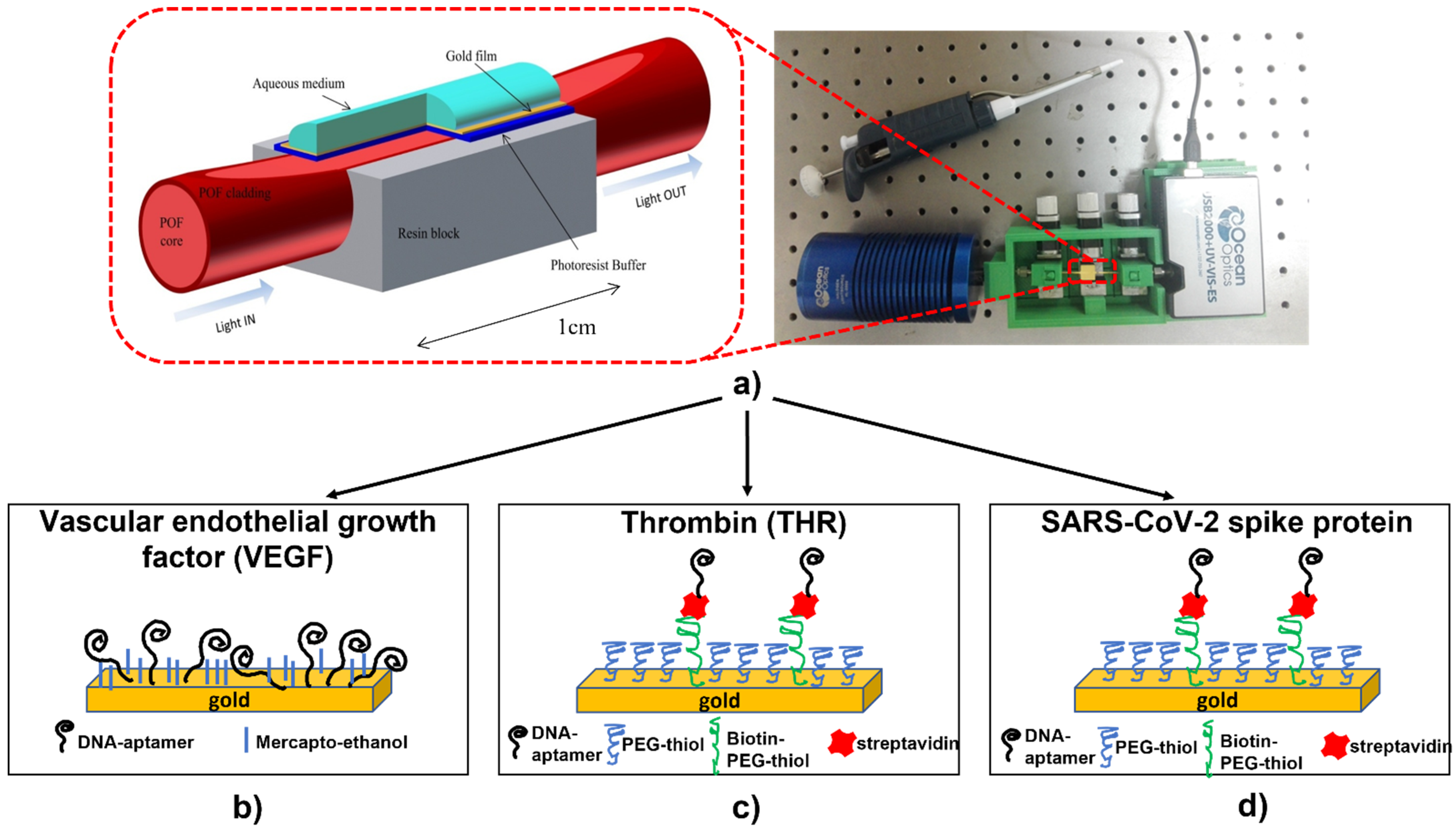
2.1. VEGF-Aptasensor Preparation

The aptasensor for VEGF detection was prepared according to the procedure reported in [13]. Briefly, after cleaning the gold surface through an argon plasma (6.8 W of power to the RF coil for one minute), a 1 μM aptamer (5′-HO-(CH2)3-S-S-(CH2)3-CC CGT CTT CCA GAC AAG AGT GCA GGG-3′) solution in 1 M potassium phosphate buffer pH 7 for one hour was applied followed by passivation in 1 mM mercaptoethanol solution in the same buffer for 30 min (Figure 1b).

2.2. THR-Aptasensor Preparation

The THR-aptasensor was prepared according to [14]. Briefly, after cleaning the gold surface (argon plasma, 6.8 W of power to the RF coil for one minute), a water solution of 0.2 mM of PEGthiol:BiotinPEGlipo in an 8:2 molar ratio was incubated overnight. After washing, 5 μg/mL streptavidin solution in phosphate buffer (10 mM phosphate buffer, 138 mM NaCl, 2.7 mM KCl, pH 7.4) was applied for one hour. Finally, 10 μM of biotin-TBA29 aptamer (5′-/5BiotinTEG/AG TCC GTG GTA GGG CAG GTT GGG GTG ACT-3′) was incubated for three hours in the same phosphate buffer (Figure 1c).

2.3. SARS-CoV-2 Spike Protein Aptasensor Preparation

A protocol similar to the one developed for THR was applied for the SARS-CoV-2 spike protein aptasensor implementation [15]. The only difference was related to the aptamer sequence which, in this case, was the following: 5′-/5BiotinTEG/CAG CAC CGA CCT TGT GCT TTG GGA GTG CTG GTC CAA GGG CGT TAA TGG ACA-3’ (Figure 1d).

2.4. Optical Measurements

The optical platform used for all the measurements is the one shown in Figure 1a. The polymer cladding over the PMMA core (980 μm) of the POF was removed and a Microposit S1813 photoresist was spun on it. A 60 nm thick gold layer was finally sputtered onto the photoresist layer. A halogen lamp (360 nm to 1700 nm) was used as light source and an Ocean Optics USB2000+VIS–NIR spectrometer (330 nm to 1100 nm) was employed to analyze the transmitted spectrum. The sensing experiments on the SPR-POF aptasensor were performed by dropping about 70 μL of solution over the sensing region. After 10 min, the solution was removed and washing in buffer was performed. After the dropping of fresh buffer, the transmission spectrum was recorded and normalized to air.

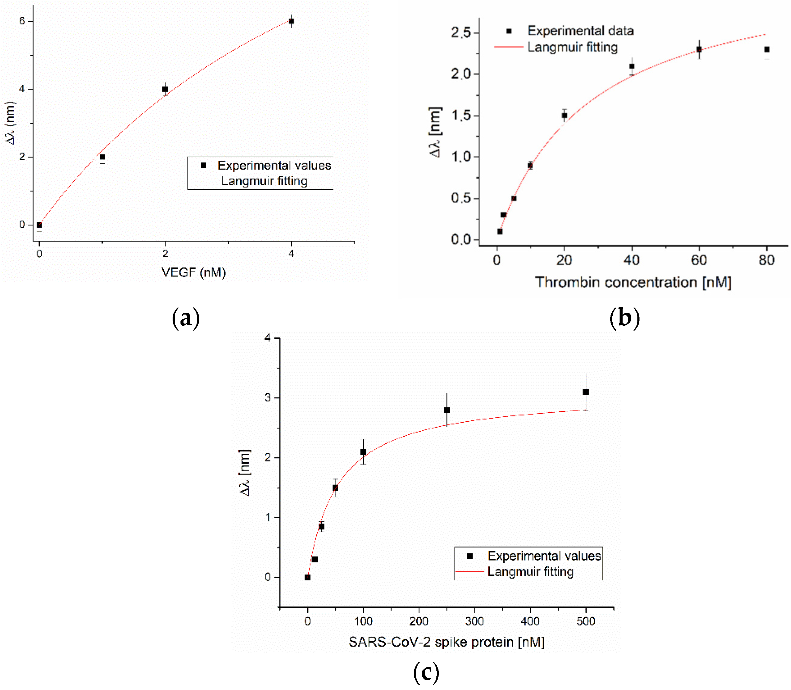
3. Results and Discussion

In the last decade, different MREs, such as antibodies, molecularly imprinted polymers (MIP), and aptamers, have been exploited on POF-based devices so proving the high versatility of this kind of sensing platform [2]. With respect to conventional MREs, aptamers represent a new emerging class increasingly employed to realize biosensors. Up to now, very few examples of coupling between aptamers and POF-based biosensors have been reported in the literature [13,14,15,16,17,18], and most of them are from our research group.
In 2015, we developed an aptamer-based POF-SPR sensor for the detection of vascular endothelial growth factor (VEGF), selected as a circulating protein potentially associated with cancer [13]. A thiolated aptamer was directly immobilized on the gold film deposited in the POF’s sensing region. Typical dose-response transmission spectra, achieved by incubating different amounts of VEGF in 20 mM Tris–HCl buffer pH 7.4, are reported in Figure 2a. The obtained results suggested that the direct immobilization of the aptamers on the gold surface can negatively affect their recognition capability and that passivation is an important step of the interface layer build-up. In fact, proper passivation assures a better aptamer conformation and allows a limit of detection of 0.8 nM [13] to be reached. Even if our aptasensor exhibits similar performances with respect to other detection systems, the dissociation constant measured on our sensor was two orders of magnitude lower. This is probably caused by a loss of affinity in the immobilization procedure.
For the above reason, in the subsequent works, we changed the approach and developed an interface based on short polyethylene glycol (PEG). The idea was to keep away the aptamer from the surface in order to ensure a better sequence conformation. So, the detection of thrombin (THR), a clinical marker of the blood coagulation cascade, was performed modifying the gold-coated POF with a mixed interface (a short-PEG and a biotinylated-PEG) and immobilizing, through avidin-biotin chemistry, a THR binding aptamer [14] (see Figure 1c). Figure 2b reports an example of the dose-response curve recorded by POF-SPR measurement for different THR concentrations in buffer (Tris 50 mM, EDTA 1 mM, MgCl2 1 mM, KCl 150 mM pH 7.4) [14]. The good performance of the obtained interface were confirmed, resulting in a detection limit of 1.6 nM and in the increase of the dissociation constant of one order of magnitude.
Recently, we modified the PEG-based interface, previously developed for thrombin detection, in order to detect the receptor-binding domain (RBD) of the SARS-CoV-2 spike glycoprotein [15]. To this purpose, we changed the aptamer sequence, as reported in Section 2.3. Figure 2c reports an example of the dose-response curve obtained by incubating different amounts of protein in buffer (136.8 mM NaCl, 10.1 mM Na2HPO4, 2.7 mM KCl, 1.8 mM KH2PO4, 0.55 mM MgCl2, pH 7.4). The aptasensor was tested, not only on the specific target, but also on aspecific targets (BSA, AH1H1 hemagglutinin protein and MERS spike protein) and in diluted human serum (50%). A limit of detection in the nanomolar range was achieved, confirming the good performance of this aptamer-based optical sensor.

4. Conclusions

Aptamers represent an emerging class of biorecognition elements, more and more exploited in the development of optical biosensors. Their performance makes them the ideal elements to be immobilized on plasmonic optical fiber-based devices. On the other hand, the POF-based platform exhibits excellent flexibility, making it extremely interesting for coupling with aptamers in order to develop sensitive biosensors, which can be easily integrable in portable, small-size, simple-to-manufacture devices for clinical applications.

Author Contributions

Conceptualization, writing—original draft, writing—review and editing, L.P. and N.C.; writing—review and editing L.Z. All authors have read and agreed to the published version of the manuscript.

Funding

This research received no external funding.

Institutional Review Board Statement

Not applicable.

Informed Consent Statement

Not applicable.

Data Availability Statement

The data are available on reasonable request from the corresponding author.

Acknowledgments

This work was supported by the VALERE program of the University of Campania “Luigi Vanvitelli” (Italy).

Conflicts of Interest

The authors declare no conflict of interest.

References

  1. Jin, Y.; Granville, A. Polymer Fiber Optic Sensors—A Mini Review of their Synthesis and Applications. J. Biosens. Bioelectron. 2016, 7, 194. [Google Scholar] [CrossRef]
  2. Cennamo, N.; Pesavento, M.; Zeni, L. A review on simple and highly sensitive plastic optical fiber probes for bio-chemical sensing. Sens. Actuators B Chem. 2021, 331, 129393. [Google Scholar] [CrossRef]
  3. Kadhim, R.A.; Abdul, A.K.K.; Yuan, L. Advances in Surface Plasmon Resonance-Based Plastic Optical Fiber Sensors. IETE Tech. Rev. 2020, 1–18. [Google Scholar] [CrossRef]
  4. Tombelli, S.; Minunni, M.; Mascini, M. Analytical applications of aptamers. Biosens. Bioelectron. 2005, 20, 2424–2434. [Google Scholar] [CrossRef] [PubMed]
  5. Yan, S.R.; Foroughi, M.M.; Safaei, M.; Jahani, S.; Ebrahimpour, N.; Borhani, F.; Rezaei Zade Baravati, N.; Aramesh-Boroujeni, Z.; Foong, L.K. A review: Recent advances in ultrasensitive and highly specific recognition aptasensors with various detection strategies. Int. J. Biol. Macromol. 2020, 155, 184–207. [Google Scholar] [CrossRef] [PubMed]
  6. Feng, C.; Dai, S.; Wang, L. Optical aptasensors for quantitative detection of small biomolecules: A review. Biosens. Bioelectron. 2014, 59, 64–74. [Google Scholar] [CrossRef] [PubMed]
  7. Chen, W.; Lei, W.; Xue, M.; Xue, F.; Meng, Z.H.; Zhang, W.B.; Qu, F.; Shea, K.J. Protein recognition by a surface imprinted colloidal array. J. Mater. Chem. A 2014, 2, 7165–7169. [Google Scholar] [CrossRef]
  8. Cunha, I.; Biltes, R.; Sales, M.G.F.; Vasconcelos, V. Aptamer-based biosensors to detect aquatic phycotoxins and cyanotoxins. Sensors 2018, 18, 2367. [Google Scholar] [CrossRef] [PubMed] [Green Version]
  9. Hassan, E.M.; DeRosa, M.C. Recent advances in cancer early detection and diagnosis: Role of nucleic acid based aptasensors. Trends Anal. Chem. 2020, 124, 115806. [Google Scholar] [CrossRef]
  10. Citartan, M.; Tang, T.H. Recent developments of aptasensors expedient for point-of-care (POC) diagnostics. Talanta 2019, 199, 556–566. [Google Scholar] [CrossRef] [PubMed]
  11. Majdinasab, M.; Hayat, A.; Marty, J.L. Aptamer-based assays and aptasensors for detection of pathogenic bacteria in food samples. Trends Anal. Chem. 2018, 107, 60–77. [Google Scholar] [CrossRef]
  12. Safarpour, H.; Dehghani, S.; Nosrati, R.; Zebardast, N.; Alibolandi, M.; Mokhtarzadeh, A.; Ramezani, M. Optical and electrochemical-based nano-aptasensing approaches for the detection of circulating tumor cells (CTCs). Biosens. Bioelectron. 2020, 148, 111833. [Google Scholar] [CrossRef] [PubMed]
  13. Cennamo, N.; Pesavento, M.; Lunelli, L.; Vanzetti, L.; Pederzolli, C.; Zeni, L.; Pasquardini, L. An easy way to realize SPR aptasensor: A multimode plastic optical fiber platform for cancer biomarkers detection. Talanta 2015, 140, 88–95. [Google Scholar] [CrossRef] [PubMed]
  14. Cennamo, N.; Pasquardini, L.; Arcadio, F.; Vanzetti, L.E.; Bossi, A.M.; Zeni, L. D-shaped plastic optical fibre aptasensor for fast thrombin detection in nanomolar range. Sci. Rep. 2019, 9, 18740. [Google Scholar] [CrossRef] [PubMed]
  15. Cennamo, N.; Pasquardini, L.; Arcadio, F.; Lunelli, L.; Vanzetti, L.; Carafa, V.; Altucci, L.; Zeni, L. SARS-CoV-2 spike protein detection through a plasmonic D-shaped plastic optical fiber aptasensor. Talanta 2021, 233, 122532. [Google Scholar] [CrossRef] [PubMed]
  16. Galatus, R.M.; Cristea, C.; Feier, B.; Cennamo, N.; Zeni, L. SPR based hybrid electro-optic biosensor for β-lactam antibiotics determination in water. In Remote Sensing and Modeling of Ecosystems for Sustainability XIV; International Society for Optics and Photonics: Bellingham, WA, USA, 2017; Volume 10405. [Google Scholar] [CrossRef]
  17. Sun, J.; Jiang, S.; Xu, J.; Li, Z.; Li, C.; Jing, Y.; Zhao, X.; Pan, J.; Zhang, C.; Man, B. Sensitive and selective surface plasmon resonance sensor employing a gold-supported graphene composite film/D-shaped fiber for dopamine detection. J. Phys. D Appl. Phys. 2019, 51, 195402. [Google Scholar] [CrossRef]
  18. Sanjay, M.; Singh, N.K.; Ngashangva, L.; Goswami, P. A smartphone-based fiber-optic aptasensor for label-free detection of: Plasmodium falciparum glutamate dehydrogenase. Anal. Methods 2020, 12, 1333–1341. [Google Scholar] [CrossRef]
Figure 1. (a) Image of the sensor system with a zoom of the plasmonic optical sensor and different aptamer-based interfaces: (b) vascular endothelial growth factor (VEGF) [13], (c) thrombin [14], and (d) SARS-CoV-2 spike protein [15].
Engproc 11 00019 g001
Figure 2. Dose-response curves obtained incubating different concentrations of VEGF (a), of thrombin, reprinted from [14] (b), and of SARS-CoV-2 spike protein (c). Langmuir fits are reported as well.
Engproc 11 00019 g002
Publisher’s Note: MDPI stays neutral with regard to jurisdictional claims in published maps and institutional affiliations.

