Proceeding Paper

Comparison of Modifications of Cellulose for the Extraction and Elution of DNA †

Biotechnology Innovation Centre, Rhodes University, Makhanda 6139, South Africa
*
Author to whom correspondence should be addressed.
Presented at the Micro Manufacturing Convergence Conference, Stellenbosch, South Africa, 7–9 July 2024.
Eng. Proc. 2025, 109(1), 5; https://doi.org/10.3390/engproc2025109005
Published: 11 September 2025
(This article belongs to the Proceedings of Micro Manufacturing Convergence Conference)

Abstract

The extraction of DNA from biological samples precedes many research and commercial applications. This study compares surface treatments of cellulose (a low-cost binding matrix) to enhance binding and elution of DNA to paper-based dipsticks. Cellulose paper was modified with poly-L-lysine, silica, or guanidine, as well as subjected to TEMPO-based oxidation. Subsequently, binding and elution behaviour of fragmented salmon sperm DNA to dipsticks was evaluated. Qubit fluorimetry and agarose gel electrophoresis measurements indicated that TEMPO-based oxidation significantly increased the binding of DNA and its elution from dipsticks, while silica modifications bound DNA efficiently, but strongly retained it. Leaching of select modifiers (guanidine, silica and poly-L-lysine) was indicated by UV/Vis spectroscopy, indicating that further optimization of attachment processes is required. This study is the first to compare multiple cellulose surface treatments for their influence on DNA binding and elution, especially the use of TEMPO-based oxidation for this purpose, and highlights some means of identifying leaching of modifiers during DNA capture at these surfaces and subsequent elution. While TEMPO-based oxidation proves a promising treatment to enhance DNA elution, further refinement of the approach is needed to ensure compatibility with molecular biology techniques.

1. Introduction

The extraction of DNA from biological materials is an essential process in both biological research [1,2,3,4] and commercial applications, e.g., pathogen identification [5], forensics [6], agriculture [7], etc. The double-stranded form of DNA (dsDNA) has multiple functional groups, which potentially allow it to interact with numerous surfaces, permitting extraction by noncovalent bond formation: electrostatic interactions, hydrophobic forces, and specific bonding [8,9,10]. These forces are strongly influenced by external factors such as pH, ionic strength, and buffer composition [8,10,11,12,13].
Cellulose-based papers have recently emerged as low-cost and portable matrices for nucleic acid purification, offering an accessible alternative to traditional DNA extraction methods [14]. Unmodified cellulose reportedly binds to nucleic acids effectively and can elute them directly into aqueous solutions, following washing [14]. Cellulose is a linear polymer of β-1,4-linked glucopyranose residues, permitting DNA adsorption by offering sites for hydrogen bond formation and hydrophobic interactions [15].
Cellulose is highly modifiable due to its abundant primary hydroxyl groups, enabling the attachment of various functional groups [15]. The study reported in this short communication sought to determine which existing modifications of cellulose produce improved DNA binding ability and subsequent ease-of-elution into a low-salt aqueous solution. Two approaches were initially pursued: modification with cationic groups (poly-L-lysine, guanidine) [16,17] to neutralize the electrostatic repulsion caused by DNA’s anionic phosphate, and modifications with hydrophobic moieties via silica sol–gel casting to improve hydrophobic bond formation [18]. TEMPO-oxidized cellulose [16] was included as an intermediate surface for the covalent modification of cellulose with cationic groups.
The main objective of this study was to compare the influence of different cellulose surface modifications on DNA binding and elution at cellulose-based dipsticks, with an emphasis on the strengths and limitations of each modification. Notably, the work explores the role of TEMPO-based oxidation as an under-research approach to facilitate DNA elution.

2. Materials and Methods

2.1. Materials

A full list of materials, and detailed methodology, is provided in S1A.

2.2. Methods

2.2.1. Salmon Sperm Sonication

DNA extracted from salmon sperm was selected as a source of dsDNA [19] and ultrasonicated for 60 min to create a range of DNA fragment sizes [20].

2.2.2. Paper Treatments

Sections of Whatman® qualitative filter paper no. 1 measured at 7 × 9 cm were used as cellulose surfaces. For untreated cellulose samples, paper was rinsed with ddH2O. For silica-modified cellulose, a silica sol was produced by combining a silica solution consisting of 43.39 mL TEOS and 46.14 mL MTES and was mixed with 10.16 mL colloidal SiO2 nanoparticle. This was dip-coated for 1 min onto paper and cured in the oven for 30 min at 55 °C [18]. TEMPO-based oxidation of the cellulose surface was conducted using previously reported protocols [16]. For Poly-L-Lysine (PLL) cellulose modification, paper was TEMPO-oxidized, then functionalised with Poly-L-Lysine via covalent attachment using EDC and NHS conjugation [16]. For guanidine cellulose treatment, paper was TEMPO-oxidized [16] followed by guanidine hydrochloride noncovalent electrostatic attachment [17]. All papers were rinsed with ddH2O and dried in the oven at 55 °C for 1 h before use and/or subsequent modifications.

2.2.3. Paper-DNA Interaction Analyses

Cellulose dipsticks were fabricated by cutting the paper surfaces detailed in Section 2.2.2. into 0.5 × 1 cm rectangles and attaching them to notched pipette tips. Each dipstick was dipped 15 times for 1 s each into a sample of 450 ng/µL ultrasonicated salmon sperm DNA (dissolved in ddH2O), followed by dipping it 5 times for 1 s each into a Tris wash buffer (10 mM, pH 8). DNA attached to paper was subsequently eluted by dipping it 15 times for 1 s each in nuclease-free H2O [14].
The DNA content of all samples was quantified using the following: NanoDrop UV/VIS spectroscopy; Qubit fluorometric assays (Qubit™ dsDNA BR Assay Kit—Thermo Fisher Scientific: Waltham, MA, USA), and agarose gel electrophoresis (AGE) densitometry.

3. Results and Discussion

DNA sonicated for 60 min was selected for the cellulose experiment based on this treatment providing a broad range of DNA fragment sizes (S1B—Figure S1).
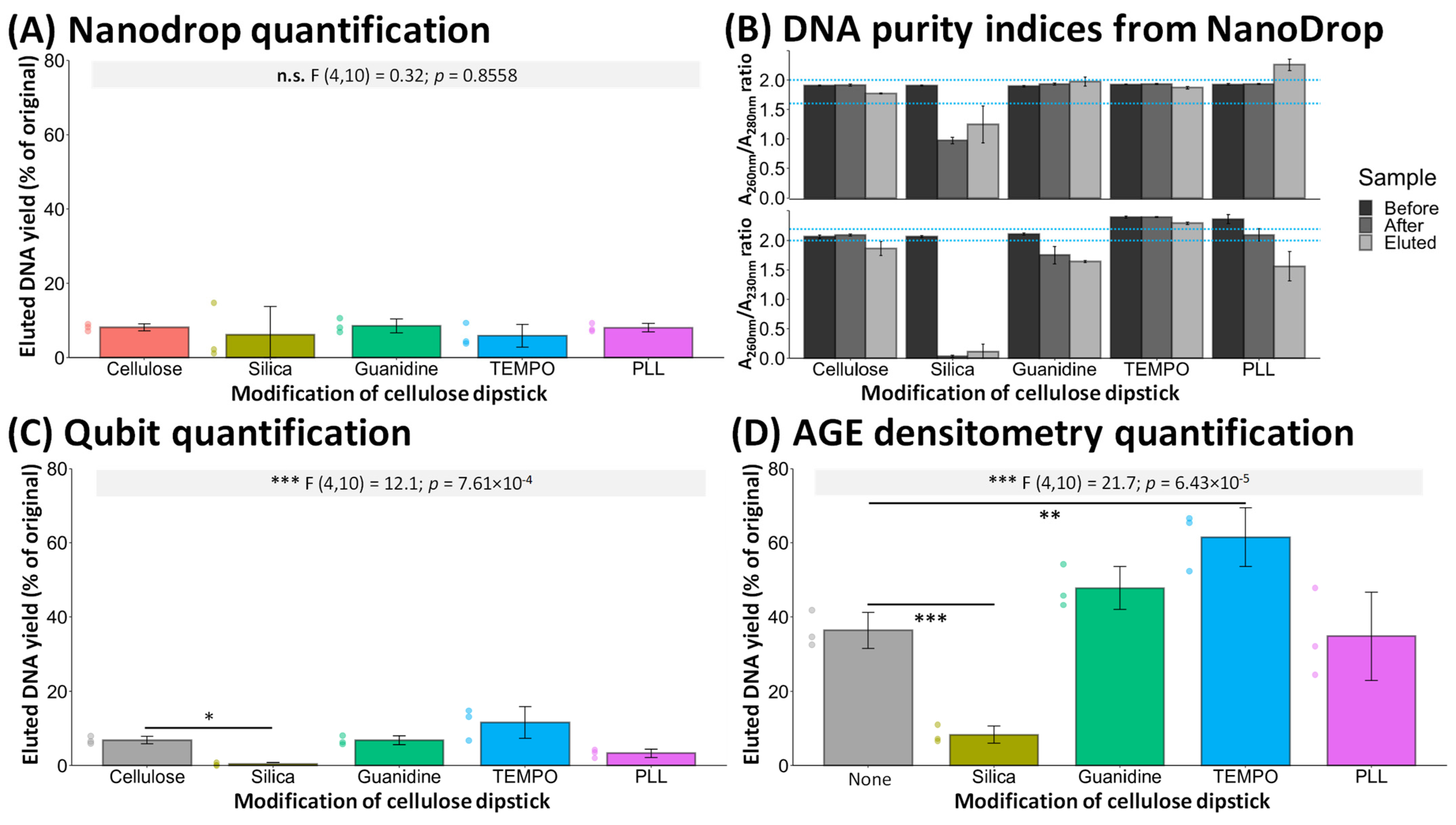
The DNA content of samples was quantified using three different measurement approaches: NanoDrop UV/VIS spectroscopy; Qubit fluorometric assays, and agarose gel electrophoresis (AGE) densitometry. DNA content of each sample was measured before exposure to the dipstick, after exposure to the dipstick, and following elution from the dipstick; this data is presented in the Supplementary Materials (S1B—Figure S2). The eluted DNA, as a percentage of the original DNA content of individual samples, are compared in Figure 1.
The direct evaluation of eluted yields using NanoDrop UV/VIS quantification (Figure 1A) was prevented by variations in the quality of the DNA after exposure to some of the modifications (Figure 1B). The consistent decreases in both A 260   nm A 280   nm and A 260   nm A 230   nm ratios following exposure to silica-modified surfaces is consistent with UV/Vis spectra of silica [21] indicating dissolution of this into the DNA solution. Similarly, the concomitant increases of A 260   nm A 280   nm and decreases of A 260   nm A 230   nm are consistent with the presence of guanidine [22] and lysine residues [23] in the DNA solutions after exposure to these modified cellulose surfaces (Figure 1B). For these reasons, the retention and elution of DNA from the modified cellulose surfaces was evaluated using a combination of Qubit and AGE.
Figure 1. Quantification of the DNA yields eluted from cellulose dipstick tests, evaluating the effect of cellulose modification on DNA extraction. (A) NanoDrop UV/VIS spectroscopic-based quantification of eluted DNA. (B) DNA purity indices calculated from individual wavelength absorbances measured by NanoDrop. Wavelength absorbance indices indicating good-quality DNA ranges (dotted blue lines in (B)) were obtained from [24]. (C) Qubit fluorometric analysis of eluted DNA. (D) AGE analysis of eluted DNA. All yields are calculated as the percentage of initial DNA quantified for each sample before exposure to the cellulose dipsticks. Annotated text in (A,C,D) indicate results from one-way ANOVA, followed by Tukey HSD post hoc tests to identify significant differences. N = 3. *—p ≤ 0.05, **—p ≤ 0.01, ***—p ≤ 0.001.
Engproc 109 00005 g001
Silica modification consistently eluted significantly less DNA than untreated cellulose (Figure 1C,D). TEMPO-oxidation of cellulose eluted significantly more DNA than untreated cellulose by AGE analysis, with a non-significant improvement by Qubit analysis. Guanidine treatment is shown to elute slightly more DNA than untreated cellulose, but not to statistically significant extents. Similarly, PLL treatment showed slightly, but not significantly, lower DNA elution. The lack of significant DNA elution with guanidine and PLL treatments might be due to the instability of these treatments, resulting in the loss of the cations during exposure to the DNA solution, as indicated in the NanoDrop analysis (Figure 1B). Figure 2 presents examples of AGE, examining whether a size-dependent DNA binding and/or elution took place at the different dipstick surfaces tested.
As reported by others [14] and by this study (Figure 1), unmodified cellulose bound DNA and eluted it (Figure 2A); however, only a small amount of DNA was considered to bind, evident by the lack of visible change in the electrophoretic profile of DNA before and after exposure to the dipstick but appeared to be effectively eluted from the cellulose subsequently. All samples exhibiting elution of DNA elute higher molecular weight DNA fragments, evidenced by the visible bands in the Elution 1 lanes occurring from approximately ~48.5 kbp onwards.
Apart from a slight increase in the eluted DNA intensity for guanidine, neither PLL nor guanidine treatments appear to visibly alter the underlying binding capacity of unmodified cellulose (Figure 2C,E), in line with the slight effects in eluted yields in Figure 1. Conversely, by AGE, silica modification showed extensive binding of DNA (substantial removal of DNA after exposure to the dipstick) but appeared to fail to subsequently elute the DNA. Unexpectedly, oxidation of cellulose by TEMPO produced a surface that both efficiently bound high MW DNA (yellow arrow showing the absence of the high MW region) and exhibited visibly better elution compared to untreated cellulose, (Figure 1D). While silica was effective in absorbing DNA due to hydrophobic interactions [25], it did not efficiently release the DNA afterward (Figure 2B). In contrast, TEMPO-oxidized cellulose also bound DNA, likely through similar hydrophobic interactions found for silica surfaces [26]. However, unlike silica, TEMPO-oxidized cellulose efficiently released DNA, covering a broad range of molecular weights (Figure 2D).

4. Conclusions

This study indicated that TEMPO-based oxidation of the cellulose significantly improved DNA binding and elution, providing a simple method for treating cellulose dipsticks. The precise mechanism by which this occurs is to be explored in follow-up studies. The possible dissolution of modifiers—specifically, guanidine and PLL—from the cellulose might decrease the surface’s ability to retain DNA at the surface; optimization of the attachment processes will be evaluated and physicochemical characterization of the surfaces, e.g., using Raman spectroscopy, should be conducted to evaluate the stability of modification of the cellulose using these approaches. UV/VIS spectroscopic characterization proved an effective method to analyze sample contamination by the modifiers and should be retained for studies examining surface–DNA interactions; conversely, Qubit and AGE proved more robust methods for quantifying DNA, overall. Additionally, AGE densitometry allowed for size-dependent analysis of DNA binding and elution. Further refinement is essential to optimize these methods and ensure compatibility with molecular biology techniques, e.g., qPCR. Additionally, future studies examining the ability of cellulose to effectively concentrate DNA in complex samples, such as cell lysate, should also be conducted to determine if cellulose-based surfaces can effectively serve as DNA extraction matrices.

Supplementary Materials

The following supporting information can be downloaded at: https://www.mdpi.com/article/10.3390/engproc2025109005/s1, 1. (A) Full List of Materials and Experimental Details of Methods; (B) Supporting Figures. Figure S1: Salmon Sperm Sonicated for Different Time Intervals; Figure S2: DNA Quantification of the Different Cellulose Dipstick Tests Using Different Methods; Figure S3: Representative Examples Of AGE Analysis of Size-Dependent DNA Binding and/or Elution by the Tested Cellulose Modification Dipsticks.

Author Contributions

Conceptualization, R.F., J.L., and S.M.R.; methodology, S.M.R. and R.F.; validation, S.M.R. and R.F.; formal analysis, S.M.R. and R.F.; investigation, S.M.R.; resources, S.M.R. and R.F.; data curation, S.M.R.; writing—original draft preparation, S.M.R.; writing—review and editing, R.F. and J.L.; visualization, S.M.R.; supervision, R.F. and J.L.; project administration, J.L.; funding acquisition, J.L. All authors have read and agreed to the published version of the manuscript.

Funding

This research was funded by the DSI/NRF South African Research Chair in Biotechnology Innovation and Engagement, grant number 95319.

Institutional Review Board Statement

Not applicable.

Informed Consent Statement

Not applicable.

Data Availability Statement

Dataset available on request from the authors.

Acknowledgments

This research was supported by access to the Nano-Micro Manufacturing Facility, funded by the Department of Science and Innovation (South African Research Infrastructure Roadmap). SR acknowledges the Pearson Young Memorial Trust and NRF for postgraduate funding.

Conflicts of Interest

The authors declare no conflicts of interest. The funders had no role in the design of the study; in the collection, analyses, or interpretation of data; in the writing of the manuscript; or in the decision to publish the results.

References

  1. Huan, T.; Joehanes, R.; Song, C.; Peng, F.; Guo, Y.; Mendelson, M.; Yao, C.; Liu, C.; Ma, J.; Richard, M.; et al. Genome-wide identification of DNA methylation QTLs in whole blood highlights pathways for cardiovascular disease. Nat. Commun. 2019, 10, 4267. [Google Scholar] [CrossRef]
  2. Ferguson, L.R.; Han, D.Y.; Fraser, A.G.; Huebner, C.; Lam, W.J.; Morgan, A.R.; Duan, H.; Karunasinghe, N. Genetic factors in chronic inflammation: Single nucleotide polymorphisms in the STAT-JAK pathway, susceptibility to DNA damage and Crohn’s disease in a New Zealand population. Mutat. Res. Mol. Mech. Mutagen. 2010, 690, 108–115. [Google Scholar] [CrossRef] [PubMed]
  3. Tettamanti, L.; Gaudio, R.M.; Iapichino, A.; Mucchi, D.; Tagliabue, A. Genetic susceptibility and periodontal disease: A retrospective study on a large Italian sample. Oral Implant. 2017, 10, 20–27. [Google Scholar] [CrossRef] [PubMed]
  4. Song, M.; Hwang, G.T. DNA-Encoded Library Screening as Core Platform Technology in Drug Discovery: Its Synthetic Method Development and Applications in DEL Synthesis. J. Med. Chem. 2020, 63, 6578–6599. [Google Scholar] [CrossRef]
  5. Luna, R.A.; Fasciano, L.R.; Jones, S.C.; Boyanton, B.L.; Ton, T.T.; Versalovic, J. DNA Pyrosequencing-Based Bacterial Pathogen Identification in a Pediatric Hospital Setting. J. Clin. Microbiol. 2007, 45, 2985–2992. [Google Scholar] [CrossRef]
  6. Chong, K.W.Y.; Thong, Z.; Syn, C.K.-C. Recent trends and developments in forensic DNA extraction. Wiley Interdiscip. Rev. Forensic Sci. 2021, 3, e1395. [Google Scholar] [CrossRef]
  7. Johnson, M.; Lee, K.; Scow, K. DNA fingerprinting reveals links among agricultural crops, soil properties, and the composition of soil microbial communities. Geoderma 2003, 114, 279–303. [Google Scholar] [CrossRef]
  8. Freitas, S.S.; Santos, J.A.; Prazeres, D.M. Plasmid purification by hydrophobic interaction chromatography using sodium citrate in the mobile phase. Sep. Purif. Technol. 2009, 65, 95–104. [Google Scholar] [CrossRef]
  9. Xiao, F.; Chen, Z.; Wei, Z.; Tian, L. Hydrophobic Interaction: A Promising Driving Force for the Biomedical Applications of Nucleic Acids. Adv. Sci. 2020, 7, 2001048. [Google Scholar] [CrossRef]
  10. Katevatis, C.; Fan, A.; Klapperich, C.M. Low concentration DNA extraction and recovery using a silica solid phase. PLoS ONE 2017, 12, e0176848. [Google Scholar] [CrossRef]
  11. Koo, K.M.; Sina, A.A.I.; Carrascosa, L.G.; Shiddiky, M.J.A.; Trau, M. DNA–bare gold affinity interactions: Mechanism and applications in biosensing. Anal. Methods. 2015, 7, 7042–7054. [Google Scholar] [CrossRef]
  12. Park, J.S.; Goo, N.-I.; Kim, D.-E. Mechanism of DNA Adsorption and Desorption on Graphene Oxide. Langmuir 2014, 30, 12587–12595. [Google Scholar] [CrossRef]
  13. Zhai, H.; Wang, L.; Putnis, C.V. Molecular-Scale Investigations Reveal Noncovalent Bonding Underlying the Adsorption of Environmental DNA on Mica. Environ. Sci. Technol. 2019, 53, 11251–11259. [Google Scholar] [CrossRef] [PubMed]
  14. Zou, Y.; Mason, M.G.; Wang, Y.; Wee, E.; Turni, C.; Blackall, P.J.; Trau, M.; Botella, J.R.; Misteli, T. Nucleic acid purification from plants, animals and microbes in under 30 seconds. PLoS Biol. 2017, 15, e2003916. [Google Scholar] [CrossRef]
  15. Figueiredo, J.A.; Ismael, M.I.; Anjo, C.M.S.; Duarte, A.P. Cellulose and Derivatives from Wood and Fibers as Renewable Sources of Raw-Materials. In Carbohydrates in Sustainable Development I; Springer: Berlin/Heidelberg, Germany, 2010; pp. 117–128. [Google Scholar] [CrossRef]
  16. Zhang, S.; Chen, H.; Shi, Z.; Liu, Y.; Liu, L.; Yu, J.; Fan, Y. Preparation of amino cellulose nanofiber via ε-poly-L-lysine grafting with enhanced mechanical, anti-microbial and food preservation performance. Ind. Crop. Prod. 2023, 194, 116288. [Google Scholar] [CrossRef]
  17. Mi, X.; Albukhari, S.M.; Heldt, C.L.; Heiden, P.A. Virus and chlorine adsorption onto guanidine modified cellulose nanofibers using covalent and hydrogen bonding. Carbohydr. Res. 2020, 498, 108153. [Google Scholar] [CrossRef]
  18. Castro, Y.; Durán, A. Control of degradation rate of Mg alloys using silica sol–gel coatings for biodegradable implant materials. J. Sol-Gel Sci. Technol. 2018, 90, 198–208. [Google Scholar] [CrossRef]
  19. Hui, C.-Y.; Guo, Y.; Zhang, X.; Shao, J.-H.; Yang, X.-Q.; Zhang, W. Oligodeoxyribonucleotides derived from salmon sperm DNA: An alternative to defibrotide. Biologicals 2013, 41, 190–196. [Google Scholar] [CrossRef]
  20. Mann, T.L.; Krull, U.J. The application of ultrasound as a rapid method to provide DNA fragments suitable for detection by DNA biosensors. Biosens. Bioelectron. 2004, 20, 945–955. [Google Scholar] [CrossRef]
  21. Lucena-Aguilar, G.; Sánchez-López, A.M.; Barberán-Aceituno, C.; Carrillo-Ávila, J.A.; López-Guerrero, J.A.; Aguilar-Quesada, R. DNA Source Selection for Downstream Applications Based on DNA Quality Indicators Analysis. Biopreservation Biobanking 2016, 14, 264–270. [Google Scholar] [CrossRef]
  22. Biradar, A.I.; Sarvalkar, P.D.; Teli, S.B.; Pawar, C.; Patil, P.; Prasad, N.R. Photocatalytic degradation of dyes using one-step synthesized silica nanoparticles. Mater. Today Proc. 2021, 43, 2832–2838. [Google Scholar] [CrossRef]
  23. Heptinstall, J.; Rapley, R. Spectrophotometric Analysis of Nucleic Acids. In The Nucleic Acid Protocols Handbook; Rapley, R., Ed.; Springer Protocols Handbooks: Berlin/Heidelberg, Germany; Humana Press: Totowa, NJ, USA, 2000. [Google Scholar] [CrossRef]
  24. Stagi, L.; Malfatti, L.; Caboi, F.; Innocenzi, P. Thermal Induced Polymerization of l-Lysine forms Branched Particles with Blue Fluorescence. Macromol. Chem. Phys. 2021, 222, 2100242. [Google Scholar] [CrossRef]
  25. Shi, B.; Shin, Y.K.; Hassanali, A.A.; Singer, S.J. DNA Binding to the Silica Surface. J. Phys. Chem. B 2015, 119, 11030–11040. [Google Scholar] [CrossRef]
  26. Lindman, B.; Medronho, B.; Alves, L.; Norgren, M.; Nordenskiöld, L. Hydrophobic interactions control the self-assembly of DNA and cellulose. Q. Rev. Biophys. 2021, 54, e3. [Google Scholar] [CrossRef]
Figure 2. Representative examples of AGE analysis of size-dependent DNA binding and/or elution by the tested cellulose modification dipsticks. Samples were electrophoresed on 1% w/v agarose gels containing 1× GelRed for 30 min at 100 V prior to visualization. (A) Untreated cellulose. (B) Silica treated. (C) Guanidine treated. (D) TEMPO-oxidized treated; the substantial decrease of a high-MW band present in the other samples is indicated by the annotated yellow arrow. (E) Poly-L-Lysine (PPL) treated. Lane annotations: High range DNA Ladder, Negative controls (NC—ddH2O sample), sample before cellulose dip, sample after cellulose dip, and a sample taken after elution. The first sample of a triplicate sample is presented here in a contrast-enhanced format—complete, unenhanced, gels are presented in S1B (Figure S3).
Engproc 109 00005 g002
Disclaimer/Publisher’s Note: The statements, opinions and data contained in all publications are solely those of the individual author(s) and contributor(s) and not of MDPI and/or the editor(s). MDPI and/or the editor(s) disclaim responsibility for any injury to people or property resulting from any ideas, methods, instructions or products referred to in the content.