Proceeding Paper

In Vitro Cytotoxicity of Single Walled Carbon Nanotube Bioconjugates on Cancer Cells †

by
Zvikomborero T. Gwanzura
1,
Willem J. Perold
1,* and
Anna-Mart Engelbrecht
2
1
Department of Electrical and Electronic Engineering, Faculty of Engineering, Stellenbosch University, Stellenbosch 7600, South Africa
2
Department of Physiological Sciences, Faculty of Science, Stellenbosch University, Stellenbosch 7600, South Africa
*
Author to whom correspondence should be addressed.
Presented at the Micro Manufacturing Convergence Conference, Stellenbosch, South Africa, 7–9 July 2024.
Eng. Proc. 2025, 109(1), 6; https://doi.org/10.3390/engproc2025109006
Published: 11 September 2025

Abstract

Carbon nanotubes have shown great promise in drug delivery systems as they can easily penetrate the cell membrane. Herein, carbon nanotubes functionalized with polyethylene glycol and folic acid were used to improve target specificity in breast and colon cancer cells. The functionalized carbon nanotubes were bioconjugated with bioactive compounds from plant extracts. In vitro cytotoxicity studies were conducted to demonstrate cellular uptake and apoptosis due to bioconjugate cellular internalization. The bioconjugates were able to preserve normal cells and induce cell death in cancer cells. The efficacy of the carbon nanotube bioconjugates in this study shows great potential in cancer therapy applications.

1. Introduction

Nanoparticles have played a crucial role in cancer research as they can effectively deliver drugs and easily enter cells [1]. They have the capacity to increase drug efficacy and improve target specificity [2]. The use of metallic nanoparticles as nanocarriers has faced significant obstacles due to their heightened toxicity to biological systems. This drawback arises from specific properties, such as their shape, surface chemistry and composition [3]. As a result, researchers have sought alternative options to address this concern. One promising solution has been the exploration of carbon nanotubes as nanocarriers [4]. Carbon nanotubes exhibit lower toxicity compared to metallic nanoparticles, making them a better choice for drug delivery and nanomedicine applications [5].
Carbon nanotube uptake can occur in various ways. The pathways include passive uptake, direct penetration through the cell membrane and active uptake or endocytosis pathways [6]. A new area of research in nanomedicine is the incorporation of carbon nanotubes with plant-based drugs. Since plant-based drugs are readily available in extract form, they are suitable candidates for incorporation into nanocarriers as they possess the property of bioavailability [7]. Plant-based drugs may either be packed inside the CNT or attached to the CNT surface for efficient drug delivery to the target site, thereby minimizing drug loss and off-target effects [8]. It is due to the structure of CNT that the drug is delivered. This study utilized carbon nanotubes bioconjugated with plant extracts to demonstrate cellular uptake in cancer cells.

2. Materials and Methods

Cell Lines

For in vitro cytotoxicity studies, the following cell lines were utilized: HT-29, a human colorectal adenocarcinoma cell line, MCF-7 and MDA-MB-231, human breast cancer cell lines (American Type Culture Collection, Rockville, MD, USA) and MCF12A, a human, non-tumorigenic breast epithelial cell line (University of Cape Town, Cape Town, South Africa). The procedure for splitting, seeding and treatment of cell lines is further described in the Supplementary Information (S1).

3. Results

In vitro cytotoxicity studies were carried out using the WST-1 assay, which is a colorimetric method.

3.1. Heatmaps of Carbon Nanotubes and Bioconjugates

The heatmaps were used to display cell viability variations in cell lines. The different color intensities indicate the degree of efficacy of the carbon nanotubes and bioconjugates.

3.2. Inverted Microscopy Imaging

Inverted microscopy is a useful technique for live cell imaging. Cells are observed through the bottom of a cell culture vessel. This technique was used to assess interactions of bioconjugates with cancer cells.

3.3. Fluorescence Microscopy

Fluorescence microscopy was used to demonstrate whether the bio-conjugates are internalized by the cancer cells.

4. Discussion

The cytotoxicity of pure and functionalized carbon nanotubes is displayed in Figure 1a heatmap. To ensure minimal cytotoxicity to normal cells, a low nanotube concentration of 100 μg/mL was used, which induced a cell viability of over 90% across cell lines. A concentration of 100 μg/mL was sufficient to offer low cytotoxicity and to also load high amounts of bioactive compounds from the plant extracts. In Figure 1b, the viability in cancer cells was significantly decreased in the SWCNTPEG-FA-TV, SWCNT-PEG-FA-DC, SWCNT-PEG-FA-AM, SWCNT-PEG-FA-DV and SWCNT-PEG-FA-AM-DC-DV-TV groups when compared to the control. Furthermore, cell viability was significantly decreased when comparing the combination SWCNT-PEGFA-AM-DC-DV-TV group to the SWCNT-PEG-FA-TV, SWCNT-PEG-FA-DC, SWCNTPEG-FA-AM and SWCNT-PEG-FA-DV groups. The images in Figure 2a–c demonstrate the interaction of SWCNTPEG- FA-AM-DC-DV-TV bioconjugate with cancer cells. In Figure 2a, the MCF-7 cells are engulfed by the bioconjugate. This ensures that the bioactive compounds are released, and apoptosis is initiated. In Figure 2b, engulfing of MDA-MB-231 cells and cell shrinkage are demonstrated. A “halo” effect was also observed in one of the cells, indicating the first stages of apoptosis. In Figure 2c, attractive Van der Waals forces are displayed between the bioconjugate and the HT-29 cancer cells, which caused the cancer cells to surround the bioconjugate and clump together. Since folates interact with cancer cells by folate receptor α and β, the folate part of the SWCNT-PEG-FA-AM-DC-DV-TV bioconjugate acted as the tumour-targeting ligand. A strong attractive bond to α or β folate receptors on the HT-29 cancer cells resulted in the subsequent interaction observed in the image.
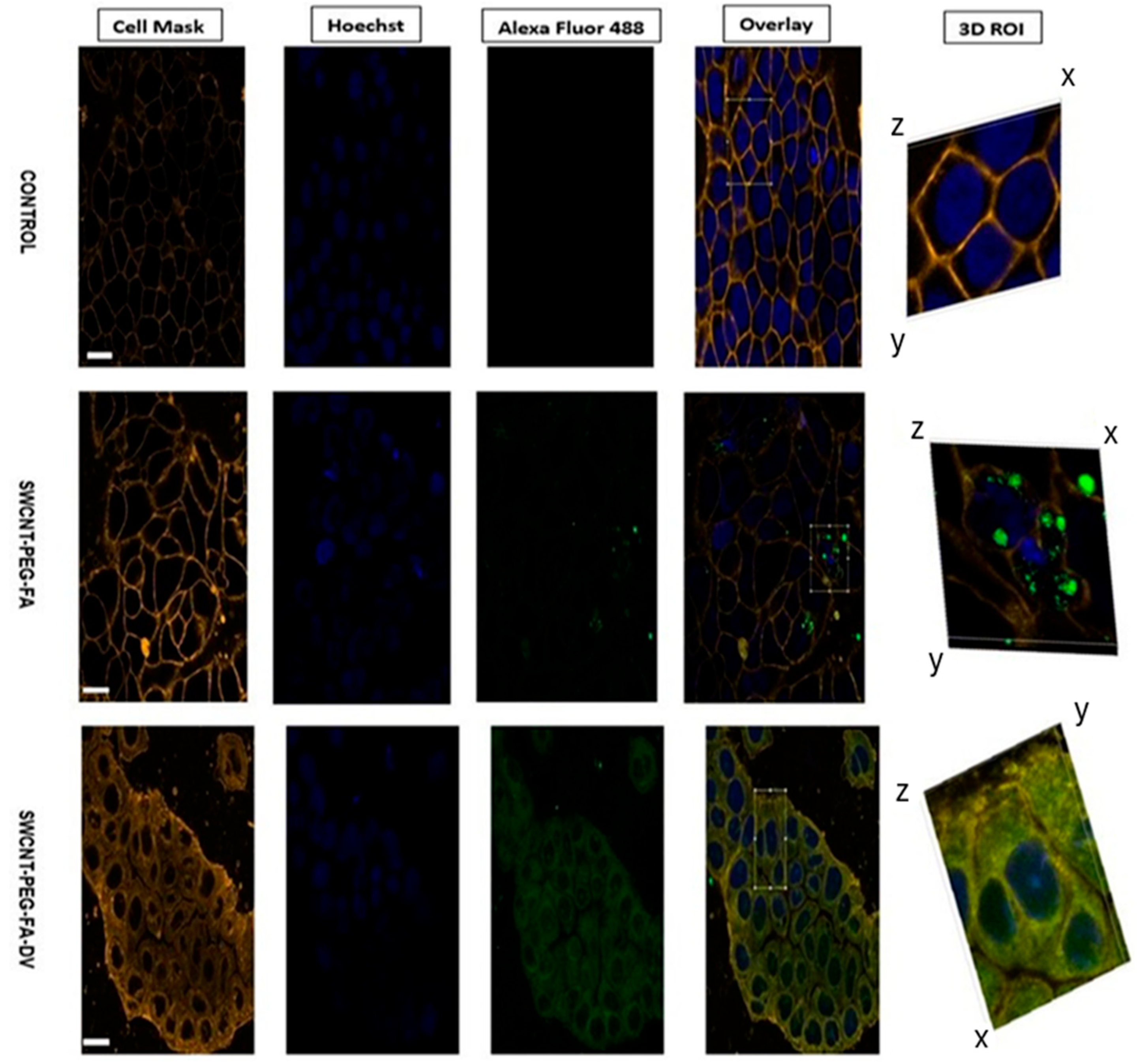
The fluorescence microscopy images displayed in Figure 3 demonstrate carbon nanotube internalization by cancer cells. Untreated cancer cells made up the control group and they showed normal distribution and morphology across all cancer cell lines. For the SWCNT-PEG-FA without the plant extract, the nanotubes entered the cell membrane and some of the nanotubes went further and encompassed the nuclear membrane. The bioconjugated nanotubes showed cellular uptake and damage to the cell membrane. In comparison with the SWCNT-PEG-FA conjugate, the cellular uptake in the bioconjugates was higher as observed by the intensity of green stains inside the cells. From the images, it can be concluded that the bioconjugates can be internalized by the cancer cells, and they can also interact around the cell membrane and deliver bioactive compounds to destroy cancer cells.

5. Patents

Patent Application No: PCT/ZA2024/050020; Title: Conjugate and composition for treating cancer.

Supplementary Materials

The following supporting information can be downloaded at: https://www.mdpi.com/article/10.3390/engproc2025109006/s1, Methodology S1: Experimental procedures for in vitro cytotoxicity studies, including details on cell lines, culture conditions, splitting and seeding, treatment protocols, WST-1 cell viability assay, and fluorescence microscopy imaging.

Author Contributions

Conceptualization, Z.T.G., W.J.P. and A.-M.E.; methodology, Z.T.G.; software, Z.T.G.; validation, Z.T.G., W.J.P. and A.-M.E.; formal analysis, Z.T.G., W.J.P. and A.-M.E.; investigation, Z.T.G.; resources, W.J.P. and A.-M.E.; data curation, Z.T.G.; writing—original draft preparation, Z.T.G.; writing—review and editing, W.J.P. and A.-M.E.; visualization, Z.T.G.; supervision, W.J.P. and A.-M.E.; project administration, W.J.P. and A.-M.E.; funding acquisition, W.J.P. and A.-M.E. All authors have read and agreed to the published version of the manuscript.

Funding

This research received no external funding.

Institutional Review Board Statement

Not applicable.

Informed Consent Statement

Not applicable.

Data Availability Statement

The data presented in this study are available on request from the corresponding author.

Conflicts of Interest

The authors declare no conflicts of interest.

References

  1. Astruc, D. Introduction to Nanomedicine. Molecules 2016, 21, 4. [Google Scholar] [CrossRef] [PubMed]
  2. Sarvarian, P.; Gholipour, E.; Asenjan, K.S.; Hojjat-Farsangi, M.; Motavalli, R.; Khiavi, F.M.; Yousefi, M. Application of emerging plant-derived nanoparticles as a novel approach for nano-drug delivery systems. Immunol. Investig. 2022, 51, 1039–1059. [Google Scholar] [CrossRef] [PubMed]
  3. Zhang, N.; Xiong, G.; Liu, Z. Toxicity of metal-based nanoparticles: Challenges in the nano era. Front. Bioeng. Biotechnol. 2022, 10, 1001572. [Google Scholar] [CrossRef] [PubMed]
  4. Zhang, W.; Zhang, Z.; Zhang, Y. The application of carbon nanotubes in target drug delivery systems for cancer therapies. Nanoscale Res. Lett. 2011, 6, 555. [Google Scholar] [CrossRef] [PubMed]
  5. Thiruvengadam, M.; Rajakumar, G.; Swetha, V.; Ansari, M.A.; Alghamdi, S.; Almehmadi, M.; Halawi, M.; Kungumadevi, L.; Raja, V.; Sarbudeen, S.S.; et al. Recent insights and multifactorial applications of carbon nanotubes. Micromachines 2021, 12, 1502. [Google Scholar] [CrossRef] [PubMed]
  6. Ahmed, I.; Javeed, A.; Ahmed, W.; Phoenix, D.A.; Elhissi, A.; Jackson, M.J. Delivery of anticancer molecules using carbon nanotubes. In Surgical Tools and Medical Devices; Ahmed, W., Jackson, M.J., Eds.; Springer International Publishing: Cham, Switzerland, 2016; pp. 563–572. ISBN 978-3-319-33489-9. [Google Scholar] [CrossRef]
  7. Hafez, D.A.; Elkhodairy, K.A.; Teleb, M.; Elzoghby, A.O. Nanomedicine-based approaches for improved delivery of phyto-therapeutics for cancer therapy. Expert Opin. Drug Deliv. 2020, 17, 279–285. [Google Scholar] [CrossRef] [PubMed]
  8. Abdifetah, O.; Na-Bangchang, K. Pharmacokinetic studies of nanoparticles as a delivery system for conventional drugs and herb-derived compounds for cancer therapy: A systematic review. Int. J. Nanomed. 2019, 14, 5659–5677. [Google Scholar] [CrossRef] [PubMed]
Figure 1. Heatmaps of (a) functionalized SWCNT at 100 μg/mL; (b) bio-conjugated SWCNT after 72 h. SWCNT—single walled carbon nanotubes; PEG—polyethylene glycol; FA—folic acid; AM—Annona muricata; DC—Dicoma capensis; DV—Dodoneae viscosa; TV—Tulbaghia violacia.
Engproc 109 00006 g001
Figure 2. Mechanisms of surface interaction between SWCNT-PEG-FA-AM-DC-DV-TV (black colour) and (a) MCF-7; (b) MDA-MB-231; (c) HT-29 cancer cell lines.
Engproc 109 00006 g002
Figure 3. Live cell imaging of carbon nanotubes stained (green), nucleus (blue), cell membrane (orange) in a HT-29 colon cancer cell line. Scale bar = 20 μm.
Engproc 109 00006 g003
Disclaimer/Publisher’s Note: The statements, opinions and data contained in all publications are solely those of the individual author(s) and contributor(s) and not of MDPI and/or the editor(s). MDPI and/or the editor(s) disclaim responsibility for any injury to people or property resulting from any ideas, methods, instructions or products referred to in the content.