Proceeding Paper

Emerging Trends in Paper-Based Electrochemical Biosensors for Healthcare Applications †

1
Department of Pharmaceutical Sciences, Dibrugarh University, Dibrugarh 786004, India
2
Department of Mechanical Engineering, Dibrugarh University, Dibrugarh 786004, India
3
Pratiksha Institute of Pharmaceutical Sciences, Panikhaiti, Guwahati 781026, India
4
School of Pharmacy, Assam Kaziranga University, Jorhat 785006, India
*
Author to whom correspondence should be addressed.
Presented at the 5th International Electronic Conference on Biosensors, 26–28 May 2025; Available online: https://sciforum.net/event/IECB2025.
Eng. Proc. 2025, 106(1), 8; https://doi.org/10.3390/engproc2025106008
Published: 11 September 2025

Abstract

Paper-based electrochemical biosensors have emerged as a revolutionary technology in healthcare diagnostics due to their affordability, portability, ease of use, and environmental sustainability. These biosensors utilize paper as the primary material, capitalizing on its unique properties such as high porosity, flexibility, and capillary action, which make it an ideal candidate for low-cost, functional, and reliable diagnostic devices. The simplicity and cost-effectiveness of paper-based biosensors make them especially suitable for point-of-care (POC) applications, particularly in resource-limited settings where traditional diagnostic tools may be inaccessible. Their lightweight nature and ease of operation allow non-specialized users to perform diagnostic tests without the need for complex laboratory equipment, making them suitable for emergency, field, and remote applications. Technological advancements in paper-based biosensors have significantly enhanced their capabilities. Integration with microfluidic systems has improved fluid handling and reagent storage, resulting in enhanced sensor performance, including greater sensitivity and specificity for target biomarkers. The use of nanomaterials in electrode fabrication, such as reduced graphene oxide and gold nanoparticles, has further elevated their sensitivity, allowing for the precise detection of low-concentration biomarkers. Moreover, the development of multiplexed sensor arrays has enabled the simultaneous detection of multiple biomarkers from a single sample, facilitating comprehensive and rapid diagnostics in clinical settings. These biosensors have found applications in diagnosing a wide range of diseases, including infectious diseases, cancer, and metabolic disorders. They are also effective in genetic analysis and metabolic monitoring, such as tracking glucose, lactate, and uric acid levels, which are crucial for managing chronic conditions like diabetes and kidney diseases. In this review, the latest advancements in paper-based electrochemical biosensors are explored, with a focus on their applications, technological innovations, challenges, and future directions.

1. Introduction

Paper-based electrochemical biosensors offer numerous advantages, such as low-cost production, ease of use, portability, and environmental sustainability, which make them particularly well-suited for point-of-care (POC) testing. The unique properties of paper, such as its high porosity, flexibility, and ability to facilitate capillary action, make it an ideal material for fabricating biosensors that are both functional and cost-effective [1,2]. As a result, these sensors have found applications in detecting a range of medical conditions, from infectious diseases to metabolic disorders.
Paper-based biosensors offer notable advantages due to their lightweight, low-cost, and biodegradable nature, making them highly suitable for use in resource-limited and rural settings [1,3]. Their simple design enables diagnostics without sophisticated equipment, allowing non-specialists to conduct tests effectively in field or emergency conditions [4,5]. Since they are primarily cellulose-based, these devices also provide an environmentally sustainable alternative to conventional diagnostic tools, reducing waste from disposables [2,6]. Leveraging properties like porosity and capillarity, paper-based sensors deliver sensitive and rapid results when combined with modern electrochemical methods [4,6,7]. Their affordability, scalability, and eco-friendliness strengthen their role in advancing accessible and sustainable healthcare technologies [6,8].
The development and optimization of paper-based electrochemical biosensors hold significant potential for transforming healthcare delivery. Their affordability and scalability make them highly adaptable for widespread use in low-resource and underserved communities, addressing long-standing disparities in diagnostic access [4,6,7]. Their portability and minimal infrastructure requirements allow for timely disease detection in emergency situations, outbreak zones, and rural healthcare settings. Moreover, their biodegradable nature aligns with global sustainability goals by reducing waste and the environmental impact of disposable healthcare devices [6,8]. By bridging the gap between advanced diagnostic capabilities and accessibility, ePADs have the potential to improve patient outcomes, enhance disease surveillance, and support public health initiatives worldwide.
This review provides a comprehensive overview of paper-based electrochemical biosensors, with a particular focus on recent advancements and persisting challenges. It reviews recent progress in paper-based electrochemical biosensors, focusing on fabrication strategies such as printing innovations, nanomaterial integration, and microfluidic incorporation to enhance performance. Applications in diagnosing HIV, tuberculosis, COVID-19, and malaria highlight improvements in sensitivity, specificity, and rapid detection. Additionally, issues of scalability, reproducibility, and environmental impact are examined, providing guidance for future research and commercialization. By addressing both opportunities and limitations, this review supports continued innovation in biosensor technology.

2. Fabrication of Paper-Based Biosensors

The performance and reliability of paper-based biosensors are highly influenced by the fabrication techniques employed to develop functional, consistent, and highly sensitive sensing platforms. Owing to their low cost, lightweight design, and compatibility with environmentally sustainable materials, these devices have gained prominence for point-of-care testing in both developed and resource-limited settings. Central to their success is the variety of fabrication strategies employed, which enable precise microfluidic control, the effective immobilization of reagents, and robust transduction mechanisms. Among these, wax printing has emerged as one of the most common approaches due to its simplicity and effectiveness. By creating hydrophobic barriers on paper substrates, wax printing enables the controlled flow of liquids along defined microchannels without the need for external pumps [9]. This process provides an inexpensive yet efficient means to produce reproducible paper-based platforms, thereby making it highly attractive for large-scale use.
Beyond wax printing, other advanced fabrication techniques, such as screen printing, inkjet printing, laser printing, and photolithography, have been increasingly adopted for developing biosensors with higher precision [9]. These methods enable the accurate deposition of biomolecules, reagents, and conductive inks onto paper substrates, allowing integration with electrochemical and optical detection mechanisms. Such approaches enhance sensitivity, enable multiplexed detection, and broaden the scope of clinical applications. In addition to these conventional strategies, more cost-effective and flexible methods have been proposed. The pen-on-paper (PoP) technique, for example, employs custom ink pens to manually draw electrodes and channels on paper, offering on-demand fabrication without sophisticated infrastructure [10]. Similarly, the pencil-drawing method, which uses graphite pencils, has been successfully utilized for producing conductive electrodes with remarkable cost efficiency. This ultra-simple approach has even demonstrated applicability in detecting pathogens such as Escherichia coli, highlighting its potential for low-cost field diagnostics [11].
Recent advances in fabrication have also introduced bottom-up printing approaches, wherein cellulose microfibers are assembled into translucent, flexible structures through wettability patterning. This method enables the fabrication of microfluidic devices with improved structural resolution and functional transparency [12]. Such innovations not only enhance device aesthetics but also improve analytical performance, particularly in optical biosensing applications. Collectively, these fabrication methods have paved the way for the widespread use of paper-based biosensors in detecting clinically relevant biomarkers such as glucose, lactate, cholesterol, infectious disease agents (HIV, influenza, and hepatitis), and cancer markers [4,13]. Their minimal sample preparation requirements and portability make them particularly valuable in resource-limited environments and during public health emergencies [14,15]. The development of microfluidic paper-based analytical devices (µPADs) has significantly expanded the functionality of these systems by integrating lab-on-a-chip capabilities. These platforms allow multistep biochemical reactions and multiplexed detection on a single sheet of paper, thereby providing a low-cost alternative to conventional diagnostic tools [2].
Despite such advancements, technical and commercial challenges persist. For instance, the large-scale commercialization of paper-based biosensors continues to be hindered by issues of performance variability, limited shelf life, and reduced sensitivity when detecting ultra-trace levels of biomarkers [7]. To overcome these challenges, recent studies have emphasized integrating paper-based platforms with digital technologies, particularly smartphones, which can enhance signal acquisition, automate interpretation, and enable cloud-based data sharing for real-time epidemiological monitoring [15]. In parallel, the incorporation of novel materials such as metal-organic frameworks (MOFs) into paper matrices has shown promise in improving sensor stability, selectivity, and signal transduction efficiency [16]. Likewise, the use of nanostructured materials, including reduced graphene oxide and gold nanoparticles, has significantly enhanced sensitivity, enabling the reliable detection of low-concentration biomarkers with high precision [17].
Another critical aspect in the development of paper-based biosensors is reproducibility, particularly in scalable manufacturing. For screen-printed devices, parameters such as humidity, temperature, squeegee firmness, mesh density, and curing conditions directly influence conductivity, resolution, and overall device performance [18,19,20,21]. For instance, adjusting squeegee angle and printing pressure has been shown to affect ink deposition and sheet resistance [22]. Advances in metallization pastes and high-resolution screens now allow conductive features as fine as 40–50 µm, making screen printing highly compatible with mass production [20]. Similarly, wax printing demands precise control of nozzle temperature, print speed, and layer thickness to achieve dimensional accuracy. Optimal conditions—such as nozzle temperatures around 64–85 °C and print speeds between 40 and 60 mm/s—have been reported to yield consistent patterns [23,24]. Moreover, experimental optimization approaches, including the Taguchi design and orthogonal testing, have been successfully applied to refine critical wax printing parameters such as nozzle diameter, shrinkage angle, and die length [25]. These improvements support scalability, particularly when using household 3D printers for the batch fabrication of µPADs [26]. Furthermore, the adoption of non-cellulosic substrates like modified polyethylene terephthalate (mPET) provides structural stability at elevated temperatures, enabling high-throughput production without compromising device integrity [27]. Optimized printing conditions—such as a 0.6 mm nozzle diameter, 80° shrinkage angle, and controlled environmental parameters—have also been shown to minimize deformation and enhance fabrication accuracy [28]. Table 1 shows that fabrication methods such as wax/screen printing and pencil drawing are low-cost and eco-friendly, while advanced methods like robotic printing, laser-induced graphene, and roll-to-roll processing provide higher performance and scalability but require complex setups.

3. Microfluidic Integration in Paper-Based Biosensors

Recent advancements in paper-based electrochemical biosensors include their integration with microfluidic devices. This enhancement allows for more sophisticated fluid handling, reagent storage, and improved sensor performance, such as increased sensitivity and selectivity for target analytes. This integration has been pivotal in making paper-based sensors more versatile for clinical diagnostics [38,39]. The integration of microfluidics into paper-based biosensors has significantly advanced the capabilities of low-cost, portable, and user-friendly diagnostic tools. Since the introduction of microfluidic paper-based analytical devices (µPADs) in 2007, these platforms have gained widespread attention for their potential in point-of-care testing (POCT) and decentralized diagnostics. Their primary advantage lies in their affordability and fabrication simplicity, as paper serves as an inexpensive, biodegradable, and readily available substrate. This enables µPADs to be manufactured without the need for complex instruments or highly specialized labor [40,41]. Moreover, their lightweight and portable nature, combined with the ability to operate with small sample volumes, make them especially suited for use in resource-limited and field settings [42,43,44]. Their design inherently facilitates capillary-driven flow, eliminating the need for external pumps and enabling fluid handling in a simple and autonomous manner. Recent advances have further enhanced the analytical performance of µPADs. Innovations in fabrication methods such as stamping, laser ablation, and chemical surface modification have improved colorimetric detection by minimizing issues such as uneven color distribution, leading to more accurate visual interpretation [45]. The integration of advanced materials like graphene, transition metal nanomaterials, and MXenes has significantly improved signal transduction and sensitivity in electrochemical µPADs, broadening their application range for detecting a variety of biological and chemical targets [43,44]. Emerging approaches now integrate deep learning algorithms to analyze time-sequence colorimetric data, with TimePAD enhancing the speed and accuracy of ELISA-style diagnostics through AI-based signal interpretation [46]. Additionally, structural innovations such as programmable binary valve matrices have enabled complex fluid manipulation and multiplexed biochemical assays on paper substrates, facilitating high-throughput testing [47]. µPADs have also shown great promise in wearable biosensing applications. Devices designed for the continuous monitoring of biomolecules, such as those for disease surveillance or athletic performance, underscore the adaptability of paper-based microfluidics for real-time, non-invasive diagnostics [48]. These applications align well with growing demand for point-of-care and home-based monitoring, particularly in regions with limited access to centralized healthcare. However, the path to commercialization remains challenging. The reproducibility of fluid flow across devices, precise flow control mechanisms, and consistent batch quality are critical aspects that must be resolved to meet commercial and regulatory standards [7,49]. Many µPADs still rely on laboratory-grade detection systems, which limit their stand-alone utility in real-world scenarios. Integrating µPADs with emerging technologies such as 3D printing, smart materials, and AI-driven detection systems represents a promising direction for the development of next-generation diagnostic tools. Tanjaya&Harito [50] emphasize that the fusion of microfluidics with biosensor platforms can lead to intelligent, autonomous devices capable of high precision and rapid decision-making, especially when coupled with mobile health (mHealth) systems. These efforts may enable µPADs to move beyond laboratory prototypes into scalable, smart diagnostic platforms capable of addressing global health challenges, environmental monitoring, and food safety with minimal infrastructure requirements.
Multiplexed detection represents a significant advancement in paper-based biosensor technology, enabling the simultaneous analysis of multiple biomarkers from a single sample. This approach provides real-time, comprehensive diagnostic information, which is particularly valuable in clinical settings requiring rapid, multi-target detection [38,51]. By capturing several analytes at once, multiplexed sensor arrays enhance sensitivity, diagnostic accuracy, and analytical efficiency, while reducing analysis time, sample volume, and operational costs [52,53]. The ability to perform internal referencing and cross-validation within the same assay improves signal-to-noise ratios and overall reliability, making these platforms especially advantageous for early-stage disease diagnostics and monitoring complex medical conditions. The incorporation of functional nanomaterials, such as zinc oxide nanowires (ZnO NWs), has been shown to enhance fluorescence signals up to fivefold when integrated into fluorogenic immunodevices, thus improving the limit of detection for biomarkers such as cardiac proteins [54]. Similarly, the use of hierarchical nanomaterials and metal-organic frameworks (MOFs) increases the active sensing surface and improves electron transfer, both of which contribute to more sensitive electrochemical readings [55]. Another innovative approach is the bipolar electrode electrochemiluminescence (BPE-ECL) platform, which enables the simultaneous detection of multiple microRNAs while effectively reducing spatial signal interference and cross-reactivity through distinct dual ECL probes [56]. This not only improves signal clarity but also enhances the system’s ability to distinguish between closely related targets.
Advancements in microfluidic design further support the sensitivity of multiplexed biosensors. Devices such as vertical-flow immunoassays with wax-patterned nitrocellulose membranes allow for precise fluid control, which reduces background noise and prevents signal dilution [57]. Additionally, optical and electrochemical methods continue to play a critical role. For example, paper-based electrochemical immunodevices that employ amplification-by-polymerization reactions can detect cancer biomarkers at extremely low concentrations, thanks to improved electron transfer kinetics and signal amplification [58]. Optical systems utilizing wavelength-dependent absorbance and transmittance can also differentiate signals across multiple channels, offering transparent, visually intuitive multiplexed assays [59].
Another key enabler of sensitivity is the integration of nanomaterials such as single-walled carbon nanotubes (SWNTs), quantum dots, and conductive polymers. These materials significantly expand the electroactive surface area and improve signal conductivity, which in turn enhances the sensitivity and selectivity of the sensor platform [60,61]. Microfluidic integration helps define clear sensing zones, reduces cross-contamination between test regions, and facilitates high-throughput parallel analysis—factors that collectively contribute to reproducible and high-sensitivity results. The convergence of these features makes multiplexed detection highly suitable for point-of-care diagnostics, particularly in infectious disease screening, cancer biomarker profiling, and cardiovascular risk assessment [62].

4. Nanomaterials and Surface Engineering

The integration of nanomaterials and surface engineering has significantly revolutionized the performance and capabilities of paper-based biosensors, particularly in healthcare, environmental monitoring, and food safety applications. These biosensors benefit from the intrinsic advantages of paper—such as its low cost, biodegradability, and capillary-driven fluidics—while nanomaterials and engineered surfaces offer enhanced sensitivity, selectivity, and functional versatility. A wide variety of nanomaterials, as shown in Figure 1, have been employed to improve biosensor performance. Notably, gold nanoparticles, carbon nanotubes (CNTs), magnetic nanoparticles, and quantum dots are frequently incorporated due to their exceptional optical, electrical, and magnetic properties [62,63,64]. These nanostructures not only increase the surface area available for immobilizing biorecognition elements but also facilitate efficient signal transduction and amplification. Further enhancement comes from the use of nanowires and nanorods, such as zinc oxide and polymer-based nanowires, which offer superior electron transport capabilities and biocompatibility [63,65]. Electrospun nanofibers are increasingly utilized to create porous, high-surface-area substrates that significantly improve signal-to-noise ratios in biosensing systems. These nanomaterials are commonly employed in both electrochemical and optical biosensors. In electrochemical applications, nanomaterials enhance conductivity and provide efficient anchoring sites for enzymes or antibodies, leading to higher sensitivity and lower detection limits [62,66]. In optical platforms, materials like quantum dots and metallic nanoparticles enable enhanced fluorescence or surface-enhanced Raman scattering (SERS), improving visual detection in low-concentration scenarios [67].
Complementing the role of nanomaterials, surface engineering techniques play a crucial role in optimizing the biosensor interface for biomolecular recognition. Various methods such as physical adsorption, covalent bonding, and electrochemical polymerization are employed to immobilize biorecognition elements like antibodies, enzymes, or nucleic acids onto the paper substrate [64,67,68]. These immobilization strategies enhance specificity by promoting strong and stable interactions between the sensor and the target analyte. Moreover, chemical surface modifications using polymers, silanes, or nanostructured coatings have been shown to fine-tune properties such as hydrophilicity, porosity, and electroactive surface area, all of which are critical for signal enhancement [44,68]. Advanced nanostructuring techniques—including ultrasonic cavitation and graphene transfer to microstructured surfaces—have also been utilized to produce tailored sensor topographies that enhance mass transfer and detection efficiency [68].
The integration of nanomaterials and surface engineering yields several key advantages. First, it enables high sensitivity and selectivity, allowing the detection of trace-level analytes relevant in clinical diagnostics, such as cancer markers, viruses, or glucose in saliva and blood [69]. Second, the combination of nanostructures with low-cost paper substrates results in affordable and scalable biosensors, which are particularly suited for resource-limited and point-of-care settings [7]. Third, the portability and ease of use of these devices make them attractive for real-time, on-site testing, bypassing the need for centralized laboratories or trained personnel [70]. However, the integration of nanomaterials is not without challenges. Issues such as batch-to-batch reproducibility, material stability, and potential cytotoxicity and environmental hazards remain key concerns [60]. The synthesis and disposal of engineered nanomaterials must therefore be approached cautiously to ensure biosafety and ecological sustainability. The addition of graphene, carbon nanotubes, and gold nanoparticles improves the electrical conductivity, surface area, and signal amplification capabilities of the sensors. These modifications contribute to lower detection limits and increased sensor durability under varied conditions [71,72,73].
Environmental disposal strategies of nanomaterial-loaded paper-based biosensors: The environmental disposal of nanomaterial-loaded paper-based biosensors is an emerging area of concern, as the integration of nanostructured materials into biodegradable paper substrates introduces unique sustainability and safety challenges. While paper is generally biodegradable and recyclable, the presence of engineered nanomaterials (ENMs) such as metal nanoparticles, carbon nanostructures, or metal oxides makes it complicated due to their persistence, potential, and toxicity [74]. Consequently, the development of environmentally responsible disposal strategies requires a combination of recycling, safe destruction, regulatory compliance, and green remediation approaches.
Incineration and thermal treatment: Thermal disposal via controlled incineration remains a viable option for nanomaterial-loaded paper-based biosensors, as high-temperature combustion effectively destroys organic binders and paper fibers while concentrating nanomaterials in bottom ash [75]. Most studies indicate that nanomaterials are not fully vaporized during incineration but are instead immobilized in ash fractions, which can then be handled as hazardous waste [75]. To mitigate airborne nanoparticle emissions, incinerators equipped with electrostatic precipitators or high-efficiency particulate air (HEPA) filtration are recommended. However, post-incineration ash management protocols must be developed to prevent nanomaterial leaching into soil or water systems.
Biodegradation and green remediation technologies: Innovative research is exploring the biodegradation of nanomaterials or their transformation into less harmful forms through microbial action, enzymatic processes, or photodegradation [76]. Similarly, sustainable nanotechnology-based remediation strategies—such as biosorption or green-synthesized nanocatalysts—are being developed to mitigate pollutants from pulp and paper industry effluents, potentially extending to nanomaterial-containing sensor waste streams [77]. Biorefinery approaches could also convert wastepaper from biosensors into biofuels or high-value biomaterials, provided the nanomaterials are safely removed or stabilized beforehand [77].
Cytotoxicity of nanomaterial-loaded paper-based biosensors: The cytotoxicity of nanomaterial-loaded paper-based biosensors is an important consideration in their design, application, and environmental management. These devices, which integrate engineered nanomaterials (ENMs) such as metal nanoparticles, carbon nanostructures, and metal oxides into paper-based platforms, offer enhanced analytical performance but also introduce potential risks to human health and the environment. Understanding the biological interactions and toxicological profiles of the nanomaterials used is essential for safe and responsible deployment [61]. Nanomaterials in biosensors can interact with cells through various mechanisms, resulting in oxidative stress, genotoxicity, protein denaturation, and the disruption of membrane integrity [78]. For example, nano-copper has been shown to generate hydrogen peroxide as a primary source of toxicity, which triggers oxidative damage to cellular components [78]. Some nanomaterials are inherently biocompatible, the addition of cationic functional groups can render them cytotoxic by promoting membrane disruption and intracellular stress responses [79]. Paper-based biosensors incorporating nanomaterials benefit from low cost, ease of fabrication, and the capability for multi-step or multiplexed assays [77]. However, the release of nanomaterials during use or disposal could pose health risks if not properly contained. Biocompatible materials such as nanocellulose offer a safer alternative, as they exhibit low cytotoxicity and high cytocompatibility, making them attractive substrates for sensor fabrication [80].
Lifecycle assessment of nanomaterial-loaded paper-based biosensors: The lifecycle assessment (LCA) of nanomaterial-loaded paper-based biosensors is a critical process for evaluating the environmental, health, and sustainability implications of these emerging diagnostic technologies. Given the growing use of engineered nanomaterials (ENMs) in point-of-care devices, LCAs serve as decision-support tools that examine environmental burdens across the entire product lifecycle—from raw material extraction to end-of-life management [81]. LCA provides a structured framework to quantify environmental and human health impacts associated with nanomaterial-enabled products. This is particularly relevant for nanomaterials, which possess unique size-, shape-, and surface-dependent properties that can alter toxicity, persistence, and environmental mobility compared to bulk materials [81]. By evaluating every stage of the product lifecycle—including synthesis, integration into paper-based substrates, usage, and disposal—LCA helps identify potential “hotspots” where environmental and safety risks are most significant [82]. Standard LCA methodologies were developed for bulk materials and often lack the nano-specific inventory data needed to model ENM behavior accurately. Key challenges include limited ecotoxicity data, insufficient exposure metrics for occupational and consumer use, and uncertainties in environmental fate models [81]. These limitations necessitate the integration of emerging nano-specific impact assessment frameworks that incorporate particle size distribution, surface reactivity, and dissolution rates into environmental modeling [82].
Performance of paper-based electrochemical biosensors across different environmental conditions: Paper-based electrochemical biosensors are increasingly employed in point-of-care diagnostics and environmental monitoring due to their low cost, portability, and ease of use. However, their performance can be significantly influenced by environmental variables such as temperature, humidity, and combined field conditions. Understanding these effects is critical for ensuring reliable sensor performance in real-world applications. Temperature variations can directly affect the electrochemical processes within biosensors. For example, increasing the temperature from 5 °C to 45 °C accelerates redox reaction rates, resulting in faster activation and higher output performance in paper-based microfluidic fuel cells (PMFCs). Beyond 45 °C, performance plateaus, and at 65 °C no further gains were observed, indicating that excessive heat may not improve functionality and could risk thermal degradation [83]. The thermal stability of biosensors is also a critical consideration for storage and transport. Humidity is another key environmental factor influencing biosensor reliability [84]. Sensors using paper and potato starch substrates exhibit high humidity sensitivity but suffer from longer response and recovery times, as well as pronounced hysteresis near 100% relative humidity (RH). In contrast, sensors fabricated on PET and PLA substrates showed faster responses and reduced hysteresis, offering better stability under fluctuating humidity levels [85]. Temperature sensors had the lowest average hysteresis at around 50% RH, suggesting that maintaining moderate humidity can enhance signal stability and reduce drift. Beyond isolated effects, combined environmental factors—temperature, humidity, oxygen exposure, and light—can induce chemical and physical transformations in sensor materials [86]. Material selection and sensor design play pivotal roles in environmental robustness. For instance, the incorporation of Prussian Blue-mediated electrodes has been shown to improve selectivity and minimize interference under varying field conditions [87].

5. Signal Acquisition, Noise Reduction, and Amplification in Paper-Based Electrochemical Biosensors

5.1. Signal Acquisition

Signal acquisition in ePADs involves the detection, conversion, and processing of biochemical interactions into quantifiable electrochemical signals, typically in the form of current, potential, or impedance changes. A key component of the signal acquisition process is the electrochemical reader or potentiostat, which serves as the interface between the sensing electrodes and the data processing system. Portable, custom-designed potentiostats are widely used for ePADs, enabling multi-analyte detection and high-throughput analysis [38,51]. These devices often incorporate multichannel amperometric boards with high-accuracy analog-to-digital converters (ADCs) capable of digitizing low-current signals and transmitting them to user interfaces via USB connections or integrated touchscreens [88]. Signal amplification strategies are critical for extending the detection limits of ePADs, particularly in trace-level analyte detection. Nanomaterial integration plays a pivotal role in this domain. For example, graphene-modified immunodevice surfaces accelerate electron transfer kinetics, while silica nanoparticles can function as tracing tags for labeling antibodies, amplifying the generated electrochemical signal [58,89]. Similarly, gold nanorods and spherical cerium dioxide nanoparticles have been employed to enhance conductivity, facilitate catalytic reactions, and improve electron transfer efficiency [90,91,92]. Such modifications not only improve signal intensity but also enhance the stability and reproducibility of the sensor response.
Electrode design and surface modification significantly influence signal acquisition quality in paper-based platforms. Innovations include the use of screen-printed graphene electrodes (SPGNE) and hybrid materials such as PEDOT:PSS/reduced graphene oxide composites to improve both sensitivity and selectivity in analyte detection [93,94]. Other fabrication methods, such as pencil-drawn carbon transfer, offer low-cost yet effective approaches for creating conductive pathways [95]. Carbon black/Prussian Blue nanocomposites have also been applied to tailor the electrocatalytic properties of electrodes for specific analytes [96]. Such modifications not only increase the electroactive surface area but also reduce the charge transfer resistance, resulting in improved signal-to-noise ratios during acquisition. In multiplexed detection, ePADs have been designed to simultaneously measure multiple biomarkers from a single sample, which is invaluable for complex disease diagnostics and metabolic profiling [38,51]. In POC diagnostics, the portability and cost-efficiency of ePADs make them indispensable in low-resource settings, offering rapid and accurate diagnostics without extensive laboratory infrastructure [1,4,97]. Furthermore, in environmental and food safety monitoring, paper-based electrochemical biosensors have been used to detect contaminants, pathogens, and toxins, expanding their utility beyond medical diagnostics [98,99].

5.2. Noise Reduction

Noise reduction is a critical factor in the performance optimization of paper-based electrochemical biosensors (ePADs), as unwanted electrical, chemical, and environmental noise can mask weak electrochemical signals, reduce measurement accuracy, and increase detection limits. In the context of ePADs, noise can originate from instrumentation, electrode materials, fluidic dynamics within the paper substrate, and environmental interference such as temperature fluctuation or electromagnetic coupling. Effective noise suppression involves a combination of hardware-based improvements, signal-processing techniques, and innovative device fabrication strategies, each of which plays a role in improving the signal-to-noise ratio (SNR) and ensuring robust analytical performance. From a hardware perspective, modifying the sensor interface circuitry is one of the most effective strategies. The use of modified transimpedance amplifiers with current compensation has been shown to reduce current noise at the converter input, yielding a higher SNR and significantly lowering measurement error, particularly in amperometric measurements for analytes such as dissolved oxygen [100]. Another effective approach involves the use of noise-canceling architectures combined with chopper stabilization in the front-end circuit, which suppresses low-frequency 1/f noise while enhancing linearity and dynamic range—demonstrated to improve dynamic range by over 10 dB [101]. In bio-signal sensing contexts, capacitive grounding electrodes and driven-right-leg (DRL) electrodes have been integrated to reject common-mode interference from power lines, with DRL configurations offering superior performance in attenuating ambient electrical noise [102]. Additionally, electrode material choice and geometry influence noise characteristics; thin-film electrodes of gold or conductive polymers have been reported to effectively balance amplifier noise against resistance noise, leading to more stable signal acquisition [103]. Signal-processing techniques further contribute to noise suppression after signal acquisition. Bandwidth optimization through low-pass and high-pass filtering in transimpedance circuits has been shown to reduce broadband noise by up to 14.3% compared to standard systems [100]. Advanced adaptive filtering, such as the use of Wiener filters with noise-replica generation from unconventional electric field sensors, can attenuate noise by as much as 40.8 dB in bio-signal recordings, enabling the detection of low-amplitude electrochemical events [104]. More sophisticated digital noise reduction algorithms, such as Haar wavelet decomposition, have also been successfully applied to analytical data (e.g., in process gas chromatography), enhancing detection repeatability and lowering the minimum detectable levels [39].

5.3. Signal Amplification

Signal amplification plays a pivotal role in enhancing the sensitivity, selectivity, and overall analytical performance of paper-based electrochemical biosensors (ePADs), enabling the detection of ultra-low analyte concentrations in clinical, environmental, and point-of-care (POC) settings. A wide range of amplification strategies have been developed to strengthen the electrochemical signal by increasing electron transfer efficiency, enhancing catalytic activity, or introducing multiple signal-generating events per target molecule. One prominent approach involves graphene surface modification, where graphene’s exceptional conductivity and large surface area accelerate electron transfer and improve electrode kinetics, thereby boosting the electrochemical output. This has been effectively demonstrated in the ultrasensitive detection of cancer biomarkers using paper-based microfluidic immunodevices [91]. Complementary to graphene modification, nanoparticle tagging—particularly with silica nanoparticles—has been used to label signal antibodies, dramatically increasing the number of detectable electrochemical events per analyte binding event, which is especially advantageous in multiplexed assays for cancer diagnostics [58,89].
Beyond surface modification, controlled radical polymerization, such as activators generated by electron transfer atom transfer radical polymerization (AGET ATRP), has been applied to grow long-chain polymers after target capture, introducing multiple binding sites for enzyme coupling and enabling a significant amplification of the final electrochemical signal [59]. Enzymatic recycling offers another powerful biochemical amplification pathway, wherein enzymes like diaphorase and NADH oxidase regenerate cofactors in situ, allowing repeated catalytic turnover of redox-active species and thus extending the linear range and lowering detection limits, as shown in microcystin detection for water quality monitoring [90]. Nanomaterial-based probes, including gold nanorods and composite catalysts, such as cerium dioxide-gold conjugated with glucose oxidase (CeO2-Au@GOx), further enhance conductivity and promote catalytic activity, enabling highly sensitive detection of microRNA targets [76]. Similarly, electrocatalytic nanostructures like Pd@hollow Zn/Co core–shell ZIF67/ZIF8 nanoparticles have been used to improve redox catalysis and signal output, supporting applications such as the detection of prostate-specific antigen [91]. The benefits of these amplification strategies are most evident in high-sensitivity diagnostic applications, where they allow the quantification of analytes at femtomolar or even attomolar concentrations, thereby expanding the clinical utility of ePADs for early disease detection [58,90,91]. Their integration into low-cost, portable, and disposable paper-based platforms makes them ideal for POC testing, particularly in resource-limited settings [92]. Nevertheless, challenges remain, particularly in mitigating background noise and cross-reactivity, which can reduce signal specificity, and in ensuring seamless integration with sample handling modules for real-world deployment [93]. Addressing these limitations through improved surface chemistry, microfluidic design, and hybrid amplification methods is likely to further expand the applicability of ePADs in healthcare, environmental monitoring, and food safety testing.

6. Applications in Medical Diagnostics

Paper-based electrochemical biosensors have been successfully used for the detection of a wide array of disease biomarkers, including those related to cancer, infectious diseases (such as COVID-19, dengue, and Zika), and metabolic disorders. These biosensors offer rapid, reliable, and cost-effective methods for diagnosing conditions in diverse environments. In addition to disease biomarkers, these biosensors have been employed in the detection of nucleic acids and genetic mutations, facilitating the diagnosis of genetic disorders and the monitoring of genetic interactions [105]. This has profound implications for personalized medicine and the early detection of genetic predispositions. Paper-based biosensors have demonstrated rapid, accurate detection for pathogens responsible for diseases such as COVID-19, tuberculosis (TB), and malaria. For TB, gold nanoparticle-decorated graphene-based paper biosensors have enabled sensitive detection of the Hsp16.3 antigen [106]. In COVID-19 detection, PBBs have been optimized for viral RNA and protein targets, enabling on-site testing [107]. Similarly, MoS2 nanosheet-mediated fluorescence resonance energy transfer (FRET) aptasensors have been developed for rapid malaria diagnostics [108]. PBBs also facilitate the monitoring of metabolic biomarkers such as glucose, lactate, and uric acid—critical for diagnosing diabetes, cardiovascular diseases, and kidney disorders [41,42]. Multiplexed detection platforms allow the simultaneous analysis of several metabolites, improving diagnostic precision and enabling comprehensive health screening [109]. Advances in PBB technology have improved the sensitivity and specificity of cancer biomarker detection, allowing identification at very low analyte concentrations. Devices for detecting cytokines and tumor-associated antigens are enabling earlier diagnosis and better patient prognosis [3,110].

Paper-Based Biosensors for the Diagnosis of HIV, Tuberculosis (TB), COVID-19, and Malaria

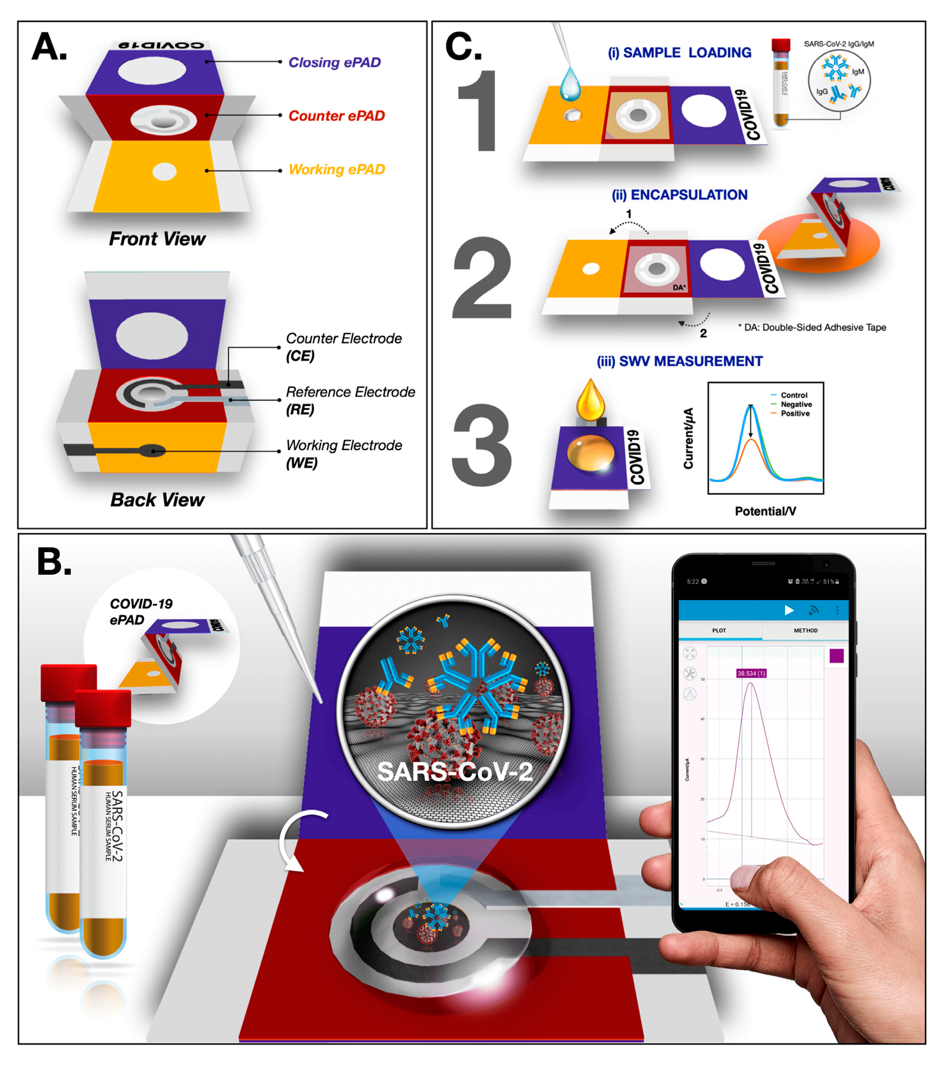
Paper-based biosensors (PBBs) have revolutionized diagnostic capabilities for infectious diseases such as HIV, tuberculosis (TB), COVID-19, and malaria, offering cost-effective, rapid, and portable alternatives to conventional laboratory diagnostics. Their ease of use and ability to operate without complex instrumentation make them especially valuable for point-of-care (POC) applications in low-resource and remote settings. The COVID-19 pandemic accelerated the development of PBBs for rapid SARS-CoV-2 detection. Figure 2 illustrates the operation of ePADs in COVID-19 diagnostics, depicting (A) device components, (B) detection principle, and (C) detection method.
HIV diagnosis: For HIV, early and accurate detection is critical to controlling viral transmission and initiating timely treatment. Paper-based biosensors have been developed to target multiple diagnostic biomarkers, with notable advancements in sensitivity and detection speed. Lateral flow immunoassays (LFIAs) utilizing nanoparticle catalyst labels have achieved ultrabroad dynamic ranges in detecting p24, a key early HIV biomarker, using porous platinum core–shell nanocatalysts. This design enables low femtomolar detection, significantly reducing the acute-phase diagnostic window [112]. Electrochemical impedance spectroscopy (EIS) biosensors enhanced with zinc oxide nanowires (ZnO NWs) have demonstrated a detection limit of 0.4 pg/mL for p24 antigen, allowing for rapid and ultrasensitive detection. Nucleic acid-based platforms integrating paper-based extraction and isothermal amplification techniques have shown high specificity for HIV RNA detection in clinical samples, providing a viable solution for decentralized testing [113].
Tuberculosis (TB) diagnosis: The slow turnaround of traditional TB culture tests has driven the need for PBB-based rapid diagnostics. Interdigitated electrode (IDE) biosensors functionalized with green-synthesized silver nanoparticles (AgNPs) and multi-walled carbon nanotube–zinc oxide (MWCNT-ZnO) composite nanofibers have achieved highly sensitive detection of TB lipoarabinomannan (LAM) antigen, with a limit of detection (LOD) as low as 25.79 fg/mL [114]. Electrochemical biosensors, including amperometric and impedimetric types, have been designed to detect key antigens such as CFP-10 and ESAT-6, offering high accuracy and reproducibility in POC applications [115]. Graphene-based field-effect transistor (FET) biosensors targeting MPT64 protein have demonstrated ultra-low detection capabilities down to 1 fg/mL, highlighting their potential for early-stage TB diagnostics [116].
COVID-19 diagnosis: The COVID-19 pandemic accelerated the innovation of PBBs for the rapid detection of SARS-CoV-2. Figure 2 shows the rapid function of ePADs in COVID-19 diagnostics. Lateral flow immunoassays have been optimized to detect viral antigens and antibodies with high specificity, enabling deployment in mass screening scenarios [107,117]. Surface-enhanced Raman scattering (SERS) biosensors have demonstrated the detection of SARS-CoV-2 spike protein in untreated saliva at concentrations as low as 6.07 fg/mL, allowing non-invasive and ultra-sensitive screening [117]. CRISPR-Cas12a-integrated electrochemical platforms combined with a toehold-mediated strand displacement reaction (TSDR) have reached a detection limit of 40 aM for SARS-CoV-2 RNA, offering high specificity and rapid turnaround [62].
Malaria diagnosis: Rapid malaria diagnosis remains essential in endemic regions to prevent severe disease progression. Aptamer-based biosensors for Plasmodium lactate dehydrogenase (pLDH) have achieved a detection limit of 17 nM, providing a robust platform for specific malaria detection even in mixed infections [118]. Microfluidic paper-based analytical devices (μPADs) combined with recombinase polymerase amplification (RPA) enable the detection of Plasmodium species at concentrations as low as 28 parasites/mL, with a total assay time of approximately 35 min, making them highly suitable for rapid field diagnostics [119].
Alzheimer’s disease diagnosis: The early and accurate diagnosis of Alzheimer’s disease (AD) remains a significant challenge, as current clinical methods often depend on costly imaging technologies or invasive cerebrospinal fluid analysis. Paper-based biosensors (PBBs) provide a low-cost, accessible alternative for detecting established and emerging AD biomarkers. A key focus has been the detection of amyloid beta (Aβ) and tau proteins, which are hallmark indicators of AD pathology. One study demonstrated a multi-chamber paper-based platform using wax printing and copper-enhanced gold immunoblotting to detect amyloid beta 42 oligomers (AβO42) at concentrations as low as 23.7 pg/mL, with results processed via smartphone imaging [120]. Another investigation developed a paper-based electrochemical sensor incorporating an in situ molecularly imprinted polymer for β-amyloid peptide (Aβ-42) detection, achieving a linear response down to 0.1 ng/mL [121]. Beyond Aβ and tau, there is growing interest in non-Aβ-tau biomarkers to enable broader and potentially earlier diagnostic coverage [120,122]. The integration of nanomaterials has further enhanced detection sensitivity and facilitated multiplexed biomarker analysis, offering significant potential for improved prognosis prediction and therapeutic monitoring [123].
Sexually transmitted infections diagnosis: Paper-based biosensors (PBBs) have shown strong potential for the rapid diagnosis of sexually transmitted infections (STIs), where timely detection is critical to reducing transmission and enabling early treatment. Chlamydia trachomatis, one of the most common bacterial STIs, has been a major target for PBB development. A minimally instrumented paper-based platform integrating cell lysis, isothermal nucleic acid amplification, and lateral flow visual detection achieved a detection limit over 100 times more sensitive than current rapid immunoassays [124]. Another approach demonstrated a paper-based molecular diagnostic capable of detecting C. trachomatis with high sensitivity in under an hour, making it suitable for point-of-care applications [124]. Beyond single-pathogen testing, multiplexed detection capabilities are emerging. An impedimetric immunochip capable of simultaneously detecting C. albicans, S. agalactiae, and C. trachomatis has been developed, enabling comprehensive genital tract infection screening from a single sample [125]. Such multiplexing is particularly valuable in reproductive health, where co-infections are common and accurate pathogen differentiation is essential for effective treatment. The appeal of PBBs in STI diagnostics lies in their rapid turnaround—often under an hour—combined with low manufacturing costs and portability [126]. Table 2 presents a comparison of recent key research studies on paper-based biosensors, highlighting multiple aspects of their development and performance. For each study, the table outlines the fabrication techniques employed, the specific electrode modification strategies implemented, and the resulting biosensor performance metrics. It also summarizes the unique advantages of each approach, such as enhanced sensitivity, portability, or cost-effectiveness, alongside any identified limitations that may affect practical application.

7. Challenges and Future Directions

Despite the rapid evolution and potential of paper-based biosensors, several critical challenges must be addressed to ensure their full clinical and commercial realization. One of the foremost technical limitations lies in their sensitivity and specificity. While advances in material science and signal amplification have led to improved detection capabilities, these devices still fall short when compared to traditional laboratory-based assays in terms of accuracy and reliability. This gap is especially pronounced when detecting trace-level biomarkers in complex biological fluids. Enhancing the selectivity of biorecognition elements and minimizing false positives remains a significant area for future research [1,3,52,137]. In addition to analytical performance, commercialization presents another formidable hurdle. Scaling up the production of paper-based biosensors while maintaining consistent quality, low cost, and device performance requires robust and reproducible fabrication protocols. Issues such as batch-to-batch variability, a lack of standardized testing methods, and regulatory constraints pose major barriers to mass production and market adoption [7].
The development of wearable paper-based biosensors represents an emerging frontier in point-of-care (POC) diagnostics. These flexible, skin-conformable devices have the potential to enable real-time, continuous health monitoring for chronic disease management, physical performance tracking, and preventive healthcare applications [138]. However, the practical realization of such systems demands innovation in sensor integration, data transmission, and power management, all while preserving the disposability and affordability that paper-based platforms are known for. Furthermore, future perspectives highlight the integration of paper-based electrochemical analytical devices (ePADs) with advanced digital technologies such as machine learning, artificial intelligence, and 3D printing. These combinations could transform ePADs into intelligent, autonomous biosensing systems capable of self-calibration, pattern recognition, and remote data interpretation, thus greatly enhancing their clinical and operational relevance [100]. To fully harness these opportunities, interdisciplinary collaboration and innovation in biosensor design, signal processing, and regulatory compliance will be essential in shaping the next generation of sustainable, scalable, and high-performance paper-based biosensors.

8. Conclusions

Paper-based electrochemical biosensors (ePADs) have emerged as a transformative diagnostic platform, offering an exceptional combination of affordability, portability, environmental sustainability, and ease of use. Their inherent properties—such as capillarity, flexibility, and biodegradability—make paper an ideal substrate for point-of-care applications, particularly in resource-limited settings. By integrating microfluidics, nanomaterials, and surface engineering, ePADs have evolved into highly sensitive and selective tools capable of detecting a broad range of biomarkers, including those for infectious diseases, cancer, metabolic disorders, and genetic conditions. Advancements in fabrication techniques, including wax printing, screen printing, and pencil-drawing methods, have enabled the creation of highly functional biosensors at low cost and with scalable reproducibility. Microfluidic integration has enhanced sample handling and reduced reagent volumes, while innovations in multiplexed detection have allowed for the simultaneous analysis of multiple biomarkers, significantly improving diagnostic throughput and efficiency. Moreover, the incorporation of nanomaterials like gold nanoparticles, carbon nanotubes, and graphene has revolutionized signal amplification, leading to lower detection limits and greater sensor stability. Despite these advancements, challenges remain. Sensitivity, specificity, and reproducibility under real-world conditions continue to hinder widespread clinical adoption. Additionally, issues related to large-scale manufacturing, standardization, and regulatory approval must be addressed for commercial viability. Nevertheless, future directions are promising. The integration of artificial intelligence, smartphone interfaces, and wearable formats holds the potential to revolutionize healthcare diagnostics, enabling real-time monitoring and data-driven decision-making.

Author Contributions

Conceptualization, A.D. and P.P.B.; methodology, D.D.; software, D.D.; validation, D.D., A.D. and P.P.B.; formal analysis, D.D.; investigation, D.D.; resources, P.K.; data curation, D.D.; writing—original draft preparation, D.D.; writing—review and editing, P.P.B., J.J.S. and K.P.; visualization, D.D.; supervision, P.P.B.; project administration, P.P.B.; funding acquisition, K.P. All authors have read and agreed to the published version of the manuscript.

Funding

This research received no external funding.

Institutional Review Board Statement

Not applicable.

Informed Consent Statement

Not applicable.

Data Availability Statement

No new data were created or analyzed in this study. Data sharing is not applicable to this article.

Conflicts of Interest

The authors declare no conflicts of interest.

References

  1. Karuppannan, P.G.; Sudha, D.; Banupriya, K.; Karthik, K.; Arumugam, R. Emerging diagnostic methods using paper-based electrochemical biosensors. J. Environ. Nanotechnol. 2024, 13, 418–423. [Google Scholar] [CrossRef]
  2. Shrikrishna, N.S.; Sharma, R.; Gandhi, S. Paper-based biosensors: Overview from past to future. In Paper-Based Diagnostic Devices for Infectious Diseases; IOP Publishing Ltd.: Bristol, UK, 2023; pp. 1-1–1-18. [Google Scholar] [CrossRef]
  3. Loo, S.W.; Pui, T.-S. Cytokine and cancer biomarkers detection: The dawn of electrochemical paper-based biosensor. Sensors 2020, 20, 1854. [Google Scholar] [CrossRef] [PubMed]
  4. Ataide, V.N.; Pradela-Filho, L.A.; Ameku, W.A.; Negahdary, M.; Oliveira, T.G.; Santos, B.G.; Paixão, T.R.L.C.; Angnes, L. Paper-based electrochemical biosensors for the diagnosis of viral diseases. Microchim. Acta 2023, 190, 276. [Google Scholar] [CrossRef]
  5. Liu, B.; Du, D.; Hua, X.; Yu, X.-Y.; Lin, Y. Paper-based electrochemical biosensors: From test strips to paper-based microfluidics. Electroanalysis 2014, 26, 1214–1223. [Google Scholar] [CrossRef]
  6. Puiu, M.; Mirceski, V.; Bala, C. Paper-based diagnostic platforms and devices. Curr. Opin. Electrochem. 2021, 27, 100726. [Google Scholar] [CrossRef]
  7. Kuswandi, B.; Ensafi, A.A. Perspective—Paper-based biosensors: Trending topic in clinical diagnostics developments and commercialization. J. Electrochem. Soc. 2020, 167, 037509. [Google Scholar] [CrossRef]
  8. Zhang, Z.; Du, M.; Cheng, X.; Dou, X.; Zhou, J.; Wu, J.; Xie, X.; Zhu, M. A disposable paper-based electrochemical biosensor decorated by electrospun cellulose acetate nanofibers for highly sensitive bio-detection. Analyst 2024, 149, 2436–2444. [Google Scholar] [CrossRef]
  9. Liu, Y.; Lu, S.; Zhang, Z.; Yang, Z.; Cui, X.; Liu, G. Printable biosensors towards next-generation point-of-care testing: Paper substrate as an example. Lab A Chip 2023, 23, 3328–3352. [Google Scholar] [CrossRef]
  10. Li, Z.; Li, F.; Xing, Y.; Liu, Z.; You, M.; Li, Y.; Wen, T.; Qu, Z.; Ling Li, X.; Xu, F. Pen-on-paper strategy for point-of-care testing: Rapid prototyping of fully written microfluidic biosensor. Biosens. Bioelectron. 2017, 98, 478–485. [Google Scholar] [CrossRef]
  11. Ozoglu, O.; Korukluoglu, M.; Uzunoglu, A. Electrochemical detection of foodborne Escherichia coli using carbon nanotube-incorporated pencil-drawn paper-based disposable biosensors. Anal. Chim. Acta 2025, 1369, 344344. [Google Scholar] [CrossRef]
  12. Ma, P.; Wang, S.; Wang, J.; Wang, Y.; Dong, Y.; Li, S.; Su, H.; Chen, P.; Feng, X.; Li, Y.; et al. Rapid assembly of cellulose microfibers into translucent and flexible microfluidic paper-based analytical devices via wettability patterning. Anal. Chem. 2022, 94, 13332–13341. [Google Scholar] [CrossRef]
  13. Aydın, E.B.; Aydın, M.; Sezgintürk, M.K. Paper-based devices. In Commercial Biosensors and Their Applications: Clinical, Food, and Beyond; Elsevier: Amsterdam, The Netherlands, 2020; pp. 107–166. [Google Scholar] [CrossRef]
  14. Bilgen, E.; Suvacı, Z.; Persil Çetinkol, Ö.; Forough, M. Disposable paper-based sensors. In Fundamentals of Sensor Technology: Principles and Novel Designs; Woodhead Publishing: London, UK, 2023; pp. 803–860. [Google Scholar] [CrossRef]
  15. Dillon, M.J.; Campbell, K. Hyphenating paper-based biosensors with smartphones. In Comprehensive Analytical Chemistry; Elsevier: Amsterdam, The Netherlands, 2023; Volume 101, pp. 109–141. [Google Scholar] [CrossRef]
  16. Huang, J.; Pan, J.; Song, Y.; Lin, Q.; Xu, Y.; Dai, Z.; Liu, S.-Y. MOF-functionalized paper-based biosensors: Fabrications, mechanisms and applications. TrAC—Trends Anal. Chem. 2024, 173, 117619. [Google Scholar] [CrossRef]
  17. Torul, H.; Yarali, E.; Eksin, E.; Ganguly, A.; Benson, J.; Tamer, U.; Papakonstantinou, P.; Erdem, A. Paper-based electrochemical biosensors for voltammetric detection of miRNA biomarkers using reduced graphene oxide or MoS2 nanosheets decorated with gold nanoparticle electrodes. Biosensors 2021, 11, 236. [Google Scholar] [CrossRef]
  18. Somarathna, K.U.S.; Garakani, B.; Weerawarne, D.L.; Khinda, G.S.; Burns, A.; Alizadeh, A.; Poliks, M.D. Screen-printed water-soluble resistors for wearable electronics: An analysis of the fabrication process. In Proceedings of the Electronic Components and Technology Conference, San Diego, CA, USA, 1 June–4 July 2021; pp. 2285–2292. [Google Scholar] [CrossRef]
  19. Kołodziej, L.; Ostrowski, S.; Maciejewski, A.; Jakubowska, M.; Wróblewski, G. The influence of screen-printing parameters on properties of conductive layers for application in biomedical electrodes. In Advances in Intelligent Systems and Computing; Springer: Berlin/Heidelberg, Germany, 2020; Volume 1044, pp. 406–413. [Google Scholar] [CrossRef]
  20. Campos-Arias, L.; Peřinka, N.; Lau, Y.C.; Castro, N.; Pereira, N.; Correia, V.M.G.; Costa, P.; Vilas-Vilela, J.L.; Lanceros-Mendez, S. Improving definition of screen-printed functional materials for sensing application. ACS Appl. Electron. Mater. 2024, 6, 2152–2160. [Google Scholar] [CrossRef]
  21. Ali, M.; Lin, L.; Faisal, S.; Sahito, I.A.; Ali, S.I. Optimisation of screen printing process for functional printing. Pigment Resin Technol. 2019, 48, 456–463. [Google Scholar] [CrossRef]
  22. Pan, J.; Keif, M.; Ledgerwood, J.; Rong, X.; Wang, X. Screen printing fine pitch stretchable silver inks onto a flexible substrate for wearable applications. J. Microelectron. Electron. Packag. 2018, 15, 179–186. [Google Scholar] [CrossRef]
  23. Kuo, C.-C.; You, Z.-Y. Development of injection molding tooling with conformal cooling channels fabricated by optimal process parameters. Int. J. Adv. Manuf. Technol. 2018, 96, 1003–1013. [Google Scholar] [CrossRef]
  24. Prianto, E.; Herianto; Herliansyah, M.K. Optimization of wax model printing using a DLP 3D printer for castable wax resin material. E3S Web Conf. 2024, 517, 05022. [Google Scholar] [CrossRef]
  25. Yadav, A.C.; Navin Kumar, N.; Raja, K.; Naiju, C.D. Investigations on Dimensional Analysis of Fused Filament Fabrication of Wax Filament by Taguchi Design; SAE Technical Papers; SAE International: Warrendale, PA, USA, 2019. [Google Scholar] [CrossRef]
  26. Yan, Y.; Huang, X.; Yuan, L.; Tang, Y.; Zhu, W.; Du, H.; Nie, J.; Zhang, L.; Liao, S.; Tang, X.; et al. Single-step batch fabrication of microfluidic paper-based analytical devices with a 3D printer and their applications in nanoenzyme-enhanced visual detection of dopamine. Anal. Bioanal. Chem. 2024, 416, 4131–4141. [Google Scholar] [CrossRef] [PubMed]
  27. Qamar, A.Z.; Amar, K.; Kohli, P.; Chowdhury, F.; Shamsi, M.H. Wax patterned microwells for stem cell fate study. RSC Adv. 2016, 6, 104919–104924. [Google Scholar] [CrossRef]
  28. Cui, X.-J.; Lian, J.; Zhang, X.-P. Process of investment casting wax pattern formed by fused deposition modeling based on finite element method. Zhuzao/Foundry 2020, 69, 1203–1206. Available online: https://www.scopus.com/inward/record.uri?eid=2-s2.0-85103447717&partnerID=40&md5=70898bdcd90124e5d4987e5c54cebdce (accessed on 15 January 2025).
  29. Colozza, N.; Caratelli, V.; Moscone, D.; Arduini, F. Origami paper-based electrochemical (Bio)sensors: State of the art and perspective. Biosensors 2021, 11, 328. [Google Scholar] [CrossRef]
  30. Dong, X.; Song, P.; Liu, X. Rapid prototyping of paper-based electronics by robotic printing and micromanipulation. In Proceedings of the IEEE International Conference on Automation Science and Engineering (CASE), Xi’an, China, 20–23 August 2017; pp. 586–591. [Google Scholar] [CrossRef]
  31. Yang, M.; Liu, M.; Cheng, J.; Wang, H. A movable type bioelectronics printing technology for modular fabrication of biosensors. Sci. Rep. 2021, 11, 22323. [Google Scholar] [CrossRef] [PubMed]
  32. Liu, S.; Cao, R.; Wu, J.; Guan, L.; Li, M.; Liu, J.; Tian, J. Directly writing barrier-free patterned biosensors and bioassays on paper for low-cost diagnostics. Sens. Actuators B Chem. 2019, 285, 529–535. [Google Scholar] [CrossRef]
  33. Gao, P.; Kasama, T.; Shin, J.; Huang, Y.; Miyake, R. A mediated enzymatic electrochemical sensor using paper-based laser-induced graphene. Biosensors 2022, 12, 995. [Google Scholar] [CrossRef]
  34. Gandhiraman, R.P.; Nordlund, D.; Jayan, V.; Meyyappan, M.; Koehne, J.E. Scalable low-cost fabrication of disposable paper sensors for DNA detection. ACS Appl. Mater. Interfaces 2014, 6, 22751–22760. [Google Scholar] [CrossRef]
  35. Cagnani, G.R.; Ibáñez-Redín, G. Fundamentals concepts of the large-scale deposition techniques applied to biodevices manufacturing. In Advances in Bioelectrochemistry Volume 2: Biomimetic, Bioelectrocatalysis and Materials Interfaces; Springer: Berlin/Heidelberg, Germany, 2022; pp. 55–70. [Google Scholar] [CrossRef]
  36. Cheng, Q.; Zhang, J.; Wang, H.; Wang, D.; Feng, X.; Jiang, L. High-performance flexible bioelectrocatalysis bioassay system based on a triphase interface. Adv. Mater. Interfaces 2020, 7, 1902172. [Google Scholar] [CrossRef]
  37. Castro, L.F.; Silva-Neto, H.A.; Sousa, L.R.; de Araujo, W.R.; Coltro, W.K.T. Silicone glue-based graphite ink incorporated on paper platform as an affordable approach to construct stable electrochemical sensors. Talanta 2022, 251, 123812. [Google Scholar] [CrossRef]
  38. Zhao, C.; Thuo, M.M.; Liu, X. A microfluidic paper-based electrochemical biosensor array for multiplexed detection of metabolic biomarkers. Sci. Technol. Adv. Mater. 2013, 14, 054402. [Google Scholar] [CrossRef]
  39. Chen, Z.; Liu, X. A paper-based microfluidic device for multiplexed electrochemical detection of biomarkers. In Proceedings of the 2013 International Conference on Manipulation, Manufacturing and Measurement on the Nanoscale, 3M-NANO 2013, Suzhou, China, 26–30 August 2013; pp. 162–165. [Google Scholar] [CrossRef]
  40. Hasanzadeh, A.; Hashemzadeh, I. Microfluidic paper-based devices. In Biomedical Applications of Microfluidic Devices; Elsevier: Amsterdam, The Netherlands, 2020; pp. 257–274. [Google Scholar] [CrossRef]
  41. Vereshchagina, E. Paper microfluidics. In Microfluidics for Biologists: Fundamentals and Applications; Springer: Berlin/Heidelberg, Germany, 2016; pp. 165–190. [Google Scholar] [CrossRef]
  42. Wang, S.; Guan, X.; Sun, S. Microfluidic biosensors: Enabling advanced disease detection. Sensors 2025, 25, 1936. [Google Scholar] [CrossRef]
  43. Limbut, W.; Promsuwan, K.; Kongkaew, S.; Thavarungkul, P.; Mak, W.C. Emerging functional materials for microfluidic biosensors. In Microfluidic Biosensors; Elsevier: Amsterdam, The Netherlands, 2022; pp. 195–231. [Google Scholar] [CrossRef]
  44. Kumar, R.; Chauhan, G.; Martinez-Chapa, S.O. Metal oxides and their composites as flow-through biosensors for biomonitoring. In Metal Oxides for Biomedical and Biosensor Applications; Elsevier: Amsterdam, The Netherlands, 2021; pp. 291–319. [Google Scholar] [CrossRef]
  45. Gabriel, E.F.M.; Garcia, P.T.; Evans, E.; Cardoso, T.M.G.; Garcia, C.D.; Coltro, W.K.T. Enhanced performance of colorimetric biosensing on paper microfluidic platforms through chemical modification and incorporation of nanoparticles. In Methods in Molecular Biology; Humana Press: Totowa, NJ, USA, 2017; Volume 1571, pp. 327–341. [Google Scholar] [CrossRef]
  46. Zhu, J.; Hoettges, K.; Wang, Y.; Ma, H.; Song, P.; Hu, Y.; Lim, E.G.; Zhang, Q. TimePAD—Unveiling temporal sequence ELISA signal by deep learning for rapid readout and improved accuracy in a microfluidic paper-based analytical platform. Anal. Chem. 2025, 97, 4515–4523. [Google Scholar] [CrossRef]
  47. Jia, Y.; Sun, H.; Dong, H.; Wang, C.; Lin, X.; Dong, D. Scalable and parallelized biochemical assays in paper devices integrated with a programmable binary valve matrix. Sens. Actuators B Chem. 2020, 321, 128466. [Google Scholar] [CrossRef]
  48. Suvanasuthi, R.; Chimnaronk, S.; Promptmas, C. 3D printed hydrophobic barriers in a paper-based biosensor for point-of-care detection of dengue virus serotypes. Talanta 2022, 237, 122962. [Google Scholar] [CrossRef]
  49. Lim, H.; Jafry, A.T.; Lee, J. Fabrication, flow control, and applications of microfluidic paper-based analytical devices. Molecules 2019, 24, 2869. [Google Scholar] [CrossRef] [PubMed]
  50. Tanjaya, H.; Harito, C. Integrating microfluidic and biosensors: A mini review. J. Phys. Conf. Ser. 2024, 2705, 012018. [Google Scholar] [CrossRef]
  51. Chen, D.; Chen, N.; Liu, F.; Wang, Y.; Liang, H.; Yang, Y.; Yuan, Q. Flexible point-of-care electrodes for ultrasensitive detection of bladder tumor-relevant miRNA in urine. Anal. Chem. 2023, 95, 1847–1855. [Google Scholar] [CrossRef] [PubMed]
  52. Kumari, S.; Islam, M.; Gupta, A. Paper-based multiplex biosensors for inexpensive healthcare diagnostics: A comprehensive review. Biomed. Microdevices 2023, 25, 17. [Google Scholar] [CrossRef]
  53. Valero, M.; De Leon, I.; Ray, M.; Berini, P. Multiplexed biosensors based on interference of surface plasmons in multimode nanoslits. Appl. Opt. 2025, 64, 50–63. [Google Scholar] [CrossRef]
  54. Guo, X.; Zong, L.; Jiao, Y.; Han, Y.; Zhang, X.; Xu, J.; Li, L.; Zhang, C.-W.; Liu, Z.; Ju, Q.; et al. Signal-enhanced detection of multiplexed cardiac biomarkers by a paper-based fluorogenic immunodevice integrated with zinc oxide nanowires. Anal. Chem. 2019, 91, 9300–9307. [Google Scholar] [CrossRef]
  55. Tian, R.; Li, Y.; Bai, J. Hierarchical assembled nanomaterial paper-based analytical devices for simultaneously electrochemical detection of microRNAs. Anal. Chim. Acta 2019, 1058, 89–96. [Google Scholar] [CrossRef]
  56. Wang, F.; Liu, Y.; Fu, C.; Li, N.; Du, M.; Zhang, L.; Ge, S.; Yu, J. Paper-based bipolar electrode electrochemiluminescence platform for detection of multiple miRNAs. Anal. Chem. 2021, 93, 1702–1708. [Google Scholar] [CrossRef]
  57. Lee, Y.; Ma, H.; Jun, B.-H.; Kim, S.-J. Automated paper-based immunoassay device for ultrasensitive multibiomarker detection with an integrated mechanical timer actuator. Sens. Actuators B Chem. 2025, 442, 138099. [Google Scholar] [CrossRef]
  58. Wu, Y.; Xue, P.; Hui, K.M.; Kang, Y. A paper-based microfluidic electrochemical immunodevice integrated with amplification-by-polymerization for the ultrasensitive multiplexed detection of cancer biomarkers. Biosens. Bioelectron. 2014, 52, 180–187. [Google Scholar] [CrossRef] [PubMed]
  59. Zong, L.; Han, Y.; Gao, L.; Du, C.; Zhang, X.; Li, L.; Huang, X.; Liu, J.; Yu, H.-D.; Huang, W. A transparent paper-based platform for multiplexed bioassays by wavelength-dependent absorbance/transmittance. Analyst 2019, 144, 7157–7161. [Google Scholar] [CrossRef] [PubMed]
  60. Shen, Y.; Tran, T.-T.; Modha, S.; Tsutsui, H.; Mulchandani, A. A paper-based chemiresistive biosensor employing single-walled carbon nanotubes for low-cost, point-of-care detection. Biosens. Bioelectron. 2019, 130, 367–373. [Google Scholar] [CrossRef] [PubMed]
  61. Caratelli, V.; Di Meo, E.; Colozza, N.; Fabiani, L.; Fiore, L.; Moscone, D.; Arduini, F. Nanomaterials and paper-based electrochemical devices: Merging strategies for fostering sustainable detection of biomarkers. J. Mater. Chem. B 2022, 10, 9021–9039. [Google Scholar] [CrossRef]
  62. Shi, K.; Yi, Z.; Han, Y.; Chen, J.; Hu, Y.; Cheng, Y.; Liu, S.; Wang, W.; Song, J. PAM-free cascaded strand displacement coupled with CRISPR-Cas12a for amplified electrochemical detection of SARS-CoV-2 RNA. Anal. Biochem. 2023, 664, 115046. [Google Scholar] [CrossRef]
  63. Haarindra Prasad, R.P.; Hashim, U. A review on the label free nanowire based biosensor. J. Appl. Sci. Res. 2012, 8, 4759–4769. [Google Scholar]
  64. Seddaoui, N.; Colozza, N.; Gullo, L.; Arduini, F. Paper as smart support for bioreceptor immobilization in electrochemical paper-based devices. Int. J. Biol. Macromol. 2023, 253, 127409. [Google Scholar] [CrossRef]
  65. Chalupniak, A.; Merkoçi, A. Recent trends in nanomaterials integration into simple biosensing platforms. In Nanotechnology in Biology and Medicine: Methods, Devices, and Applications, 2nd ed.; CRC Press: Boca Raton, FL, USA, 2017; pp. 389–405. [Google Scholar] [CrossRef]
  66. Siebe, H.S.; Chen, Q.; Li, X.; Xu, Y.; Browne, W.R.; Bell, S.E.J. Filter paper based SERS substrate for the direct detection of analytes in complex matrices. Analyst 2021, 146, 1281–1288. [Google Scholar] [CrossRef]
  67. Thirugnanasambandan, T. Functionalization on sensing surfaces for efficient biomolecular capturing. In Nanobiosensors for Biomolecular Targeting; Elsevier: Amsterdam, The Netherlands, 2018; pp. 51–94. [Google Scholar] [CrossRef]
  68. Rocha, J.F.; de Oliveira, J.C.; Bettini, J.; Strauss, M.; Selmi, G.S.; Okazaki, A.K.; de Oliveira, R.F.; Lima, R.S.; Santhiago, M. Tuning the chemical and electrochemical properties of paper-based carbon electrodes by pyrolysis of polydopamine. ACS Meas. Sci. Au 2024, 4, 188–200. [Google Scholar] [CrossRef]
  69. Roy, L.; Buragohain, P.; Borse, V. Strategies for sensitivity enhancement of point-of-care devices. Biosens. Bioelectron. X 2022, 10, 100098. [Google Scholar] [CrossRef]
  70. Hasanzadeh, M.; Shadjou, N. Electrochemical and photoelectrochemical nano-immunesensing using origami paper based method. Mater. Sci. Eng. C 2016, 61, 979–1001. [Google Scholar] [CrossRef]
  71. Rahman, M.A.; Pal, R.K.; Islam, N.; Freeman, R.; Berthiaume, F.; Mazzeo, A.; Ashraf, A. A facile graphene conductive polymer paper based biosensor for dopamine, TNF-α, and IL-6 detection. Sensors 2023, 23, 8115. [Google Scholar] [CrossRef]
  72. Hosseini, Z.; Yuan, G.J. Flexible electrochemical biosensor with graphene and gold nanoparticle modification for enhanced e-ELISA point-of-care biomarker detection. In 2024 IEEE Sensors; IEEE: Piscataway, NJ, USA, 2024. [Google Scholar] [CrossRef]
  73. Hunt, A.; Torati, S.R.; Slaughter, G. Paper-based DNA biosensor for rapid and selective detection of miR-21. Biosensors 2024, 14, 485. [Google Scholar] [CrossRef]
  74. Rai, M.; Nguyen, T.A. Nanomaterials Recycling; Elsevier: Amsterdam, The Netherlands, 2022. [Google Scholar] [CrossRef]
  75. Holder, A.L.; Vejerano, E.P.; Zhou, X.; Marr, L.C. Nanomaterial disposal by incineration. Environ. Sci. Process. Impacts 2013, 15, 1652–1664. [Google Scholar] [CrossRef] [PubMed]
  76. Pandey, J.K.; Bobde, P.; Patel, R.K.; Manna, S. Disposal and Recycling Strategies for Nano-Engineered Materials; Elsevier: Amsterdam, The Netherlands, 2023. [Google Scholar] [CrossRef]
  77. Gupta, Y.; Sharma, A.; Pandey, C.M. Nanopapers-based biosensors for point-of-care diagnostics. In Handbook of Nanobioelectrochemistry: Application in Devices and Biomolecular Sensing; Springer: Berlin/Heidelberg, Germany, 2023; pp. 383–411. [Google Scholar] [CrossRef]
  78. Saravanan, J.; Nair, A.; Krishna, S.S.; Viswanad, V. Nanomaterials in biology and medicine: A new perspective on its toxicity and applications. Drug Chem. Toxicol. 2024, 47, 767–784. [Google Scholar] [CrossRef] [PubMed]
  79. Vankayala, R.; Kalluru, P.; Tsai, H.-H.; Chiang, C.-S.; Hwang, K.C. Effects of surface functionality of carbon nanomaterials on short-term cytotoxicity and embryonic development in zebrafish. J. Mater. Chem. B 2014, 2, 1038–1047. [Google Scholar] [CrossRef] [PubMed]
  80. Langari, M.M.; Nikzad, M.; Labidi, J. Nanocellulose-based sensors in medical/clinical applications: The state-of-the-art review. Carbohydr. Polym. 2023, 304, 120509. [Google Scholar] [CrossRef]
  81. González-Gálvez, D.; Janer, G.; Vilar, G.; Vílchez, A.; Vázquez-Campos, S. The life cycle of engineered nanoparticles. In Advances in Experimental Medicine and Biology; Demidov, A.V., Ed.; Springer: Berlin/Heidelberg, Germany, 2017; Volume 947, pp. 41–69. [Google Scholar] [CrossRef]
  82. Gallagher, M.J.; Allen, C.; Buchman, J.T.; Qiu, T.A.; Clement, P.L.; Krause, M.O.P.; Gilbertson, L.M. Research highlights: Applications of life-cycle assessment as a tool for characterizing environmental impacts of engineered nanomaterials. Environ. Sci. Nano 2017, 4, 276–281. [Google Scholar] [CrossRef]
  83. Ma, J.; Zhang, C.; Xi, F.; Chen, W.; Jiao, K.; Du, Q.; Bai, F.; Liu, Z. Experimental study on the influence of environment conditions on the performance of paper-based microfluidic fuel cell. Appl. Therm. Eng. 2023, 219, 119487. [Google Scholar] [CrossRef]
  84. Salehnezhad, Z.; Soroosh, M.; Mondal, H. A Highly Sensitive Plasmonic Graphene-Based Structure for Deoxyribonucleic Acid Detection. Photonics 2024, 11, 549. [Google Scholar] [CrossRef]
  85. Wawrzynek, E.; Baumbauer, C.; Arias, A.C. Characterization and comparison of biodegradable printed capacitive humidity sensors. Sensors 2021, 21, 6557. [Google Scholar] [CrossRef]
  86. Allarà, C.; Vasquez, S.; Ahmad, M.; Ibba, P.; Angeli, M.A.C.; Altana, A.; Rivadeneyra, A.; Benson, N.; Lugli, P.; Petti, L. Temperature sensor fabricated via direct laser writing on copper nanoparticles coated paper substrates. In Proceedings of the IEEE Sensors, Kobe, Japan, 20–23 October 2024. [Google Scholar] [CrossRef]
  87. Nota, G.; Cimmino, W.; Singh, S.; Darwish, I.A.; La Rocca, C.; Carbone, F.; Matarese, G.; Cinti, S. A portable and ecological paper-based device for glucose monitoring in peripheral blood mononuclear cell lysates. Anal. Methods 2025, 17, 2529–2535. [Google Scholar] [CrossRef]
  88. Papadimitriou, K.I.; Evans, D.; Morgan, H.; Prodromakis, T. A PCB-based electronic ELISA system for rapid, portable infectious disease diagnosis. In Proceedings of the 2016 IEEE Biomedical Circuits and Systems Conference (BioCAS 2016), Shanghai, China, 17–19 October 2016; pp. 252–255. [Google Scholar] [CrossRef]
  89. Wu, Y.; Xue, P.; Kang, Y.; Hui, K.M. Paper-based microfluidic electrochemical immunodevice integrated with nanobioprobes onto graphene film for ultrasensitive multiplexed detection of cancer biomarkers. Anal. Chem. 2013, 85, 8661–8668. [Google Scholar] [CrossRef] [PubMed]
  90. Campàs, M.; Olteanu, M.G.; Marty, J.-L. Enzymatic recycling for signal amplification: Improving microcystin detection with biosensors. Sens. Actuators B Chem. 2008, 129, 263–267. [Google Scholar] [CrossRef]
  91. Zhou, C.; Cui, K.; Liu, Y.; Hao, S.; Zhang, L.; Ge, S.; Yu, J. Ultrasensitive microfluidic paper-based electrochemical/visual analytical device via signal amplification of Pd@hollow Zn/Co core–shell ZIF67/ZIF8 nanoparticles for prostate-specific antigen detection. Anal. Chem. 2021, 93, 5459–5467. [Google Scholar] [CrossRef]
  92. Baharfar, M.; Rahbar, M.; Tajik, M.; Liu, G. Engineering strategies for enhancing the performance of electrochemical paper-based analytical devices. Biosens. Bioelectron. 2020, 167, 112506. [Google Scholar] [CrossRef]
  93. Rahul, P.K.; Kummari, S.; Krishnan, S. Signal amplification strategies for biosensing of clinically important analytes. In Biosensors for Personalized Healthcare; Springer: Berlin/Heidelberg, Germany, 2024; pp. 115–193. [Google Scholar] [CrossRef]
  94. Settu, K.; Huang, Y.-M.; Zhou, S.-X. A facile approach for the electrochemical sensing of dopamine using paper-based PEDOT:PSS/RGO graphene biosensor. ECS J. Solid State Sci. Technol. 2020, 9, 121002. [Google Scholar] [CrossRef]
  95. Ataide, V.N.; Arantes, I.V.S.; Mendes, L.F.; Rocha, D.S.; Baldo, T.A.; Coltro, W.K.T.; Paixão, T.R.L.C. Review—A pencil drawing overview: From graphite to electrochemical sensors/biosensors applications. J. Electrochem. Soc. 2022, 169, 047524. [Google Scholar] [CrossRef]
  96. Cinti, S.; Minotti, C.; Moscone, D.; Palleschi, G.; Arduini, F. Fully integrated ready-to-use paper-based electrochemical biosensor to detect nerve agents. Biosens. Bioelectron. 2017, 93, 46–51. [Google Scholar] [CrossRef]
  97. Benjamin, S.R.; de Lima, F.; Nascimento, V.A.D.; de Andrade, G.M.; Oriá, R.B. Advancement in paper-based electrochemical biosensing and emerging diagnostic methods. Biosensors 2023, 13, 689. [Google Scholar] [CrossRef] [PubMed]
  98. Lee, V.B.C.; Mohd-Naim, N.F.; Tamiya, E.; Ahmed, M.U. Trends in paper-based electrochemical biosensors: From design to application. Anal. Sci. 2018, 34, 7–18. [Google Scholar] [CrossRef]
  99. Pingarrón, J.M.; Yáñez-Sedeño, P.; Campuzano, S. New tools of electrochemistry at the service of (bio)sensing: From rational designs to electrocatalytic mechanisms. J. Electroanal. Chem. 2021, 896, 115097. [Google Scholar] [CrossRef]
  100. Umar, L.; Setiadi, R.N.; Arfianti, A. Noise reduction of amperometric electrochemical sensor using current compensation on transimpedance amplifier for dissolved oxygen measurement. Fluct. Noise Lett. 2025, 24, 2550015. [Google Scholar] [CrossRef]
  101. Maleki, M.J.; Soroosh, M.; AL-Shammri, F.K.; Alkhayer, A.G.; Mondal, H. Design and simulation of a compact subwavelength graphene-based switch for surface plasmon polariton transmission in integrated optoelectronic circuits. Plasmonics 2025, 20, 3605–3617. [Google Scholar] [CrossRef]
  102. Bednar, T.; Babusiak, B.; Labuda, M.; Smetana, M.; Borik, S. Common-mode voltage reduction in capacitive sensing of biosignal using capacitive grounding and DRL electrode. Sensors 2021, 21, 2568. [Google Scholar] [CrossRef]
  103. Larsen, S.T.; Heien, M.L.; Taboryski, R. Amperometric noise at thin film band electrodes. Anal. Chem. 2012, 84, 7744–7749. [Google Scholar] [CrossRef] [PubMed]
  104. Aviles-Espinosa, R.; Dore, H.; Rendon-Morales, E. An experimental method for bio-signal denoising using unconventional sensors. Sensors 2023, 23, 3527. [Google Scholar] [CrossRef]
  105. Hashkavayi, A.B.; Raoof, J.B. Nucleic acid-based electrochemical biosensors. In Electrochemical Biosensors; Elsevier: Amsterdam, The Netherlands, 2019; pp. 253–276. [Google Scholar] [CrossRef]
  106. Pornprom, T.; Phusi, N.; Thongdee, P.; Pakamwong, B.; Sangswan, J.; Kamsri, P.; Punkvang, A.; Suttisintong, K.; Leanpolchareanchai, J.; Hongmanee, P.; et al. Toward the early diagnosis of tuberculosis: A gold particle-decorated graphene-modified paper-based electrochemical biosensor for Hsp16.3 detection. Talanta 2024, 267, 125210. [Google Scholar] [CrossRef] [PubMed]
  107. Antiochia, R. Paper-based biosensors: Frontiers in point-of-care detection of COVID-19 disease. Biosensors 2021, 11, 110. [Google Scholar] [CrossRef] [PubMed]
  108. Geldert, A.; Kenry; Lim, C.T. Paper-based MoS2 nanosheet-mediated FRET aptasensor for rapid malaria diagnosis. Sci. Rep. 2017, 7, 17510. [Google Scholar] [CrossRef]
  109. Liu, Y.; Wang, F.; Hu, H.; Zhang, Z.; Yang, H.; Yao, X.; Liu, H.; Zheng, J. A paper-based label-free plasmonic nanosensor for portable pre-diagnosis of multiple metabolic diseases. Biosens. Bioelectron. 2025, 275, 117231. [Google Scholar] [CrossRef] [PubMed]
  110. Bhardwaj, P.; Arora, B.; Saxena, S.; Singh, S.; Palkar, P.; Goda, J.S.; Banerjee, R. Paper-based point of care diagnostics for cancer biomarkers. Sens. Diagn. 2024, 3, 504–535. [Google Scholar] [CrossRef]
  111. Yakoh, A.; Pimpitak, U.; Rengpipat, S.; Hirankarn, N.; Chailapakul, O.; Chaiyo, S. Paper-based electrochemical biosensor for diagnosing COVID-19: Detection of SARS-CoV-2 antibodies and antigen. Biosens. Bioelectron. 2021, 176, 112912. [Google Scholar] [CrossRef]
  112. Loynachan, C.N.; Thomas, M.R.; Gray, E.R.; Richards, D.A.; Kim, J.; Miller, B.S.; Brookes, J.C.; Agarwal, S.; Chudasama, V.; McKendry, R.A.; et al. Platinum nanocatalyst amplification: Redefining the gold standard for lateral flow immunoassays with ultrabroad dynamic range. ACS Nano 2018, 12, 279–288. [Google Scholar] [CrossRef]
  113. Adedokun, G.; Sidhu, G.; Wang, G.P.; Hugh Fan, Z. Development of paper-based RNA amplification devices for point-of-care detection of HIV. In Proceedings of the ASME International Mechanical Engineering Congress and Exposition, Proceedings (IMECE), New Orleans, LA, USA, 29 October–2 November 2023; 5p. [Google Scholar] [CrossRef]
  114. Rotake, D.R.; Zalke, J.B.; Anjankar, S.C.; Singh, S.G. A novel chemiresistive biosensor utilizing green-synthesized AgNPs and MWCNT-ZnO nanofibers for rapid detection of tuberculosis Lipoarabinomannan (LAM) antigen. Anal. Chim. Acta 2025, 1358, 344092. [Google Scholar] [CrossRef]
  115. Sundaram, K.; Subramani, S.; Prabhu, V. Nanoparticle-based biosensor—From existing to advanced tools in diagnosis of tuberculosis. Indian J. Tuberc. 2025, in press. [Google Scholar] [CrossRef]
  116. Seo, G.; Lee, G.; Kim, W.; An, I.; Choi, M.; Jang, S.; Park, Y.-J.; Lee, J.-O.; Cho, D.; Park, E.C. Ultrasensitive biosensing platform for Mycobacterium tuberculosis detection based on functionalized graphene devices. Front. Bioeng. Biotechnol. 2023, 11, 1313494. [Google Scholar] [CrossRef]
  117. Rasmi, Y.; Li, X.; Khan, J.; Ozer, T.; Choi, J.R. Emerging point-of-care biosensors for rapid diagnosis of COVID-19: Current progress, challenges, and future prospects. Anal. Bioanal. Chem. 2021, 413, 4137–4159. [Google Scholar] [CrossRef]
  118. Ogunmolasuyi, A.M.; Fogel, R.; Hoppe, H.; Goldring, D.; Limson, J. A microfluidic paper analytical device using capture aptamers for the detection of PfLDH in blood matrices. Malar. J. 2022, 21, 174. [Google Scholar] [CrossRef] [PubMed]
  119. Siriyod, N.; Prabowo, M.H.; Cheeveewattanagul, N.; Manopwisedjaroen, K.; Nguitragool, W.; Sattabongkot Prachumsri, J.; Surareungchai, W.; Rijiravanich, P. Microfluidic paper-based analytical device for point-of-care nucleic acid quantification of malaria. Microchem. J. 2025, 212, 113139. [Google Scholar] [CrossRef]
  120. Phan, L.M.T.; Hoang, T.X.; Vo, T.A.T.; Kim, J.Y.; Lee, S.-M.; Cho, W.W.; Kim, Y.H.; Choi, S.H.; Cho, S. Nanobiosensors for non-amyloidbeta-tau biomarkers as advanced reporters of Alzheimer’s disease. Diagnostics 2020, 10, 913. [Google Scholar] [CrossRef]
  121. Pereira, M.V.; Marques, A.C.; Oliveira, D.; Martins, R.; Moreira, F.T.C.; Sales, M.G.F.; Fortunato, E. Paper-based platform with an in situ molecularly imprinted polymer for β-amyloid. ACS Omega 2020, 5, 12057–12066. [Google Scholar] [CrossRef]
  122. Karki, H.P.; Jang, Y.; Jung, J.; Oh, J. Advances in the development paradigm of biosample-based biosensors for early ultrasensitive detection of Alzheimer’s disease. J. Nanobiotechnol. 2021, 19, 72. [Google Scholar] [CrossRef]
  123. Parihar, A.; Gaur, K.; Khan, R. Rapid diagnostic assays for the detection of Alzheimer’s and Parkinson’s diseases. In Smart Diagnostics for Neurodegenerative Disorders: Neuro-Sensors; Elsevier: Amsterdam, The Netherlands, 2023; pp. 221–250. [Google Scholar] [CrossRef]
  124. Linnes, J.C.; Klapperich, C.M. Minimally instrumented paper-based molecular diagnostic for sexually transmitted infections. In Proceedings of the 18th International Conference on Miniaturized Systems for Chemistry and Life Sciences, MicroTAS 2014, San Antonio, TX, USA, 26–30 October 2014; pp. 1589–1591. [Google Scholar]
  125. Chiriacò, M.S.; Primiceri, E.; De Feo, F.; Montanaro, A.; Monteduro, A.G.; Tinelli, A.; Megha, M.; Carati, D.; Maruccio, G. Simultaneous detection of multiple lower genital tract pathogens by an impedimetric immunochip. Biosens. Bioelectron. 2016, 79, 9–14. [Google Scholar] [CrossRef]
  126. Rozand, C. Paper-based analytical devices for point-of-care infectious disease testing. Eur. J. Clin. Microbiol. Infect. Dis. 2014, 33, 147–156. [Google Scholar] [CrossRef] [PubMed]
  127. Kumalasari, M.R.; Alfanaar, R.; Andreani, A.S. Gold nanoparticles (AuNPs): A versatile material for biosensor application. Talanta Open 2024, 9, 100327. [Google Scholar] [CrossRef]
  128. Bahl, S.; Bagha, A.K.; Rab, S.; Javaid, M.; Haleem, A.; Singh, R.P. Advancements in biosensor technologies for medical field and COVID-19 pandemic. J. Ind. Integr. Manag. 2021, 6, 175–191. [Google Scholar] [CrossRef]
  129. Hosseini, M.S.; Padhye, R.; Wang, X.; Houshyar, S. Advances in nanoparticle-enhanced paper sensor for detecting toxic metals in water. Talanta 2025, 293, 128146. [Google Scholar] [CrossRef] [PubMed]
  130. Yang, D.; Hu, C.; Zhang, H.; Geng, S. Recent developments in paper-based sensors with instrument-free signal readout technologies (2020–2023). Biosensors 2024, 14, 36. [Google Scholar] [CrossRef]
  131. Sharma, A.; Kashyap, B.K.; Puranik, N. State-of-the-art of paper-based technology and challenges in its commercialization. In Paper-Based Diagnostic Devices for Infectious Diseases; IOP Publishing: Bristol, UK, 2023; pp. 11-1–11-15. [Google Scholar] [CrossRef]
  132. Adil, O.; Shamsi, M.H. Transformative biomedical devices to overcome biomatrix effects. Biosens. Bioelectron. 2025, 279, 117373. [Google Scholar] [CrossRef] [PubMed]
  133. Ratajczak, K.; Stobiecka, M. High-performance modified cellulose paper-based biosensors for medical diagnostics and early cancer screening: A concise review. Carbohydr. Polym. 2020, 229, 115463. [Google Scholar] [CrossRef] [PubMed]
  134. Yang, S.; Yang, X.; Wang, B.; Wang, L. Design and applications of fluorogenic nucleic acid-based paper biosensors. Prog. Chem. 2021, 33, 2309–2315. [Google Scholar] [CrossRef]
  135. Sinha, A.; Basu, M.; Chandna, P. Paper-based microfluidics: A forecast toward the most affordable and rapid point-of-care devices. Prog. Mol. Biol. Transl. Sci. 2022, 186, 109–158. [Google Scholar] [CrossRef]
  136. Hosain, M.N.; Kwak, Y.-S.; Lee, J.; Choi, H.; Park, J.; Kim, J. IoT-enabled biosensors for real-time monitoring and early detection of chronic diseases. Phys. Act. Nutr. 2024, 28, 60–69. [Google Scholar] [CrossRef]
  137. Ivanišević, I.; Milardović, S.; Kassal, P. Recent advances in (bio)chemical sensors for food safety and quality based on silver nanomaterials. Food Technol. Biotechnol. 2021, 59, 216–237. [Google Scholar] [CrossRef] [PubMed]
  138. Ukirade, N.; Kolhe, P. Recent advancement in electrochemical biosensor based on redox enzyme composite modified electrode for the detection of biomolecules. Suranaree J. Sci. Technol. 2024, 31, 030215. [Google Scholar] [CrossRef]
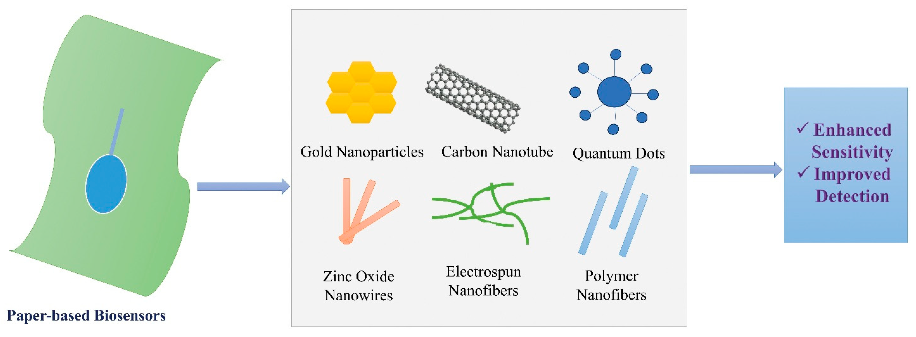
Figure 1. Incorporation of nanomaterials in paper-based biosensors.
Engproc 106 00008 g001
Figure 2. COVID-19 ePAD schematic diagram showing (A) device components, (B) detection principle, and (C) detection technique. Reprinted with permission from [111]. Copyright 2021, Elsevier.
Engproc 106 00008 g002
Table 1. Fabrication of paper-based biosensors: comparative features.
Fabrication TechniqueDescriptionAdvantagesLimitationsApplications
Wax and Screen Printing [29].Printing methods to pattern hydrophobic and conductive regions on paper substrates.Simple, cost-effective, eco-friendly, adaptable to origami structures.Limited design complexity; may require post-processing.Multi-analyte detection, origami devices, point-of-care diagnostics.
Robotic Printing and Micromanipulation [30].Uses robotic systems to integrate semiconductor microtubes into paper devices.High precision, automated, versatile for electronics and biosensors.Complex setup; requires advanced control systems.Field-effect transistor (FET) biosensors, microelectronics.
Movable Type Bioelectronics Printing [31].Transfers bioelectronic materials via modular, pre-fabricated molds.Flexible, low-cost, direct transfer of bioactive compounds.Limited throughput; best for small-scale prototyping.Continuous glucose and lactate monitoring.
Pen-Writing Technique [32].Employs rollerball pens with reagent inks to directly draw sensing patterns.Affordable, customizable, barrier-free fabrication.Limited to simple, manually drawn structures.On-site bioassays, multi-analyte diagnostics.
Laser-Induced Graphene (LIG) [33].Generates conductive graphene directly on paper via laser irradiation.High conductivity, flexible, disposable.Requires specialized laser equipment; two-step processing.Glucose biosensors, enzymatic electrochemical devices.
Aerosol-Assisted PECVD [34].Deposits bioreactive and biorepellent layers using plasma-enhanced chemical vapor deposition.Fast, stable, adherent, reproducible coatings.Expensive and equipment-intensive.DNA detection, nucleic acid biosensors.
Roll-to-Roll Processing [35].Employs continuous printing and deposition methods for large-scale production.Scalable, high-speed, low-cost manufacturing.Requires integration of multiple techniques; equipment intensive.Mass production of fully printed biosensors.
Pencil Drawing Method [11,36].Uses graphite pencils to create conductive traces on paper.Extremely low-cost, reproducible, simple fabrication.Restricted to graphite-based electrodes.Electrochemical detection of E. coli and pathogens.
Bottom-Up Wax-Patterned Microchip Printing [12,37].Builds translucent microfluidic arrays from cellulose fibers with wettability patterning.High resolution, rapid prototyping, transparent structures.Paper porosity limits resolution; requires careful optimization.Glucose monitoring, flexible wearable biosensors.
Table 2. Comparison of recent key research studies on paper-based biosensors.
YearStudy TitleStudy Person(s)Findings
2023“Printable biosensors towards next-generation point-of-care testing: Paper substrate as an example”Liu, Y., Lu, S., Zhang, Z., & Liu, G.Showed printing technologies (wax, screen, photolithography, inkjet, and laser) improve precision, efficiency, and scalability of paper biosensors [9].
2022“Nanomaterials and paper-based electrochemical devices: Emerging strategies for detection of biomarkers”Caratelli, V., Di Meo, E., Colozza, N., & Arduini, F.Integration of nanomaterials enhanced sensitivity, selectivity, and sustainability in biomarker detection [61].
2024“Gold nanoparticles (AuNPs): A versatile material for biosensor application”Kumalasari, M.R., Alfanaar, R., & Andreani, A.S.Demonstrated that AuNPs increase electroactive surface area and detection accuracy [127].
2024“Paper-based DNA biosensor for rapid and selective detection of miR-21”Hunt, A., Torati, S.R., & Slaughter, G.Developed inkjet-printed gold biosensor for rapid miR-21 cancer biomarker detection [73].
2021“Advancements in biosensor technologies for medical field and COVID-19 pandemic”Bahl, S., Bagha, A.K., Rab, S., & Singh, R.P.Showed rapid electrochemical biosensors for COVID-19 detection and medical diagnostics [128].
2025“Advances in nanoparticle-enhanced paper sensor for detecting toxic metals in water”Hosseini, M.S., Padhye, R., Wang, X., & Houshyar, S.Nanoparticle functionalization enabled sensitive on-site detection of toxic metals in water [129].
2024“MOF-mediated paper-based (bio)sensors for detecting food and environmental pollutants”Huang, D., Ma, H., Wang, J., & Li, R.Demonstrated MOFs integrated with paper substrates for rapid and accurate detection of food/environmental contaminants [16].
2024“Recent developments in paper-based sensors with instrument-free signal readout technologies (2020–2023)”Yang, D., Hu, C., Zhang, H., & Geng, S.Innovated distance-based, counting-based, and text-based readout methods for instrument-free detection [130].
2023“State-of-the-art of paper-based technology and challenges in its commercialization”Sharma, A., Kashyap, B.K., & Puranik, N.Highlighted commercialization barriers such as large-scale reproducibility and clinical validation challenges [131].
2025“Transformative biomedical devices to overcome biomatrix effects”Adil, O., & Shamsi, M.H.Addressed biomatrix interference in clinical samples with improved biosensor designs [132].
2020“High-performance modified cellulose paper-based biosensors for medical diagnostics and early cancer screening: A concise review”Ratajczak, K., & Stobiecka, M.Reviewed modified cellulose paper substrates enhancing performance in medical diagnostics and early cancer detection [133].
2023“Paper-based biosensors: Overview from past to future”Shrikrishna, N.S., Sharma, R., & Gandhi, S.Provided a comprehensive overview of paper-based biosensors, highlighting trends, applications, and future directions [2].
2021“Design and applications of fluorogenic nucleic acid-based paper biosensors”Yang, S., Yang, X., Wang, B., & Wang, L.Developed fluorescent biosensors with functional nucleic acids for sensitive and real-time detection [134].
2023“Paper-based electrochemical biosensors for the diagnosis of viral diseases”Ataide, V.N., Pradela-Filho, L.A., Ameku, W.A., & Angnes, L.Presented electrochemical biosensors for diagnosing viral diseases such as COVID-19, dengue, and Zika [4].
2022“Paper-based microfluidics: A forecast toward the most affordable and rapid point-of-care devices”Sinha, A., Basu, M., & Chandna, P.Predicted future trends of paper-based microfluidics for rapid and affordable point-of-care diagnostics [135].
2024“IoT-enabled biosensors for real-time monitoring and early detection of chronic diseases”Hosain, M.N., Kwak, Y.-S., Lee, J., & Kim, J.Developed IoT-enabled biosensors for real-time monitoring and early diagnosis of chronic diseases [136].
2021“Recent advances in (bio)chemical sensors for food safety and quality based on silver nanomaterials”Ivanišević, I., Milardović, S., & Kassal, P.Reviewed silver nanomaterial-based sensors for food safety and quality monitoring [137].
2024“Emerging diagnostic methods using paper-based electrochemical biosensors”Karuppannan, P.G., Sudha, D., Banupriya, K., Arumugam, R.Highlighted emerging diagnostic methods using paper-based electrochemical biosensors [1].
2020“Cytokine and cancer biomarkers detection: The dawn of electrochemical paper-based biosensor”Loo, S.W., Pui, T.-S.Showed sensitive detection of cytokines and cancer biomarkers using electrochemical paper-based biosensors [3].
Disclaimer/Publisher’s Note: The statements, opinions and data contained in all publications are solely those of the individual author(s) and contributor(s) and not of MDPI and/or the editor(s). MDPI and/or the editor(s) disclaim responsibility for any injury to people or property resulting from any ideas, methods, instructions or products referred to in the content.