Proceeding Paper

Antibacterial Activity of Ag2O/SrO/CaO Nanocomposite †

Department of Chemistry, Iran University of Science and Technology, Tehran 16846-13114, Iran
*
Author to whom correspondence should be addressed.
Presented at the 26th International Electronic Conference on Synthetic Organic Chemistry, 15–30 November 2022; Available online: https://sciforum.net/event/ecsoc-26.
Chem. Proc. 2022, 12(1), 77; https://doi.org/10.3390/ecsoc-26-13577
Published: 14 November 2022

Abstract

:
The increase in bacterial resistance to one or several antibiotics has become a global health problem. Nanocomposites have become a tool against multidrug-resistant bacteria. A nanocomposite, Ag2O/SrO/CaO, was prepared from AgNO3, SrCl2·6H2O, CaCl2, and a solution of Na2CO3 via the calcination of the salts mixture. The nanocomposite was successfully prepared by the co-precipitation method and completely according to green chemistry, in terms of synthesis method, solvent and precursors. The nanocomposite was characterized by XRD, XRF, and FESEM analyses. Afterwards, the nanocomposite was applied for antibacterial activity against gram-positive and gram-negative bacteria including PS Aeruginosa, Keleb peneumonia, Staph coccus aureus, Staph sapropphyticus, and Escherichia coli.

1. Introduction

The mixture of metal oxide nanoparticles and the combination of two or more metal oxides has an improved set of properties. In this way, we can modify the physical, chemical, biological, and morphological properties of the oxides. The combination and calcination of metallic compounds can create diverse properties and applications in the product; for example, it is shown that silver compounds are severe against bacteria.
Metal oxides, especially CuO, NiO, CoO, ZnO, and Cu2O in their nano-forms, have been considered as potential biocide agents. Most of these metal oxides, and their antibacterial activity, have been often related to the production of reactive oxygen species (ROS) [1,2].
Metals and metal oxides show antibacterial properties in the following ways: protein dysfunction, production of ROS and antioxidant depletion, impaired membrane function, interference with nutrient uptake, and genotoxicity [1].

2. Experimental Section

2.1. Peparation of Ag2O/SrO/CaO Nanocomposite

The Ag2O/SrO/CaO nanocomposite metal was prepared by the co-precipitation of corresponding carbonates from the aqueous solution of metal salts. Initially, 0.25 M, 30 mL solution of each of AgNO3 (1.274 g), SrCl2·6H2O (1.999 g), CaCl2 (0.832 g), and a solution (1.00 M, 50 mL) of Na2CO3 (5.299 g) were prepared with distilled water. Next, AgNO3, SrCl2·6H2O, and CaCl2 solutions were mixed, and the resulted mixture was stirred vigorously at room temperature for a few minutes. After this, the solution of 1.00 M Na2CO3 was added slowly to the above mixture with agitation until the precipitation of the carbonates was complete. The final mixture was stirred for 4 h at 55–60 °C with constant stirring. Then, the white metallic precipitate was filtered and washed several times with distilled water. Then the produced compound was dried at room temperature. To obtain a multi-metal nanocomposite (Ag2O/SrO/CaO nanocomposite), the obtained dried precipitate was calcinated in a muffle furnace at 600 °C for five hours. As a result of the calcination, a metal oxide nanocomposite was formed from the carbonates [3].

2.2. Characterization

The XRD patterns of the title composite, shown in Figure 1, was applied for the investigation of crystalline structure of nanomaterials. In Figure 1, the 2θ peaks appear at 25.67°, 27.705°, 33.246°, 36.55°, 41.8°, 42°, 45°, 51.849°, 54.724°, 57.8°, 62° and 76.5°, respectively, due to the formation of monoclinic (P21/C) Ag2O in this nanocomposite. The diffraction peaks observed at 32°, 35.8°, 37.5° 38.2°, 48°, 54.5°, and 57.9° are for the crystallographic planes of SrO, the cubic structure of SrO nanoparticles was detected (JCPDS file#6-520). The peaks at 32.5°, 36.6°, 38.2°, 44.5°, 45.4°, 64.2°, 67.8°, 77.4°, and 81.8° (JCPDS card no.00-004-0777) resulted from the cubic structure of CaO. The phase purity of SrO, CaO, and Ag2O in the Ag2O/SrO/CaO seen in the XRD pattern proves the presence of all three oxides in the nanocomposite.
In the XRF analysis of Ag2O/SrO/CaO, shown in Table 1, the percentages of pure CaO, SrO, and Ag2O are calculated equal to 10.77, almost 45.16, and 42.7, respectively.
The SEM micrographs of Ag2O/SrO/CaO Nanocomposite are presented in Figure 2, showing that the spherical shaped average particles of the Ag2O/SrO/CaO Nanocomposite have the size of 5 μm and 200 nm

2.3. Antibacterial Activity

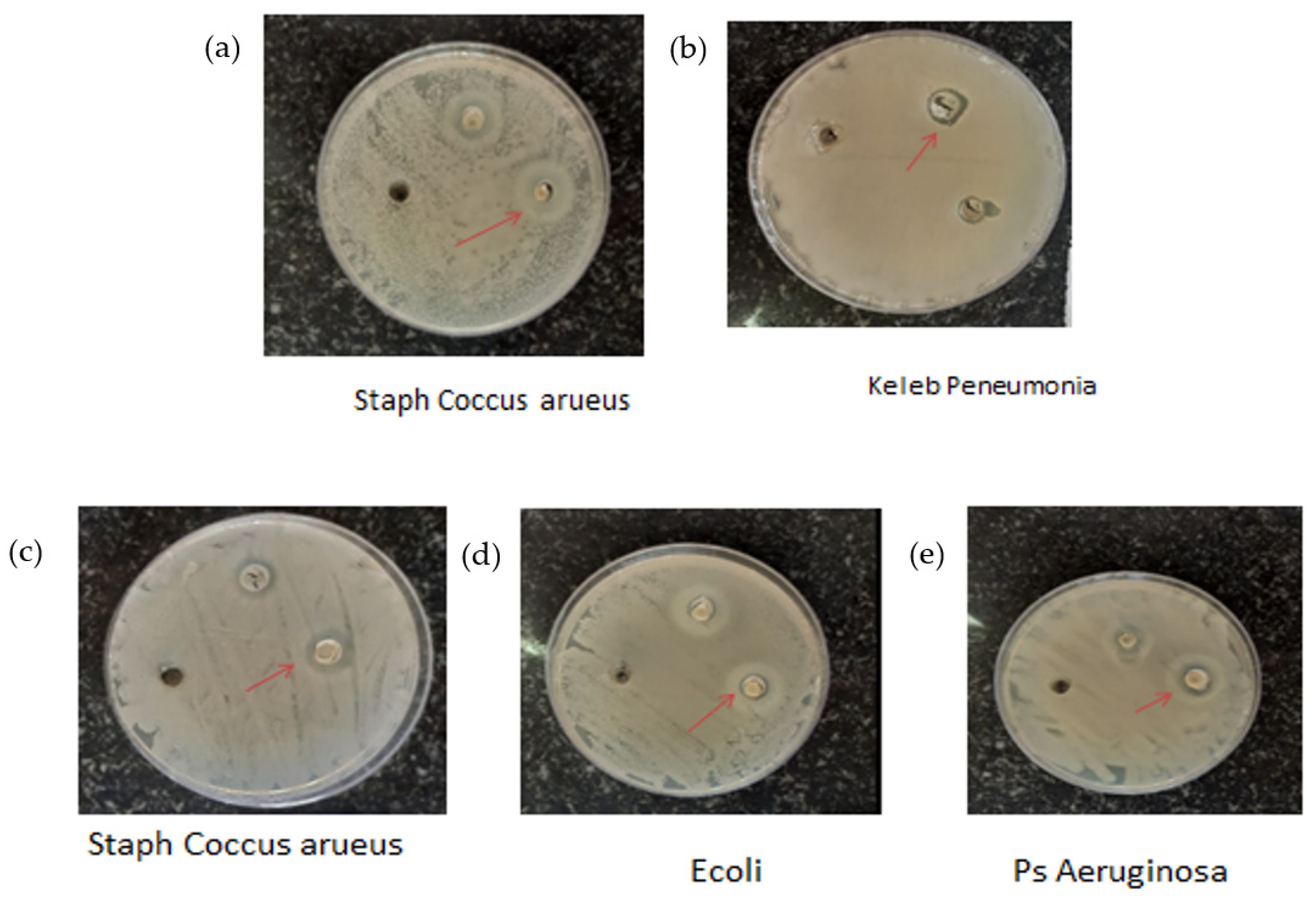
Antibacterial activity of the title nanocomposite against gram-positive and gram-negative bacteria were tested. The bacteria involve PS. Aeruginosa, Keleb peneumonia, Staph coccus aureus, Staph sapropphyticus, and Esherichia. The results are shown in Figure 3a–e and summarized in Table 2. It can be seen that the inhibition zone diameter from Ag2O/SrO/CaO is varied from 7.876 to 18.991 mm.
Antibacterial effects of the nanocomposite may be through the following properties:
Cell membrane damage: Silver nanoparticles have physicochemical and biological properties that are different from the properties of silver in the from of bulk. In order to have toxic effects of silver nanoparticles, they must be able to interact with bacteria on the surface and even in the cytoplasm. Silver nanoparticles release silver ions that must be able to cross the bacteria cell membrane and this is not very easy. Bacteria are divided into two groups: gram-positive (layers of peptidoglycans) and gram-negative (layers of lipopolysaccharides). In both groups, silver ions must be able to cross the membrane. Silver nanoparticles have their first encounter with the outermost part of the membrane, which is composed of protein with electron donors that include oxygen, phosphorus, nitrogen, and sulfur atoms. Thiol-containing agents can block silver nanoparticles and inhibit their antibacterial activity. In some papers, it is stated that silver nanoparticles are adsorbed on cell membranes and the accumulation of nanoparticles in bacterial membranes causes abnormal structure, gaps are created in the surface of the membrane, and the cell membrane is destroyed with the expansion of these cavities. Finally, the silver nanoparticles reach the cytoplasm and react with proteins, enzymes, and also DNA [4].
DNA interaction: The effect of silver Np on DNA was not understood in detail. Oxidative pressure was expressed as important mechanism proposed to damage DNA. Silver Np inhibit respiratory enzymes that conduct ROS formation. Silver ions react with bases in DNA with higher affinity than phosphate groups, although AgNp also have an antibacterial effect without releasing silver ions. AgNP can penetrate bacterial cells; AgNp invade the surface of cell membranes and disport permeability by modifying cell potential and by inhibiting cellular respiration. AgNp cations join to thiol groups in bacterial proteins, disrupting their activity. and causing cell death. The most important part of the mechanism of AgNp against bacterial DNA is oxidative stress [4].

3. Conclusions

In this work, a nanocomposite (Ag2O/SrO/CaO) was synthesized by an almost green method and green solvent, i.e., water and used environmentally friendly salts of calcium, strontium and silver. The product was characterized by XRD, XRF, and FESEM analyses, then the nanocomposite was examined as antibacterial activities against gram-positive and gram-negative bacteria, which included PS. Aeruginosa, Keleb peneumonia, Staph coccus aureus, Staph sapropphyticus, and E. coli.

Author Contributions

Conceptualization, F.M.. methodology, F.M..; software, M.A., F.M.; validation, F.M..; formal analysis, F.M.; investigation, F.M.; resources, F.M.; data curation, F.M.; writing—original draft preparation, M.A.; writing—review and editing, F.M.; visualization, F.M..; supervision, F.M.; project administration, F.M..; funding acquisition, aero. All authors have read and agreed to the published version of the manuscript.

Funding

This research received no external funding.

Institutional Review Board Statement

Not applicable.

Informed Consent Statement

Not applicable.

Data Availability Statement

Not applicable.

Conflicts of Interest

The authors declare no conflict of interest.

References

  1. Wyszogrodzka, G.; Marszałek, B.; Gil, B.; Dorożyński, P. Metal-organic frameworks: Mechanisms of antibacterial action and potential applications. Drug Discov. Today 2016, 21, 1009–1018. [Google Scholar] [CrossRef] [PubMed]
  2. Durán, N.; Durán, M.; De Jesus, M.B.; Seabra, A.B.; Fávaro, W.J.; Nakazato, G. Silver nanoparticles: A new view on mechanistic aspects on antimicrobial activity. Nanomed. Nanotechnol. Biol. Med. 2016, 12, 789–799. [Google Scholar] [CrossRef] [PubMed]
  3. Subhan, M.A.; Rifat, T.P.; Saha, P.C.; Alam, M.M.; Asiri, A.M.; Rahman, M.M.; Uddin, J. Enhanced visible light-mediated photocatalysis, antibacterial functions and fabrication of a 3-chlorophenol sensor based on ternary Ag2O·SrO·CaO. RSC Adv. 2020, 10, 11274–11291. [Google Scholar] [CrossRef] [PubMed]
  4. Lemire, J.A.; Harrison, J.J.; Turner, R.J. Antimicrobial activity of metals: Mechanisms, molecular targets and applications. Nat. Rev. Microbiol. 2013, 11, 371–384. [Google Scholar] [CrossRef] [PubMed]
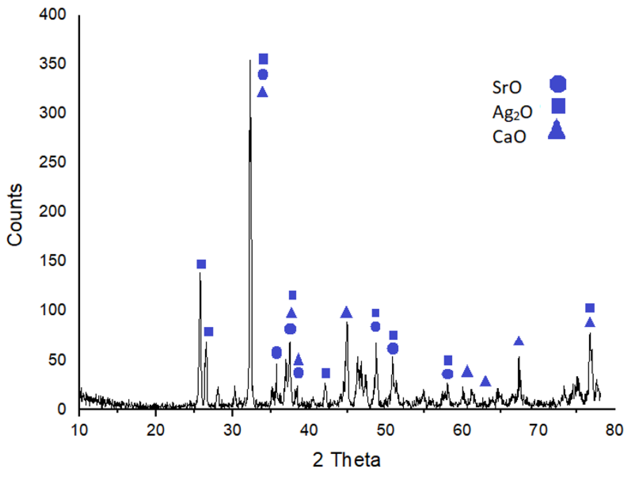
Figure 1. XRD pattern of Ag2O/SrO/CaO.
Chemproc 12 00077 g001
Figure 2. The FESEM images Ag2O/SrO/CaO.
Chemproc 12 00077 g002
Figure 3. Inhibition zones of different bacteria effected by the title nanocomposite.
Chemproc 12 00077 g003
Table 1. The XRF results as percentage of elements.
ElementsAgNaAl2O3SiO2P2O5SO2K2OCaOTiO2
wt%56.062>>9.627
ElementsFe2O3V2O5MnOCr2O3BaSrZnSeNb
wt%31.985>>
ElementsFCrClCeCoMoCaCuHo
wt%2.325>>
Table 2. Inhibition zone diameters of different bacteria effected by the title nanocomposite.
Test BacteriaInhibition Zone Diameter (mm)
Ag2O/SrO/CaO Nano Composite
Ps. Aeruginosa18.991
Keleb Peneumonia7.876
Staph Coccus aureus13.785
Staph Sapropphyticus12.723
E. coli16.456
Disclaimer/Publisher’s Note: The statements, opinions and data contained in all publications are solely those of the individual author(s) and contributor(s) and not of MDPI and/or the editor(s). MDPI and/or the editor(s) disclaim responsibility for any injury to people or property resulting from any ideas, methods, instructions or products referred to in the content.