Review

Diabetic Cardiomyopathy: Distinct Clinical Entity or Manifestation of Metabolic Heart Disease?

1
Cardiology Unit, AOU Luigi Vanvitelli, 80138 Naples, Italy
2
Department of Advanced Medical and Surgical Sciences, University of Campania “Luigi Vanvitelli”, Piazza Luigi Miraglia, 2, 80138 Naples, Italy
3
Department of Translational Medical Sciences, Section of Cardiology, University of Campania “Luigi Vanvitelli”, 80131 Naples, Italy
4
Vanvitelli Cardiology and Intensive Care Unit, Monaldi Hospital, 80131 Naples, Italy
5
Department of Life Science, Health, and Health Professions, Link Campus University, 00165 Rome, Italy
*
Author to whom correspondence should be addressed.
Diabetology 2026, 7(8), 160; https://doi.org/10.3390/diabetology7080160
Submission received: 1 April 2026 / Revised: 7 August 2026 / Accepted: 10 August 2026 / Published: 18 August 2026
(This article belongs to the Section Complications and Comorbidities of Diabetes)

Abstract

Background/Objectives: Type 2 diabetes mellitus (T2DM) is a global epidemic strongly associated with an increased risk of heart failure, independent of coronary artery disease or hypertension. This condition, historically termed diabetic cardiomyopathy (DCM) and recently redefined as “diabetic myocardial disorder,” remains frequently underdiagnosed in its subclinical stages. The objective of this non-systematic review is to synthesize current evidence on the pathophysiological mechanisms, diagnostic advancements, and evolving therapeutic strategies for diabetic myocardial involvement. Methods: A comprehensive review of contemporary literature was conducted, focusing on recent consensus statements from the ESC and AHA, large-scale epidemiological data (IDF/WHO), and pivotal clinical trials (EMPA-REG, DAPA-HF, and LEADER). We analyzed the role of multimodal imaging—specifically speckle-tracking echocardiography (STE) and multiparametric cardiac magnetic resonance (CMR)—and circulating biomarkers in early phenotyping. Results: Pathophysiological drivers include lipotoxicity, oxidative stress, and AGE-mediated fibrosis. Advanced imaging techniques, such as global longitudinal strain (GLS) and CMR T1-mapping/ECV quantification, demonstrate superior sensitivity over LVEF in detecting early subendocardial dysfunction and diffuse fibrosis. Furthermore, NT-proBNP serves as a robust prognostic marker for the HFpEF-like trajectory typical of diabetes. Clinically, the therapeutic landscape has shifted with SGLT2 inhibitors and GLP-1 receptor agonists, which provide significant cardioprotection and reduction in heart failure hospitalizations through mechanisms beyond glycemic control. Conclusions: Diabetic myocardial disorder represents a complex continuum within the cardiometabolic spectrum. Early detection through multimodal imaging and biomarkers is essential for risk stratification. Integrating novel glucose-lowering therapies with proven cardiovascular benefits is now mandatory to alter the natural history of the disease and prevent progression to overt heart failure.

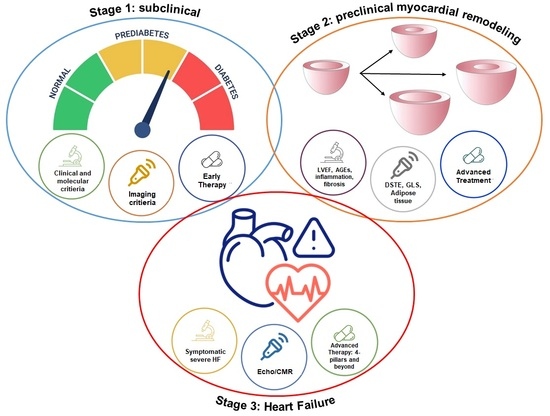
Graphical Abstract

1. Literature Sources and Search Strategy

We performed a non-systematic review of the literature by applying the search strategy in different electronic databases (MEDLINE, EMBASE, Cochrane Register of Controlled Trials, and Web of Science). Original reports, meta-analyses, and review articles in peer-reviewed journals up to March 2026 regarding adipose tissue, cardiac adipose tissue, obesity, metabolic syndrome, and adipocyte molecular pathways were incorporated into the search strategy. The references of all identified articles were reviewed to look for additional papers of interest and to extrapolate the more recent available data on DCM.

2. Introduction

T2DM is a major and rapidly escalating global health problem, driven by demographic transitions, urbanization, and rising obesity. The 11th edition of the International Diabetes Federation (IDF) Diabetes Atlas reports that diabetes affects a substantial share of the adult population worldwide, with prevalence rising across nearly all regions [1]. Pooled analyses from the NCD Risk Factor Collaboration, supported by the World Health Organization (WHO), show that the number of adults living with diabetes worldwide has now exceeded 800 million, with prevalence roughly doubling from 7% in 1990 to about 14% in 2022 [2]. This increase has been particularly marked in low- and middle-income countries, where rapid epidemiological transition has been accompanied by limited access to diagnosis and treatment [2]. A large share of diabetes cases also remain undiagnosed or inadequately treated, a gap in care that contributes to excess morbidity and mortality [2]. Diabetes remains among the leading contributors to global mortality and healthcare expenditure [1]. T2DM accounts for more than 90% of diabetes cases and is strongly tied to modifiable risk factors such as obesity, sedentary behavior, and an unhealthy diet; the global rise in adiposity is a central driver of the epidemic and links T2DM closely to the broader spectrum of cardiometabolic disease [3]. Taken together, these figures point to a disease that is not only becoming more common but is also unevenly distributed, poorly treated in many settings, and tightly bound to metabolic risk factors—all reasons for why prevention and management need to be addressed together.

2.1. Cardiovascular Complications of Diabetes

Cardiovascular disease (CVD) remains the predominant cause of morbidity and mortality in individuals with type 2 diabetes mellitus (T2DM) despite therapeutic advances in glucose lowering and risk factor management [4]. The link between diabetes and cardiovascular complications is multifactorial. Beyond traditional risk factors such as hypertension, dyslipidemia, and obesity, diabetes-specific mechanisms—chronic hyperglycemia, insulin resistance, endothelial dysfunction, inflammation, and oxidative stress—contribute directly to vascular injury and myocardial dysfunction [5]. These processes drive both macrovascular and microvascular damage, accelerating atherosclerosis while also affecting the myocardium independently of epicardial coronary disease. Diabetes is also associated with intrinsic myocardial abnormalities, such as fibrosis, metabolic derangements, and impaired contractility, which can occur without significant epicardial coronary artery disease or hypertension [6,7]. These diabetes-related myocardial changes form the basis of the concept of diabetic myocardial disorder, which can predispose patients to heart failure (HF), even in the absence of overt ischemic heart disease [7]. Consensus statements of the Heart Failure Association (HFA) of the ESC and the ESC Working Group on Myocardial and Pericardial Diseases recognize that diabetic myocardial disorder can manifest as structural and functional cardiac abnormalities independent of atherosclerotic disease, supporting a multifactorial cardiovascular risk profile in T2DM that encompasses both vascular and myocardial components [7]. Recent European Society of Cardiology (ESC) guidelines note that patients with diabetes are at high or very high cardiovascular risk and recommend comprehensive management of metabolic and cardiovascular factors, including risk stratification with tools such as SCORE2-Diabetes and early detection of subclinical cardiac dysfunction using biomarkers like NT-proBNP [4].
This makes comprehensive cardiovascular risk assessment in patients with diabetes important, including strategies for early identification of subclinical myocardial dysfunction.

2.2. Increased Risk of Heart Failure in Diabetic Patients

Type 2 diabetes mellitus (T2DM) is a major risk factor not only for atherosclerotic cardiovascular disease but also for heart failure (HF). Epidemiological evidence consistently shows a substantially higher incidence of HF in people with T2DM than in those without diabetes, in populations with and without established cardiovascular disease [8,9]. A recent systematic review and meta-analysis found that individuals with T2DM had an approximately 1.6- to 2.0-fold higher risk of developing HF compared with non-diabetic controls, regardless of the presence of established ASCVD [9]. HF in T2DM often presents as HF with a preserved ejection fraction (HFpEF) and subclinical left ventricular dysfunction, conditions that are frequently underdiagnosed in this population [10]. Metabolic derangements, such as insulin resistance and chronic hyperglycemia, contribute to myocardial remodeling, diastolic dysfunction, and increased myocardial stiffness, which may precede overt symptomatic HF [10,11]. These manifestations reflect the concept of diabetic myocardial disorder, as introduced above (Section 2.1), including structural and functional myocardial abnormalities—fibrosis, metabolic derangements, impaired contractility, and increased stiffness—that can occur independently of epicardial coronary artery disease or hypertension. The relationship between diabetes and HF is also bidirectional: patients with established HF face a higher risk of developing glucose abnormalities and incident diabetes, while T2DM, in turn, worsens one’s HF prognosis, with higher rates of hospitalization and mortality [11]. HF should therefore be considered a major clinical endpoint in diabetic patients—one that occurs frequently, often early, and with pathophysiological features distinct from non-diabetic HF. NT-proBNP is increasingly used as a biomarker for the early identification of subclinical myocardial dysfunction in T2DM: in asymptomatic patients, elevated NT-proBNP values have classified a significant portion as at risk of “heart stress,” corresponding to early cardiac involvement and prompting further evaluation [12,13].

2.3. Introduction of the Concept of Diabetic Cardiomyopathy

The term “diabetic cardiomyopathy” (DCM) was first introduced in the early 1970s to describe a form of myocardial disease occurring in patients with diabetes mellitus without significant coronary artery disease, hypertension, or valvular heart disease. Seminal pathological observations by Rubler [14] and colleagues in 1972 identified adverse structural myocardial changes in diabetic subjects without overt atherosclerotic disease, pointing to a primary cardiac effect of diabetes itself. Subsequent epidemiological data from the Framingham Heart Study confirmed an independent association between diabetes and congestive heart failure, even after accounting for common comorbidities, which reinforced the case for a distinct diabetic myocardial phenotype [15]. Despite decades of research, the clinical utility and precise definition of DCM have remained controversial. Traditional definitions emphasize the presence of myocardial dysfunction directly attributable to diabetes-related metabolic disturbances—insulin resistance, hyperglycemia-induced glucotoxicity, lipotoxicity, and microvascular impairment—after exclusion of other major cardiac pathology. However, the specificity of these criteria has been challenged by the frequent coexistence of obesity, arterial hypertension, coronary artery disease, and chronic kidney disease in patients with type 2 diabetes, complicating efforts to attribute myocardial dysfunction exclusively to diabetic mechanisms [7]. Expert consensus has recently pushed the field toward a different framework: the 2024 clinical consensus statement from the European Society of Cardiology’s Heart Failure Association and the ESC Working Group on Myocardial and Pericardial Diseases proposes the term “diabetic myocardial disorder” as a more pragmatic and inclusive descriptor. It defines diabetic myocardial disorder as systolic and/or diastolic myocardial dysfunction in the presence of diabetes, on the premise that diabetes rarely acts alone and usually adds to myocardial impairment alongside other cardiometabolic comorbidities [7]. This definition also fits contemporary models of heart failure progression better than a binary presence/absence concept: myocardial involvement in diabetes is increasingly viewed as part of a continuum of early structural and functional cardiac abnormalities, potentially representing pre-heart failure (stage B) when asymptomatic, and progressing to overt heart failure as remodeling and dysfunction advance. This framework helps risk stratification, early detection, and possible intervention before symptomatic heart failure develops [7]. However, evidence gaps remain. How often isolated diabetic myocardial dysfunction occurs independently of comorbid conditions in real-world populations is unclear, and large-scale human histopathological and longitudinal mechanistic data are still limited. Preclinical models and imaging studies point to distinct myocardial remodeling patterns in diabetes, but whether these translate into a universally accepted clinical entity is still debated [16].
This non-systematic review aims to summarize and critically appraise current evidence on myocardial involvement in diabetes, focusing on structural, functional, and clinical characteristics. Drawing on recent human and preclinical studies, it outlines the cardiological features of diabetic myocardial dysfunction, including changes in myocardial geometry, diastolic and systolic performance, fibrosis, and microvascular involvement.
It also aims to clarify the evolving definition and terminology of DCM, placing historical perspectives alongside contemporary evidence, with particular attention given to advanced imaging, circulating biomarkers, and mechanistic studies that shed light on diabetic myocardial impairment.
The goal is a cardiology-focused framework for diabetic myocardial disease that outlines where consensus exists, where debate remains, and which knowledge gaps persist—informing both clinical risk stratification and future research, with an emphasis on early detection and potential therapeutic intervention.

3. Pathophysiological Mechanisms

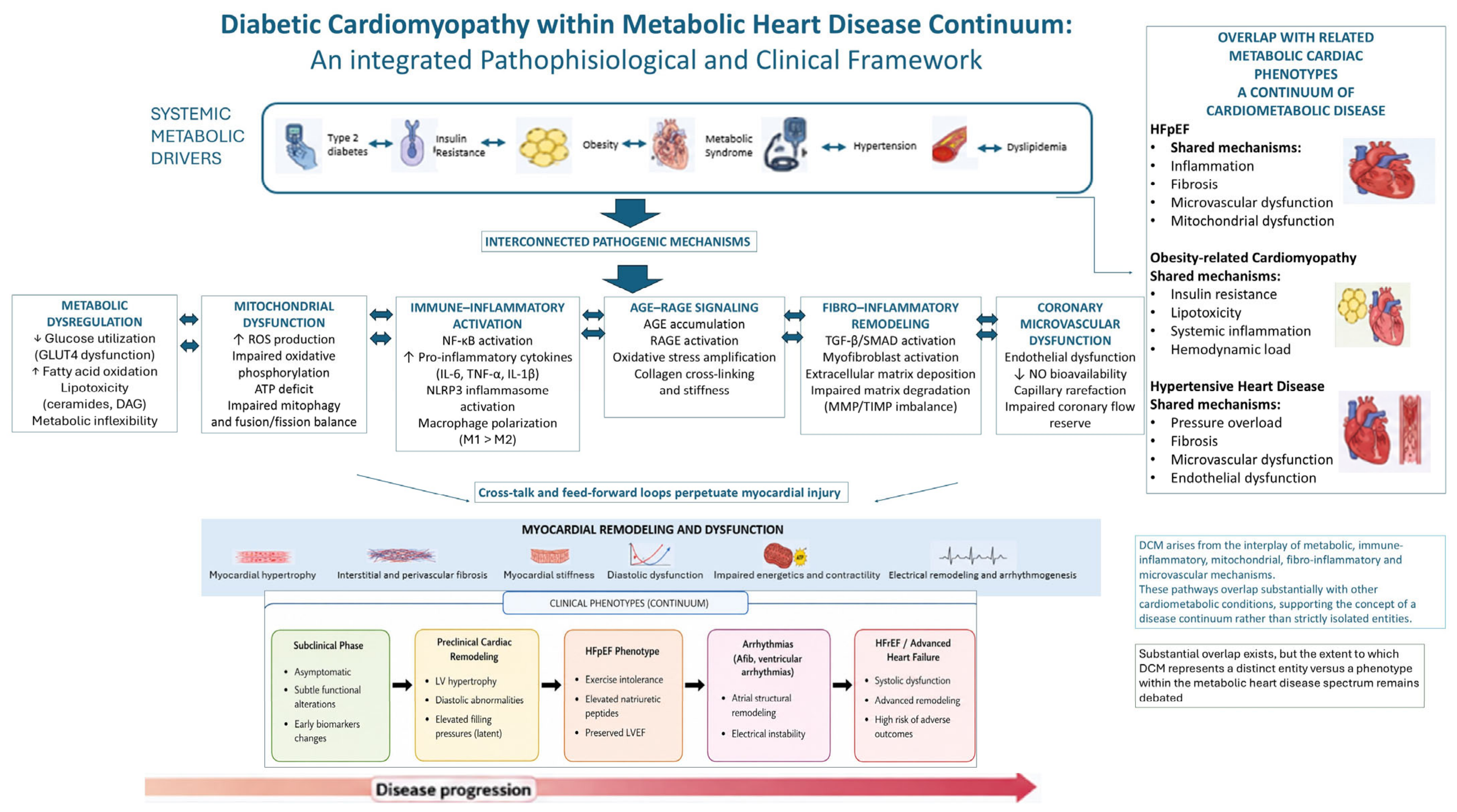
DCM arises from a multifactorial process involving metabolic disturbances, oxidative stress, inflammatory signaling, and microvascular dysfunction. Chronic hyperglycemia and insulin resistance trigger a cascade of molecular and cellular alterations that progressively affect myocardial structure and function. These interconnected mechanisms ultimately contribute to myocardial remodeling, impaired relaxation, and the development of heart failure in patients with diabetes (see Figure 1).

3.1. Metabolic Alterations

Alterations in myocardial energy metabolism represent one of the earliest hallmarks of DCM. In patients with type 2 diabetes mellitus, systemic metabolic dysregulation progressively affects cardiac substrate utilization and energy production, contributing to the development of structural and functional myocardial abnormalities [17,18]. Under normal conditions, the heart is metabolically flexible, drawing on both fatty acids and glucose depending on demand. In diabetes, insulin resistance and chronic hyperglycemia impair myocardial glucose uptake, mainly by reducing activity and translocation of the glucose transporter GLUT4; thus, cardiomyocytes use less glucose [19,20]. The diabetic myocardium compensates by relying more heavily on fatty acid oxidation to sustain ATP production. Fatty acids normally supply a large share of cardiac energy, but excessive dependence on this pathway costs efficiency: it takes more oxygen to generate the same amount of ATP from fatty acids than from glucose, so myocardial oxygen demand rises while metabolic efficiency falls [21]. This shift also causes lipid intermediates to build up in cardiomyocytes. When fatty acid uptake exceeds mitochondrial oxidative capacity, toxic lipid species such as ceramides and diacylglycerols accumulate—a state known as lipotoxicity that disrupts intracellular signaling—worsening insulin resistance and injuring cardiomyocytes [22]. These metabolic abnormalities do not act alone; they interact dynamically with inflammatory, mitochondrial, and microvascular pathways as part of a broader cardiometabolic remodeling process, which helps explain the substantial overlap between DCM and other metabolic heart disease phenotypes, including HFpEF and obesity-related cardiomyopathy (CM) [17,18].

3.2. Oxidative Stress and Inflammation

Oxidative stress and chronic low-grade inflammation represent key mechanisms contributing to the development and progression of DCM. Metabolic changes in diabetes, including hyperglycemia and increased fatty acid oxidation, promote excessive production of reactive oxygen species (ROS) in cardiomyocytes. Several cellular sources feed this ROS generation in the diabetic heart, including mitochondrial dysfunction, activation of NADPH oxidases, and increased flux through alternative glucose metabolic pathways [17,18]. The resulting imbalance between ROS production and antioxidant defenses damages proteins, lipids, and nucleic acids. In cardiomyocytes, oxidative stress impairs mitochondrial function, alters calcium handling, and reduces contractile efficiency, all of which contribute to myocardial dysfunction [23]. ROS also act as signaling molecules, activating intracellular pathways involved in inflammation and cellular stress responses.
Among these pathways, activation of nuclear factor kappa B (NF-κB) drives the expression of pro-inflammatory cytokines such as tumor necrosis factor-α (TNF-α) and interleukin-6 (IL-6). Persistent inflammatory signaling contributes to endothelial dysfunction, myocardial remodeling, and progressive impairment of cardiac function in diabetes; emerging evidence points to chronic low-grade inflammation and immune-metabolic interactions as central mechanisms linking diabetes to myocardial remodeling and HFpEF-like phenotypes [17,24]. Chronic inflammation can also worsen oxidative stress, creating a self-perpetuating cycle that amplifies myocardial injury. Oxidative stress and inflammation together link metabolic disturbances to structural and functional changes in the diabetic myocardium, and they favor the formation of advanced glycation end products and extracellular matrix remodeling that add to myocardial fibrosis. Rather than acting as isolated mechanisms, they form part of a complex pathogenic network alongside metabolic dysregulation, mitochondrial dysfunction, endothelial injury, and fibro-inflammatory remodeling [17,18,23,24].

3.3. Advanced Glycation End Products (AGEs) and Fibrosis

Advanced glycation end products (AGEs) represent a key link between chronic hyperglycemia and structural remodeling in DCM [24,25,26]. AGEs form through non-enzymatic reactions between reducing sugars and proteins, lipids, or nucleic acids, a process that is significantly accelerated under hyperglycemic conditions typical of diabetes [25,26]. Their progressive accumulation in myocardial tissue contributes substantially to both cellular dysfunction and extracellular matrix remodeling [25,27], which increases myocardial stiffness, reduces ventricular compliance, and contributes to diastolic dysfunction—an early manifestation of DCM [27,28,29]. AGE-modified collagen is more resistant to degradation, promoting its accumulation and favoring interstitial and perivascular fibrosis [27,29]. Beyond these structural effects, AGEs act as bioactive molecules through binding to their specific receptor, RAGE (receptor for advanced glycation end products). RAGE is expressed on cardiomyocytes, endothelial cells, and inflammatory cells [26]. RAGE activation triggers multiple intracellular signaling pathways, including an increased production of reactive oxygen species and activation of pro-inflammatory transcription factors such as NF-κB [24,26], which enhances the expression of cytokines, adhesion molecules, and profibrotic mediators and amplifies inflammation and oxidative stress in the myocardium [24,30]. The AGE–RAGE interaction promotes activation of cardiac fibroblasts and their differentiation into myofibroblasts, which are key mediators of extracellular matrix synthesis and deposition [30]. Beyond collagen cross-linking, several profibrotic signaling pathways contribute to matrix remodeling in the diabetic myocardium. Among these, transforming growth factor-β (TGF-β) is a central regulator of fibroblast activation and matrix synthesis: hyperglycemia, oxidative stress, and AGE–RAGE signaling can all stimulate TGF-β expression, which promotes differentiation of cardiac fibroblasts into activated myofibroblasts and boosts production of structural matrix proteins, particularly type I and type III collagen [31].
This imbalance between matrix synthesis and degradation leads to progressive accumulation of extracellular matrix components and disruption of normal myocardial architecture. Increased collagen deposition and interstitial fibrosis contribute to ventricular stiffening, impaired myocardial relaxation, and reduced ventricular compliance—all characteristic features of DCM [17,18,31]. AGE-induced microvascular dysfunction, including endothelial damage and reduced nitric oxide bioavailability, further exacerbates tissue hypoxia and fibrotic remodeling [27,30]. AGE accumulation and its downstream signaling pathways are thus central to the development of myocardial fibrosis and increased ventricular stiffness in DCM, interacting with metabolic disturbances, oxidative stress, and inflammatory pathways to drive adverse cardiac remodeling and progression toward heart failure [30,32]. Myocardial fibrosis in diabetes should be interpreted as part of a broader fibro-inflammatory remodeling process, in which inflammatory signaling, oxidative stress, AGE–RAGE activation, and fibroblast dysregulation act synergistically to promote extracellular matrix expansion and ventricular stiffening [24,26,30,31].

3.4. Microvascular Dysfunction

Microvascular dysfunction represents a critical aspect in the pathophysiology of DCM, contributing to myocardial remodeling and functional deterioration, even in the absence of obstructive coronary artery disease [17,33]. In patients, chronic hyperglycemia, insulin resistance, and low-grade inflammation impair the integrity and function of the coronary microcirculation [17]. Coronary microvascular dysfunction may act as a mechanistic bridge between early diabetic myocardial impairment and the later development of HFpEF-related phenotypes, consistent with a progressive cardiometabolic continuum [7,33].

3.5. Endothelial Dysfunction

Endothelial dysfunction is one of the earliest alterations observed in DCM and plays a pivotal role in its progression [17]. Under physiological conditions, the endothelium regulates vascular tone, inflammation, and hemostasis through the balanced release of vasoactive mediators, including nitric oxide (NO). In diabetes, hyperglycemia increases the production of reactive oxygen species (ROS), resulting in reduced NO bioavailability and increased oxidative stress [32]. Advanced glycation end products (AGEs) and the activation of protein kinase C (PKC) pathways further exacerbate endothelial damage [32]. This results in impaired vasodilation, increased vascular stiffness, and the development of a pro-inflammatory, pro-thrombotic endothelial phenotype [1]. Upregulation of adhesion molecules promotes leukocyte recruitment and sustains vascular inflammation [17]. Together, these changes compromise myocardial perfusion at the microcirculatory level, causing inadequate oxygen delivery even without epicardial coronary stenoses [33], and are increasingly seen as central mechanisms linking diabetic myocardial dysfunction to the HFpEF phenotype, where endothelial inflammation, impaired nitric oxide signaling, and coronary microvascular dysfunction increase myocardial stiffness and impair ventricular relaxation [7,10,17,33].

3.6. Diabetic Cardiomyopathy Within the Metabolic Heart Disease Spectrum

Although DCM has traditionally been described as a distinct myocardial complication of diabetes mellitus, increasing evidence suggests substantial overlap with other cardiometabolic conditions, particularly heart failure with a preserved ejection fraction (HFpEF), obesity-related cardiac dysfunction, and hypertensive heart disease [17,18,24]. Many of its pathophysiological mechanisms, including metabolic dysregulation, chronic low-grade inflammation, mitochondrial dysfunction, fibro-inflammatory remodeling, endothelial injury, and coronary microvascular impairment, are shared across the broader spectrum of metabolic heart disease [17,18,23,24].
DCM may be interpreted not only as a diabetes-specific myocardial disorder, but also as part of a multidimensional cardiometabolic continuum characterized by interacting metabolic, inflammatory, vascular, and structural remodeling pathways [17,18].
The coexistence of insulin resistance, obesity, endothelial dysfunction, and systemic inflammation likely explains much of this phenotypic overlap, particularly in patients with diastolic dysfunction and HFpEF-like phenotypes. Whether DCM is a truly distinct clinical entity remains debated, largely because no universally accepted diagnostic criteria exist and it is difficult to separate diabetes-specific myocardial changes from the contribution of comorbid risk factors such as hypertension, obesity, and metabolic syndrome. Future studies combining molecular profiling, advanced cardiac imaging, and phenotype-based stratification may help refine this framework and improve diagnosis and treatment in this complex setting [18,24].

3.7. Coronary Microcirculation Impairment

Coronary microcirculation impairment is a hallmark of DCM and is associated with significant structural and functional changes [17]. Key modifications include capillary rarefaction, basement membrane thickening, and perivascular fibrosis, which reduce capillary density and increase oxygen diffusion distance [17]. Functionally, there is a reduction in coronary flow reserve (CFR) and impaired autoregulation of blood flow, limiting the heart’s ability to adapt to increased metabolic demands [33,34]. This mismatch between oxygen supply and demand leads to chronic microvascular ischemia, which, over time, contributes to cardiomyocyte dysfunction, interstitial fibrosis, and the development of diastolic and systolic dysfunction [17,33].

3.8. Mitochondrial Dysfunction

Mitochondrial dysfunction is a key mechanism in DCM and underlies myocardial energy deficit [17,23]. Because the myocardium has such a high energy demand, intact mitochondrial function is essential for maintaining contractility and cellular homeostasis [35].

3.9. Altered Oxidative Phosphorylation

In diabetes, mitochondrial oxidative phosphorylation is markedly impaired [36]. A shift toward increased fatty acid oxidation and reduced glucose oxidation leads to metabolic inflexibility [36]; fatty acid oxidation generates ATP, but it is less oxygen-efficient compared to glucose metabolism [36]. Excess fatty acid uptake also promotes the accumulation of toxic lipid intermediates (lipotoxicity), adding further mitochondrial damage [23]. The electron transport chain becomes impaired, resulting in increased electron leakage and enhanced ROS production. Oxidative damage to mitochondrial DNA, proteins, and lipids further amplifies mitochondrial dysfunction, establishing a vicious cycle of reduced energy production and increased oxidative stress [23,37].

Energy Deficit

Together, these changes produce a myocardial energy deficit, marked by reduced ATP availability and an altered phosphocreatine/ATP ratio [35]—a ratio that is essential for contraction and relaxation processes.
Energy deprivation particularly affects high-ATP-demand processes such as intracellular calcium homeostasis and sarcomere function [35]. Impaired calcium reuptake into the sarcoplasmic reticulum contributes to diastolic dysfunction, often an early manifestation of DCM [17]. Over time, a chronic energy deficit promotes contractile dysfunction, cardiomyocyte apoptosis, and adverse ventricular remodeling [23,35]. Mitochondrial dysfunction, then, is not just a metabolic derangement but a real determinant of how DCM progresses structurally and functionally.

4. Clinical Manifestations and Phenotypes

DCM is a disease defined by myocardial dysfunction and/or structural abnormalities in patients affected by diabetes mellitus, which is not determined by other factors such as hypertension, valvular, or coronary disease [17]. According to the American Heart Association (AHA), diabetic patients are considered “at risk” for HF (stage A), and diabetes can promote the worsening from a preclinical stage to symptomatic HF (stage C). A higher incidence of HF is observed in diabetic men than in women, especially in middle-aged adults [38], while women present higher levels of N-terminal pro-B-type natriuretic peptide (NT-proBNP) and more preserved left ventricular (LV) function [39]. Traditional risk factors increase the probability of developing HF in diabetic patients [38], and among patients with HF, those with type 2 diabetes mellitus face a higher risk of adverse outcomes [39]. Diabetes and HF are linked through several mechanisms—abnormal calcium signaling, disrupted glucose metabolism, and inflammatory pathway activation—that result in myocardial fibrosis and hypertrophy. Hormonal overactivation also plays a role, with excessive renal sodium reabsorption causing fluid overload and poor diuretic responsiveness.
DCM was originally described as reduced LV systolic function (heart failure with reduced ejection fraction, HFrEF) with eccentric hypertrophy, caused microscopically by replacement of sarcomeres with extracellular fibrosis. Current evidence also strongly links it to diastolic LV dysfunction (heart failure with a preserved ejection fraction, HFpEF) and LV concentric hypertrophy, with hypertrophied cardiomyocytes and increased collagen deposition in the extracellular matrix [38]. Whether diastolic dysfunction always precedes systolic dysfunction is still unclear: a rat model study suggests a linear progression from diastolic to systolic dysfunction, but 20–30% of diabetic patients show decreased systolic strain—a more sensitive parameter of LV function—before diastolic dysfunction appears [40]. Several studies demonstrate the link between diabetes and left ventricular hypertrophy (LVH), which is responsible for reduced LV compliance, increased diastolic filling pressure, and diastolic dysfunction [38]. LVH seems also to be partially reversible with adequate glycemic control. LV diastolic dysfunction is also observed in diabetic patients independently of LVH, with a prevalence of 54% [41]. Diabetes is also associated with progression from HFpEF to HFrEF. Indeed, it is related to global longitudinal strain (GLS) reduction, also seen in young adults and adolescents. Furthermore, diabetes also has an impact on diastolic and systolic right ventricular (RV) function independently of LV deterioration [40]. Given the prevalence of this condition, several scores have been proposed to determine the risk of developing HF. One of them, proposed by Pandey et al. [42], is based on the determination of highly sensitive T-Troponin, C-reactive protein, NT-proBNP and LVH signs on ECG [38]. Because HF is often subclinical in its early stages, American Diabetes Association (ADA) guidelines recommend early screening [39].

5. Imaging and Biomarkers

Imaging and circulating biomarkers are essential for identifying DCM early since subclinical myocardial alterations precede overt heart failure—a pattern first shown by the Framingham study [43]. A multimodal approach that combines imaging, such as cardiac magnetic resonance (CMR) and speckle-tracking echocardiography (STE), with biochemical markers like NT-proBNP, supports proactive risk stratification, disease monitoring, and targeted interventions to slow DCM progression [44,45].

5.1. Advanced Echocardiographic Modalities: Speckle-Tracking Echocardiography and Global Longitudinal Strain

In diabetic patients, transthoracic echocardiography remains the first-line tool for evaluating cardiac structure and function; however, conventional approaches often lack sensitivity, requiring advanced echocardiography modalities to detect DCM early-stage disease, as the key parameters frequently remain within normal ranges until advanced stages. Advanced echocardiography modalities, specifically speckle-tracking echocardiography (STE) and diastolic stress echocardiography (DSTE), have emerged as superior methodologies that are capable of detecting subtle impairments in myocardial relaxation and systolic strain before the left ventricular ejection fraction (LVEF) declines [46,47]. The global longitudinal strain (GLS), a key STE-derived parameter, is a highly sensitive marker of DCM, enabling the detection of subclinical myocardial dysfunction even when conventional parameters, such as left ventricular ejection fraction (LVEF), remain preserved [47]. While STE excels in detecting myocardial deformation at rest, DSTE may provide additional information on diastolic function under stress conditions by unmasking exertional elevations in filling pressures and impaired diastolic reserve [46]. This integrated evaluation, combining conventional indices with advanced echocardiography modalities, promotes the early identification of functional abnormalities, supporting proactive management to prevent progression towards symptomatic heart failure. Transthoracic echocardiography may also be useful in differentiating DCM from other pathological conditions, such as long-standing hypertensive heart disease and storage disorders, such as amyloidosis. In particular, in long-standing hypertensive heart disease with restrictive filling, concentric LVH is marked, E/A and E/e′ are high, the LAVI is increased, but tissue Doppler e′ may not be severely reduced, and GLS is often preserved, without apical sparing or other infiltrative features [48]. Cardiac amyloidosis typically shows markedly reduced GLS with relative apical sparing; speckle-tracking pattern recognition (apical sparing) is highlighted as an amyloidosis marker, distinguishing infiltrative hypertrophy from DbCM or pure hypertensive remodeling, where such a pattern is absent [49,50]. To mathematically and clinically quantify these myocardial deformations, global longitudinal strain (GLS) has evolved since the early 2000s into the most robust and reproducible STE-derived indicator of early subclinical systolic dysfunction.
Recent studies demonstrate that GLS is often impaired by 2–3% in asymptomatic patients with T2DM and a preserved LVEF, reflecting the vulnerability of subendocardial longitudinal fibers to metabolic stress, oxidative damage, and microvascular impairment [51,52]. Meta-analyses have consistently confirmed significantly lower GLS values in diabetic cohorts, with direct correlations to diabetes duration and poor glycemic control [53,54]. Recently, the integration of GLS with non-invasive blood pressure measurements has been used to calculate the pressure–strain loop (PSL) of the LV, giving an estimation of the myocardial work (MW) as a novel imaging parameter [55]. In T2DM patients, a reduced global myocardial work index (GWI) and global work efficiency (GWE), alongside increased global wasted work (GWW), indicate early myocardial inefficiency due to inefficient systolic contraction. Incorporating these metrics into routine screening enhances the monitoring of therapeutic responses and outperforms traditional LVEF in the subclinical phase [55,56].

5.2. Circulating Biomarkers and the Role of NT-proBNP

Circulating biomarkers provide critical insights into the metabolic stress, fibrotic remodeling, and inflammatory pathways underlying DCM. Among these, protein biomarkers such as galectin-3 and adiponectin (APN), together with regulatory molecules including circulating and exosome-derived microRNAs, reflect the complex pathophysiological mechanisms of the disease and hold promise as potential diagnostic and therapeutic targets [56,57]. Among more established markers, HbA1c, cardiac troponin I (cTnI) (elevated in DCM vs. diabetes alone), inflammatory mediators (TNF-α, IL-6, C-reactive protein), fibrotic factors (TGF-β1, IGFBP7), and antioxidant indicators (reduced leptin, adiponectin, and bilirubin) also aid earlier detection [56,58].
Circulating microRNAs (miRNAs) are promising epigenetic biomarkers for the early diagnosis and risk stratification of DCM. Specific dysregulated miRNAs mirror distinct pathophysiological processes in the diabetic myocardium: miR-1 and miR-133a are closely linked to cardiomyocyte hypertrophy and metabolic remodeling [59], while miR-21 and members of the miR-29 family regulate profibrotic pathways and extracellular matrix deposition [60]. Elevated circulating levels of miR-423-5p have also been strongly correlated with subclinical left ventricular diastolic dysfunction and progression toward a clinically overt heart failure phenotype [61].
While elevated HbA1c is a ubiquitous hallmark of diabetes mellitus, its absolute baseline value often lacks the specificity required to predict the onset of DCM. Emerging clinical evidence indicates that glycemic variability—characterized by wide fluctuations in blood glucose over time—and the cumulative duration of hyperglycemic exposure are far more accurate predictors of subclinical myocardial damage than a single HbA1c measurement [62,63]. Pronounced fluctuations in glucose profiles trigger intermittent peaks of oxidative stress, accelerating the formation of advanced glycation end products (AGEs) and worsening coronary microvascular endothelial dysfunction [64]. Consequently, high HbA1c variability has been directly associated with a progressive decline in global longitudinal strain (GLS) and a higher risk of developing a heart failure phenotype, independent of mean HbA1c levels [65].
However, natriuretic peptides, particularly NT-proBNP, show a prominent and emerging diagnostic role in evaluating cardiovascular risk in diabetes. NT-proBNP levels are frequently elevated in subclinical DCM, correlating with diastolic dysfunction and wall stress even when LVEF is preserved. This marker correlates tightly with the LV diastolic function parameters (such as increased E/e′ ratio), poorer exercise tolerance on cardiopulmonary testing, and impaired right ventricular systolic function, all of which are typical of the HFpEF-like trajectory seen in DCM [13]. Current guidelines suggest a threshold of ≥125 pg/mL as an indicator of cardiac dysfunction, necessitating further diagnostic imaging [66]. Diabetes itself can cause modest non-specific increases in NT-proBNP (through hyperglycemia, renal effects, or low-grade inflammation), which somewhat limits specificity in the earliest phases. Nevertheless, its prognostic strength is not diminished: raised levels independently predict new-onset heart failure, major cardiovascular events, and death in diabetic patients—often more powerfully than HbA1c or albuminuria [45,67,68]. Combining NT-proBNP with advanced imaging, such as GLS or CMR T1 mapping, provides the most comprehensive strategy for early detection and targeted cardiovascular care in the diabetic population.

6. Multiparametric Cardiac Magnetic Resonance Imaging in DCM: From Pathophysiology to Advanced Phenotyping

Being that DCM is a progressive cardiac disorder that is defined by the most recent literature as “systolic and/or diastolic myocardial dysfunction in the presence of diabetes that is not explained by coronary artery disease, hypertension, or other known structural heart diseases” [7], cardiac magnetic resonance (CMR) imaging represents the gold standard for the non-invasive phenotyping of myocardial tissue. The pathophysiology of DCM is driven by a complex metabolic shift induced by hyperglycemia and insulin resistance: a deviation toward fatty acid oxidation promotes lipotoxicity, mitochondrial dysfunction, and oxidative stress (ROS). This biochemical cascade triggers chronic inflammation, ultimately resulting in cardiomyocyte apoptosis, interstitial fibrosis, and cellular hypertrophy [17]. Concurrently, microvascular alterations—including capillary rarefaction and endothelial dysfunction—impair myocardial perfusion reserve, accelerating the transition from subclinical derangements to manifest ventricular dysfunction. Leveraging its multiparametric approach, CMR enables the quantification of these processes, proving essential for diagnosis, risk stratification, and the monitoring of emerging therapeutic interventions.
Myocardial fibrosis is a hallmark of DCM and a potent predictor of adverse cardiovascular outcomes. Fibrotic remodeling in the diabetic heart manifests through a dual morphological pattern: diffuse interstitial fibrosis and focal replacement fibrosis [69]. Late gadolinium enhancement (LGE) exhibits high sensitivity in detecting focal lesions, which are often the sequelae of silent myocardial infarctions derived from endothelial dysfunction and microvascular occlusions. LGE remains a robust predictor of systolic dysfunction, malignant arrhythmias, cardiovascular death, and heart failure risk [70]. However, LGE has intrinsic limitations in detecting the diffuse fibrotic component.
To overcome this constraint, T1 mapping and the quantification of the extracellular volume (ECV) fraction provide accurate quantitative measures of diffuse interstitial fibrosis. An increase in native T1 and ECV reflects the expansion of the interstitial space due to incongruous collagen deposition. Elevated ECV is recognized as an independent predictor of mortality and the development of heart failure (HF) in the diabetic population. To overcome this limit, T1 mapping and quantification of the extracellular volume fraction (ECV) provide accurate quantitative measures of diffuse interstitial fibrosis. A rise in native T1 and ECV reflects expansion of the interstitial space from abnormal collagen deposition. Elevated ECV is recognized as an independent predictor of mortality and heart failure (HF) in the diabetic population, making it a key biomarker for myocardial risk phenotyping [71].
Innovative techniques are emerging that promise enhanced diagnostic accuracy, most notably T1ρ mapping, which is capable of detecting myocardial fibrosis without the need for contrast agents [72]. Nevertheless, standardization of protocols for routine clinical use remains necessary.
Another early biomarker of DCM is coronary microvascular dysfunction (CMD), a functional microcirculatory impairment that precedes macroscopic structural changes. First-pass perfusion CMR is the non-invasive method of choice for evaluating CMD. Through the administration of gadolinium-based contrast agents and dynamic imaging during stress-induced hyperemia (typically with adenosine or regadenoson), CMR can quantify myocardial blood flow (MBF) in absolute terms (mL/min/g). The key parameter is the myocardial flow reserve (MFR), calculated as the ratio of stress to rest MBF. Patients with diabetes exhibit a marked reduction in myocardial perfusion reserve (MPR), reflecting the inability of the microvascular bed to dilate adequately in response to metabolic demand. Furthermore, a direct correlation has been documented between MPR deficits and interstitial fibrosis expansion (measured via ECV), suggesting a tight pathophysiological synergy between vascular damage and tissue remodeling [73]. Beyond standard protocols, cutting-edge approaches such as adenosine-stress T1 mapping—which estimates the myocardial blood volume reserve—and Blood Oxygen Level-Dependent (BOLD) imaging—which assesses tissue oxygenation without contrast—are currently under investigation, though they require rigorous standardization [74].
Ectopic intramyocardial lipid accumulation (myocardial steatosis) and the expansion of epicardial adipose tissue (EAT) represent the metabolic determinants of cardiotoxicity in DCM. Myocardial steatosis, quantifiable via proton spectroscopy (1H-MRS), correlates closely with the severity of systolic and diastolic dysfunction, serving as a sensitive marker of functional damage [75]. Parallel to this, CMR allows for an accurate volumetric assessment of EAT; in diabetic patients, the EAT volume is significantly increased, and its expansion is linked to microvascular dysfunction, ventricular hypertrophy, and interstitial fibrosis [76,77].
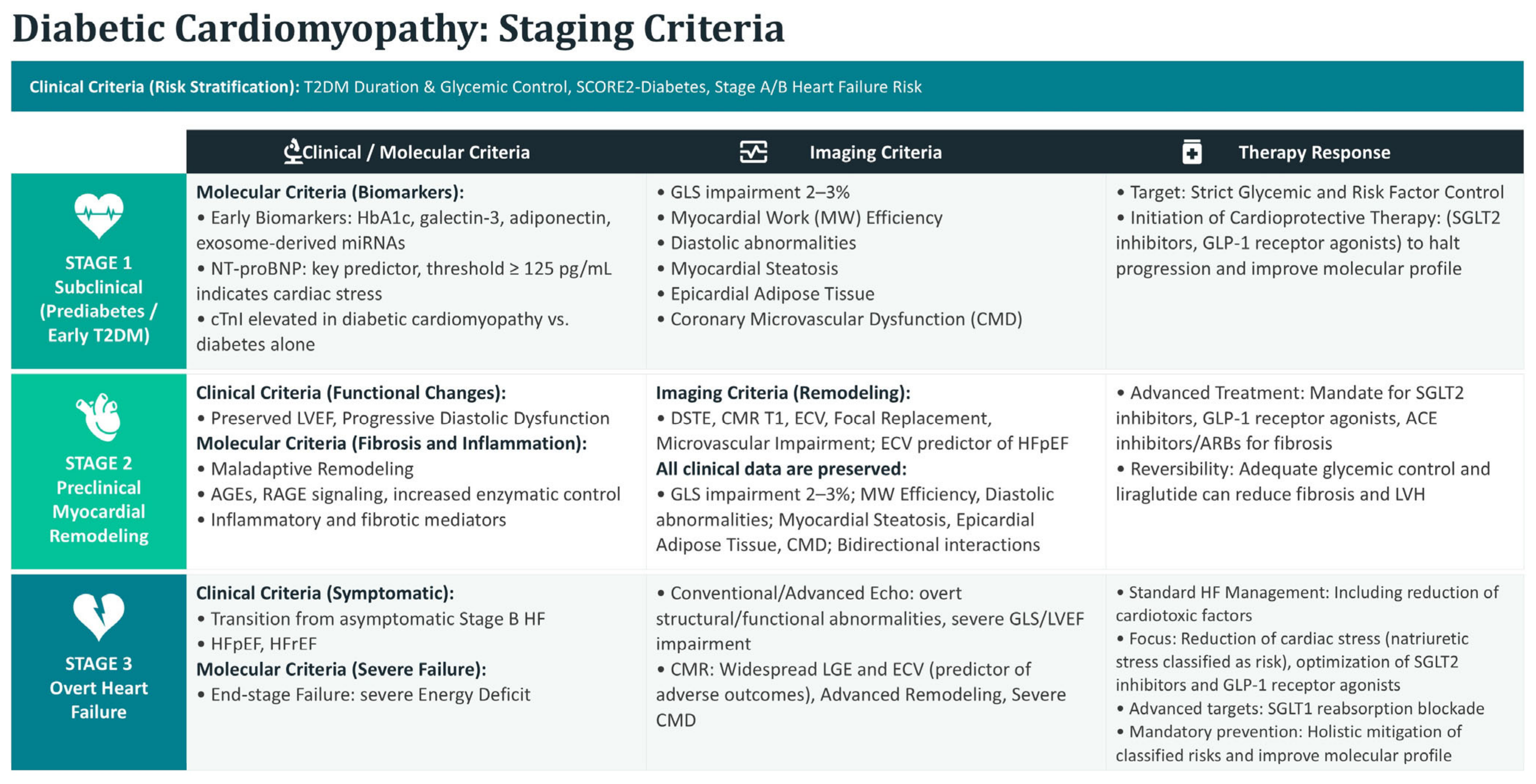
In the early stages of DCM, the left ventricular ejection fraction (LVEF) is frequently preserved, rendering overt systolic dysfunction a late-stage event. In this subclinical scenario, CMR with myocardial strain analysis (global longitudinal strain, GLS) emerges as a superior tool for the detection of incipient subendocardial impairment. Concurrently, the characterization of diastolic dysfunction finds a fundamental ally in CMR Feature Tracking (CMR-FT). The pivotal parameter, peak diastolic strain rate (PDSR), enables the early quantification of increased myocardial stiffness and prolonged isovolumetric relaxation [78]. Integrating the ventricular framework, impaired left atrial contractility emerges as a significant subclinical manifestation, capable of preceding manifest symptoms or ventricular systolic dysfunction [79]. In summary, the multiparametric approach of CMR provides an exhaustive characterization of DCM, proving indispensable for early diagnosis, risk stratification, and the tailoring of personalized therapeutic strategies. (Figure 2).

6.1. Therapeutic Implications

Hyperglycemia and insulin resistance represent the primary drivers of DCM (DCM), making targeted metabolic therapies the cornerstone of clinical management [80,81]. Glucose-lowering agents such as sodium-glucose cotransporter 2 inhibitors (SGLT2i) and glucagon-like peptide-1 receptor agonists (GLP-1RAs) were originally evaluated mainly through their renal and pancreatic physiology—blocking tubular glucose reabsorption and enhancing glucose-dependent insulin secretion, respectively [82,83]. But a purely glucose-centered view cannot explain their strong cardioprotective effects, which has prompted a reassessment of their role within the broader metabolic heart disease continuum [84].
The ongoing debate over how to classify DCM—either as an isolated clinical entity or as part of a broader cardiometabolic spectrum—has real implications for treatment. A compartmentalized view of DCM historically led to a narrow search for treatments targeting isolated, diabetes-specific myocardial shifts, such as manipulating substrate utilization or advanced glycation end-product pathways. A more modern view within the metabolic CM continuum shifts the therapeutic paradigm from protecting an isolated organ to modulating the shared pathophysiological networks systemically.
Under this broader view, the cardioprotective effects of SGLT2i and GLP-1RAs are no longer seen as merely glucose-lowering or narrowly myocardial. Their clinical efficacy instead comes from addressing the overlapping comorbidities that underlie metabolic heart disease [85]. Rather than treating DCM as a binary condition, therapeutic strategies are evolving toward phenotype-driven stratification. In this context, defining where a patient lies along the fibro-inflammatory and metabolic spectrum allows for targeted deployment of these agents to suppress systemic low-grade inflammation, mitigate microvascular endothelial dysfunction, and reverse adverse mechanical remodeling before advanced myocardial failure becomes irreversible [86].

6.2. Impact of Glucose-Lowering Therapies on Cardiovascular Outcomes

DCM treatment traditionally focused on strict glycemic targets, and optimal glycemic control has shown benefit in reducing microvascular complications. According to the UK Prospective Diabetes Study (UKPDS), a 1% reduction in HbA1c was associated with a 16% reduction in the risk of developing heart failure (HF). But other studies found that tight glycemic control in T2DM does not prevent macrovascular disease such as myocardial infarction (MI) and HF; lowering blood glucose alone is not enough to prevent DCM progression and subsequent HF. The Action to Control Cardiovascular Risk in Diabetes (ACCORD) trial found that intensive glucose lowering (targeting HbA1c < 6.0%) paradoxically increased cardiovascular mortality, and other observational studies found no reduction in HF hospitalizations with glucose-lowering therapies. A 2007 meta-analysis also linked rosiglitazone, a thiazolidinedione-class glucose-lowering drug, to a possible increased risk of MI. As a result, the FDA and the European Medicines Agency (EMA) now require cardiovascular outcome trials (CVOTs) for new antidiabetic agents. The cardiovascular benefits of newer drugs such as SGLT2i and certain GLP-1RAs, however, are independent of their glucose-lowering efficacy [82,83,87,88].

6.3. Evidence from SGLT2i

The Empagliflozin Cardiovascular Outcome Event Trial in T2DM Patients (EMPA-REG) trial showed reduced rates for three-point major adverse cardiac events (MACE, including non-fatal MI, non-fatal stroke, and cardiovascular death) in T2DM patients with a high cardiovascular risk who were assigned to empagliflozin compared to standard of care. Hospitalization for heart failure, a secondary endpoint of the trial, was also lower in patients assigned to empagliflozin. The Canagliflozin Cardiovascular Assessment Study (CANVAS) also demonstrated reduced events in patients treated with canagliflozin using three-point MACE [83,87]. The EMPagliflozin compaRative effectIveness and SafEty (EMPRISE) trial demonstrated that empagliflozin compared with sitagliptin decreased the risk of HF among T2DM patients with or without established cardiovascular disease [82]. Finally, the Dapagliflozin and Prevention of Adverse Outcomes in Heart Failure (DAPA-HF) and the Empagliflozin Outcome Trial in Patients with Chronic Heart Failure and a Reduced Ejection Fraction (EMPEROR-Reduced) trials enrolled patients with preexisting HF (LVEF < 40%) and showed fewer hospitalization rates for HF and lower cardiovascular death, both in patients with and without T2DM with either dapagliflozin or empagliflozin, respectively [18,80,83,87]. Regarding the cardioprotective effects of GLP-1ra, in the MAGNetic resonance Assessment of VICTOza efficacy in the Regression of cardiovascular dysfunction In type 2 dIAbetes mellitus (MAGNA VICTORIA) study, T2DM patients without cardiovascular disease treated with liraglutide showed an improvement in diastolic function. In contrast, the impact of Liraglutide on cardiac function and structure in Young adults with type 2 DIAbetes (LYDIA) study failed to demonstrate differences in diastolic function using MRI with treatment with either liraglutide or sitagliptin [83]. In the LEADER trial, liraglutide was shown to improve LVEF and reduce the left ventricular end-systolic volume (LVESV) by echocardiographic assessment. Furthermore, MRI revealed a decrease in myocardial fibrosis [40] Preclinical studies demonstrated that GLP-1ra effects counteract the increase in myocardial oxidative stress and cardiac myocyte apoptosis related to T2DM, which play a key role in the progression of DCM. GLP-1ra also determines an improvement in cardiac energetics through an increase in myocardial glucose oxidation, which is impaired in T2DM. In this regard, the liraglutide improvement in diastolic function is associated with increased myocardial pyruvate dehydrogenase activity. Finally, liraglutide may decrease inflammation and fibrosis, which also characterize DCM. In fact, liraglutide has been shown to reduce TNFα, interleukin, and CD163 levels [80,81,83]. Exchange protein activated by cyclic-AMP (Epac-2) is a guanine nucleotide exchange factor that plays an important role in cAMP-mediated signal transduction. GLP-1ra showed the possibility of activating Epac-2, thus increasing ANP secretion and troponin I phosphorylation, resulting in increased myocyte contractility. Treatment with GLP-1ra also showed a reduction in infarct dimensions, improved cardiac function, and left ventricular systolic function [80,87]. GLP-1ra has also beneficial actions on endothelial cells, reducing glucose-induced oxidative stress and apoptosis in endothelial cells of diabetic rats and stimulating proliferation and NO synthase activity in human endothelial cells [87]. Due to their cardioprotective effects that are independent of their glucose-lowering efficacy, GLP-1ra and SGLT2i represent important weapons against the development and progression of DCM.

7. Controversies, Future Perspectives and Conclusions

7.1. Is DCM a Distinct Entity?

DCM has traditionally been defined as ventricular dysfunction occurring in patients with diabetes in the absence of significant coronary artery disease, hypertension, or valvular heart disease—a concept first described by Rubler et al. in 1972 [14]. This definition implies that DCM represents a distinct clinical entity that is driven by diabetes-specific myocardial alterations.
A substantial body of experimental and clinical evidence supports this concept. Diabetes induces characteristic molecular and cellular changes: altered myocardial substrate utilization with a shift toward fatty acid oxidation, accumulation of advanced glycation end products (AGEs), increased oxidative stress, mitochondrial dysfunction, and activation of pro-fibrotic signaling pathways. These processes promote myocardial stiffness, interstitial fibrosis, and cardiomyocyte apoptosis, leading initially to diastolic dysfunction and, in later stages, systolic impairment [16,17,18].
However, despite this strong biological rationale, the classification of DCM as a distinct clinical entity remains controversial. A major limitation lies in its definition as a diagnosis of exclusion, which is difficult to apply in real-world clinical settings, where patients with type 2 diabetes frequently present with coexisting hypertension, obesity, and subclinical coronary artery disease. Furthermore, many of the pathophysiological mechanisms implicated in DCM are not specific to diabetes but are shared across a wide spectrum of cardiovascular diseases.
From a clinical perspective, no universally accepted diagnostic criteria exist, and the absence of specific biomarkers or imaging signatures uniquely attributable to DCM further complicates its identification. This lack of specificity challenges the utility of DCM as a standalone diagnostic category.
Imaging and biomarker studies reinforce the case for DCM as a distinct phenotype: subclinical diastolic dysfunction, increased left ventricular mass, and diffuse myocardial fibrosis have been consistently observed in asymptomatic diabetic patients without overt cardiovascular disease [89]. However, the recognition of DCM as a truly independent disease entity remains controversial. A major limitation is its definition as a diagnosis of exclusion, which is difficult to apply in real-world settings where most diabetic patients also have hypertension, obesity, or subclinical coronary artery disease [90]. Recent perspectives have therefore shifted toward a more integrative interpretation. DCM retains distinct biological features, but it is increasingly viewed as part of a broader spectrum of cardiometabolic disorders rather than a fully isolated disease entity [91]. The 2024 Heart Failure Association consensus of the European Society of Cardiology introduces the broader term “diabetic myocardial disorder,” defined as systolic and/or diastolic dysfunction in the presence of diabetes, on the basis that myocardial dysfunction in diabetes is rarely attributable to a single mechanism and is typically multifactorial [7]. Similarly, the 2022 AHA/ACC/HFSA heart failure guidelines consider diabetes to be a major contributor to heart failure development and progression, without classifying DCM as a distinct CM [92].
Taken together, current evidence suggests DCM is best understood not as a strictly independent disease but as a phenotype within a broader cardiometabolic spectrum, where diabetes is a key but not exclusive driver of myocardial dysfunction. As Table 1 summarizes, this debate reflects a tension between strong biological plausibility and limited clinical specificity—driven less by a lack of biological evidence than by the absence of clinically actionable, specific diagnostic criteria.

7.2. Overlap with Metabolic Heart Disease

The concept of metabolic heart disease has emerged to describe myocardial dysfunction driven by systemic metabolic derangements, including insulin resistance, obesity, dyslipidemia, and type 2 diabetes. Within this framework, DCM is increasingly viewed as part of a cardiometabolic continuum rather than an isolated condition.
DCM shares multiple pathophysiological mechanisms with other metabolic cardiac phenotypes, including impaired insulin signaling, altered substrate metabolism, lipotoxicity, mitochondrial dysfunction, oxidative stress, and chronic low-grade inflammation. These converge on common structural and functional changes, such as cardiomyocyte hypertrophy, interstitial fibrosis, and increased myocardial stiffness [17,95]. Similar myocardial changes are observed in obesity-related CM and metabolic syndrome, even in the absence of overt diabetes, suggesting that metabolic dysregulation per se plays a central role in myocardial remodeling [96]. Clinically, patients with type 2 diabetes rarely present with isolated hyperglycemia but rather with a cluster of metabolic abnormalities, making it difficult to disentangle the relative contribution of each factor. Moreover, the cardiovascular benefits of therapies targeting metabolic pathways, such as SGLT2 inhibitors, extend beyond glucose lowering, reinforcing the concept of a shared pathophysiological substrate [97,98].
Beyond classical metabolic alterations, omics-based studies offer deeper insight into diabetic myocardial disease. Transcriptomic and proteomic analyses reveal substantial alterations in mitochondrial metabolic networks, including disrupted fatty acid oxidation pathways and impaired mitophagy, reflecting how complex energy substrate dysregulation is in the diabetic myocardium. Growing evidence also points to immune–metabolic interactions in myocardial remodeling, with chronic low-grade inflammation, NLRP3 inflammasome activation, and NF-κB-dependent fibro-inflammatory signaling contributing to cardiomyocyte dysfunction and interstitial fibrosis [99,100]. These findings support a model in which DCM is one expression of a broader metabolic myocardial disease, with overlapping and interdependent mechanisms rather than clearly separable entities.

7.3. Relationship with HFpEF Syndrome

The relationship between DCM and heart failure with a preserved ejection fraction (HFpEF) further supports the concept of a cardiometabolic continuum.
Advanced imaging studies have demonstrated that even in early stages of diabetes, subtle impairments in myocardial relaxation and an increased extracellular volume fraction are present, suggesting a continuum from subclinical DCM to overt HFpEF [101]. Both conditions share key pathophysiological mechanisms, including microvascular dysfunction, systemic inflammation, impaired myocardial energetics, and increased ventricular stiffness. These processes promote a phenotype characterized by concentric remodeling, diastolic dysfunction, and preserved systolic function [102]. In patients with type 2 diabetes, excessive collagen deposition, abnormal protein glycosylation, lipotoxicity, and chronic pro-inflammatory signaling converge to increase left ventricular filling pressure and reduce diastolic compliance, driving the transition toward a clinical HFpEF phenotype [102].
Cardiac magnetic resonance imaging has confirmed a high burden of myocardial fibrosis, quantified by extracellular volume fraction, in patients with both type 2 diabetes and HFpEF, further supporting the pathophysiological continuum between these two conditions [103,104].
The 2024 ESC consensus on diabetic myocardial disorder reinforces this view, emphasizing that myocardial involvement in diabetes evolves along a continuum, often becoming clinically indistinguishable from HFpEF in the presence of comorbidities such as obesity and hypertension [7].
This fits the emerging view of HFpEF itself as a systemic, multisystem disorder driven by cardiometabolic stress, in which diabetes-related increases in stiffness, low-grade inflammation, and fibrosis are one contributing pathway, especially alongside obesity, hypertension, and chronic kidney disease. Clinically, this overlap matters: it points to the need for early phenotyping and targeted intervention before overt heart failure develops [105]. DCM and HFpEF are best understood not as separate conditions but as overlapping phenotypes within a shared cardiometabolic continuum, with DCM potentially representing an early or diabetes-predominant manifestation of HFpEF. This integrated view supports early phenotyping, risk stratification, and timely intervention in diabetic patients to prevent or delay progression to overt heart failure and to improve long-term cardiovascular outcomes (Table 2).

7.4. Role of Multimodal Imaging

In line with the evolving view of DCM as part of a broader cardiometabolic continuum and its close relationship with HFpEF, multimodal imaging has become essential for the early detection, phenotyping, and longitudinal assessment of myocardial involvement.
Subclinical cardiac alterations often precede overt heart failure, as first highlighted by the Framingham Study [43,106,107], which underscores the value of combining advanced imaging with circulating biomarkers for timely diagnosis and risk stratification [44]. Echocardiography remains the first-line modality; however, conventional parameters such as left ventricular ejection fraction (LVEF) lack sensitivity in early disease stages. Advanced echocardiographic techniques, particularly speckle-tracking echocardiography (STE), enable the detection of subclinical myocardial dysfunction through global longitudinal strain (GLS), which is consistently reduced in asymptomatic diabetic patients despite preserved LVEF [46]. GLS impairment reflects early subendocardial damage and correlates with glycemic control and disease duration [53,54]. Combining strain analysis with pressure-strain loops also allows estimation of myocardial work, which can reveal early systolic inefficiency, even without overt dysfunction [55]. Meta-analyses have consistently demonstrated that global longitudinal strain is reduced in all myocardial directions in patients with subclinical DCM despite a preserved ejection fraction, confirming the superior sensitivity of speckle-tracking echocardiography over conventional parameters for early detection [53,108].
Cardiac magnetic resonance (CMR) represents the gold standard for non-invasive myocardial tissue characterization and advanced phenotyping. Its multiparametric approach enables the identification of key pathological features of DCM, including diffuse and focal fibrosis, microvascular dysfunction, and myocardial steatosis [7]. Late gadolinium enhancement (LGE) detects focal replacement fibrosis and predicts adverse cardiovascular outcomes, while T1 mapping and extracellular volume (ECV) quantification provide sensitive measures of diffuse interstitial fibrosis, which is strongly associated with heart failure development and mortality [71]. Emerging techniques such as T1ρ mapping further enhance fibrosis detection without contrast administration [72]. T1 mapping and extracellular volume fraction quantification enable non-invasive detection of diffuse interstitial fibrosis and myocardial steatosis in asymptomatic diabetic patients, providing tissue-level characterization beyond what is achievable with echocardiography alone [74,101]. CMR also enables the evaluation of coronary microvascular dysfunction through stress perfusion imaging and myocardial flow reserve quantification, revealing impaired microvascular vasodilatory capacity in diabetic patients and its association with fibrotic remodeling [73]. In addition, proton spectroscopy allows quantification of myocardial lipid accumulation, while a volumetric assessment of epicardial adipose tissue provides insight into cardiometabolic risk, both of which are closely linked to myocardial dysfunction and structural remodeling [75,77]. Finally, strain analysis by CMR and feature tracking techniques enable the detection of subtle systolic and diastolic abnormalities, including an impaired peak diastolic strain rate, which reflects increased myocardial stiffness and early diastolic dysfunction [78]. These alterations often precede symptomatic disease and are complemented by early changes in left atrial function, further supporting the concept of a progressive cardiometabolic remodeling process [79].
A key clinical challenge when evaluating patients with suspected DCM, particularly with left ventricular hypertrophy or a HFpEF phenotype, is ruling out alternative causes of myocardial disease. Differential diagnosis should include infiltrative and storage disorders such as cardiac amyloidosis, Fabry disease, and hemochromatosis, along with hypertensive heart disease and ischemic cardiomyopathy. Multimodal imaging is central here: cardiac magnetic resonance and nuclear imaging techniques allow tissue characterization and identification of disease-specific patterns. Recognizing these conditions matters, since they may call for disease-specific therapies and carry distinct prognostic implications. (Table 3).

8. Conclusions

CM is a well-supported pathophysiological construct, backed by extensive experimental and clinical evidence. Its classification as a distinct clinical entity, however, is limited by the lack of specific diagnostic criteria and by significant overlap with other forms of metabolic heart disease.
Current evidence favors moving from a binary definition to a continuum-based model, in which DCM sits within a broader spectrum of cardiometabolic myocardial dysfunction, closely linked to HFpEF and other metabolic cardiac phenotypes.
We therefore propose viewing DCM as a diabetes-enriched phenotype within the broader spectrum of cardiometabolic heart disease. In this model, diabetes acts as a major amplifier of metabolic, inflammatory, vascular, and fibro-inflammatory pathways shared with HFpEF and other metabolic cardiomyopathies, while also contributing disease-specific mechanisms such as chronic hyperglycemia and AGE accumulation.
A priority for future research is a unifying framework that integrates molecular mechanisms, myocardial remodeling, and clinical phenotypes into a coherent model of cardiometabolic heart disease—one that could redefine DCM not as a binary diagnostic category but as a spectrum condition shaped by varying contributions of metabolic, inflammatory, and hemodynamic factors.
Resolving this conceptual ambiguity has direct clinical stakes. A clearer definition of DCM could improve diagnostic accuracy, help identify at-risk patients earlier, and sharpen therapeutic targeting; distinguishing between predominant phenotypes within the cardiometabolic spectrum, in particular, could guide personalized treatment.
Moving from a descriptive to a mechanistic, phenotype-driven classification of diabetic myocardial disease is a necessary step toward better outcomes for patients with diabetes.
The challenge is no longer to demonstrate the existence of DCM, but to define its precise role within the evolving framework of cardiometabolic heart disease.

Author Contributions

Conceptualization, G.C., F.N. and S.D.; methodology, P.G., G.C. and S.D.; resources, E.L., A.S., C.S., R.F., M.M., G.T., G.M., A.B. and F.L.; data curation, A.S., C.S., R.F., M.M., G.T., G.M., A.B. and F.L.; writing—original draft preparation, S.D., E.L., A.S., R.F., C.S., M.M., G.T., G.M., A.B. and F.L.; writing—review and editing, P.G., F.N. and G.C.; supervision, P.G. All authors have read and agreed to the published version of the manuscript.

Funding

This research received no external funding.

Institutional Review Board Statement

Not applicable.

Informed Consent Statement

Not applicable.

Data Availability Statement

No new data were created or analyzed in this study. Data sharing is not applicable to this article.

Acknowledgments

During the preparation of this manuscript, the authors used Gemini (Gemini 3.6 Flash version, Google) for the purposes of translation and language editing. The authors have reviewed and edited the output and take full responsibility for the content of this publication.

Conflicts of Interest

The authors declare no conflicts of interest.

References

  1. Genitsaridi, I.; Salpea, P.; Salim, A.; Sajjadi, S.F.; Tomic, D.; James, S.; Thirunavukkarasu, S.; Issaka, A.; Chen, L.; Basit, A.; et al. 11th edition of the IDF Diabetes Atlas: Global, regional, and national diabetes prevalence estimates for 2024 and projections for 2050. Lancet Diabetes Endocrinol. 2026, 14, 149–156. [Google Scholar] [CrossRef] [Scilit] [PubMed]
  2. NCD Risk Factor Collaboration. Worldwide trends in diabetes prevalence and treatment from 1990 to 2022: A pooled analysis of 1108 population-representative studies with 141 million participants. Lancet 2024, 404, 2077–2093. [Google Scholar] [CrossRef] [Scilit] [PubMed]
  3. Zheng, Y.; Ley, S.H.; Hu, F.B. Global aetiology and epidemiology of type 2 diabetes mellitus and its complications. Nat. Rev. Endocrinol. 2018, 14, 88–98. [Google Scholar] [CrossRef] [Scilit]
  4. Marx, N.; Federici, M.; Schutt, K.; Muller-Wieland, D.; Ajjan, R.A.; Antunes, M.J.; Christodorescu, R.M.; Crawford, C.; Di Angelantonio, E.; Eliasson, B.; et al. 2023 ESC Guidelines for the management of cardiovascular disease in patients with diabetes. Eur. Heart J. 2023, 44, 4043–4140. [Google Scholar] [CrossRef] [Scilit] [PubMed]
  5. Siam, N.H.; Snigdha, N.N.; Tabasumma, N.; Parvin, I. Diabetes Mellitus and Cardiovascular Disease: Exploring Epidemiology, Pathophysiology, and Treatment Strategies. Rev. Cardiovasc. Med. 2024, 25, 436. [Google Scholar] [CrossRef] [Scilit] [PubMed]
  6. Giraldo-Gonzalez, G.C.; Roman-Gonzalez, A.; Canas, F.; Garcia, A. Molecular Mechanisms of Type 2 Diabetes-Related Heart Disease and Therapeutic Insights. Int. J. Mol. Sci. 2025, 26, 4548. [Google Scholar] [CrossRef] [Scilit] [PubMed]
  7. Seferovic, P.M.; Paulus, W.J.; Rosano, G.; Polovina, M.; Petrie, M.C.; Jhund, P.S.; Tschope, C.; Sattar, N.; Piepoli, M.; Papp, Z.; et al. Diabetic myocardial disorder. A clinical consensus statement of the Heart Failure Association of the ESC and the ESC Working Group on Myocardial & Pericardial Diseases. Eur. J. Heart Fail. 2024, 26, 1893–1903. [Google Scholar] [CrossRef] [Scilit] [PubMed]
  8. Jerkins, T.; McGill, J.B.; Bell, D.S.H. Heart failure and diabetes: Clinical significance and epidemiology of this two-way association. Diabetes Obes. Metab. 2023, 25, 3–14. [Google Scholar] [CrossRef] [Scilit] [PubMed]
  9. Panchal, K.; Lawson, C.; Chandramouli, C.; Lam, C.; Khunti, K.; Zaccardi, F. Diabetes and risk of heart failure in people with and without cardiovascular disease: Systematic review and meta-analysis. Diabetes Res. Clin. Pract. 2024, 207, 111054. [Google Scholar] [CrossRef] [Scilit] [PubMed]
  10. Hoek, A.G.; Dal Canto, E.; Wenker, E.; Bindraban, N.; Handoko, M.L.; Elders, P.J.M.; Beulens, J.W.J. Epidemiology of heart failure in diabetes: A disease in disguise. Diabetologia 2024, 67, 574–601. [Google Scholar] [CrossRef] [Scilit] [PubMed]
  11. Theofilis, P.; Oikonomou, E.; Tsioufis, K.; Tousoulis, D. Diabetes Mellitus and Heart Failure: Epidemiology, Pathophysiologic Mechanisms, and the Role of SGLT2 Inhibitors. Life 2023, 13, 497. [Google Scholar] [CrossRef] [Scilit] [PubMed]
  12. Landolfo, M.; Spannella, F.; Giulietti, F.; Ortensi, B.; Stella, L.; Carlucci, M.A.; Galeazzi, R.; Turchi, F.; Luconi, M.P.; Zampa, R.; et al. Detecting heart stress using NT-proBNP in patients with type 2 diabetes mellitus and hypertension or high-normal blood pressure: A cross-sectional multicentric study. Cardiovasc. Diabetol. 2024, 23, 297. [Google Scholar] [CrossRef] [Scilit] [PubMed]
  13. Gastouniotis, I.; Fragoulis, C.; Antonopoulos, A.; Kouroutzoglou, A.; Noutsou, M.; Thanopoulou, A.; Chrysohoou, C.; Tsioufis, K.P. The Implication of NT-proBNP in the Assessment of the Clinical Phenotype of Patients with Type 2 Diabetes Mellitus, Without Established Cardiovascular Disease. Biomedicines 2024, 12, 2718. [Google Scholar] [CrossRef] [Scilit] [PubMed]
  14. Rubler, S.; Dlugash, J.; Yuceoglu, Y.Z.; Kumral, T.; Branwood, A.W.; Grishman, A. New type of cardiomyopathy associated with diabetic glomerulosclerosis. Am. J. Cardiol. 1972, 30, 595–602. [Google Scholar] [CrossRef] [Scilit] [PubMed]
  15. Grubic Rotkvic, P.; Planinic, Z.; Liberati Prso, A.M.; Sikic, J.; Galic, E.; Rotkvic, L. The Mystery of Diabetic Cardiomyopathy: From Early Concepts and Underlying Mechanisms to Novel Therapeutic Possibilities. Int. J. Mol. Sci. 2021, 22, 5973. [Google Scholar] [CrossRef] [Scilit] [PubMed]
  16. Marfella, R.; Sardu, C.; Mansueto, G.; Napoli, C.; Paolisso, G. Evidence for human diabetic cardiomyopathy. Acta Diabetol. 2021, 58, 983–988. [Google Scholar] [CrossRef] [Scilit] [PubMed]
  17. Jia, G.; Hill, M.A.; Sowers, J.R. Diabetic Cardiomyopathy: An Update of Mechanisms Contributing to This Clinical Entity. Circ. Res. 2018, 122, 624–638. [Google Scholar] [CrossRef] [Scilit] [PubMed]
  18. Tan, Y.; Zhang, Z.; Zheng, C.; Wintergerst, K.A.; Keller, B.B.; Cai, L. Mechanisms of diabetic cardiomyopathy and potential therapeutic strategies: Preclinical and clinical evidence. Nat. Rev. Cardiol. 2020, 17, 585–607. [Google Scholar] [CrossRef] [Scilit] [PubMed]
  19. Dillmann, W.H. Diabetic Cardiomyopathy. Circ. Res. 2019, 124, 1160–1162. [Google Scholar] [CrossRef] [Scilit] [PubMed]
  20. Abel, E.D.; O’Shea, K.M.; Ramasamy, R. Insulin resistance: Metabolic mechanisms and consequences in the heart. Arterioscler. Thromb. Vasc. Biol. 2012, 32, 2068–2076. [Google Scholar] [CrossRef] [Scilit] [PubMed]
  21. Stanley, W.C.; Recchia, F.A.; Lopaschuk, G.D. Myocardial substrate metabolism in the normal and failing heart. Physiol. Rev. 2005, 85, 1093–1129. [Google Scholar] [CrossRef] [Scilit] [PubMed]
  22. Unger, R.H.; Scherer, P.E. Gluttony, sloth and the metabolic syndrome: A roadmap to lipotoxicity. Trends Endocrinol. Metab. 2010, 21, 345–352. [Google Scholar] [CrossRef] [Scilit] [PubMed]
  23. Bugger, H.; Abel, E.D. Molecular mechanisms of diabetic cardiomyopathy. Diabetologia 2014, 57, 660–671. [Google Scholar] [CrossRef] [Scilit] [PubMed]
  24. Westermann, D.; Lindner, D.; Kasner, M.; Zietsch, C.; Savvatis, K.; Escher, F.; von Schlippenbach, J.; Skurk, C.; Steendijk, P.; Riad, A.; et al. Cardiac inflammation contributes to changes in the extracellular matrix in patients with heart failure and normal ejection fraction. Circ. Heart Fail. 2011, 4, 44–52. [Google Scholar] [CrossRef] [Scilit] [PubMed]
  25. Singh, V.P.; Bali, A.; Singh, N.; Jaggi, A.S. Advanced glycation end products and diabetic complications. Korean J. Physiol. Pharmacol. 2014, 18, 1–14. [Google Scholar] [CrossRef] [Scilit] [PubMed]
  26. Ramasamy, R.; Yan, S.F.; Schmidt, A.M. Advanced glycation endproducts: From precursors to RAGE: Round and round we go. Amino Acids 2012, 42, 1151–1161. [Google Scholar] [CrossRef] [Scilit] [PubMed]
  27. Goldin, A.; Beckman, J.A.; Schmidt, A.M.; Creager, M.A. Advanced glycation end products: Sparking the development of diabetic vascular injury. Circulation 2006, 114, 597–605. [Google Scholar] [CrossRef] [Scilit] [PubMed]
  28. Kass, D.A.; Shapiro, E.P.; Kawaguchi, M.; Capriotti, A.R.; Scuteri, A.; deGroof, R.C.; Lakatta, E.G. Improved arterial compliance by a novel advanced glycation end-product crosslink breaker. Circulation 2001, 104, 1464–1470. [Google Scholar] [CrossRef] [Scilit] [PubMed]
  29. Asbun, J.; Villarreal, F.J. The pathogenesis of myocardial fibrosis in the setting of diabetic cardiomyopathy. J. Am. Coll. Cardiol. 2006, 47, 693–700. [Google Scholar] [CrossRef] [Scilit] [PubMed]
  30. Forbes, J.M.; Cooper, M.E. Mechanisms of diabetic complications. Physiol. Rev. 2013, 93, 137–188. [Google Scholar] [CrossRef] [Scilit] [PubMed]
  31. Russo, I.; Frangogiannis, N.G. Diabetes-associated cardiac fibrosis: Cellular effectors, molecular mechanisms and therapeutic opportunities. J. Mol. Cell. Cardiol. 2016, 90, 84–93. [Google Scholar] [CrossRef] [Scilit] [PubMed]
  32. Brownlee, M. The pathobiology of diabetic complications: A unifying mechanism. Diabetes 2005, 54, 1615–1625. [Google Scholar] [CrossRef] [Scilit] [PubMed]
  33. Taqueti, V.R.; Di Carli, M.F. Coronary Microvascular Disease Pathogenic Mechanisms and Therapeutic Options: JACC State-of-the-Art Review. J. Am. Coll. Cardiol. 2018, 72, 2625–2641. [Google Scholar] [CrossRef] [Scilit] [PubMed]
  34. Murthy, V.L.; Naya, M.; Foster, C.R.; Gaber, M.; Hainer, J.; Klein, J.; Dorbala, S.; Blankstein, R.; Di Carli, M.F. Association between coronary vascular dysfunction and cardiac mortality in patients with and without diabetes mellitus. Circulation 2012, 126, 1858–1868. [Google Scholar] [CrossRef] [Scilit] [PubMed]
  35. Neubauer, S. The failing heart—An engine out of fuel. N. Engl. J. Med. 2007, 356, 1140–1151. [Google Scholar] [CrossRef] [Scilit] [PubMed]
  36. Lopaschuk, G.D.; Karwi, Q.G.; Tian, R.; Wende, A.R.; Abel, E.D. Cardiac Energy Metabolism in Heart Failure. Circ. Res. 2021, 128, 1487–1513. [Google Scholar] [CrossRef] [Scilit] [PubMed]
  37. Rosca, M.G.; Hoppel, C.L. Mitochondrial dysfunction in heart failure. Heart Fail. Rev. 2013, 18, 607–622. [Google Scholar] [CrossRef] [Scilit] [PubMed]
  38. Oktay, A.A.; Paul, T.K.; Koch, C.A.; Lavie, C.J. Diabetes, Cardiomyopathy, and Heart Failure. In Endotext; MDText.com, Inc.: South Dartmouth, MA, USA, 2023. [Google Scholar]
  39. Marwick, T.H.; Lam, C.; Liu, Y.; Del Prato, S.; Rosenstock, J.; Butler, J.; Ezekowitz, J.; Ibrahim, N.E.; Tang, W.H.W.; Zannad, F.; et al. Echocardiographic phenotypes of diabetic myocardial disorder: Evolution over 15 months follow-up in the ARISE-HF trial. Cardiovasc. Diabetol. 2025, 24, 16. [Google Scholar] [CrossRef] [Scilit] [PubMed]
  40. Rizza, V.; Tondi, L.; Patti, A.M.; Cecchi, D.; Lombardi, M.; Perone, F.; Ambrosetti, M.; Rizzo, M.; Cianflone, D.; Maranta, F. Diabetic cardiomyopathy: Pathophysiology, imaging assessment and therapeutical strategies. Int. J. Cardiol. Cardiovasc. Risk Prev. 2024, 23, 200338. [Google Scholar] [CrossRef] [Scilit] [PubMed]
  41. Dandamudi, S.; Slusser, J.; Mahoney, D.W.; Redfield, M.M.; Rodeheffer, R.J.; Chen, H.H. The prevalence of diabetic cardiomyopathy: A population-based study in Olmsted County, Minnesota. J. Card. Fail. 2014, 20, 304–309. [Google Scholar] [CrossRef] [Scilit] [PubMed]
  42. Pandey, A.; Vaduganathan, M.; Patel, K.V.; Ayers, C.; Ballantyne, C.M.; Kosiborod, M.N.; Carnethon, M.; Defilippi, C.; McGuire, D.K.; Khan, S.S.; et al. Biomarker-Based Risk Prediction of Incident Heart Failure in Pre-Diabetes and Diabetes. JACC Heart Fail. 2021, 9, 215–225. [Google Scholar] [CrossRef] [Scilit] [PubMed]
  43. Kannel, W.B.; McGee, D.L. Diabetes and cardiovascular disease. The Framingham study. JAMA 1979, 241, 2035–2038. [Google Scholar] [CrossRef] [Scilit] [PubMed]
  44. Adel, F.W.; Chen, H.H. The role of multimodality imaging in diabetic cardiomyopathy: A brief review. Front. Endocrinol. 2024, 15, 1405031. [Google Scholar] [CrossRef] [Scilit] [PubMed]
  45. Malachias, M.V.B.; Wijkman, M.O.; Bertoluci, M.C. NT-proBNP as a predictor of death and cardiovascular events in patients with type 2 diabetes. Diabetol. Metab. Syndr. 2022, 14, 64. [Google Scholar] [CrossRef] [Scilit] [PubMed]
  46. Urlic, H.; Kumric, M.; Vrdoljak, J.; Martinovic, D.; Dujic, G.; Vilovic, M.; Ticinovic Kurir, T.; Bozic, J. Role of Echocardiography in Diabetic Cardiomyopathy: From Mechanisms to Clinical Practice. J. Cardiovasc. Dev. Dis. 2023, 10, 46. [Google Scholar] [CrossRef] [Scilit] [PubMed]
  47. Burhanuddin, M.; Ahmed, Z.; Hamza, A.; Zeeshan, M.; Abdul Rehman, F.; Gillani, S.H.M.; Gul, A.; Rabbani, Z.; Omonfuegbe, O.M.; Ali, S.M. Early Detection of Diabetic Cardiomyopathy Using Speckle Tracking Echocardiography: A Systematic Review. Cureus 2025, 17, e92653. [Google Scholar] [CrossRef] [Scilit] [PubMed]
  48. Pitasari, J.; Kent, M.; Kundu, S.; Rabbani, B. Restrictive Filling Pattern in Diastolic Dysfunction: Differentiating Hypertensive Heart Disease from Restrictive Cardiomyopathy Using Transthoracic Echocardiogram. Hypertension 2025, 82. [Google Scholar] [CrossRef] [Scilit]
  49. Leitman, M.; Tyomkin, V.; Fuchs, S. Longitudinal Displacement vs. Strain in Cardiac Amyloidosis: A Speckle Tracking Echocardiography Study. J. Clin. Med. 2026, 15, 1544. [Google Scholar] [CrossRef] [Scilit] [PubMed]
  50. Lee, J.H.; Park, J.H. Clinical Applications of Speckle-Tracking Echocardiography in Heart Failure: From Diagnosis to Prognostication. Int. J. Heart Fail. 2025, 7, 201–215. [Google Scholar] [CrossRef] [Scilit] [PubMed]
  51. Ahmed Khair, L.; Ali, S.; Alansari, M. Usefulness of Two-Dimensional Speckle-Tracking Echocardiography in Detecting Subclinical Left Ventricular Systolic Dysfunction in Diabetes Mellitus. Cureus 2025, 17, e85682. [Google Scholar] [CrossRef] [Scilit] [PubMed]
  52. Nghiem, T.P.; Tran, H.M.; Nguyen, D.N.Q.; Dao, L.T.; Tran, C.C.; Vo, T.M. Subclinical Myocardial Dysfunction in Type 2 Diabetes Mellitus: Insights from Left Ventricular Diastolic Function and Global Longitudinal Strain Assessment. Med. Sci. 2025, 13, 237. [Google Scholar] [CrossRef] [Scilit] [PubMed]
  53. Ghoreyshi-Hefzabad, S.M.; Jeyaprakash, P.; Gupta, A.; Vo, H.Q.; Pathan, F.; Negishi, K. Three-Dimensional Global Left Ventricular Myocardial Strain Reduced in All Directions in Subclinical Diabetic Cardiomyopathy: A Systematic Review and Meta-Analysis. J. Am. Heart Assoc. 2021, 10, e020811. [Google Scholar] [CrossRef] [Scilit] [PubMed]
  54. Gao, J.; Xu, M.; Gong, M.; Jiang, S.; Yang, Z.; Jiang, X.; Chen, M. Left ventricular longitudinal strain in patients with type 2 diabetes mellitus is independently associated with glycated hemoglobin level. Clin. Cardiol. 2023, 46, 1578–1587. [Google Scholar] [CrossRef] [Scilit] [PubMed]
  55. Liao, L.; Shi, B.; Ding, Z.; Chen, L.; Dong, F.; Li, J.; Zhong, Y.; Xu, J. Echocardiographic study of myocardial work in patients with type 2 diabetes mellitus. BMC Cardiovasc. Disord. 2022, 22, 59. [Google Scholar] [CrossRef] [Scilit] [PubMed]
  56. Huo, J.L.; Feng, Q.; Pan, S.; Fu, W.J.; Liu, Z.; Liu, Z. Diabetic cardiomyopathy: Early diagnostic biomarkers, pathogenetic mechanisms, and therapeutic interventions. Cell Death Discov. 2023, 9, 256. [Google Scholar] [CrossRef] [Scilit] [PubMed]
  57. Gao, S.; Dong, Y.; Yan, C.; Yu, T.; Cao, H. The role of exosomes and exosomal microRNA in diabetic cardiomyopathy. Front. Endocrinol. 2023, 14, 1327495. [Google Scholar] [CrossRef] [Scilit] [PubMed]
  58. Ianos, R.D.; Cozma, A.; Lucaciu, R.L.; Hangan, A.C.; Negrean, V.; Mercea, D.C.; Ciulei, G.; Pop, C.; Procopciuc, L.M. Role of Circulating Biomarkers in Diabetic Cardiomyopathy. Biomedicines 2024, 12, 2153. [Google Scholar] [CrossRef] [Scilit] [PubMed]
  59. Guo, R.; Nair, S. Role of microRNA in diabetic cardiomyopathy: From mechanism to intervention. Biochim. Biophys. Acta Mol. Basis Dis. 2017, 1863, 2070–2077. [Google Scholar] [CrossRef] [Scilit] [PubMed]
  60. Dong, S.; Ma, W.; Hao, B.; Hu, F.; Yan, L.; Yan, X.; Wang, Y.; Chen, Z.; Wang, Z. microRNA-21 promotes cardiac fibrosis and development of heart failure with preserved left ventricular ejection fraction by up-regulating Bcl-2. Int. J. Clin. Exp. Pathol. 2014, 7, 565–574. [Google Scholar] [PubMed]
  61. Costantino, S.; Paneni, F.; Luscher, T.F.; Cosentino, F. MicroRNA profiling unveils hyperglycaemic memory in the diabetic heart. Eur. Heart J. 2016, 37, 572–576. [Google Scholar] [CrossRef] [Scilit] [PubMed]
  62. Monnier, L.; Mas, E.; Ginet, C.; Michel, F.; Villon, L.; Cristol, J.P.; Colette, C. Activation of oxidative stress by acute glucose fluctuations compared with sustained chronic hyperglycemia in patients with type 2 diabetes. JAMA 2006, 295, 1681–1687. [Google Scholar] [CrossRef] [Scilit] [PubMed]
  63. Frontoni, S.; Di Bartolo, P.; Avogaro, A.; Bosi, E.; Paolisso, G.; Ceriello, A. Glucose variability: An emerging target for the treatment of diabetes mellitus. Diabetes Res. Clin. Pract. 2013, 102, 86–95. [Google Scholar] [CrossRef] [Scilit] [PubMed]
  64. Zhou, Z.; Sun, B.; Huang, S.; Zhu, C.; Bian, M. Glycemic variability: Adverse clinical outcomes and how to improve it? Cardiovasc. Diabetol. 2020, 19, 102. [Google Scholar] [CrossRef] [Scilit] [PubMed]
  65. Huang, L.; Pan, Y.; Zhou, K.; Liu, H.; Zhong, S. Correlation Between Glycemic Variability and Diabetic Complications: A Narrative Review. Int. J. Gen. Med. 2023, 16, 3083–3094. [Google Scholar] [CrossRef] [Scilit] [PubMed]
  66. McDonagh, T.A.; Metra, M.; Adamo, M.; Gardner, R.S.; Baumbach, A.; Bohm, M.; Burri, H.; Butler, J.; Celutkiene, J.; Chioncel, O.; et al. 2021 ESC Guidelines for the diagnosis and treatment of acute and chronic heart failure. Eur. Heart J. 2021, 42, 3599–3726. [Google Scholar] [CrossRef] [Scilit] [PubMed]
  67. Clodi, M.; Resl, M.; Neuhold, S.; Hulsmann, M.; Vila, G.; Elhenicky, M.; Strunk, G.; Abrahamian, H.; Prager, R.; Luger, A.; et al. A comparison of NT-proBNP and albuminuria for predicting cardiac events in patients with diabetes mellitus. Eur. J. Prev. Cardiol. 2012, 19, 944–951. [Google Scholar] [CrossRef] [Scilit] [PubMed]
  68. Pop-Busui, R.; Repetto, E.; Baron, J.; Schumacher, D.; Vaduganathan, M.; Pandey, A. Screening Natriuretic Peptide Levels Predicts Heart Failure and Death in Individuals With Type 1 and Type 2 Diabetes Without Known Heart Failure. Diabetes Care 2025, 48, 2145–2153. [Google Scholar] [CrossRef] [Scilit] [PubMed]
  69. Salvador, D.B., Jr.; Gamba, M.R.; Gonzalez-Jaramillo, N.; Gonzalez-Jaramillo, V.; Raguindin, P.F.N.; Minder, B.; Grani, C.; Wilhelm, M.; Stettler, C.; Doria, A.; et al. Diabetes and Myocardial Fibrosis: A Systematic Review and Meta-Analysis. JACC Cardiovasc. Imaging 2022, 15, 796–808. [Google Scholar] [CrossRef] [Scilit] [PubMed]
  70. Elliott, M.D.; Heitner, J.F.; Kim, H.; Wu, E.; Parker, M.A.; Lee, D.C.; Kaufman, D.B.; Bonow, R.O.; Judd, R.; Kim, R.J. Prevalence and Prognosis of Unrecognized Myocardial Infarction in Asymptomatic Patients With Diabetes: A Two-Center Study With Up to 5 Years of Follow-up. Diabetes Care 2019, 42, 1290–1296. [Google Scholar] [CrossRef] [Scilit] [PubMed]
  71. Wong, T.C.; Piehler, K.M.; Kang, I.A.; Kadakkal, A.; Kellman, P.; Schwartzman, D.S.; Mulukutla, S.R.; Simon, M.A.; Shroff, S.G.; Kuller, L.H.; et al. Myocardial extracellular volume fraction quantified by cardiovascular magnetic resonance is increased in diabetes and associated with mortality and incident heart failure admission. Eur. Heart J. 2014, 35, 657–664. [Google Scholar] [CrossRef] [Scilit] [PubMed]
  72. Bustin, A.; Witschey, W.R.T.; van Heeswijk, R.B.; Cochet, H.; Stuber, M. Magnetic resonance myocardial T1rho mapping: Technical overview, challenges, emerging developments, and clinical applications. J. Cardiovasc. Magn. Reson. 2023, 25, 34. [Google Scholar] [CrossRef] [Scilit] [PubMed]
  73. Zhao, W.; Li, K.; Tang, L.; Zhang, J.; Guo, H.; Zhou, X.; Luo, M.; Liu, H.; Cui, R.; Zeng, M. Coronary Microvascular Dysfunction and Diffuse Myocardial Fibrosis in Patients With Type 2 Diabetes Using Quantitative Perfusion MRI. J. Magn. Reson. Imaging 2024, 60, 2395–2406. [Google Scholar] [CrossRef] [Scilit] [PubMed]
  74. Huang, S.; Li, X.; Zhang, L.; Sun, R.; Liu, H.; Zhang, X.; Shi, K.; Jiang, L.; Lamb, H.J.; Yang, Z.; et al. Cardiac MRI in diabetic cardiomyopathy: Translating imaging biomarkers into clinical practice. Cardiovasc. Diabetol. 2025, 25, 4. [Google Scholar] [CrossRef] [Scilit] [PubMed]
  75. Ng, A.C.T.; Strudwick, M.; van der Geest, R.J.; Ng, A.C.C.; Gillinder, L.; Goo, S.Y.; Cowin, G.; Delgado, V.; Wang, W.Y.S.; Bax, J.J. Impact of Epicardial Adipose Tissue, Left Ventricular Myocardial Fat Content, and Interstitial Fibrosis on Myocardial Contractile Function. Circ. Cardiovasc. Imaging 2018, 11, e007372. [Google Scholar] [CrossRef] [Scilit] [PubMed]
  76. D’Elia, S.; Luisi, E.; Solimene, A.; Serpico, C.; Morello, M.; Titolo, G.; Caso, V.M.; Loffredo, F.S.; Golino, P.; Cimmino, G.; et al. Targeting Obesity in Cardiovascular Disease Management: Cardiac Adipose Tissue Is a Real Biomarker! Targets 2025, 3, 17. [Google Scholar] [CrossRef] [Scilit]
  77. Li, Y.; Liu, B.; Li, Y.; Jing, X.; Deng, S.; Yan, Y.; She, Q. Epicardial fat tissue in patients with diabetes mellitus: A systematic review and meta-analysis. Cardiovasc. Diabetol. 2019, 18, 3. [Google Scholar] [CrossRef] [Scilit] [PubMed]
  78. Kasner, M.; Gaub, R.; Sinning, D.; Westermann, D.; Steendijk, P.; Hoffmann, W.; Schultheiss, H.P.; Tschope, C. Global strain rate imaging for the estimation of diastolic function in HFNEF compared with pressure-volume loop analysis. Eur. J. Echocardiogr. 2010, 11, 743–751. [Google Scholar] [CrossRef] [Scilit] [PubMed][Green Version]
  79. Thomas, L.; Marwick, T.H.; Popescu, B.A.; Donal, E.; Badano, L.P. Left Atrial Structure and Function, and Left Ventricular Diastolic Dysfunction: JACC State-of-the-Art Review. J. Am. Coll. Cardiol. 2019, 73, 1961–1977. [Google Scholar] [CrossRef] [Scilit] [PubMed]
  80. Ghosh, N.; Chacko, L.; Bhattacharya, H.; Vallamkondu, J.; Nag, S.; Dey, A.; Karmakar, T.; Reddy, P.H.; Kandimalla, R.; Dewanjee, S. Exploring the Complex Relationship between Diabetes and Cardiovascular Complications: Understanding Diabetic Cardiomyopathy and Promising Therapies. Biomedicines 2023, 11, 1126. [Google Scholar] [CrossRef] [Scilit] [PubMed]
  81. Balakrishnan, B.; Somashekara, D.M.; Nayak, V.; Hariharapura, R.C. Diabetic cardiomyopathy: A comprehensive review of diagnosis, management, and future directions. Diabetol. Metab. Syndr. 2025, 17, 422. [Google Scholar] [CrossRef] [Scilit] [PubMed]
  82. Li, N.; Zhou, H. SGLT2 Inhibitors: A Novel Player in the Treatment and Prevention of Diabetic Cardiomyopathy. Drug Des. Dev. Ther. 2020, 14, 4775–4788. [Google Scholar] [CrossRef] [Scilit] [PubMed]
  83. Gopal, K.; Chahade, J.J.; Kim, R.; Ussher, J.R. The Impact of Antidiabetic Therapies on Diastolic Dysfunction and Diabetic Cardiomyopathy. Front. Physiol. 2020, 11, 603247. [Google Scholar] [CrossRef] [Scilit] [PubMed]
  84. Gorica, E.; Geiger, M.A.; Di Venanzio, L.; Atzemian, N.; Kleeberger, J.A.; Grigorian, D.; Mongelli, A.; Emini Veseli, B.; Mohammed, S.A.; Ruschitzka, F.; et al. Cardiometabolic heart failure with preserved ejection fraction: From molecular signatures to personalized treatment. Cardiovasc. Diabetol. 2025, 24, 265. [Google Scholar] [CrossRef] [Scilit] [PubMed]
  85. Salvatore, T.; Galiero, R.; Caturano, A.; Rinaldi, L.; Di Martino, A.; Albanese, G.; Di Salvo, J.; Epifani, R.; Marfella, R.; Docimo, G.; et al. An Overview of the Cardiorenal Protective Mechanisms of SGLT2 Inhibitors. Int. J. Mol. Sci. 2022, 23, 3651. [Google Scholar] [CrossRef] [Scilit] [PubMed]
  86. Luna-Marco, C.; Iannantuoni, F.; Hermo-Argibay, A.; Devos, D.; Salazar, J.D.; Victor, V.M.; Rovira-Llopis, S. Cardiovascular benefits of SGLT2 inhibitors and GLP-1 receptor agonists through effects on mitochondrial function and oxidative stress. Free Radic. Biol. Med. 2024, 213, 19–35. [Google Scholar] [CrossRef] [Scilit] [PubMed]
  87. Borghetti, G.; von Lewinski, D.; Eaton, D.M.; Sourij, H.; Houser, S.R.; Wallner, M. Diabetic Cardiomyopathy: Current and Future Therapies. Beyond Glycemic Control. Front. Physiol. 2018, 9, 1514. [Google Scholar] [CrossRef] [Scilit] [PubMed]
  88. Hsuan, C.F.; Teng, S.I.F.; Hsu, C.N.; Liao, D.; Chang, A.J.; Lee, H.L.; Hee, S.W.; Chang, Y.C.; Chuang, L.M. Emerging Therapy for Diabetic Cardiomyopathy: From Molecular Mechanism to Clinical Practice. Biomedicines 2023, 11, 662. [Google Scholar] [CrossRef] [Scilit] [PubMed]
  89. Ernande, L.; Derumeaux, G. Diabetic cardiomyopathy: Myth or reality? Arch. Cardiovasc. Dis. 2012, 105, 218–225. [Google Scholar] [CrossRef] [Scilit] [PubMed]
  90. Seferovic, P.M.; Petrie, M.C.; Filippatos, G.S.; Anker, S.D.; Rosano, G.; Bauersachs, J.; Paulus, W.J.; Komajda, M.; Cosentino, F.; de Boer, R.A.; et al. Type 2 diabetes mellitus and heart failure: A position statement from the Heart Failure Association of the European Society of Cardiology. Eur. J. Heart Fail. 2018, 20, 853–872. [Google Scholar] [CrossRef] [Scilit] [PubMed]
  91. Paulus, W.J.; Tschope, C. A novel paradigm for heart failure with preserved ejection fraction: Comorbidities drive myocardial dysfunction and remodeling through coronary microvascular endothelial inflammation. J. Am. Coll. Cardiol. 2013, 62, 263–271. [Google Scholar] [CrossRef] [Scilit] [PubMed]
  92. Heidenreich, P.A.; Bozkurt, B.; Aguilar, D.; Allen, L.A.; Byun, J.J.; Colvin, M.M.; Deswal, A.; Drazner, M.H.; Dunlay, S.M.; Evers, L.R.; et al. 2022 AHA/ACC/HFSA Guideline for the Management of Heart Failure: A Report of the American College of Cardiology/American Heart Association Joint Committee on Clinical Practice Guidelines. J. Am. Coll. Cardiol. 2022, 79, e263–e421. [Google Scholar] [CrossRef] [Scilit] [PubMed]
  93. Kannel, W.B.; Hjortland, M.; Castelli, W.P. Role of diabetes in congestive heart failure: The Framingham study. Am. J. Cardiol. 1974, 34, 29–34. [Google Scholar] [CrossRef] [Scilit] [PubMed]
  94. Zhao, X.; Liu, S.; Wang, X.; Chen, Y.; Pang, P.; Yang, Q.; Lin, J.; Deng, S.; Wu, S.; Fan, G.; et al. Diabetic cardiomyopathy: Clinical phenotype and practice. Front. Endocrinol. 2022, 13, 1032268. [Google Scholar] [CrossRef] [Scilit] [PubMed]
  95. Ritchie, R.H.; Abel, E.D. Basic Mechanisms of Diabetic Heart Disease. Circ. Res. 2020, 126, 1501–1525. [Google Scholar] [CrossRef] [Scilit] [PubMed]
  96. Ren, J.; Wu, N.N.; Wang, S.; Sowers, J.R.; Zhang, Y. Obesity cardiomyopathy: Evidence, mechanisms, and therapeutic implications. Physiol. Rev. 2021, 101, 1745–1807. [Google Scholar] [CrossRef] [Scilit] [PubMed]
  97. McMurray, J.J.V.; DeMets, D.L.; Inzucchi, S.E.; Kober, L.; Kosiborod, M.N.; Langkilde, A.M.; Martinez, F.A.; Bengtsson, O.; Ponikowski, P.; Sabatine, M.S.; et al. A trial to evaluate the effect of the sodium-glucose co-transporter 2 inhibitor dapagliflozin on morbidity and mortality in patients with heart failure and reduced left ventricular ejection fraction (DAPA-HF). Eur. J. Heart Fail. 2019, 21, 665–675. [Google Scholar] [CrossRef] [Scilit] [PubMed]
  98. Zelniker, T.A.; Wiviott, S.D.; Raz, I.; Im, K.; Goodrich, E.L.; Bonaca, M.P.; Mosenzon, O.; Kato, E.T.; Cahn, A.; Furtado, R.H.M.; et al. SGLT2 inhibitors for primary and secondary prevention of cardiovascular and renal outcomes in type 2 diabetes: A systematic review and meta-analysis of cardiovascular outcome trials. Lancet 2019, 393, 31–39. [Google Scholar] [CrossRef] [Scilit] [PubMed]
  99. Wenzl, F.A.; Ambrosini, S.; Mohammed, S.A.; Kraler, S.; Lüscher, T.F.; Costantino, S.; Paneni, F. Inflammation in Metabolic Cardiomyopathy. Front. Cardiovasc. Med. 2021, 8, 742178. [Google Scholar] [CrossRef] [Scilit] [PubMed]
  100. De Blasio, M.J.; Ritchie, R.H. Inflammatory signalling in diabetic cardiomyopathy: Molecular mechanisms and potential therapeutic strategies. Nat. Rev. Cardiol. 2026. [Google Scholar] [CrossRef] [Scilit] [PubMed]
  101. Cao, Y.; Zeng, W.; Cui, Y.; Kong, X.; Wang, M.; Yu, J.; Zhang, S.; Song, J.; Yan, X.; Greiser, A.; et al. Increased myocardial extracellular volume assessed by cardiovascular magnetic resonance T1 mapping and its determinants in type 2 diabetes mellitus patients with normal myocardial systolic strain. Cardiovasc. Diabetol. 2018, 17, 7. [Google Scholar] [CrossRef] [Scilit] [PubMed]
  102. Abudureyimu, M.; Luo, X.; Wang, X.; Sowers, J.R.; Wang, W.; Ge, J.; Ren, J.; Zhang, Y. Heart failure with preserved ejection fraction (HFpEF) in type 2 diabetes mellitus: From pathophysiology to therapeutics. J. Mol. Cell Biol. 2022, 14, mjac028. [Google Scholar] [CrossRef] [Scilit] [PubMed]
  103. Kellman, P.; Wilson, J.R.; Xue, H.; Ugander, M.; Arai, A.E. Extracellular volume fraction mapping in the myocardium, part 1: Evaluation of an automated method. J. Cardiovasc. Magn. Reson. 2012, 14, 63. [Google Scholar] [CrossRef] [Scilit] [PubMed]
  104. Vasques-Novoa, F.; Ferreira, J.P.; Mancio, J.; Marques, P.; Torrinha, M.; Martins, S.; Guimaraes, J.T.; Almeida, J.; Frioes, F.; Bettencourt, N.; et al. Myocardial fibrosis in heart failure with preserved ejection fraction: Integrating circulating collagen fragments and cardiac magnetic resonance mapping. Eur. J. Heart Fail. 2026, xuaf020. [Google Scholar] [CrossRef] [Scilit] [PubMed]
  105. Anker, S.D.; Butler, J.; Filippatos, G.; Ferreira, J.P.; Bocchi, E.; Bohm, M.; Brunner-La Rocca, H.P.; Choi, D.J.; Chopra, V.; Chuquiure-Valenzuela, E.; et al. Empagliflozin in Heart Failure with a Preserved Ejection Fraction. N. Engl. J. Med. 2021, 385, 1451–1461. [Google Scholar] [CrossRef] [Scilit] [PubMed]
  106. Li, X.M.; Shi, K.; Jiang, L.; Wang, J.; Yan, W.F.; Gao, Y.; Shen, M.T.; Shi, R.; Zhang, G.; Liu, X.J.; et al. Assessment of subclinical LV myocardial dysfunction in T2DM patients with diabetic peripheral neuropathy: A cardiovascular magnetic resonance study. Cardiovasc. Diabetol. 2024, 23, 217. [Google Scholar] [CrossRef] [Scilit] [PubMed]
  107. Ioannou, M.; Karelas, D.; Kalesi, A.E.; Parpas, G.; Papanastasiou, C.A.; Papadopoulos, C.H.; Mouzarou, A.; Kadoglou, N.P.E. The Contribution of Echocardiography to the Diagnosis and Prognosis Stratification of Diabetic Cardiomyopathy. Diagnostics 2025, 15, 2587. [Google Scholar] [CrossRef] [Scilit] [PubMed]
  108. Ghoreyshi-Hefzabad, S.M.; Jeyaprakash, P.; Vo, H.Q.; Gupta, A.; Ozawa, K.; Pathan, F.; Negishi, K. Subclinical systolic dysfunction detected by 2D speckle tracking echocardiography in adults with diabetes mellitus: Systematic review and meta-analysis of 6668 individuals with diabetes mellitus and 7218 controls. Int. J. Cardiovasc. Imaging 2023, 39, 977–989. [Google Scholar] [CrossRef] [Scilit] [PubMed]
Figure 1. Pathophysiological mechanisms underlying diabetic cardiomyopathy. Systemic metabolic drivers, including type 2 diabetes, insulin resistance, obesity, metabolic syndrome, hypertension, and dyslipidemia, promote interconnected pathogenic mechanisms involving metabolic dysregulation, mitochondrial dysfunction, immune–inflammatory activation, AGE–RAGE signaling, fibro-inflammatory remodeling, and coronary microvascular dysfunction. These pathways interact through cross-talk and feed-forward loops that contribute to myocardial remodeling, fibrosis, energetic impairment, electrical instability, and progressive cardiac dysfunction. The figure also highlights the substantial overlap between DCM and related cardiometabolic phenotypes, including HFpEF, obesity-related CM, and hypertensive heart disease, supporting the concept of a cardiometabolic disease continuum rather than completely distinct pathological entities. The lower panel illustrates the progressive clinical spectrum from subclinical myocardial alterations to advanced heart failure phenotypes.
Diabetology 07 00160 g001
Figure 2. Diagnostic tools for DCM diagnosis.
Diabetology 07 00160 g002
Table 1. Arguments supporting DCM as a distinct entity [14,17,18,19,92,93,94].
Arguments Supporting DCM as a Distinct EntityArguments Against DCM as a Distinct Entity
Presence of diabetes-specific molecular alterations (e.g., AGE accumulation, altered substrate metabolism, lipotoxicity)Pathophysiological mechanisms (inflammation, fibrosis, oxidative stress) are shared with many cardiovascular diseases
Early subclinical myocardial dysfunction observed in diabetic patients without overt cardiovascular diseaseLack of specific diagnostic criteria or universally accepted definition
Consistent structural and functional cardiac changes (e.g., diastolic dysfunction, increased LV mass, diffuse fibrosis)Diagnosis largely based on exclusion of other conditions (e.g., CAD, hypertension), limiting clinical applicability
Experimental models demonstrate direct myocardial effects of hyperglycemia and insulin resistanceMost patients with type 2 diabetes have multiple comorbidities (obesity, hypertension), making isolation of diabetes-specific effects difficult
Imaging and biomarker studies identify a reproducible phenotype in diabetesAbsence of specific biomarkers or imaging features uniquely attributable to DCM
Epidemiological association between diabetes and increased risk of heart failure independent of coronary artery diseaseSignificant overlap with other cardiometabolic conditions, including HFpEF and obesity-related cardiomyopathy
Concept supported by historical and experimental literatureContemporary guidelines (ESC, AHA/ACC) do not recognize DCM as a distinct cardiomyopathy
May allow earlier identification of myocardial involvement in diabetesIncreasing shift toward viewing myocardial dysfunction in diabetes as part of a cardiometabolic continuum
Table 2. Features of DCM, HFpEF, obesity related CM, and hypertensive heart disease.
FeatureDCMHFpEFObesity-Related CMHypertensive Heart Disease
Hyperglycemia✓VariableVariableNot dominant
Insulin resistance✓Variable✓Variable
Lipotoxicity✓Variable✓Not dominant
Oxidative stress✓✓✓✓
Chronic low-grade inflammation✓Variable✓Variable
Coronary microvascular dysfunction✓✓VariableVariable
Interstitial fibrosis✓✓✓✓
LV hypertrophyVariableVariableVariable✓
Diastolic dysfunction✓✓✓✓
Preserved EF in early stages✓✓✓✓
Principal driverDiabetesMultisystem syndromeObesity/adiposityPressure overload
Table 3. Pathophysiological and clinical overlap between DCM and related cardiometabolic conditions such as HFpEF, obesity cardiomyopathy, and hypertensive cardiomyopathy. CMR: cardiac magnetic resonance; DCM: diabetic cardiomyopathy; ECV: extracellular volume; GLS: global longitudinal strain; HFpEF: heart failure with preserved ejection fraction; LV: left ventricle; LVH: left ventricular hypertrophy; ROS: reactive oxygen species.
ConditionCore PathophysiologyClinical/Imaging PhenotypeOverlap Insight
Diabetic Cardiomyopathy (Baseline)Insulin resistance and glucotoxicity.Subclinical diastolic dysfunction, low GLS.Primary Diabetic Myocardial Disorder
Overlap with HFpEFShared microvascular dysfunction and AGEs-driven fibrosis.LV remodeling, high ECV on CMR, and high NT-proBNP.Advanced continuum: HFpEF represents the clinical progression of DCM.
Overlap with Obesity CMShared lipotoxicity and pro-inflammatory epicardial fat (EAT).LV hypertrophy and loss of diastolic reserve.Metabolic drivers: synergistic and weight-dependent myocardial stress.
Overlap with Hypertensive CMShared ROS-driven fibrosis and reduced coronary flow reserve (CFR).Concentric LVH and high filling pressures (E/e’).Dual threat: real-world additive and synergistic myocardial damage.
Disclaimer/Publisher’s Note: The statements, opinions and data contained in all publications are solely those of the individual author(s) and contributor(s) and not of MDPI and/or the editor(s). MDPI and/or the editor(s) disclaim responsibility for any injury to people or property resulting from any ideas, methods, instructions or products referred to in the content.