
Article

Linking Real-World Glycemic Control to Circulating Levels of Angiogenic T Cells in Young Adults with Type 1 Diabetes

1
Department of Life Science, Health, and Health Professions, Link Campus University, 00165 Rome, Italy
2
Division of Endocrinology and Metabolic Diseases, Department of Advanced Medical and Surgical Sciences, University of Campania “Luigi Vanvitelli”, 80138 Naples, Italy
3
Department of Experimental Medicine, University of Campania “Luigi Vanvitelli”, 80138 Naples, Italy
4
Unit of Endocrinology and Metabolic Diseases, University Hospital Luigi Vanvitelli, 80138 Naples, Italy
*
Author to whom correspondence should be addressed.
Diabetology 2026, 7(6), 113; https://doi.org/10.3390/diabetology7060113
Submission received: 30 March 2026 / Revised: 26 May 2026 / Accepted: 4 June 2026 / Published: 11 June 2026

Abstract

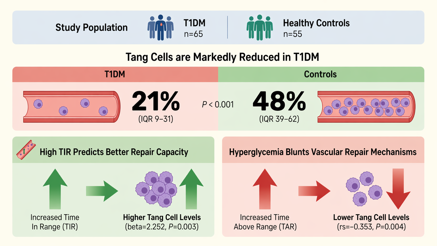
Background/Objectives: Angiogenic T (Tang) cells support endothelial repair and vascular homeostasis. This cross-sectional study compared circulating Tang cell levels in young adults with T1DM vs. healthy controls, and assessed associations between Tang cells and continuous glucose monitoring (CGM) metrics. Methods: Sixty-five young adults with T1DM and 55 healthy controls were enrolled at the University of Campania “Luigi Vanvitelli,” Naples, Italy. Clinical and biochemical data were collected. Tang cells (CD3+CD31+CD184+) were quantified by flow cytometry as absolute counts and percentage of CD3+ T cells. In T1DM, CGM metrics from the preceding 14 days were analyzed, including time in range (TIR), time above range (TAR), and time below range (TBR). Results: Individuals with T1DM had higher fasting glucose and HbA1c than controls. Total CD3+ T cell counts were lower in T1DM. Tang cells were significantly reduced in T1DM both as absolute number and percentage (21% [10–31] vs. 48% [39–62]; p < 0.001). In multivariable analyses, Tang cell percentage was positively associated with TIR and inversely associated with HbA1c and TAR. Conclusions: Young adults with T1DM exhibit significantly reduced circulating Tang cells. Associations with CGM metrics support a link between real-world glucose control and endothelial vascular health.

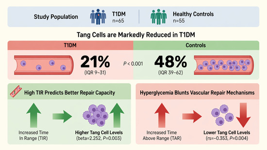
Graphical Abstract

1. Introduction

Type 1 diabetes mellitus (T1DM) is a lifelong chronic disease characterized by the selective destruction of pancreatic islet β-cells, resulting in absolute insulin deficiency and persistent hyperglycemia. Worldwide, the incidence of T1DM is rising; affected individuals are exposed to a multifaceted risk profile encompassing both microvascular and macrovascular complications, with cardiovascular disease (CVD) representing the principal cause of premature morbidity and mortality in this population [1]. Longitudinal data from landmark studies, such as the Diabetes Control and Complications Trial (DCCT), unequivocally demonstrate that poor glycemic control accelerates the onset of vascular complications and long-term cardiovascular risk [2]. Epidemiological studies further indicate that individuals with T1DM face a 4- to 10-fold higher risk of CVD than their age-matched counterparts in the general population [3,4]. The pathogenesis of diabetic vascular disease is multifactorial and involves chronic hyperglycemia, insulin resistance, dyslipidemia, immune dysregulation and endothelial inflammation [5]. Chronic hyperglycemia promotes endothelial dysfunction through the formation of advanced glycation end products (AGEs), oxidative stress and inflammatory activation [6]. Endothelial progenitor cells (EPCs) support vascular repair, and their reduction is considered a marker of impaired endothelial homeostasis. Young adults with T1DM already show lower EPC levels than healthy peers [7], and this deficit persists over time and relates to poorer glycemic control [7]. Angiogenic T (Tang) cells, a specialized subset of T lymphocytes co-expressing endothelial surface molecules (CD31) and T cell markers (CD3), along with chemokine receptor CXCR4 (CD184), have garnered attention for their critical role in promoting endothelial repair and neovascularization [8]. Tang cells exert their proangiogenic effects by interacting with EPCs. Dysfunction or impaired levels of Tang have been reported in multiple diseases characterized by endothelial dysfunction, including type 2 diabetes [9], systemic sclerosis [10] and hypertension-related cerebral small vessel disease [11]. However, data on Tang cells in T1DM are scarce, and their relationship with glycemic control remains poorly defined. Glycemic control is a key determinant of vascular health in individuals with T1DM. Continuous glucose monitoring (CGM) overcomes the limitations of self-monitoring of blood glucose and HbA1c, by capturing daily glucose fluctuations and exposure to hypo- and hyperglycemia, through specific metrics like time in range (TIR), time above range (TAR), time below range (TBR), and indices of glycemic variability [12,13,14]. Importantly, greater exposure to hyperglycemia and glucose swings is linked to endothelial dysfunction and impaired vascular repair [15,16,17], whereas higher TIR and lower variability are associated with improved endothelial function and reduced inflammation [16,18]. Given the central role of Tang cells in vascular repair and homeostasis, it is plausible that suboptimal glycemic control may adversely affect Tang cell levels, thereby contributing to the early vascular impairment observed in T1DM. However, their role in type 1 diabetes and their relationship with glycemic control remain poorly understood. The primary aim of this study was to compare circulating levels of Tang cells in young adults with T1DM versus healthy controls. A secondary aim was to assess the associations between Tang cells and CGM-derived key glucose control metrics within the T1DM cohort. The study was focused on early vascular alterations rather than on established microvascular or macrovascular complications. By integrating cellular and metabolic indices, this study seeks to advance the understanding of early vascular impairment in T1DM and to identify novel targets.

2. Materials and Methods

This is a cross-sectional study of adults with type 1 diabetes consecutively recruited at the Diabetes Unit of the University of Campania “Luigi Vanvitelli” (Naples, Italy) between 2020 and 2025. Inclusion criteria comprised: age between 18 and 30 years, a stable lifestyle, adherence to diet and insulin therapy for at least 6 months, and the absence of clinically evident cardiovascular complications. Stable lifestyle was defined as the absence of major changes in diet, physical activity, smoking status, sleep–wake routine, and medication use during the 3 months preceding enrollment. Exclusion criteria included the presence of acute or chronic diseases (other than diabetes), pregnancy or breastfeeding, and use of drugs able to increase angiogenic cells. All participants signed an informed consent declaration before enrollment.

2.1. Study Measurements

All participants underwent a comprehensive physical examination, including measurements of weight, body mass index (BMI), and blood pressure. Laboratory evaluations were performed centrally and included HbA1c, fasting plasma glucose and lipid profile. All individuals with T1DM were treated either with multiple daily insulin injections (MDI) or continuous subcutaneous insulin infusion (CSII) and were monitored using a CGM system for 14 consecutive days. CGM data were obtained using commercially available CGM system. Following the 14-day monitoring period, TIR (defined as the percentage of time within the target glucose range of 70–180 mg/dL), time below range (TBR, glucose < 70 mg/dL), and time above range (TAR, glucose > 180 mg/dL) were derived from the ambulatory glucose profile (AGP) [19].

2.2. Quantification of Circulating Angiogenic Progenitor Cells

Assessment of circulating levels of Tang was performed on fresh blood samples collected in citrate tubes after overnight fasting at least 8 h; water intake was permitted, and participants continued their usual basal insulin therapy. Peripheral blood mononuclear cells (PBMCs) were isolated by standard density-gradient centrifugation. Cells were incubated with anti-CD3 (eBioscience, Milan, Italy), anti-CD184 (eBioscience, Milan, Italy), and CD31 (Biorbyt, Segrate, Milan, Italy) antibodies [10]. Quantitative analysis was performed on a BD FACSCalibur cytometer, acquiring a minimum of 500,000 cells per sample. Data were processed with the use of Macintosh CellQuest software (version 3.5.1) program (Becton Dickinson, Franklin Lakes, NJ, USA). Tang cells were expressed as a percentage of the total T cell population (CD3+) or as absolute number (cells/mL).

2.3. Statistical Analysis

An a priori sample size calculation was not performed, as this was the first study to investigate circulating Tang cell levels and their relationship with CGM-derived metrics in individuals with T1DM, and no prior data were available for effect size estimation. Therefore, the sample size was determined by consecutive enrollment of all eligible individuals attending our center during the study period. The observed difference in Tang cell percentage between individuals with T1DM and healthy controls corresponded to a large effect size (Cohen’s d ≈ 1.8), indicating a marked separation between groups and supporting the adequacy of the achieved sample size for the primary comparison.
Descriptive statistics were used to characterize the study sample. Continuous variables are presented as mean and SD unless otherwise specified, when normally distributed. Variables that appeared to be non-normally distributed upon the univariate Kolmogorov–Smirnov test were presented as median and IQR that was defined as the interval between the 25th and 75th percentiles. Categorical variables are summarized as number and percentages. Comparative analyses between groups were performed using Student’s t test or the Mann–Whitney U test, as appropriate. The χ2 test was used for comparing dichotomous variables. Statistical associations between circulating levels of Tang and variables were assessed using Spearman’s correlation test. A multiple linear regression analysis was performed to identify independent factors associated with circulating Tang cell percentage. Tang cell percentage was entered as the dependent variable, while demographic, anthropometric, biochemical, and CGM-derived glycemic metrics were included as independent variables. Variables were entered simultaneously, and multicollinearity was assessed using variance inflation factors (VIF < 2 for all variables). All analysis were performed using SPSS software (version 27.0, SPSS, Chicago, IL, USA). p values of <0.05 were taken to indicate statistical significance.

3. Results

Sixty-five young adults with T1DM and 55 age- and sex-matched healthy controls were enrolled. Table 1 summarizes the characteristics of the study population. Key parameters including age, sex, weight, BMI, lipid profile, blood pressure, heart rate and percentage of smokers were well balanced between the two groups; as expected, significantly higher levels of fasting glucose and HbA1c were observed in people with T1DM compared to healthy controls [153.0 (106.5, 225.7) vs. 84.0 (74.0, 90.0) mg/dL; 7.4 (6.7, 8.3) vs. 4.8 (4.5, 5.2)% respectively; both p < 0.001]. Higher levels of creatinine and lower levels of eGFR were observed in people with T1DM compared to healthy controls [0.83 (0.75, 0.90) vs. 0.77 (0.72, 0.81) mg/dL; p = 0.011; 109.6 (95.5, 122.7) vs. 120.1 (116.6, 124.8) mg/dL respectively; p < 0.001].
Among the 65 patients with T1DM, the mean disease duration was 14.6 ± 6.1 years, and the average daily insulin requirement was 0.7 ± 0.2 IU/kg. Thirty patients were treated with CSII, whereas 35 received MDI. Microvascular complications were observed in six patients (9%): specifically, four (6%) had diabetic retinopathy, one (2%) had neuropathy, and one (2%) had nephropathy. CGM-derived metrics showed a median TIR of 56% (IQR 42–67), a median TAR of 32% (IQR 24.7–45.5), a median TBR of 6.5% (IQR 2–13) and a median coefficient of variation (CV) of 37.3% (IQR 28.1–43.5).
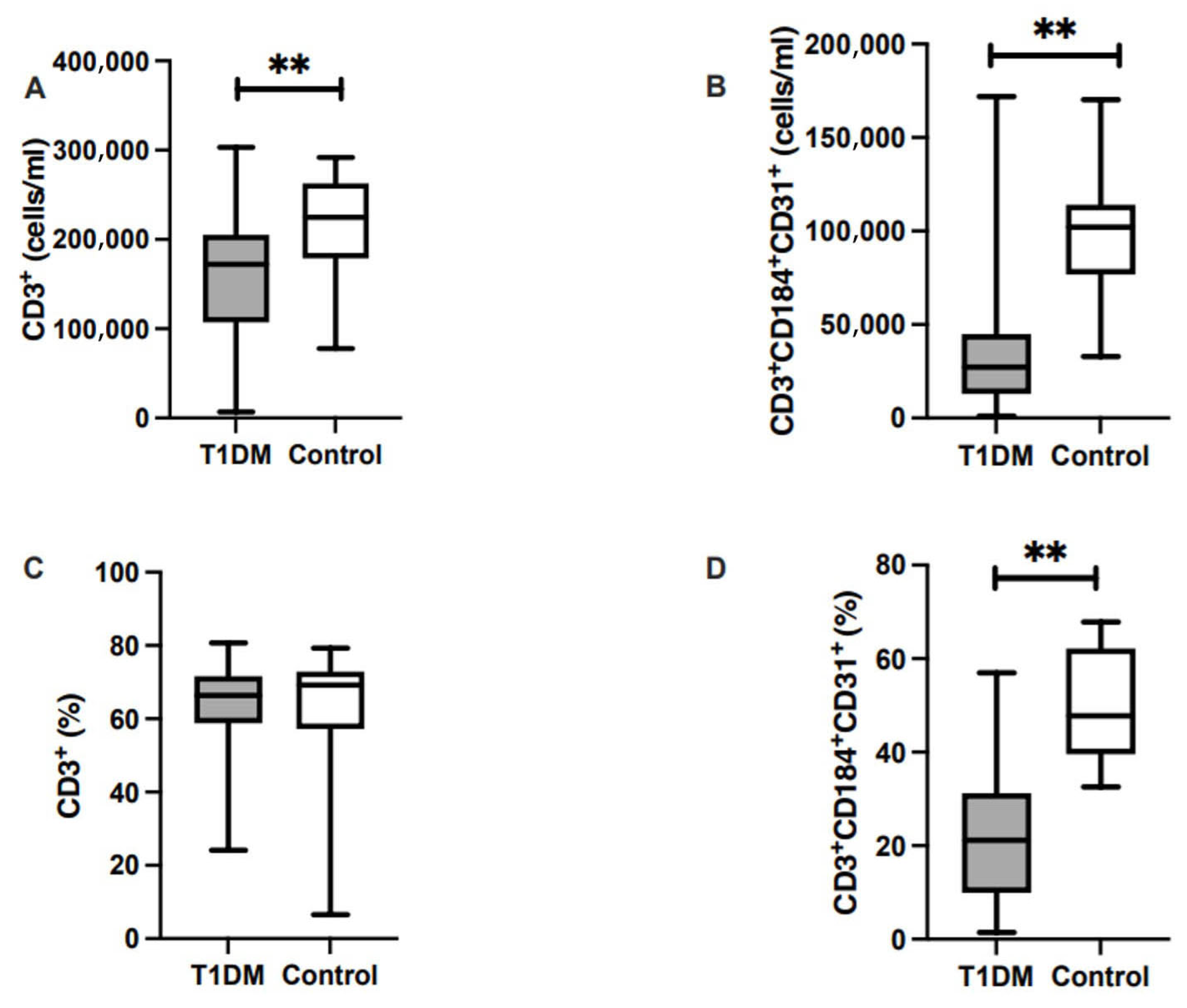
Patients with T1DM exhibited significantly lower absolute count of CD3+ T cells levels compared to the control group [172,000 (107,000–205,000) vs. 225,000 (183,000–263,000) cells/mL, p < 0.001], with no difference in percentage of CD3+ cells. Similarly, circulating CD3+CD31+CD184+ Tang cell levels were markedly reduced, both in absolute count and percentage, in young adults with T1DM [27,000 (13,000–43,000) vs. 102,000 (80,000–114,000) cells/mL, p < 0.001; and 21% (9–31) vs. 48% (39–62), p < 0.001] than in the control group (Figure 1).
In the overall study population, Tang cell percentage correlated negatively with fasting plasma glucose (rsp = −0.358, p = <0.001), HbA1c (rsp = −0.529, p < 0.001), creatinine (rsp = −0.228, p = 0.023), and triglycerides (rsp = −0.225, p = 0.021), but positively with eGFR (rsp = 0.262, p = 0.008) (Table 2).
Correlation analysis within the T1DM cohort revealed a positive association between TIR and circulating Tang cell percentages (rsp = 0.331, p = 0.032), and a negative association with TAR (rsp = −0.353, p = 0.004) (Table 3). HbA1c exhibited a negative correlation with Tang cells (rsp = −0.293, p = 0.042). No significant associations were identified with other clinical variables. No direct markers of endothelial injury were measured; therefore, the associations observed with CGM-derived metrics should not be interpreted as direct evidence of endothelial damage.
In the multivariable analysis restricted to the T1DM population (Table 4), Tang cell percentage was independently associated with fasting glucose (β coefficient = −0.098, p = 0.036), HbA1c (β coefficient = −1.014, p = 0.023), TIR (β coefficient = 2.252, p = 0.003) and TAR (β = −1.947, p = 0.006). No significant associations were observed with age, anthropometric parameters, or other metabolic variables. Creatinine and eGFR were included in the multivariable model to account for potential renal-related confounding, but neither parameter was independently associated with Tang cell percentage.

4. Discussion

To our knowledge, this cross-sectional study provides novel evidence that young adults with long-standing T1DM exhibit a marked reduction in circulating Tang cells levels as compared with age- and sex-matched healthy individuals, despite similar classical cardiovascular risk profiles. Moreover, circulating levels of Tang cells were robustly and positively associated with specific CGM-derived glucose control metrics and, specifically, higher TIR and lower levels of HbA1c, whereas no significant correlations emerged with BMI, blood pressure, or lipid parameters. In multivariable linear regression analyses, fasting plasma glucose showed a significant inverse association with Tang cell percentage, indicating that higher fasting glucose levels were independently associated with lower Tang values. Similarly, HbA1c was negatively associated with Tang cell percentage, supporting a detrimental effect of chronic hyperglycemia on angiogenic immune cell availability. Notably, CGM-derived metrics emerged as the strongest independent predictors of Tang cell levels. Specifically, TIR was positively and strongly associated with Tang cell percentage, suggesting that better overall glycemic control is independently related to preserved angiogenic T cell availability. Conversely, TAR was inversely associated with Tang cell percentage, consistent with a negative impact of hyperglycemic exposure. Collectively, these findings suggest that CGM metrics capture clinically relevant dimensions of glycemic control that extend beyond conventional biomarkers and may better reflect the biological impact of glucose dysregulation on vascular health.
The behavior of Tang cells appears to be highly context-dependent across different pathological states. Reduced Tang cell levels have been reported in type 2 diabetes [9] and hypertension-related cerebral small vessel disease [11], where they have been interpreted as markers of impaired or exhausted vascular repair capacity. On the other hand, in systemic sclerosis [10] Tang cells are expanded and correlate with more severe peripheral vascular damage, suggesting a maladaptive or compensatory activation in chronic inflammatory vasculopathies. Within this heterogeneous scenario, the profound reduction in Tang cells observed in our group of young adults with T1DM, together with their tight relationship with CGM metrics, supports the hypothesis that chronic hyperglycemia blunts proangiogenic T cell availability. Consistent with this interpretation, previous randomized controlled trials of subjects with type 2 diabetes [9] reported significantly lower Tang cell levels in people with diabetes compared with healthy controls (11.5 vs. 20.4%); similarly, in the present study Tang cells were markedly reduced in both absolute counts and percentage in patients with T1DM vs. controls (15 vs. 45%).
Our findings are consistent with previous evidence suggesting that diabetes-related hyperglycemia may be associated with alterations in vascular repair-related cellular pathways, including EPCs. However, EPCs and endothelial function markers were not directly assessed in the present study; therefore, these comparisons should be interpreted with caution. Although glycemic variability and hyperglycemic exposure have been previously linked to endothelial dysfunction and vascular injury, endothelial injury markers were not directly measured in the present study. Therefore, our data do not demonstrate a direct association between Tang cells and endothelial injury. Rather, they suggest that Tang cells are associated with CGM-derived indicators of glycemic exposure, such as TIR and TAR, which may indirectly reflect biological pathways involved in endothelial homeostasis. In both type 1 and type 2 diabetes, poor glycemic control is associated with reduced circulating EPC counts and impaired endothelial repair capacity [8]. Higher EPC levels have been linked to better function in different vascular districts [20,21]. Our results extend these findings to Tang cells, emphasizing their sensitivity to real-world glucose patterns and may provide additional insight into early vascular injury in T1DM.
Maintaining a higher TIR in people with T1DM may not only reduce metabolic complications but also preserve the immune–endothelial mechanisms responsible for vascular repair [22]. Interestingly, in our cohort, no significant associations were found between Tang cells and classical cardiovascular risk factors such as BMI, blood pressure, or lipid profile, suggesting that alterations in angiogenic T cell levels may precede overt metabolic or hemodynamic abnormalities. This finding reinforces the concept that endothelial dysfunction is an early and potentially reversible phenomenon in young adults with T1DM, primarily driven by glycemic dysregulation rather than traditional risk factors.
From a translational perspective, these findings may have potential clinical relevance. In this context, Tang cell quantification may represent a promising biomarker of subclinical vascular alterations and could be explored as a tool to assess the vascular impact of interventions aimed at improving glycemic stability, such as advanced insulin delivery systems and CGM-guided therapeutic strategies.
This study has some limitations. Due to its observational design, causal relationships cannot be established. In addition, the relatively small sample size represents a major limitation and warrants confirmation of these findings in larger cohorts. Longitudinal studies are warranted to determine whether restoring Tang cell number or function through optimal glycemic control or targeted therapies translates into measurable improvements in vascular outcomes. Despite these limitations, our study presents several strengths. First, we used advanced multiparameter flow cytometry to characterize angiogenic T cell subsets, and integrated these immunophenotyping data with clinical and glycemic parameters, including HbA1c and CGM-derived metrics. Second, the strict selection criteria resulted in a homogeneous cohort of young adults free from clinically evident cardiovascular complications. Finally, the use of continuous glucose monitoring (CGM) rather than sole reliance on HbA1c provided real-world, granular data on glycemic excursions (TIR, TAR), enabling us to identify associations between CGM-derived glucose exposure metrics and angiogenic T cell availability that conventional markers may not fully capture. In summary, this study provides novel evidence that circulating angiogenic T cells are significantly reduced in young adults with T1DM and that their levels are closely associated with CGM-derived metrics of glycemic control. These findings suggest that Tang cells may represent a potential early marker of altered vascular homeostasis in T1DM and highlight the critical role of stable and intensive glucose control in preserving endothelial repair mechanisms from the early stages of the disease.

Author Contributions

Conceptualization, M.L., A.M., M.I.M. and K.E.; methodology, M.L. and A.M.; formal analysis, A.M., M.T., M.D.N. and D.F.; investigation, D.F., F.C., P.C. and L.S.; data curation, M.I.M., G.B. and K.E.; writing—original draft preparation, M.L. and A.M.; writing—review and editing, M.L., A.M., M.I.M., G.B. and K.E.; supervision, G.B. and K.E. All authors have read and agreed to the published version of the manuscript.

Funding

This research received no external funding.

Institutional Review Board Statement

The study was conducted in accordance with the Declaration of Helsinki, and approved by the Local Ethics Committee Campania 2 (Protocol number 33_24.01.2014 on 14 February 2014).

Informed Consent Statement

Informed consent was obtained from all subjects involved in the study.

Data Availability Statement

The data presented in this study are available on request from the corresponding author due to privacy and ethical restrictions. The data are not publicly available because they contain sensitive clinical information from human participants.

Conflicts of Interest

The authors declare no conflicts of interest.

Abbreviations

The following abbreviations are used in this manuscript:
T1DMType 1 diabetes mellitus
TangAngiogenic T cells
CGMContinuous glucose monitoring
TIRTime in range
TARTime above range
TBRTime below range

References

  1. Petersmann, A.; Müller-Wieland, D.; Müller, U.A.; Landgraf, R.; Nauck, M.; Freckmann, G.; Heinemann, L.; Schleicher, E. Definition, classification and diagnosis of diabetes mellitus. Exp. Clin. Endocrinol. Diabetes 2019, 127, S1–S7. [Google Scholar] [CrossRef] [Scilit] [PubMed]
  2. Diabetes Control and Complications Trial (DCCT)/Epidemiology of Diabetes Interventions and Complications (EDIC) Study Research Group. Intensive diabetes treatment and cardiovascular outcomes in type 1 diabetes: The DCCT/EDIC study 30-year follow-up. Diabetes Care 2016, 39, 686–693.
  3. Viñals, C.; Conget, I.; Granados, M.; Giménez, M.; Amor, A.J. Evaluation of cardiovascular risk in people with type 1 diabetes: A comprehensive and specific proposed practical approach. Diabetes Ther. 2024, 15, 1831–1844. [Google Scholar] [CrossRef] [Scilit] [PubMed]
  4. Ruiz, P.L.D.; Chen, L.; Morton, J.I.; Salim, A.; Carstensen, B.; Gregg, E.W.; Pavkov, M.E.; Mata-Cases, M.; Mauricio, D.; Nichols, G.A.; et al. Mortality trends in type 1 diabetes: A multicountry analysis of six population-based cohorts. Diabetologia 2022, 65, 964–972. [Google Scholar] [CrossRef] [Scilit]
  5. Xue, C.; Chen, K.; Gao, Z.; Bao, T.; Dong, L.; Zhao, L.; Tong, X. Common mechanisms underlying diabetic vascular complications: Focus on the interaction of metabolic disorders, immuno-inflammation, and endothelial dysfunction. Cell Commun. Signal. 2023, 21, 298. [Google Scholar] [CrossRef] [Scilit]
  6. Yamagishi, S.I.; Matsui, T. Role of hyperglycemia-induced advanced glycation end product accumulation in atherosclerosis. Ann. Vasc. Dis. 2018, 11, 253–258. [Google Scholar] [CrossRef] [Scilit]
  7. Longo, M.; Scappaticcio, L.; Bellastella, G.; Pernice, V.; Cirillo, P.; Maio, A.; Castaldo, F.; Giugliano, D.; Esposito, K.; Maiorino, M.I.; et al. Alterations in the levels of circulating and endothelial progenitor cells in young adults with type 1 diabetes: A 2-year follow-up from the observational METRO study. Diabetes Metab. Syndr. Obes. 2020, 13, 777–784. [Google Scholar] [CrossRef] [Scilit]
  8. Hur, J.; Yang, H.M.; Yoon, C.H.; Lee, C.S.; Park, K.W.; Kim, J.H.; Kim, T.-Y.; Kim, J.-Y.; Kang, H.-J.; Chae, I.-H.; et al. Identification of a novel role of T cells in postnatal vasculogenesis: Characterization of endothelial progenitor cell colonies. Circulation 2007, 116, 1671–1682. [Google Scholar] [CrossRef] [Scilit]
  9. de Boer, S.A.; Reijrink, M.; Abdulahad, W.H.; Hoekstra, E.S.; Slart, R.H.; Heerspink, H.J.; Westra, J.; Mulder, D.J. Angiogenic T cells are decreased in people with type 2 diabetes mellitus and recruited by the dipeptidyl peptidase-4 inhibitor linagliptin: A subanalysis from a randomized, placebo-controlled trial (RELEASE study). Diabetes Obes. Metab. 2020, 22, 1220–1225. [Google Scholar]
  10. Manetti, M.; Pratesi, S.; Romano, E.; Bellando-Randone, S.; Rosa, I.; Guiducci, S.; Fioretto, B.S.; Ibba-Manneschi, L.; Maggi, E.; Matucci-Cerinic, M. Angiogenic T cell expansion correlates with severity of peripheral vascular damage in systemic sclerosis. PLoS ONE 2017, 12, e0183102. [Google Scholar] [CrossRef] [Scilit] [PubMed]
  11. Rouhl, R.P.; Mertens, A.E.; van Oostenbrugge, R.J.; Damoiseaux, J.G.; Debrus-Palmans, L.L.; Henskens, L.H.; Kroon, A.A.; de Leeuw, P.W.; Lodder, J.; Tervaert, J.W.C. Angiogenic T-cells and putative endothelial progenitor cells in hypertension-related cerebral small vessel disease. Stroke 2012, 43, 256–258. [Google Scholar]
  12. Ajjan, R.A. The clinical importance of measuring glycaemic variability: Utilising new metrics to optimise glycaemic control. Diabetes Obes. Metab. 2024, 26, 3–16. [Google Scholar] [CrossRef] [Scilit] [PubMed]
  13. Yapanis, M.; James, S.; Craig, M.E.; O’Neal, D.; Ekinci, E.I. Complications of diabetes and metrics of glycemic management derived from continuous glucose monitoring. J. Clin. Endocrinol. Metab. 2022, 107, e2221–e2236. [Google Scholar] [CrossRef] [Scilit]
  14. Mo, Y.; Lu, J.; Zhou, J. Glycemic variability: Measurement, target, impact on complications of diabetes and does it really matter? J. Diabetes Investig. 2024, 15, 5–14. [Google Scholar] [CrossRef] [Scilit]
  15. Buscemi, S.; Re, A.; Batsis, J.A.; Arnone, M.; Mattina, A.; Cerasola, G.; Verga, S. Glycaemic variability using continuous glucose monitoring and endothelial function in the metabolic syndrome and in type 2 diabetes. Diabet. Med. 2010, 27, 872–878. [Google Scholar] [CrossRef] [Scilit]
  16. Valente, T.; Valente, F.; Lucchesi, M.B.B.; Punaro, G.R.; Mouro, M.G.; Gabbay, M.A.L.; Higa, E.M.S.; Dib, S.A. Relationship between short- and long-term glycemic variability and oxidative stress in type 1 diabetes mellitus under daily life insulin treatment. Arch. Endocrinol. Metab. 2021, 65, 570–578. [Google Scholar] [CrossRef] [Scilit]
  17. Huang, Y.; Yue, L.; Qiu, J.; Gao, M.; Liu, S.; Wang, J. Endothelial dysfunction and platelet hyperactivation in diabetic complications induced by glycemic variability. Horm. Metab. Res. 2022, 54, 419–428. [Google Scholar] [CrossRef] [Scilit]
  18. Fedulovs, A.; Janevica, J.; Kruzmane, L.; Sokolovska, J. Glucose control and variability assessed by continuous glucose monitoring in patients with type 1 diabetes and diabetic kidney disease. Biomed. Rep. 2024, 22, 23. [Google Scholar] [CrossRef] [Scilit]
  19. Martens, T.W.; Simonson, G.D.; Bergenstal, R.M. Using continuous glucose monitoring data in daily clinical practice. Clevel. Clin. J. Med. 2024, 91, 611–620. [Google Scholar] [CrossRef] [Scilit] [PubMed]
  20. Longo, M.; Di Meo, I.; Caruso, P.; Muscio, M.F.; Scappaticcio, L.; Maio, A.; Maiorino, M.I.; Bellastella, G.; Signoriello, G.; Knop, F.K.; et al. Circulating levels of endothelial progenitor cells are associated with better cognitive function in older adults with glucagon-like peptide-1 receptor agonist-treated type 2 diabetes. Diabetes Res. Clin. Pract. 2023, 200, 110688. [Google Scholar] [CrossRef] [Scilit] [PubMed]
  21. Maio, A.; Maiorino, M.I.; Longo, M.; Scappaticcio, L.; Pernice, V.; Cirillo, P.; Caruso, P.; Paglionico, V.A.; Bellastella, G.; Esposito, K. Change in circulating levels of endothelial progenitor cells and sexual function in women with type 1 diabetes. J. Clin. Endocrinol. Metab. 2022, 107, e3910–e3918. [Google Scholar] [CrossRef] [Scilit] [PubMed]
  22. Beck, R.W.; Bergenstal, R.M.; Riddlesworth, T.D.; Kollman, C.; Li, Z.; Brown, A.S.; Close, K.L. Validation of time in range as an outcome measure for diabetes clinical trials. Diabetes Care 2019, 400, 405. [Google Scholar] [CrossRef] [Scilit]
Figure 1. Circulating levels of CD3+ and CD3+CD31+CD184+ in patients with type 1 diabetes and healthy controls. Significant differences are indicated. ** p < 0.001. (A) absolute number of CD3+ cells in patients with T1DM and controls; (B) absolute number of Tang in patients with T1DM and controls; (C) percentage of CD3+ cells in patients with T1DM and controls; (D) percentage of Tang in patients with T1DM and controls.
Diabetology 07 00113 g001
Table 1. Clinical characteristics of subjects with type 1 diabetes and healthy controls.
ParametersPatients with T1DM (n = 65)Healthy Controls
(n = 55)
p
Age (years)23 (20, 27)24 (23, 27)0.126
Males/females n33/3221/340.231
Smoke n (%)24 (37)12 (22)0.993
Weight (Kg)65.0 (59.8, 73.5)65.0 (59.0, 75)0.797
BMI (Kg/m2)23.4 (21.6, 25.0)23.2 (21.6, 26.3)0.769
Fasting glucose (mg/dL)153.0 (106.5, 225.7)84.0 (74.0, 90.0)<0.001
HbA1c (%)7.4 (6.7, 8.3)4.8 (4.5, 5.2)<0.001
HbA1c (mmol/mol)57 (50, 67)29 (26, 33)<0.001
Total cholesterol (mg/dL)159.0 (146.7, 180.0)168.0 (146.0, 181.0)0.433
LDL-cholesterol (mg/dL)94 (78, 115.5)99 (82, 120)0.378
HDL-cholesterol (mg/dL)53 (48, 60)55 (48, 63)0.311
Triglycerides (mg/dL)63.0 (51.7, 85)71.0 (52.0, 90.0)0.420
Creatinine0.83 (0.75, 0.90)0.77 (0.72, 0.81)0.011
eGFR109.6 (95.5, 122.7)120.1 (116.6, 124.8)<0.001
SBP (mmHg)110 (100, 120)110 (110, 122)0.116
DBP (mmHg)70 (66, 80)70 (70, 80)0.218
HR (bpm)76 (70, 82.7)75 (70, 80)0.207
Data are expressed as mean ± SD and as median (interquartile range). Abbreviations: BMI—body mass index; HbA1c—glycated hemoglobin A1c; LDL—low-density lipoprotein; HDL—high-density lipoprotein; eGFR—glomerular filtration rate; SBP—systolic blood pressure; DBP—diastolic blood pressure; HR—heart rate.
Table 2. Correlation between Tang and clinical characteristics in the overall populations.
ParametersCD3+D31+CD184+ (%) Overall
rsp p
Age0.1590.086
Weight−0.08310.408
BMI−0.04450.638
Fasting glucose−0.358<0.001
HbA1c−0.529<0.001
Total cholesterol (mg/dL)0.0480.632
LDL-cholesterol (mg/dL)0.0300.766
HDL-cholesterol (mg/dL)0.1870.069
Triglycerides (mg/dL)−0.2250.021
Creatinine−0.2280.023
eGFR0.2620.008
Abbreviations: BMI—body mass index; HbA1c—glycated hemoglobin A1c; LDL—low-density lipoprotein; HDL—high-density lipoprotein; eGFR—glomerular filtration rate.
Table 3. Statistical associations between CD3+CD31+CD184+ percentage and different parameters by univariate analysis in the cohort of patients with T1DM.
ParametersCD3+CD31+CD184+ (%)
rsp p
Age−0.1710.172
Weight0.1510.285
BMI−0.0450.720
Diabetes duration−0.0160.897
Fasting glucose0.1140.365
HbA1c−0.2930.042
Total cholesterol (mg/dL)0.0080.514
LDL-cholesterol (mg/dL)−0.090.466
HDL-cholesterol (mg/dL)0.0060.960
Triglycerides (mg/dL)0.0410.745
Creatinine−0.0830.506
eGFR0.0080.945
TIR0.3310.032
TAR−0.3530.004
TBR0.1250.459
CV0.0300.812
Abbreviations: BMI—body mass index; HbA1c—glycated hemoglobinA1c; LDL—low-density lipoprotein; HDL—high-density lipoprotein; eGFR—glomerular filtration rate; TIR—time in range; TAR—time above range; TBR—time below range; CV—coefficient of variation.
Table 4. Multivariable linear regression analysis of factors associated with Tang cell percentage.
β Coefficientp
Age−0.7160.240
Weight0.5120.255
BMI−1.2990.307
Fasting glucose−0.0980.036
HbA1c−1.0140.023
Triglycerides (mg/dL)−0.2090.857
Creatinine−1.040.968
TIR2.2520.003
TAR−1.9470.006
TBR−0.6110.131
eGFR0.1820.593
Abbreviations: BMI—body mass index; HbA1c—glycated hemoglbinA1c; TIR—time in range; TAR—time above range; TBR—time below range; eGFR—glomerular filtration rate.
Disclaimer/Publisher’s Note: The statements, opinions and data contained in all publications are solely those of the individual author(s) and contributor(s) and not of MDPI and/or the editor(s). MDPI and/or the editor(s) disclaim responsibility for any injury to people or property resulting from any ideas, methods, instructions or products referred to in the content.