Communication

Abscisic Acid as a Marker of Metabolic Imbalance: Serum Levels from Diabetic and Smoking Subjects

1
Department of Environmental, Biological and Pharmaceutical Sciences and Technologies, University of Campania Luigi Vanvitelli, Via Vivaldi 43, 81100 Caserta, Italy
2
NGN Healthcare—New Generation Nutraceuticals s.r.l., Torrette Via Nazionale 207, 83013 Mercogliano, Italy
3
Comegen S.c.S., Società Cooperativa Sociale di Medici di Medicina Generale, Viale Maria Bakunin 41, 80125 Naples, Italy
4
Department of Pharmacy, University of Naples Federico II, Via Domenico Montesano 59, 80131 Napoli, Italy
5
Department of Medicine and Surgery, Catholic University of the Sacred Heart, 00168 Rome, Italy
*
Author to whom correspondence should be addressed.
Diabetology 2025, 6(9), 93; https://doi.org/10.3390/diabetology6090093
Submission received: 1 August 2025 / Revised: 23 August 2025 / Accepted: 29 August 2025 / Published: 2 September 2025

Abstract

Background: Abscisic acid (ABA), a phytohormone widely distributed in nature, has recently emerged as an endogenous regulator of glucose homeostasis in humans. Specifically, scientific studies have demonstrated that exogenous ABA supplementation improves glycemic control and reduces insulin requirements, with significant advantages in prediabetic subjects. Beyond its metabolic role, growing evidence suggests that ABA is also involved in immune responses, including those associated with pulmonary diseases. Despite these promising results, the evaluation of plasma ABA levels remains largely unexplored in clinical practice. Methods: This study aimed to evaluate whether plasma ABA concentrations differ among healthy individuals, patients with type 2 diabetes, and smokers, in order to clarify the role of ABA as a potential biomarker of both metabolic imbalance and smoking-related inflammatory stress. Results: Our findings show that ABA levels were significantly higher in healthy subjects (10.9 ± 3.8 ng/mL) compared to diabetic patients (6.8 ± 4.2 ng/mL, p < 0.01 vs. healthy subjects), with the lowest levels observed in smokers (3.5 ± 2.5 ng/mL, p < 0.0001 vs. healthy subjects and p < 0.0001 vs. diabetic patients). Moreover, a significant correlation was observed between ABA plasma concentration and number of cigarettes smoked (R2 = −0.6776, p = 0.0001). Conclusions: Overall, these results highlight the relevance of measuring ABA plasma levels in both metabolic and inflammatory conditions, confirming its role as a biomarker for identifying individuals who can benefit from exogenous supplementation.

Graphical Abstract

1. Introduction

Smoking is one of the leading modifiable risk factors for numerous chronic diseases, including cardiovascular, respiratory, and metabolic disorders [1,2]. Numerous studies have demonstrated a significant association between smoking and the development of metabolic disorders, including metabolic syndrome, insulin resistance, type 2 diabetes mellitus (T2DM), and dyslipidemia [3], with a clear dose and time-dependent relationship [4]. The underlying pathophysiological mechanisms behind this association are multifaceted and include chronic inflammation, systemic oxidative stress, and endothelial dysfunction. A key mechanism is the smoking-induced impairment of insulin sensitivity, which contributes to the development of insulin resistance [5]. Another relevant mechanism is the increase in inflammatory markers, such as fibrinogen and C-reactive protein, resulting from an immune response that leads to vascular injury [6,7]. Additionally, smoking interferes with coagulation and fibrinolysis processes [8], promoting thrombosis through direct effects on platelets, the endothelium, and fibrinogen [9].
Recent years have seen growing interest in abscisic acid (ABA), a phytohormone traditionally known for its role in plant physiology, in response to environmental stress [10,11], but recently identified in the human organism as well. In fact, ABA has been implicated in metabolic and immune regulation processes [12,13,14], particularly through the activation of the LanC-like protein 2 (LANCL2) receptor [15], expressed in various tissues including the pancreas, skeletal muscle, and immune system. Activation of this pathway has been associated with beneficial effects on glucose tolerance, insulin sensitivity, and systemic inflammation [16,17,18], suggesting that ABA may function as an endogenous bioactive molecule with anti-inflammatory and insulin-sensitizing properties [13,19]. Notably, while insulin promotes glucose uptake and storage under hyperglycemic conditions, ABA facilitates glucose utilization for energy production through insulin-independent mechanisms. This complementary action suggests that ABA and insulin pathways cooperate in maintaining glucose homeostasis, with ABA reducing the exhaustion of pancreatic β-cells by enhancing peripheral glucose uptake [20]. In humans, ABA is secreted by pancreatic β-cells in response to high blood glucose concentrations [21]. It promotes glucose uptake in skeletal muscle and adipose tissue by upregulating the expression and translocation of glucose transporter 4 (GLUT4) [20,22]. This mechanism is primarily mediated through ABA’s interaction with its receptor LANCL2, which activates downstream signaling pathways, including AMPK and PGC-1α, promoting oxidative glucose metabolism independently of insulin [23]. Notably, low-dose administration of exogenous ABA has been shown to improve glucose uptake and insulin profiles in both animal models and human subjects [14,18]. Specifically, ABA facilitates peripheral glucose uptake by muscle cells and its conversion into energy. Unlike insulin, which is secreted exclusively under hyperglycemic conditions and promotes glucose storage as triglycerides in visceral adipose tissue, ABA enhances glucose utilization for energy production while sparing insulin [24]. Overall, these findings support the hypothesis that ABA supplementation could enhance glucose metabolism and reduce the burden on pancreatic β-cell insulin production [11,25,26,27].
Other evidence indicate that ABA may exert protective effects in various pulmonary pathologies characterized by inflammation and oxidative stress. In a murine model of influenza A infection, dietary ABA administration resulted in improved clinical outcomes, reduced pulmonary inflammation, and enhanced resolution of disease through activation of peroxisome proliferator-activated receptor gamma (PPAR-γ) in lung immune cells, accompanied by increased expression of interleukin-10 (IL-10) and 5-lipoxygenase pathway genes [28]. Similarly, in a lipopolysaccharide (LPS)-induced model of acute respiratory distress syndrome (ARDS), ABA mitigated lung injury by suppressing endoplasmic reticulum stress and reactive oxygen species (ROS) production via PPAR-γ and nuclear factor erythroid 2–related factor 2 (Nrf2) signaling pathways [29]. Furthermore, in an ovalbumin-induced model of allergic airway inflammation, ABA significantly attenuated airway inflammation by inhibiting NLRP3 inflammasome activation, reducing oxidative stress, and modulating mitochondrial dynamics, an effect that was abrogated by pharmacological inhibition of PPAR-γ [30]. In this context, it is plausible that smoking-mediated inflammation and oxidative stress may dysregulate ABA pathways, either by impairing its biosynthesis, accelerating its degradation, or interfering with receptor signaling. Such mechanisms could contribute to the markedly reduced ABA levels observed in smokers.
Despite increasing evidence of ABA’s role in human physiology, its potential involvement in metabolic disorders or pulmonary pathologies associated with cigarette smoking remains largely unexplored. Since smoking induces chronic inflammation and oxidative stress, and ABA is implicated in modulating these processes [31,32], we investigated whether plasma ABA levels differ among healthy individuals, smokers, and T2M patients. Our goal was to elucidate the potential role of this molecule in metabolic and inflammatory regulation under both physiological and pathological conditions.

2. Materials and Methods

2.1. Study Design and Participants

This study was conducted as a cross-sectional observational study aimed at evaluating the relationship between cigarette smoking, diabetic status, and plasma levels of ABA in human subjects. Participants were recruited from the patient registry of general practitioners affiliated with COMEGEN—Società Cooperativa Sociale (Naples, Italy). Participants were consecutively enrolled from the patient registry of general practitioners, with efforts made to balance demographic variables across study groups. The study was approved by the Ethical Committee “Campania Centro”—ASL Napoli 1 with the reference number 255 dated 6 June 2023. All procedures were conducted in accordance with the Declaration of Helsinki (1964, and subsequent revisions), and all participants provided written informed consent prior to enrollment. The study has been registered in the ISRCTN registry (www.isrctn.com, accessed on 15 May 2025) under the identifier ISRCTN90922084 (https://www.isrctn.com/ISRCTN90922084, accessed on 15 May 2025).
Eligible participants included adult men and women of Caucasian ethnicity, aged between 18 and 75 years. Subjects were assigned to one of three groups based on their clinical status: Diabetic group (patients with a diagnosis of T2DM), Smokers group (current cigarette smokers), and Control group (healthy non-smoker controls). Diagnosis of T2DM was made according to the criteria of the American Diabetes Association (ADA) [33], and confirmed by one of the following: (i) fasting plasma glucose (FPG) ≥ 126 mg/dL (7.0 mmol/L), (ii) 2-h plasma glucose ≥ 200 mg/dL (11.1 mmol/L) during a 75-g oral glucose tolerance test (OGTT), (iii) glycated hemoglobin (HbA1c) ≥ 6.5%, or (iv) random plasma glucose ≥ 200 mg/dL (11.1 mmol/L) in the presence of classic symptoms of hyperglycemia. Current smokers were defined as individuals who reported smoking at least one cigarette per day in the past 12 months and had a lifetime history of smoking ≥ 100 cigarettes, consistent with established epidemiological definitions [2]. At baseline, participants completed a structured questionnaire that included items on smoking status, history, and intensity. Specifically, subjects were asked to report the average number of cigarettes smoked per day to allow stratification by smoking intensity in subsequent analyses. Exclusion criteria included: chronic kidney disease defined as eGFR < 60 mL/min/1.73 m2 calculated from serum creatinine using the CKD-EPI (2021) equation, hepatic enzyme elevations above the laboratory upper limit of normal (ULN) for either ALT or AST, or a documented diagnosis of metabolic dysfunction–associated steatotic liver disease (MASLD/MASH), history of cardiovascular events within the last 6 months, anemia (Hb < 12 g/dL), major psychiatric illness, and current use of nutraceutical treatments (including ABA-containing supplements such as thinned-nectarine extracts, or other ABA-enriched formulations). Subjects with pregnancy or breastfeeding were also excluded.
A total of 209 participants were enrolled in the study: 101 individuals with T2DM, 56 smokers without diabetes, and 52 healthy, non-smoking controls. The three groups were matched as closely as possible for age and sex distribution. All participants underwent clinical evaluation, anthropometric measurements, and blood sampling at baseline.

2.2. Sample Collection and Metabolic Profiling

Venous blood samples were collected from all participants after an overnight fast of at least 8 h. Blood was drawn into EDTA tubes and centrifuged at 1500× g for 10 min at 4 °C to separate plasma, which was then aliquoted and stored at −80 °C until analysis. The primary outcome of the study was the change in ABA levels between healthy patients, smokers and diabetic subjects. Secondary outcomes included fasting plasma glucose (FPG), glycated hemoglobin (HbA1c), total cholesterol (TC), low-density lipoprotein cholesterol (LDL-C), high-density lipoprotein cholesterol (HDL-C), triglycerides (TG), aspartate aminotransferase (AST), alanine aminotransferase (ALT), and uric acid levels. Additionally, all participants completed a structured questionnaire designed to collect demographic, clinical, and behavioral information, including data on diabetes status, smoking habits, lifestyle, and the use of medications or supplements. The questionnaire was used to classify subjects into study groups (healthy, smokers, diabetic) and to identify potential confounding factors. ABA levels were assessed using a commercially available kit (ABclonal, Woburn, MA, USA, catalog number RK04822), HbA1c levels were measured using a spectrophotometric kit (Intermedical s.r.l., Naples, Italy), whereas plasma FPG, TC, HDL-C, LDL-C, TG, AST, ALT, and uric acid levels were evaluated using SPHERA analyzer (REF: ES00001-B, UDI-ID: 80536089400722; Edif Instruments S.R.L., Rome, Italy). Calibration and quality control procedures were performed according to the manufacturer’s specifications.

2.3. Quantification of Abscisic Acid (ABA)

Plasma levels of abscisic acid were measured using a competitive enzyme-linked immunosorbent assay (ELISA) kit (ABclonal, catalog number RK04822), specifically designed for the quantitative detection of ABA in biological fluids. Prior to large-scale testing, the validity of this assay was confirmed by comparison with results obtained using a previously validated HPLC-HESI-MS/MS method [34]. For practical reasons, ELISA was then selected for the complete sample set, as it is more suitable for high-throughput analyses. The kit had a detection range of 1.56–100 ng/mL, with a sensitivity of < 0.78 ng/mL. According to the manufacturer, the assay showed no significant cross-reactivity with ABA analogues. The intra-assay coefficient of variation (CV) was <10%, and the inter-assay CV < 15%. The assay was performed according to the manufacturer’s instructions. This assay is based on the competition between endogenous ABA in the sample and horseradish peroxidase (HRP)-labeled ABA for binding to a monoclonal antibody immobilized on a microplate. After incubation and washing steps to remove unbound components, a TMB substrate was added, and the enzymatic reaction was stopped with an acidic solution. Absorbance was measured at 450 nm using a microplate reader (EnVision 2104 Multilabel Plate Reader, PerkinElmer, Singapore). The intensity of the colorimetric signal is inversely proportional to the ABA concentration. Samples and standards were assayed in duplicate. A standard curve was constructed by using serial dilutions of a reconstituted ABA standard (100–0 ng/mL) and plotting the mean absorbance of each standard against its concentration on a log-log scale. The data were linearized by plotting the log of ABA concentrations versus the log of the optical density on a linear axis.

2.4. Statistical Analysis

All data were expressed as mean ± standard deviation (SD). Group comparisons for general demographic variables and metabolic parameters were performed using two-way analysis of variance (ANOVA), with factors being group assignment (control, diabetic, smoker) and sex, in order to account for both main effects and possible interactions. Tukey’s multiple comparisons post hoc test was then applied. Differences in plasma ABA concentrations among the three study groups were analyzed using one-way ANOVA with Tukey’s post hoc correction for pairwise comparisons. To assess the association between the number of cigarettes smoked per day and plasma ABA levels within the smokers group, Pearson’s correlation analysis was performed. A p-value of less than 0.05 was considered statistically significant. All analyses were conducted using GraphPad Prism software (version 8.4.3, GraphPad Software Inc., San Diego, CA, USA).

3. Results

3.1. Baseline Demographic and Metabolic Characteristics

As reported in Table 1, the sex distribution was similar among the groups, with a slight predominance of males: 63.5% in the control group (M/F: 33/19), 59.4% in the diabetic group (60/41), and 64.3% in the smokers group (36/20). As expected, FPG was significantly elevated in the diabetic group (127.3 ± 50.5 mg/dL), with values exceeding both the control (84.2 ± 11.1 mg/dL; **** p < 0.0001) and smoker groups (93.3 ± 14.7 mg/dL; #### p < 0.0001 vs. diabetics). Within the T2DM group, the mean disease duration was 10.2 ± 6.1 years (range 1–25 years). Interestingly, smokers without diabetes displayed slightly elevated FPG levels compared to controls, suggesting early metabolic changes in this group. Similarly, HbA1c followed a similar pattern: the diabetic group exhibited markedly increased levels (6.7 ± 1.6%), whereas both control and smoker groups had normal values (5.0 ± 0.3% and 5.0 ± 0.2%, respectively). The relatively low mean HbA1c in the diabetic group may be explained by the inclusion of patients with good glycemic control, as well as individuals with recent T2DM diagnosis who had not yet developed sustained hyperglycemia. In smokers, HbA1c values were identical to controls, despite slightly higher fasting glucose, suggesting that early metabolic alterations in this group may not yet be reflected in long-term glycemic markers.
Lipid profile analysis also revealed significant intergroup differences. Specifically, LDL-cholesterol was significantly higher in diabetics than controls (106.5 ± 20.2 vs. 83.7 ± 29.6 mg/dL, respectively, * p < 0.05). Triglyceride levels were also markedly increased in the diabetic group (167.4 ± 18.1 mg/dL) compared to both controls (92.6 ± 24.1 mg/dL; **** p < 0.0001) and smokers (106.0 ± 17.4 mg/dL; #### p < 0.0001), with high interindividual variability in all groups. Liver enzymes and uric acid levels were overall comparable among groups, with no significant differences observed.

3.2. Progressive Decline of ABA from Healthy to Diabetic and Smoking Individuals

The primary outcome of the study was the quantification of plasma ABA concentrations across the three study groups. As shown in Figure 1, ABA levels were significantly different among healthy controls, individuals with type 2 diabetes, and current smokers, indicating a potential association between ABA availability and metabolic or inflammatory status. The mean plasma concentration of ABA in the control group was 10.9 ± 3.8 ng/mL, representing the physiological baseline in the absence of metabolic or inflammatory alterations. In contrast, participants in the diabetic group exhibited significantly lower plasma ABA levels, with a mean concentration of 6.8 ± 4.2 ng/mL, corresponding to a reduction of approximately 38% compared to controls (*** p < 0.001). Notably, the lowest ABA concentrations were observed in the smokers group, with ABA plasma levels drastically reduced to 3.5 ± 2.5 ng/mL, reflecting a 68% decrease relative to healthy controls (**** p < 0.0001) and a 49% reduction compared to diabetic patients (#### p < 0.0001 vs. diabetic group). However, the relatively large standard deviation indicates high variability, which may reflect heterogeneity in smoking intensity, duration of exposure, and potential metabolic confounders. The robustness of these findings was confirmed in multivariate analyses. ANCOVA including age as a covariate demonstrated that group differences in ABA concentrations remained highly significant (p < 0.001). Linear regression models adjusted for both age and disease duration confirmed that ABA levels were significantly lower compared to controls, while disease duration itself was not significantly associated with ABA concentrations (β = −0.08, 95% CI: −0.21 to 0.05, p = 0.22). In smokers, the number of cigarettes smoked per day remained strongly and independently associated with reduced ABA concentrations (β = −0.62, 95% CI: −0.80 to −0.44, p < 0.001), even after controlling for age.

3.3. Higher Tobacco Exposure Is Associated with Lower Circulating ABA

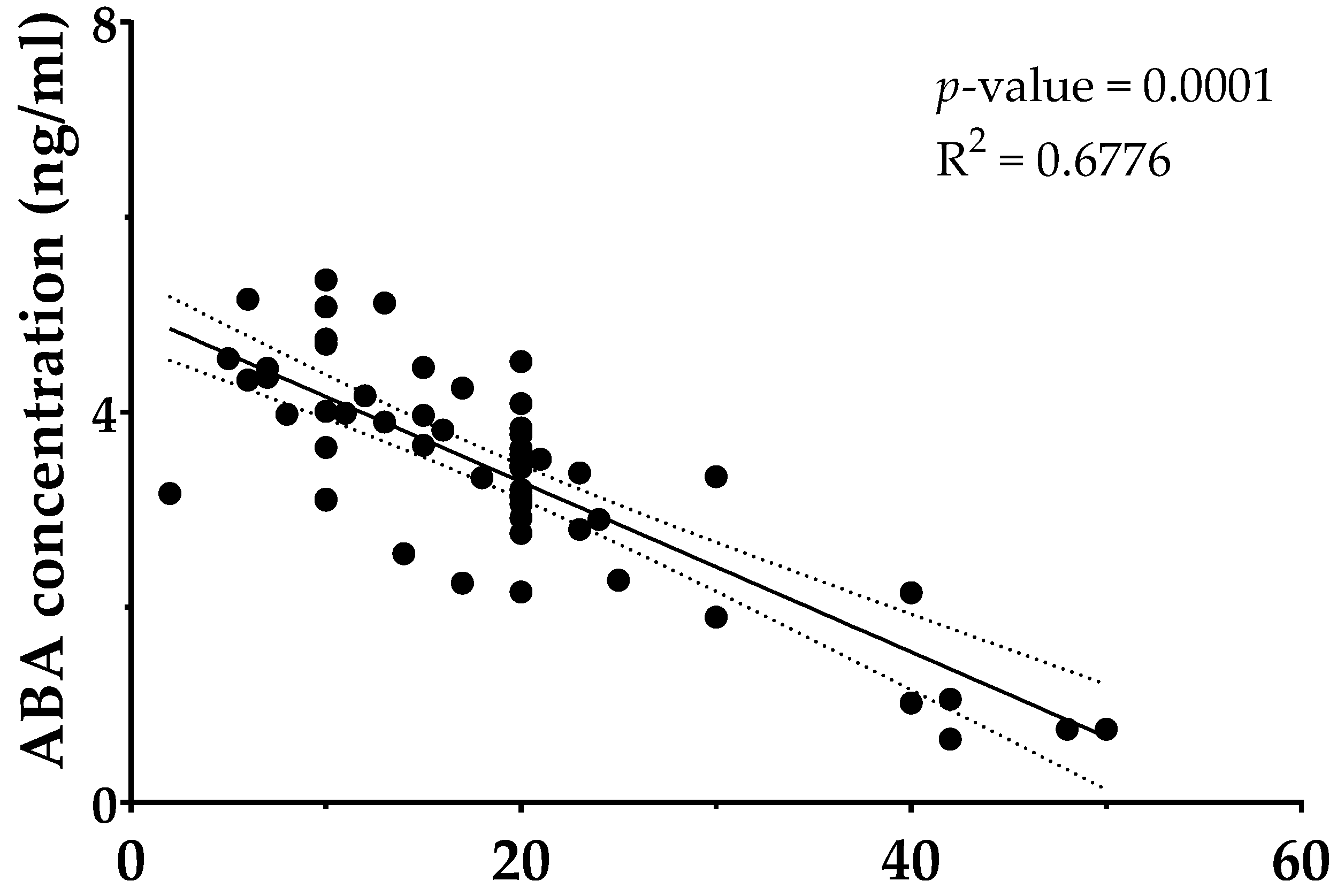
To explore whether the extent of tobacco exposure influences systemic levels of ABA, a Pearson’s correlation analysis was conducted within the smokers group (n = 56), using self-reported data on the average number of cigarettes smoked per day and plasma ABA concentrations measured via ELISA. The results revealed a statistically significant inverse correlation between the number of cigarettes smoked and circulating ABA levels (R2 = −0.6776, p = 0.0001, Figure 2). The direction and significance of the association highlight a biologically linked relationship between tobacco use and impairment of ABA homeostasis, supporting the hypothesis of a dose-dependent effect of cigarette smoke on this endogenous signaling molecule.

4. Discussion

The results obtained in this study provide strong evidence about the role of ABA as a potential biomarker for metabolic and inflammatory dysregulation. Specifically, a consistent and progressive reduction in plasma ABA concentrations was observed across three distinct population groups, e.g., healthy individuals, T2DM patients, and active smokers. First, the baseline metabolic parameters of the study participants, reported in Table 1, confirm the expected metabolic impairments in individuals with T2DM, with FPG averaging 127.3 ± 50.5 mg/dL and HbA1c at 6.7 ± 1.6%, both markedly elevated compared to healthy controls (FPG: 84.2 ± 11.1 mg/dL; HbA1c: 5.0 ± 0.3%). Interestingly, smokers, despite not having a formal diagnosis of diabetes, still exhibited slight alterations in FPG (FPG 93.3 ± 14.7 mg/dL), suggesting early metabolic dysregulation in this group. In terms of lipid metabolism, diabetic subjects had significantly higher LDL cholesterol (106.5 ± 20.2 mg/dL vs. 83.7 ± 29.6 mg/dL in controls), and triglyceride levels (167.4 ± 18.1 mg/dL vs. 92.6 ± 24.1 mg/dL in controls), further confirming their altered metabolic profile.
As regards the quantification of ABA plasmatic levels (Figure 1), healthy individuals displayed the highest ABA levels, with a mean value of 10.9 ± 3.8 ng/mL. Notably, a substantial reduction was observed in T2DM patients, with average levels decreasing to 6.8 ± 4.2 ng/mL, approximately a 38% decrease compared to healthy controls (p < 0.001). This supports previous findings that impaired glucose regulation is linked to reduced endogenous ABA production. Given that ABA is secreted by pancreatic β-cells in response to elevated blood glucose [35] and promotes peripheral glucose uptake through LANCL2-mediated pathways [23], its decreased plasma concentration in T2DM patients likely reflects β-cell dysfunction. These results support the evidence that ABA functions as a physiological modulator of glucose metabolism and may represent a sensitive indicator of pancreatic β-cells activity.
It is noteworthy to remark that the lowest ABA levels were found in smokers, a population not typically characterized by evident hyperglycemia, but known to exhibit subclinical insulin resistance, oxidative stress, and chronic low-grade inflammation [36,37]. Specifically, this population showed an average concentration of 3.5 ± 2.5 ng/mL, representing a 68% decrease compared to controls and a 49% reduction relative to T2DM patients (p < 0.0001 for both comparisons). Furthermore, the strong inverse correlation (R2 = −0.6776, p = 0.0001, Figure 2) between the number of cigarettes smoked and ABA plasma levels further strengthens the relevance of a dose-dependent relationship, indicating that plasma ABA concentrations are influenced not only by glycemic status, but also by environmental and inflammatory stressors such as smoking. In this regard, the lower levels of ABA observed in smokers suggest a potential early impairment in ABA-mediated regulatory pathways, possibly correlated to redox imbalance, systemic inflammation, or suppression of endogenous biosynthesis [38].
Overall, these finding suggest, based on current evidence, that chronic exposure to tobacco toxins may suppress ABA synthesis, promote its degradation, or interfere with its signaling cascade. Mechanistically, exposure to components of cigarette smoke, such as ROS, polycyclic aromatic hydrocarbons, and nicotine, may interfere with LANCL2 receptor activation, thereby limiting ABA bioavailability or downstream efficacy. In this regard, in vitro and toxicology studies have shown that smoke-derived ROS and xenobiotics can disrupt hormone-receptor interactions and redox-sensitive signaling cascades, providing a plausible mechanism for the dysregulation of ABA pathways observed in smokers [39,40]. Since ABA modulates insulin-independent glucose uptake via AMPK and PGC-1α activation [22,23], as well as exerts anti-inflammatory effects through PPAR-γ and Nrf2 pathways [17,29], its suppression in smokers could, in fact, impair both metabolic regulation and immunomodulation.
Further evidence supporting the immunoregulatory role of ABA derives from a study by Hoang et al., which reported lower serum ABA levels in patients with chronic obstructive pulmonary disease (COPD) compared to healthy individuals [12]. Specifically, patients with more advanced disease (GOLD stage IV), typically characterized by tissue remodeling and reduced inflammatory activity, displayed higher ABA levels than those with stage II COPD, in whom inflammation was more active. This suggests an inverse association between ABA and active inflammation. Additionally, inflammatory markers such as IL-6, TNF-α, and C-reactive protein showed negative correlations with ABA, while immunoregulatory mediators such as IL-10 and the activity of enzyme indoleamine 2,3-dioxygenase (IDO) were positively associated. Moreover, increased expression of LANCL2 and PPAR-γ in COPD patients further supports the compensatory and regulatory roles of ABA. Although this study did not directly assess pulmonary function, the marked ABA reduction in smokers paves the way for further research into whether exogenous ABA supplementation might mitigate smoking-induced lung injury or inflammation.

5. Strengths and Limits of the Study

A potential limitation of this study is the broad age range included (18–75 years), which may introduce heterogeneity related to variable disease duration. However, the actual enrolled population was more homogeneous, with a mean age of approximately 60 years across groups and a relatively low standard deviation. Moreover, the cross-sectional design prevents causal inference, and therefore longitudinal studies are required to determine whether changes in ABA precede or follow metabolic and inflammatory alterations. The relatively modest sample size may have reduced statistical power. Furthermore, the study population was composed exclusively of Caucasian subjects, limiting the generalizability of findings to other ethnic groups. Finally, mechanistic comparisons of ABA behavior under smoking-induced ER stress versus metabolic stress from diabetes were not performed, and inflammatory markers such as IL-6 or CRP were not measured, which could have provided deeper insights into the immunometabolic role of ABA.
Despite these limitations, the study offers several notable strengths. It is the first to investigate ABA plasma levels simultaneously in healthy individuals, T2DM patients, and smokers, providing novel insights into the relationship between ABA, metabolic regulation, and smoking-related inflammation. A clear dose–response relationship was observed between cigarette consumption and ABA concentrations, reinforcing biological plausibility. Also, participants were recruited from general practice settings, enhancing the real-world relevance and external applicability of the findings. Finally, the robustness of the results was supported by consistent findings across univariate and multivariate analyses, strengthening confidence in the observed associations.

6. Conclusions

Taken together, these findings confirm that ABA levels are significantly reduced in individuals with T2DM, but even more so in smokers, highlighting its potential as a biomarker of metabolic and inflammatory stress. These findings suggest that plasma ABA measurements may be utilized as a non-invasive, cost-effective tool for identifying individuals at risk of metabolic and inflammatory disorders. Moreover, individuals with low endogenous ABA, especially smokers and diabetics, might be candidates for therapeutic intervention via exogenous ABA supplementation.

Author Contributions

Conceptualization, G.C.T. and E.N.; methodology, F.A., G.P., G.C.T., and E.N.; validation, G.C.T., and E.N.; investigation, F.A., E.S., and G.P.; data curation, F.A. and E.N.; writing—original draft preparation, F.A. and E.S.; writing—review and editing, F.A., E.S. and E.N.; visualization, F.A., E.S., F.G., G.P., G.C.T., and E.N.; supervision, E.N.; funding acquisition, G.C.T. and E.N. All authors have read and agreed to the published version of the manuscript.

Funding

This research received no external funding.

Institutional Review Board Statement

The research was carried out in accordance with the principles of the Declaration of Helsinki and received approval from the Ethics Committee of “Campania Centro”—ASL Napoli 1 (protocol number 255 dated 6 June 2023). The study has been registered in the ISRCTN registry (www.isrctn.com, accessed on 15 May 2025) under the identifier ISRCTN90922084 (https://www.isrctn.com/ISRCTN90922084, accessed on 15 May 2025).

Informed Consent Statement

Informed consent was obtained from all subjects involved in the study.

Data Availability Statement

The data used to support the findings of this study are included within the article.

Acknowledgments

The assistance of the staff is gratefully appreciated.

Conflicts of Interest

Authors Elisabetta Schiano and Fabrizia Guerra are currently affiliated with NGN Healthcare—New Generation Nutraceuticals s.r.l. The company had no involvement in the design of the study, data acquisition, analysis, result interpretation, or the decision to publish the findings. The remaining authors declare no conflicts of interest.

Abbreviations

The following abbreviations are used in this manuscript:
ABAAbscisic acid
T2DM Type 2 diabetes mellitus
FPGFasting plasma glucose
TCTotal cholesterol
LDL-CLow-density lipoprotein cholesterol
HDL-CHigh-density lipoprotein cholesterol
TGTriglycerides
ALTAlanine aminotransferase
ASTAspartate aminotransferase
ROSReactive oxygen species
PPAR-γPeroxisome proliferator-activated receptor gamma
Nrf2Nuclear factor erythroid 2–related factor 2
IL-10Interleukin-10
TNF-αTumor Necrosis Factor-alpha
CRP C-Reactive Protein
LPSLipopolysacch < aride
ARDSAcute respiratory distress syndrome
OGTTOral glucose tolerance test
ELISAEnzyme-linked immunosorbent assay
LANCL2LanC-like protein 2
GLUT4Glucose transporter type 4
AMPKAMP-activated protein kinase
PGC-1αPeroxisome proliferator-activated receptor gamma coactivator 1-alpha
IDOIndoleamine 2,3-dioxygenase
SDStandard deviation
COPDChronic obstructive pulmonary disease

References

  1. Slagter, S.N.; Vliet-Ostaptchouk, J.V.V.; Vonk, J.M.; Boezen, H.M.; Dullaart, R.P.F.; Kobold, A.C.M.; Feskens, E.J.; Beek, A.P.V.; Klauw, M.M.V.D.; Wolffenbuttel, B.H.R. Associations between smoking, components of metabolic syndrome and lipoprotein particle size. BMC Med. 2013, 11, 195. [Google Scholar] [CrossRef]
  2. Leone, A.; Landini, L.; Leone, A. What is Tobacco Smoke? Sociocultural Dimensions of the Association with Cardiovascular Risk. Curr. Pharm. Des. 2010, 16, 2510–2517. [Google Scholar] [CrossRef]
  3. Lin, C.C.; Li, C.I.; Liu, C.S.; Lin, C.H.; Yang, S.Y.; Li, T.C. Relationship between tobacco smoking and metabolic syndrome: A Mendelian randomization analysis. BMC Endocr. Disord. 2025, 25, 87. [Google Scholar] [CrossRef]
  4. Kim, S.W.; Kim, H.J.; Min, K.; Lee, H.; Lee, S.H.; Kim, S.; Kim, J.S.; Oh, B. The relationship between smoking cigarettes and metabolic syndrome: A cross-sectional study with non-single residents of Seoul under 40 years old. PLoS ONE 2021, 16, e0256257. [Google Scholar] [CrossRef]
  5. Filozof, C.; Fernández Pinilla, M.C.; Fernández-Cruz, A. Smoking cessation and weight gain. Obes. Rev. 2004, 5, 95–103. [Google Scholar] [CrossRef]
  6. Woodward, M.; Rumley, A.; Lowe, G.D.O.; Tunstall-Pedoe, H. C-reactive protein: Associations with haematological variables, cardiovascular risk factors and prevalent cardiovascular disease. Br. J. Haematol. 2003, 122, 135–141. [Google Scholar] [CrossRef]
  7. Nanda, R.; Patel, S.; Ghosh, A.; Asha, K.S.; Mohapatra, E. A study of apolipoprotein A1(ApoA1) and interleukin-10(IL-10) in diabetes with foot ulcers. BioMedicine 2022, 12, 30–38. [Google Scholar] [CrossRef] [PubMed]
  8. Pretorius, E.; Oberholzer, H.M.; Van Der Spuy, W.J.; Meiring, J.H. Smoking and coagulation: The sticky fibrin phenomenon. Ultrastruct. Pathol. 2010, 34, 236–239. [Google Scholar] [CrossRef] [PubMed]
  9. Leone, A. Smoking, Haemostatic Factors, and Cardiovascular Risk. Curr. Pharm. Des. 2007, 13, 1661–1667. [Google Scholar] [CrossRef] [PubMed]
  10. Chen, K.; Li, G.J.; Bressan, R.A.; Song, C.P.; Zhu, J.K.; Zhao, Y. Abscisic acid dynamics, signaling, and functions in plants. J. Integr. Plant Biol. 2020, 62, 25–54. [Google Scholar] [CrossRef]
  11. Nambara, E.; Marion-Poll, A. Abscisic acid biosynthesis and catabolism. Annu. Rev. Plant Biol. 2005, 56, 165–185. [Google Scholar] [CrossRef]
  12. Hoang, Q.T.M.; Nguyen, V.K.; Oberacher, H.; Fuchs, D.; Hernandez-Vargas, E.A.; Borucki, K.; Waldburg, N.; Wippermann, J.; Schreiber, J.; Bruder, D.; et al. Serum Concentration of the Phytohormone Abscisic Acid Is Associated with Immune-Regulatory Mediators and Is a Potential Biomarker of Disease Severity in Chronic Obstructive Pulmonary Disease. Front. Med. 2021, 8, 676058. [Google Scholar] [CrossRef] [PubMed]
  13. Magnone, M.; Sturla, L.; Guida, L.; Spinelli, S.; Begani, G.; Bruzzone, S.; Fresia, C.; Zocchi, E. Abscisic acid: A conserved hormone in plants and humans and a promising aid to combat prediabetes and the metabolic syndrome. Nutrients 2020, 12, 1724. [Google Scholar] [CrossRef] [PubMed]
  14. Schiano, E.; Maisto, M.; Piccolo, V.; Novellino, E.; Annunziata, G.; Ciampaglia, R.; Montesano, C.; Croce, M.; Caruso, G.; Iannuzzo, F.; et al. Beneficial Contribution to Glucose Homeostasis by an Agro-Food Waste Product Rich in Abscisic Acid: Results from a Randomized Controlled Trial. Foods 2022, 11, 2637. [Google Scholar] [CrossRef]
  15. Sturla, L.; Fresia, C.; Guida, L.; Bruzzone, S.; Scarfi, S.; Usai, C.; Fruscione, F.; Magnone, M.; Millo, E.; Basile, G.; et al. LANCL2 is necessary for abscisic acid binding and signaling in human granulocytes and in rat insulinoma cells. J. Biol. Chem. 2009, 284, 28045–28057. [Google Scholar] [CrossRef]
  16. Schiano, E.; Guerra, F.; Abate, F.; Piccinocchi, G.; Tenore, G.C.; Novellino, E. Exploring the Role of AbaComplex in Managing Dysglycemia: Insights from a Randomized, Three-Arm, Placebo-Controlled Trial. Diabetology 2025, 6, 14. [Google Scholar] [CrossRef]
  17. Bassaganya-Riera, J.; Guri, A.J.; Lu, P.; Climent, M.; Carbo, A.; Sobral, B.W.; Horne, W.T.; Lewis, S.N.; Bevan, D.R.; Hontecillas, R. Abscisic acid regulates inflammation via ligand-binding domain-independent activation of peroxisome proliferator-activated receptor γ. J. Biol. Chem. 2011, 286, 2504–2516. [Google Scholar] [CrossRef]
  18. Magnone, M.; Ameri, P.; Salis, A.; Andraghetti, G.; Emionite, L.; Murialdo, G.; De Flora, A.; Zocchi, E. Microgram amounts of abscisic acid in fruit extracts improve glucose tolerance and reduce insulinemia in rats and in humans. FASEB J. 2015, 29, 4783–4793. [Google Scholar] [CrossRef]
  19. Zocchi, E.; Hontecillas, R.; Leber, A.; Einerhand, A.; Carbo, A.; Bruzzone, S.; Tubau-Juni, N.; Philipson, N.; Zoccoli-Rodriguez, V.; Sturla, L.; et al. Abscisic Acid: A Novel Nutraceutical for Glycemic Control. Front. Nutr. 2017, 4, 24. [Google Scholar] [CrossRef]
  20. Sagliocchi, S.; Schiano, E.; Acampora, L.; Iannuzzo, F.; Cicatiello, A.G.; Miro, C.; Nappi, A.; Restolfer, F.; Stornaiuolo, M.; Zarrilli, S.; et al. AbaComplex Enhances Mitochondrial Biogenesis and Adipose Tissue Browning: Implications for Obesity and Glucose Regulation. Foods 2025, 14, 48. [Google Scholar] [CrossRef] [PubMed]
  21. Bruzzone, S.; Bodrato, N.; Usai, C.; Guida, L.; Moreschi, I.; Nano, R.; Antonioli, B.; Fruscione, F.; Magnone, M.; Scarfì, S.; et al. Abscisic acid is an endogenous stimulator of insulin release from human pancreatic islets with cyclic ADP ribose as second messenger. J. Biol. Chem. 2008, 283, 32188–32197. [Google Scholar] [CrossRef]
  22. Magnone, M.; Emionite, L.; Guida, L.; Vigliarolo, T.; Sturla, L.; Spinelli, S.; Buschiazzo, A.; Marini, C.; Sambuceti, G.; De Flora, A.; et al. Insulin-independent stimulation of skeletal muscle glucose uptake by low-dose abscisic acid via AMPK activation. Sci. Rep. 2020, 10, 1454. [Google Scholar] [CrossRef]
  23. Spinelli, S.; Magnone, M.; Guida, L.; Sturla, L.; Zocchi, E. The ABA/LANCL Hormone/Receptor System in the Control of Glycemia, of Cardiomyocyte Energy Metabolism, and in Neuroprotection: A New Ally in the Treatment of Diabetes Mellitus? Int. J. Mol. Sci. 2023, 24, 1199. [Google Scholar] [CrossRef]
  24. Sturla, L.; Mannino, E.; Scarfì, S.; Bruzzone, S.; Magnone, M.; Sociali, G.; Booz, V.; Guida, L.; Vigliarolo, T.; Fresia, C.; et al. Abscisic acid enhances glucose disposal and induces brown fat activity in adipocytes in vitro and in vivo. Biochim. Biophys. Acta Mol. Cell Biol. Lipids 2017, 1862, 131–144. [Google Scholar] [CrossRef] [PubMed]
  25. Schiano, E.; Novellino, E.; Gámez Fernández, M.M.; Tiekou Lorinczova, H.; Tenore, G.C.; Iannuzzo, F.; Patel, V.B.; Somavarapu, S.; Zariwala, M.G. Antioxidant and Antidiabetic Properties of a Thinned-Nectarine-Based Nanoformulation in a Pancreatic β-Cell Line. Antioxidants 2024, 13, 63. [Google Scholar] [CrossRef] [PubMed]
  26. Schiano, E.; Piccolo, V.; Novellino, E.; Maisto, M.; Iannuzzo, F.; Summa, V.; Tenore, G.C. Thinned Nectarines, an Agro-Food Waste with Antidiabetic Potential: HPLC-HESI-MS/MS Phenolic Characterization and In Vitro Evaluation of Their Beneficial Activities. Foods 2022, 11, 1010. [Google Scholar] [CrossRef]
  27. Vigliarolo, T.; Zocchi, E.; Fresia, C.; Booz, V.; Guida, L. Abscisic acid influx into human nucleated cells occurs through the anion exchanger AE2. Int. J. Biochem. Cell Biol. 2016, 75, 99–103. [Google Scholar] [CrossRef] [PubMed]
  28. Hontecillas, R.; Roberts, P.C.; Carbo, A.; Vives, C.; Horne, W.T.; Genis, S.; Velayudhan, B.; Bassaganya-Riera, J. Dietary abscisic acid ameliorates influenza-virus-associated disease and pulmonary immunopathology through a PPARγ-dependent mechanism. J. Nutr. Biochem. 2013, 24, 1019–1027. [Google Scholar] [CrossRef]
  29. Wang, L.; Zou, H.; Xiao, X.; Wu, H.; Zhu, Y.; Li, J.; Liu, X.; Shen, Q. Abscisic acid inhibited reactive oxygen species-mediated endoplasmic reticulum stress by regulating the PPAR-γ signaling pathway in ARDS mice. Phytother. Res. 2021, 35, 7027–7038. [Google Scholar] [CrossRef]
  30. Zhao, C.C.; Xu, J.; Xie, Q.M.; Zhang, H.Y.; Fei, G.H.; Wu, H.M. Abscisic acid suppresses the activation of NLRP3 inflammasome and oxidative stress in murine allergic airway inflammation. Phyther. Res. 2021, 35, 3298–3309. [Google Scholar] [CrossRef]
  31. Antony, P.; Vijayan, R. Bioactive peptides as potential nutraceuticals for diabetes therapy: A comprehensive review. Int. J. Mol. Sci. 2021, 22, 9059. [Google Scholar] [CrossRef]
  32. Fernandes, I.; Oliveira, J.; Pinho, A.; Carvalho, E. The Role of Nutraceutical Containing Polyphenols in Diabetes Prevention. Metabolites 2022, 12, 184. [Google Scholar] [CrossRef] [PubMed]
  33. American Diabetes Association Professional Practice Committee. 2. Diagnosis and Classification of Diabetes: Standards of Care in Diabetes—2025. Diabetes Care 2025, 48 Suppl. 1, S27–S49. [Google Scholar] [CrossRef]
  34. Schiano, E.; Neri, I.; Maisto, M.; Novellino, E.; Iannuzzo, F.; Piccolo, V.; Summa, V.; Grumetto, L.; Tenore, G.C. Validation of an LC-MS/MS Method for the Determination of Abscisic Acid Concentration in a Real-World Setting. Foods 2023, 12, 1077. [Google Scholar] [CrossRef]
  35. Spinelli, S.; Humma, Z.; Magnone, M.; Zocchi, E.; Sturla, L. Role of Abscisic Acid in the Whole-Body Regulation of Glucose Uptake and Metabolism. Nutrients 2025, 17, 13. [Google Scholar] [CrossRef]
  36. Agarwal, R. Smoking, oxidative stress and inflammation: Impact on resting energy expenditure in diabetic nephropathy. BMC Nephrol. 2005, 6, 13. [Google Scholar] [CrossRef]
  37. Kopp, W. Pathogenesis of (smoking-related) non-communicable diseases—Evidence for a common underlying pathophysiological pattern. Front. Physiol. 2022, 13, 1037750. [Google Scholar] [CrossRef]
  38. Addissouky, T.A.; El Sayed, I.E.T.; Ali, M.M.A.; Wang, Y.; El Baz, A.; Elarabany, N.; Khalil, A.A. Oxidative stress and inflammation: Elucidating mechanisms of smoking-attributable pathology for therapeutic targeting. Bull. Natl. Res. Cent. 2024, 48, 16. [Google Scholar] [CrossRef]
  39. Seo, Y.S.; Park, J.M.; Kim, J.H.; Lee, M.Y. Cigarette Smoke-Induced Reactive Oxygen Species Formation: A Concise Review. Antioxidants 2023, 12, 1732. [Google Scholar] [CrossRef] [PubMed]
  40. Elisia, I.; Lam, V.; Cho, B.; Hay, M.; Li, M.Y.; Yeung, M.; Bu, L.; Jia, W.; Norton, N.; Lam, S.; et al. The effect of smoking on chronic inflammation, immune function and blood cell composition. Sci. Rep. 2020, 10, 19480. [Google Scholar] [CrossRef]
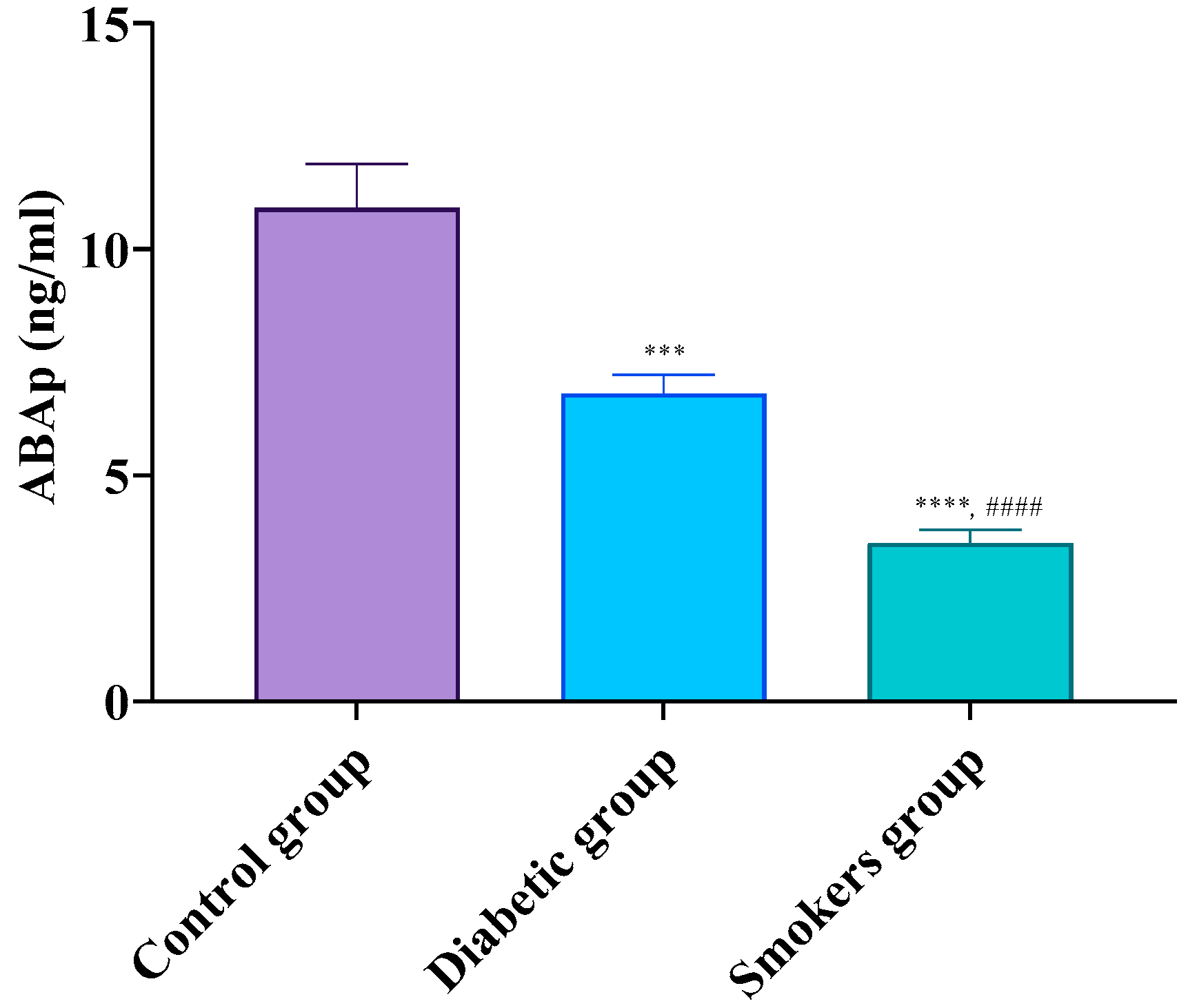
Figure 1. Abscisic acid concentrations in control, diabetic, and smoking groups. Abbreviations: ABA, abscisic acid. Data were analyzed with One-way ANOVA followed by Tukey’s multiple comparisons post hoc test; *** p < 0.01, **** p < 0.0001 significantly different vs. control group; #### p < 0.0001 significantly different vs. diabetic group.
Diabetology 06 00093 g001
Figure 2. Scatterplot showing the dose–response relationship between the number of cigarettes smoked per day and plasma ABA levels in smokers.
Diabetology 06 00093 g002
Table 1. General and metabolic parameters of the study participants.
ParametersControl GroupDiabetic GroupSmokers Group
M/F33/1960/4136/20
Age (years)61.6 ± 12.367.2 ± 10.763.5 ± 18.3
BMI (kg/m2)25.1 ± 8.229.4 ± 7.426.7 ± 9.6
Diabetes duration (years)N/A10.2 ± 6.1N/A
FPG (mg/dL)84.2 ± 11.1127.3 ± 50.5 ****93.3 ± 14.7 ####
HbA1c (%)5.0 ± 0.36.7 ± 1.65.0 ± 0.2
Total cholesterol (mg/dL)180.4 ± 25.5151.7 ± 39.9164.3 ± 30.3
HDL-cholesterol (mg/dL)44.0 ± 5.435.9 ± 7.343.5 ± 6.7
LDL-cholesterol (mg/dL)83.7 ± 29.6106.5 ± 20.2 *93.5 ± 22.5
Triglyceride (mg/dL)92.6 ± 24.1167.4 ± 18.1 ****106.0 ± 17.4 ####
ALT (UI/L)27.6 ± 11.625.0 ± 11.626.2 ± 10.7
AST (UI/L)23.4 ± 7.519.0 ± 7.522.1 ± 9.4
eGFR89.2 ± 15.378.6 ± 18.785.1 ± 14.9
Uric acid (mg/dL)4.9 ± 0.74.5 ± 1.54.8 ± 1.0
Values are means ± standard deviation. Abbreviations: ALT, Alanine aminotransferase; Aspartate aminotransferase; BMI, body mass index; eGFR, estimated glomerular filtration rate; F, females; FPG, fasting plasma glucose; HbA1c, glycated hemoglobin; M, males; N/A, not applicable. Data were analyzed with Two-way ANOVA followed by Tukey’s multiple comparisons post hoc test; * p < 0.05, **** p < 0.0001 significantly different vs. control group; #### p < 0.0001 significantly different vs. diabetic group.
Disclaimer/Publisher’s Note: The statements, opinions and data contained in all publications are solely those of the individual author(s) and contributor(s) and not of MDPI and/or the editor(s). MDPI and/or the editor(s) disclaim responsibility for any injury to people or property resulting from any ideas, methods, instructions or products referred to in the content.