Review

Exploring the Epidemiologic Burden, Pathogenetic Features, and Clinical Outcomes of Primary Liver Cancer in Patients with Type 2 Diabetes Mellitus (T2DM) and Metabolic Dysfunction-Associated Steatotic Liver Disease (MASLD): A Scoping Review

Hepatogastroenterology Division, Department of Precision Medicine, University of Campania Luigi Vanvitelli, 80138 Naples, Italy
*
Author to whom correspondence should be addressed.
Diabetology 2025, 6(8), 79; https://doi.org/10.3390/diabetology6080079 (registering DOI)
Submission received: 26 June 2025 / Revised: 12 July 2025 / Accepted: 28 July 2025 / Published: 4 August 2025

Abstract

Background/Objectives: Primary liver cancer (PLC), encompassing hepatocellular carcinoma (HCC) and cholangiocarcinoma (CCA), constitutes a growing global health concern. Metabolic dysfunction-associated Steatotic Liver Disease (MASLD) and Type 2 diabetes mellitus (T2DM) represent a recurrent epidemiological overlap. Individuals with MASLD and T2DM (MASLD-T2DM) are at a higher risk of PLC. This scoping review highlights the epidemiological burden, the classic and novel pathogenetic frontiers, and the potential strategies optimizing the management of PLC in MASLD-T2DM. Methods: A systematic search of the PubMed, Medline, and SCOPUS electronic databases was conducted to identify evidence investigating the pathogenetic mechanisms linking MASLD and T2DM to hepatic carcinogenesis, highlighting the most relevant targets and the relatively emerging therapeutic strategies. The search algorithm included in sequence the filter words: “MASLD”, “liver steatosis”, “obesity”, “metabolic syndrome”, “body composition”, “insulin resistance”, “inflammation”, “oxidative stress”, “metabolic dysfunction”, “microbiota”, “glucose”, “immunometabolism”, “trained immunity”. Results: In the MASD-T2DM setting, insulin resistance (IR) and IR-induced mechanisms (including chronic inflammation, insulin/IGF-1 axis dysregulation, and autophagy), simultaneously with the alterations of gut microbiota composition and functioning, represent crucial pathogenetic factors in hepatocarcinogenesis. Besides, the glucose-related metabolic reprogramming emerged as a crucial pathogenetic moment contributing to cancer progression and immune evasion. In this scenario, lifestyle changes, simultaneously with antidiabetic drugs targeting IR-related effects and gut-liver axis, in parallel with novel approaches modulating immunometabolic pathways, represent promising strategies. Conclusions: Metabolic dysfunction, classically featuring MASLD-T2DM, constitutes a continuously expanding global issue, as well as a critical driver in PLC progression, demanding integrated and personalized interventions to reduce the future burden of disease.

1. Background

1.1. Primary Liver Cancer in the Era of MASLD: Epidemiological Changes Guide the Etiological Shift

In the era of human cancer as the “disease of the century”, the primary liver cancer (PLC) currently constitutes a serious social-health global burden, representing the seventh most common neoplasm and the second leading cause of tumor-related mortality worldwide (5-year survival rate~18%) [1]. Epidemiological projections are dramatic, revealing an increasing trend of incidence and mortality rates in the next decade [2,3].
Hepatocellular carcinoma (HCC) is the most common type of PLC (approximately 75% of the total cases), followed by cholangiocarcinoma (CCA) [intrahepatic (ICC) and extrahepatic (ECC)] [1].
A large variety of risk factors has been associated with HCC occurrence and progression, reflecting the heterogeneity of socio-epidemiological variables featuring different geographical areas. These include the unequal availability of adequate link of care strategies with the possibility of access to proper healthcare resources, as well as the exposure to certain zone-specific environmental and infectious risk factors [3]. In Asia and sub-Saharan Africa, indeed, dietary exposure to aflatoxins through contamination of foods such as corn, exposure to microcystins (toxins of blue-green algae present in the water of ditches and ponds) [4], the habit of chewing betel nuts [5] and infection by major hepatotropic viruses [Hepatitis C infection (HCV) and Hepatitis B infection (HBV)] continue to represents prevalent risk factors [6]. On the contrary, in Western countries, alcohol abuse, smoking, and, overall, metabolic syndrome, emerge as main determinants for HCC [3].
As for CCA, similarly to HCC, different risk factors have been recognized according to the specific geographical area. In this sense, “CC high-risk” countries (i.e., Southeast Asian zones) have historically been represented by zones where parasitic infections constitute the predominant risk factors. Contrariwise, “CCA low-risk” zones (i.e., Western countries) have historically been represented by areas where metabolic comorbidities constitute the key contributors to biliary cancerogenesis [7].
In the last decade, in contrast to this traditional scenario, a significant epidemiological shift in the incidence rates of CCA has been observed, reporting a significant increase in historically low-risk countries [7], in parallel with the massive spreading of obesity (“pandemic obesity”) and type 2 diabetes mellitus (T2DM) in these industrialized areas [7].
In the panorama of liver diseases, a significant epidemiological overlap between Metabolic-Dysfunction Associated Steatotic Liver Disease (MASLD) and T2DM exists, estimating an elevated prevalence (~70% in Western countries) of T2DM in patients presenting MASLD [8]. MASLD is a chronic clinical-pathological condition defined by the evidence of hepatic steatosis (histologically ranging from “simple steatosis” to “steatohepatitis”—“MASH”, and advanced fibrosis [9]) and cardiometabolic risk factors (CMRFs) (including obesity, T2DM, arterial hypertension, hypertriglyceridemia, and dyslipidemia) [10]. MASLD represents the leading cause of chronic liver damage, cirrhosis, and hepatic cancer in Western countries [9,10,11]. According to the new diagnostic criteria, MASLD is defined by the presence of hepatic steatosis—documented by imaging or histology—in combination with at least one of five CMRFs (obesity, T2DM, arterial hypertension, hypertriglyceridemia, and dyslipidemia) [10,12].
Despite the recent important changes in nomenclature of hepatic steatosis [i.e., the progressive shift from NAFLD (Non-Alcoholic Fatty Liver Disease) to MAFLD (Metabolic-dysfunction Associated Fatty Liver Disease), and, lastly, to MASLD] with relevant modifications of relative diagnostic criteria, T2DM continues to represent a cornerstone CMRF contributing to configure the “metabolic dysfunction” (MD) in MASLD [10].
In this sense, the recent transition from “NAFLD” to “MAFLD” and, ultimately, “MASLD” with the relative changes in diagnostic criteria configuring MD marked a major conceptual shift, satisfying the need to consider this disease as a systemic condition, with a multifactorial pathogenesis [10,12]. These criteria have been demonstrated to enable earlier identification of at-risk populations. Particularly, supporting this, He et al. demonstrated that the MASLD definition more accurately identifies individuals at risk of developing T2DM [13]. Moreover, MASLD criteria have proven superior to MAFLD in stratifying lean individuals with T2DM and hepatic steatosis [14].
Interestingly, robust evidence supports a close relationship between MASLD, T2DM, and PLC, highlighting a generically increased risk of PLC onset in MASLD patients [15,16,17], twice the risk of HCC occurrence particularly in T2DM-affected patients [18], a protective role of certain antidiabetic drugs in reducing HCC onset risk [19], as well as a strongly positive association between T2DM and the increased risk of CC [20].

1.2. Primary Liver Cancer in MASLD: Impact of T2DM on Relative Occurrence Risk and Prognosis

Patients with MASLD are inherently at increased risk for developing PLC, both HCC and CCA [15,16,17]. The risk of PLC onset in adult/elderly patients, in parallel (and consistently) with the cumulative occurrence of dysmetabolic comorbidities, has been largely reported to progressively increase with aging, presenting stable epidemiological projections [21]. Alarmingly, the worldwide burden of hepatic cancer in younger MASLD populations has been only recently explored, revealing a dramatic scenario. About this, a recent review by Danpanichkul et al., analyzing the Global Burden of Disease (GBD) data from 2010 to 2019 to estimate the incidence and mortality of PLC in young adults, revealed a general decline in HCC-related mortality across major etiologies, with the notable exception of MASLD and alcohol use disorder (AUD) [22]. Specifically, among young adults with MASLD, liver cancer burden has been rising, with an estimated annual increase of 0.87% globally, considering HCC and CCA [22].
These malignancies share several features with MASLD, and MD sustained by T2DM and obesity [23,24], with relative changes in body composition, certainly represent particularly prominent risk factors, even in the case of rapid onset [25]. In support of this, a recent multicenter study by Dallio et al. evaluated the impact of acute lifestyle changes (i.e., reduced physical activity with increased sedentary daily time and improper dietary habits) occurring during the relatively short interval of the Severe Acute Respiratory Syndrome Coronavirus 2 (SARS-CoV-2) pandemic on MASLD progression [26]. Of note, the authors reported an increased occurrence of HCC overall and HCC out of the Milan criteria during this period, independently from liver disease severity (i.e., baseline fibrosis severity and subsequent fibrosis progression), highlighting the modifications in body composition (mainly, reductions in visceral fat and increases in muscle mass) and glucose metabolism-related parameters as the variables associated with the outcomes [26].
In patients with dysmetabolic liver disease, the presence of T2DM is associated with a significantly increased risk of liver-related events, including HCC occurrence, as evidenced by a recent individual participant data meta-analysis [27]. This research included 2016 adults with MASLD (736 with T2DM and 1280 without) and a median follow-up of 2.8 years, reporting a cumulative 1-year risk of HCC was 1.34% in diabetics compared to 0.09% in non-diabetics [27].
Similar associations have been observed between T2DM and the development of CCA. A large analysis from the Liver Cancer Pooling Project, which included over 1.5 million individuals, found that T2DM was associated with an 81% increased risk of CCA [23].
Moreover, in their umbrella review, Tsilidis et al. systematically assessed evidence from multiple meta-analyses of observational studies to explore associations between T2DM and cancer incidence or mortality [28].
The link between T2DM and CCA was among the most robust associations supported by high-quality evidence. Notably, the association withstood prediction interval testing, indicating potential generalizability of findings across populations [28].
In line with this, Li et al. conducted a meta-analysis of 20 observational studies (15 case-control and 5 cohort studies) and reported a significant association between T2DM and overall CCA risk (relative risk [RR] 1.74) [29]. Stratification by anatomical subtype further confirmed this association, with an RR of 1.93 for ICC and 1.66 for extrahepatic ECC [29]. Altogether, these findings suggest that T2DM may act as an independent risk factor for both major forms of CCA.
Beyond its role in carcinogenesis, T2DM also appears to influence the prognosis of MASLD patients developing PLC neoplasms [30]. Patients with either HCC or CCA and concomitant T2DM consistently exhibit worse survival outcomes compared to non-diabetic individuals. Diabetes exacerbates cirrhosis-related complications—including HCC—increasing all-cause mortality [30].
A systematic review and meta-analysis by Wang YG et al. assessed the prognostic impact of T2DM in patients with HCC [31]. Based on 16 observational studies, the authors reported significantly worse overall survival in diabetic patients (Hazard Ratio- HR 1.46) and an increased risk of recurrence (HR: 1.57) [31]. Similarly, a multivariate analysis performed by Takamatsu et al. demonstrated that both T2DM and obesity were independent negative prognostic factors for overall survival and recurrence in patients undergoing hepatic resection for HCC [32].
Although data regarding the impact of T2DM on CCA prognosis remain limited and are primarily derived from retrospective studies, emerging evidence points toward a similarly detrimental effect. A retrospective study involving 102 patients with resected ICC found that those with T2DM had significantly lower median overall survival compared to non-diabetics (20 months vs. 32 months) [33]. Another retrospective analysis of 184 patients undergoing hepatectomy for perihilar CCA reported a significantly shorter median overall survival in the diabetic subgroup (23.3 vs. 46.7 months), identifying T2DM as an independent prognostic factor (HR: 1.74) [34]. Notably, patients with T2DM and a future liver remnant (FLR) < 40% had a median survival of only 13.7 months, compared to 35.0 months in those with FLR ≥ 40%, underscoring a potential synergistic impact between metabolic and surgical risk factors [34].
Altogether, the above-presented findings propose that T2DM is increasingly recognized as a key contributor to hepatic oncogenesis and a determinant of long-term prognosis [30].

1.3. Status of the Art and Aims of the Research

In the above-presented scenario, T2DM emerges as an independent risk factor for hepatic malignancies in MASLD, revealing a not-negligible risk of hepatobiliary liver cancer occurrence even in the non-advanced hepatic fibrosis stages (i.e., not cirrhotic patients) in T2DM-affected individuals [35,36], with a significant proportion of potentially early-identifiable cases escaping from the standard surveillance/screening programs currently available for cirrhotic patients [37]. In this sense, this intersection of MD and liver malignancy calls for early risk stratification and surveillance strategies. Despite the mounting evidence, no validated predictive models are available for clinical use to guide individualized management in patients with overlapping metabolic and hepatic conditions.
In this context, multidisciplinary, patient-centered approaches remain essential for optimizing outcomes in this vulnerable population while simultaneously elucidating shared pathogenetic mechanisms to identify targets for developing tailored management strategies [30].
In line with the above-mentioned epidemiological features, MASLD and T2DM potentially share several pathogenetic mechanisms [38] contributing to the hepatic cancerogenesis (e.g., insulin resistance, inflammation, oxidative stress, gut dysbiosis, and immunometabolism). Anyway, these mechanisms remain only partially elucidated and supported by apparently disunited findings, making an overview and further clarification an absolute urgent need.
Considering this, the present research, aims to review and connect the crucial pathogenetic moments promoting hepato-cancerogenesis in the setting of MASLD-T2DM patients, overviewing classic and emerging frontiers, as well as pointing out key targets propaedeutically to the development of novel tailored strategies, potentially optimizing the routine clinical management of these individuals.

2. Methods

This scoping review is based on the Preferred Reporting Items for Systematic Reviews and Meta-analyses extension for Scoping Reviews (PRISMA-ScR) Guidelines (Supplementary File S1) [39]. To address the study aims, a systematic search was undertaken of peer-reviewed and grey literature covering the period between the years 1990 to 2025 on PubMed, Medline, and SCOPUS electronic databases.
In the first phase, original articles (including in vitro, in animals, and human evidence), guidelines, meta-analyses, trials, and systematic reviews—useful for the analysis of the present topic—were consecutively included firstly to define the epidemiological features, and, subsequently, to match the pathogenetic overlapping/shared drivers of MASLD, T2DM, and PLC (both, and separately, HCC and CCA). In the second phase, meta-analyses, guidelines, observational studies, and clinical trials reporting the clinical findings derived from modulating the identified targets were considered. Both intervention and observational studies, and those with and without a comparator/control group, were included in discussing potential therapeutic targets. Regarding meta-analyses and trials, the ones including patients affected by MASLD-T2DM reporting PLC (HCC or CCA) risk, prognostic impact, and interventions affecting liver disease outcomes were included in both phases, opportunely avoiding redundancy. Outcomes included PLC-related survival (overall and progression-free), liver-related events, and mortality in T2DM, MASLD, and MASLD-T2DM patients. Since this review aimed to collect evidence focused on HCC and CCA in MASLD-T2DM patients, the search strategy filters included MASLD, diabetes, and oncology-related terms. More specifically, also considering the recent changes in nomenclature of hepatic steatosis [10,12], the search algorithm included in sequence the filter words: “MASLD”, “MAFLD”, “NAFLD”, “liver steatosis”, “obesity”, “metabolic syndrome”, “body composition”, “insulin resistance”, “inflammation”, “oxidative stress”, “metabolic dysfunction”, “microbiota”, “glucose”, “immunometabolism”, “trained immunity”. The authors screened (titles and abstracts) a total of 232 full texts. 40 articles were excluded because in a language other than English, duplicated, did not investigate relevant outcomes, referred to the topic, or did not describe adequately the adopted methods. At the end of the process, 192 articles were included in this scoping review (Figure 1).

3. Results

3.1. Type 2 Diabetes Mellitus, MASLD, and Hepatic Cancer: From “Classic” to Novel Pathogenesis

3.1.1. Role of Insulin Resistance in Influencing Hepatobiliary Cancerogenesis

The overlap of various environmental and (epi)genetic factors configures a heterogeneous puzzle driving the progression of both T2DM and MASLD, whose multifactorial complex pathogenesis remains incompletely clarified, with significant unexplored molecular landscapes [26,40,41,42,43].
In this scenario, insulin resistance (IR), defined as a reduced responsiveness of target tissues (liver, muscle, and adipose tissue) to physiological insulin levels, constitute the cornerstone of metabolic mechanisms determining a reduction in glycogen synthesis, a failure to inhibit gluconeogenesis and lipolysis, and ultimately an increase in glucose and circulating free fatty acids (FFAs) [44], thus simultaneously representing the pivotal component contributing to TD2M (high glucose serum levels) and MASLD (hepatic fat accumulation) pathogenesis [44,45].
In addition to the above-mentioned metabolic mechanisms suggesting the relative contribution to elevated glucose levels, as well as to the hepatic fat accumulation [46], IR is pleiotropically indirectly (i.e., hyperinsulinemia and hyperglycemia, among others) and directly involved in promoting cancerogenesis [47,48].
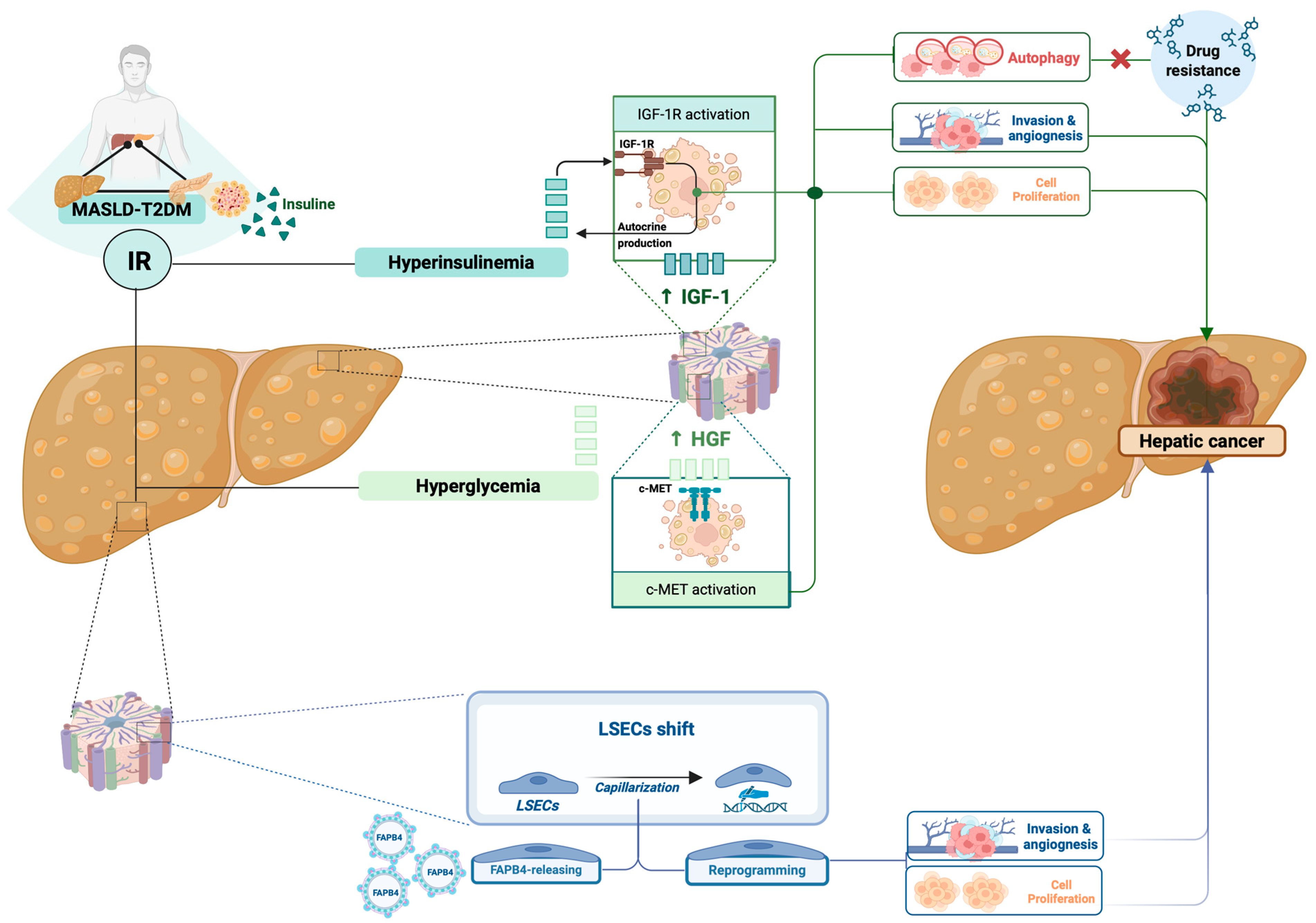
Firstly, IR-related compensatory hyperinsulinemia leads to an enhanced production of insulin-like growth factor 1 (IGF-1) in the liver [49]. IGF-1 is a well-known insulin-metabolism-related mediator whose role, by binding to its receptor (IGF1-R) and leading to downstream upregulation of several oncogenes, in promoting cell proliferation, invasion, angiogenesis, and reducing apoptosis, has been largely investigated in PLC [49].
Recent research has revealed significant alterations within the insulin-like growth factor (IGF) signaling axis that contribute to the molecular pathogenesis of HCC. These include autocrine production of IGFs, dysregulated expression of IGF-binding proteins (IGFBPs), heightened activity of IGFBP-specific proteases, and upregulation of IGF receptors [50]. Among the most characteristic changes observed in HCC tissues and hepatoma cell lines are the overexpression of IGF-1 receptor, which appears to be the key driver of malignant transformation and tumor progression [50]. In parallel, reduced IGFBP expression and increased proteolytic degradation enhance the pool of bioactive IGFs, further supporting oncogenic signaling and uncontrolled cell proliferation [50].
Moreover, IGF-1 has recently been shown to contribute to HCC development in T2DM patients, also by promoting autophagy [51].
Autophagy represents a way for tumor cells to survive in hostile environments, such as hypoxia or nutrient starvation, commonly observed in growing solid tumors [52]. In these cases, autophagy provides recycled metabolic substrates that help tumor cells resist stress, promoting their survival and resistance to chemotherapy regimens [52]. In a relevant in vivo-in vitro study on this topic, Shan et al. enrolled thirty-three HCC patients with T2DM and 33 age-matched patients with HCC without T2DM, assessed the IGF-1 levels and the mRNA expression of autophagy-related molecules (LC3 and p62 mRNA), and evaluated the prognosis of the two groups [51]. Relevantly, a higher expression of IGF-1 in HCC patients with T2DM was observed, as well as elevated IGF-1 was associated with a poor prognosis in patients with HCC [51]. In vitro, after stimulating HepG2 cells with IGF-1 and subsequently detecting changes in autophagy and cell proliferation in the presence/absence of wortmannin (an autophagy inhibitor), the authors reported that IGF-1 promoted autophagy, resulting in inhibition of apoptosis and induction of growth of HepG2 cells, as well as inhibition of autophagy by wortmannin impaired IGF-1 function [51].
Furthermore, the activation of IGF-1R has been also demonstrated to downregulate the sensitivity of liver cancer to tyrosine kinase inhibitors (TKI) through the PI3K/Akt and RAS/ERK signaling pathways [53], as well as the alteration of IGF/IGF1R signaling may stimulate the expression of cancer stem cell traits, contributing to TKI-resistance, increasing the risk of tumor relapse in patients with advanced HCC [54].
As with HCC, elevated levels of IGF-1 induced by hyperinsulinemia seem to play an important role in the development of CCA in the setting of T2DM-MASLD as well [29].
In support of this, Alvaro et al. highlighted significantly higher expression levels of IGF-1 or IGF1-R on immunohistochemical staining of human cholangiocytes from biopsies of cholangiocarcinoma (n = 18), in contrast to cholangiocytes from biopsies of normal livers (n = 10) [55]. In vitro observation revealed that the transfection of IGF-1R anti-sense oligonucleotides in cholangiocarcinoma cell lines (HuH-28 cells) markedly decreased cell proliferation, proposing IGF-1R modulation as an encouraging target [55].
Besides the above-presented relevant findings, IR has also been demonstrated to directly influence and promote the early capillarization of liver sinusoidal endothelial cells (LSECs), a physiopathological moment crucially contributing to creating a pro-oncogenic microenvironment, significantly increasing the risk of PLC in this dysmetabolic context, particularly HCC [56].
LSECs are strategically located at the interface between circulating blood and the hepatic parenchyma [57], representing highly specialized cells, distinguished by the presence of transcellular pores called “fenestrae” [57]. In physiological conditions, LSECs exert anti-inflammatory and anti-fibrotic functions. However, during the IR-guided progression of hepatic steatosis, LSECs undergo capillarization and acquire traits similar to vascular endothelial cells, thereby actively contributing to key pathological features of the disease, such as steatosis, inflammation, and fibrosis [57].
The dysfunction of LSECs plays a pivotal role in the transition to MASH and HCC, whereas restoring their homeostasis has emerged as a promising strategy to reverse liver tissue damage [56,57].
Relevantly, the loss of key LSEC markers such as stabilin-1, stabilin-2, and CD32b has been recognized as a hallmark of HCC progression in both murine and human studies [58,59,60]. Moreover, LSECs associated with HCC display enhanced angiogenic potential compared to their physiological counterparts [58,59,60]. In line with this, in vitro experiments demonstrated that these cells exhibit significantly greater adhesion to tumor cells, while their adhesion to human peripheral blood leukocytes was markedly reduced compared to healthy LSECs [61], suggesting that their changes represent a significant step in promoting the extrahepatic dissemination of cancer.
In addition to the above-presented phenotypic/functional changes, the metabolic reprogramming of LSECs appears to play a central role in driving the development of IR-related HCC via influencing various extracellular activities. In particular, in vitro studies [in human umbilical vein endothelial cells (HUVECs)] revealed that Fatty acid binding protein 4 (FABP4)—a cytosolic fatty acid chaperone implicated in metabolic syndrome-related liver carcinogenesis, whose overexpression in HCC samples from IR-MASLD in comparison to other etiologies was recently demonstrated [62]—is incorporated into endothelial-derived microvesicles, which in turn stimulate hepatocyte proliferation, migration, and enhance expression of proangiogenic genes [63]. Interestingly, the upregulation of endothelial FABP4 expression, via activation of the Mammalian Target of Rapamycin (mTOR) signaling pathway, is promoted in conditions that emulate the hepatic microenvironment typical of T2DM-MASLD, including hypoxia and elevated levels of glucose (i.e., hyperglycemia) [63].
Hyperglycemia itself plays a direct role in the pathogenesis of PLC (both in HCC and CCA) through various mechanisms of action [64].
Elevated serum glucose levels would provide abundant nutrients, leading to altered cellular energy metabolism, promoting cell growth and proliferation, especially through the activation of the c-Met pathway [65]. Met is a transmembrane receptor tyrosine kinase belonging to the MET (MNNG HOS transforming gene) family, structurally related to the insulin receptor (INSR) tyrosine kinase, whose overexpression has been reported in a variety of malignancies, including PLC [66].
Hepatocyte growth factor (HGF) represents the proper c-MET ligand, and hyperactive HGF/c-MET signaling promotes cancer growth and survival [67]. Concerning this, cornerstone research on this topic by Fafalios et al. demonstrated that the HGF-Met signaling axis plays a key regulatory role in hepatic metabolism by promoting glucose uptake and inhibiting glucose output, presenting Met as a crucial factor for a full hepatic insulin response [68]. In particular, the functioning through direct interaction with the insulin receptor (INSR) to form a Met-INSR hybrid complex, which amplifies downstream signaling, was reported, as well as the HGF-Met pathway was shown to restore insulin sensitivity in a mouse model exhibiting IR [68]. Of note, in IR-influenced conditions, an enhanced expression of HGF, c-Met, and the glucose transporter GLUT-1 has been largely reported, contributing to tumor growth, angiogenesis, and cell migration, in HCC [69] as well as in CCA [70].
Interestingly, even though solid evidence supports that HGF and its receptor MET play critical roles in liver carcinogenesis, all HGF-MET kinase activity-targeted drugs have failed in clinical trials [71]. Aiming to explain this phenomenon, in their research, Huang et al. firstly observed that HGF stimulus facilitated the Warburg effect and glutaminolysis to promote biogenesis in multiple hepatic cancer cells [71]. About this, the authors identified the pyruvate dehydrogenase complex (PDHC) and GLS/GLS1 as crucial substrates of HGF-activated MET kinase, revealing how MET-mediated phosphorylation inhibits PDHC and activates GLS to promote cancer cell metabolism. Relevantly, the key residues of kinase activity in MET (Y1234/1235) were shown as a conserved LC3-interacting region motif (Y1234-Y1235-x-V1237). Therefore, on inhibiting HGF-mediated MET kinase activation, Y1234/1235-dephosphorylated MET was revealed to induce autophagy to maintain biogenesis for hepatic cancer cell survival. Conclusively, the authors confirmed these findings by verifying that Y1234/1235-dephosphorylated MET correlated with autophagy in clinical liver cancer, as well as reporting a significant improvement in the therapeutic efficiency of PLC (in vitro and in mice) via combining MET inhibitor with autophagy suppressor, proposing thus a MET-autophagy double-targeted strategy to overcome chemotherapeutic resistance in liver cancer [71].
Finally, IR, via mechanisms increasing bile cholesterol secretion, leads to the development of cholesterol gallstones [72], another well-known risk factor for the development of CCA, supported by chronic hepatic inflammation and consequently damage to biliary epithelial cells [29].
Figure 2 summarizes the most relevant IR-related metabolic pathogenetic moments contributing to the onset and progression of hepatic cancer (Figure 2).
Altogether, the above-reported evidence supports the crucial role of IR in determining heterogeneous metabolic effects impacting the occurrence and progression of hepatobiliary cancer. Anyway, as detailly in the next section, IR has also been associated with hepatic phlogosis, constituting a vicious circle where IR and inflammation appear significantly and mutually involved in the genesis and progression of PLCs in MASLD-T2DM patients [40,44].

3.1.2. Insulin Resistance-Related Inflammation Influences Hepatobiliary Cancerogenesis

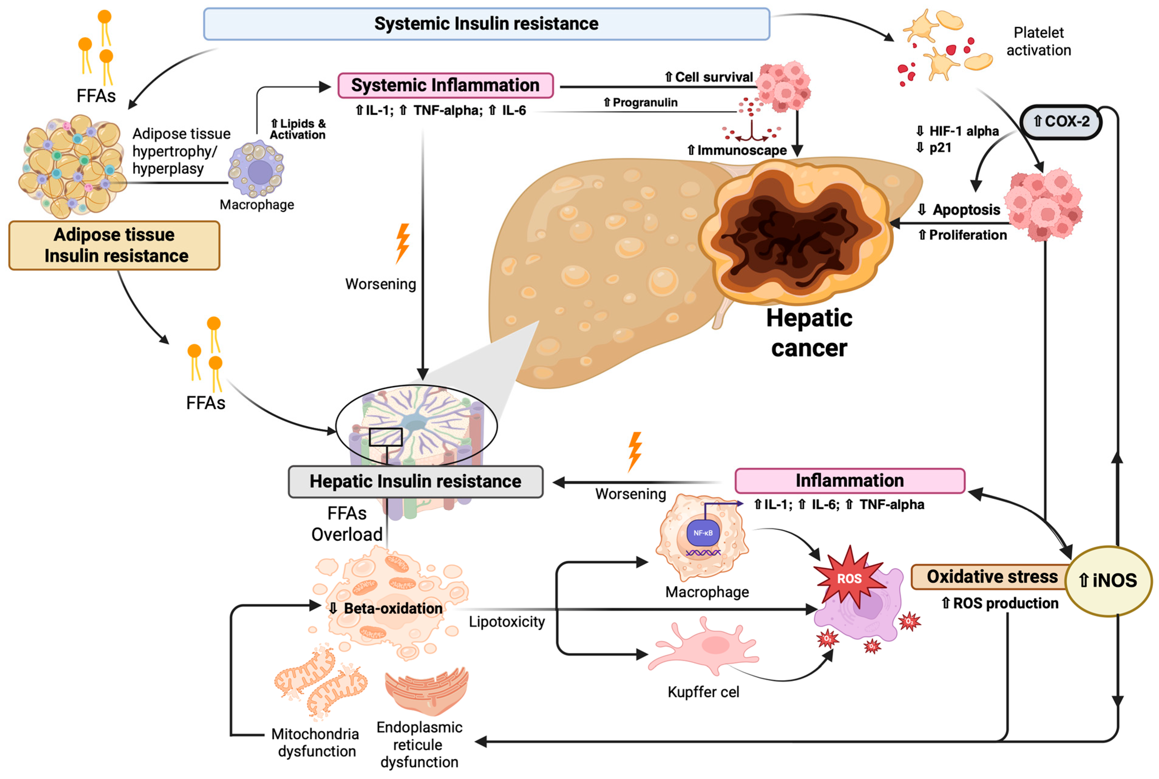
The classical pathogenetic theory identifies the IR-related excess circulation of FFAs directed to the liver as the primum movens of an entangled pathogenetic network where inflammation and oxidative stress represent the two other crucial components [40,44]. Firstly, the overload of FFAs at the hepatic level, consequent to the impairment of relative mitochondrial beta-oxidation mechanisms, results in excessive accumulation of fats as triglycerides [73]. In a second moment, when the hepatic capacity for lipid accumulation is saturated, toxic intermediates are generated and lipotoxicity occurs, triggering the activation of Kupffer cells and hepatic macrophages. The immune-mediated enhanced production of reactive oxygen species (ROS) and release of inflammatory mediators [including interleukin (IL)-1beta, IL-6, and Tumor Necrosis Factor alpha (TNF-alpha)], as well as the dysfunction of cytoplasmic organelles (including mitochondria and endoplasmic reticule), ultimately promotes a microenvironment simultaneously driving the worsening of MASLD (to MASH) and promoting the PLC onset and progression [40,44,74].
In this sense, IR could act as a proinflammatory “danger signal,” potentially triggering the activation of Kupffer cells and hepatic macrophages. In support of this, intracellular lipid accumulation, as observed in both human subjects and rodent models, has been shown to promote nuclear translocation of NF-κB (promoting IL-1beta and IL-6 release), impair mitochondrial respiratory function, induce organelle fragmentation and mitophagy, and elevate the generation of ROS [75,76].
These conditions simultaneously create a microenvironment where PLC is promoted, configuring a vicious circle where IR is furtherly worsened [76].
Looking also outside the liver, extra-hepatic IR-determined effects have been shown to indirectly impact hepatobiliary carcinogenesis, particularly via promoting systemic inflammation and oxidative stress-related pathways [40,44]. Among these, the ability to influence platelet activation certainly represents a relevant consequence of high glucose serum levels, potentially contributing to the PLC onset [77]. Activated platelets are involved in promoting chronic inflammatory processes through the release of pro-inflammatory cytokines, such as IL-1beta and IL-8, and growth factors [including transforming growth factor β (TGF-β) and vascular endothelial growth factor (VEGF)] involved in the Cyclo-Oxygenase-2 (COX-2) pathway in tumor cells [77]. COX-2 is an enzyme involved in inflammatory processes by enhancing the synthesis of prostaglandins, whose upregulation in tissue specimens from HCC and CCA has been largely demonstrated [78,79,80]. In HCC, evidence supports COX-2 overexpression, as well as the association of this with apoptosis and chemosensitivity resistance via the HIF-1alpha/PKM2 pathway and Drp-1 dependent remodeling of mitochondrial dynamics, proposing its role as a therapeutic target [78,79,80].
Similarly, elevated levels of COX-2 have been associated [81] with tumor growth promotion in CCA, as well as pharmacological inhibition of COX-2 has been shown to trigger apoptosis and suppress cell proliferation, primarily through attenuation of Akt signaling and activation of p21 along with other cyclin-dependent kinase inhibitors [82,83].
Interestingly, COX-2 expression is, in part, modulated by inducible nitric oxide synthase (iNOS), which was revealed to be overexpressed in biopsy specimens from patients with advanced CCA [84]. Interestingly, iNOS is itself upregulated in response to pro-inflammatory cytokines [84], thus configuring a vicious circle where elevated glucose levels, directly contributing to the overproduction of ROS, lead to mitochondrial dysfunction and amplify inflammation, ultimately increasing the risk of hepatocarcinogenesis [85].
In this sense, inflammation represents another crucial extra-hepatic IR-determined factor influencing PLC onset and progression even at the systemic level [85].
In an IR scenario, an inability of adipose tissue to adequately store lipids can be generated, leading to an increase in adipose tissue through hypertrophy and not hyperplasia [86]. This increase in adipocyte size leads to a reduction in insulin sensitivity, with accumulation of lipids within M1 macrophages, which enhances the release of pro-inflammatory cytokines, such as IL-6 and TNF alpha, contributing to low-grade chronic systemic inflammation [86].
Also, the hypertrophy of adipose cells typically observed in MASLD-T2DM (obese) patients can lead to hypoxia and cell death, with further recruitment of inflammatory cells in the adipose tissue, enhance in the production of pro-inflammatory cytokines, and worsening systemic inflammation, which in turn negatively impact IR, significantly increase the risk of PLC in this setting [86].
Consistent with this, Yuhua et al., in T2DM individuals, compared to healthy controls, reported higher levels of pro-inflammatory cytokines, such as IL-6 and TNF-alpha, were found, demonstrating a key role in the development of HCC and relative progression via immunoescape [87].
Similarly, in CCA, IL-6 was shown to promote cell survival by activating the transcription factor Signal transducer and activator of transcription 3 (STAT3), which in turn upregulates the anti-apoptotic protein Mcl-1 [88].
Additionally, IL-6 enhances the expression of progranulin—a precursor of granulins involved in cell growth regulation—thereby triggering the protein kinase B (PKB) signaling cascade, ultimately supporting key oncogenic processes, including cell survival, proliferation, migration, and angiogenesis [89,90].
Figure 3 summarizes the most relevant IR-related pathogenetic moments contributing to the onset and progression of hepatic cancer via promoting inflammation and oxidative stress (Figure 3).
Furthermore, in the presence of excess adipose tissue, leptin, the hormone regulating satiety, is overproduced and has been shown to promote the proliferation of cholangiocarcinoma (CCA) cells in MASLD-T2DM (obese) patients [91]. The association between MASLD-obesity/T2DM and CCA has also been confirmed by in vitro experiments, revealing that high leptin levels can stimulate both cell migration and proliferation, while preventing apoptosis in CCA cells [23]. In this sense, although the hepatocyte is the main focus of liver dysmetabolic disease, the bile ducts may become the primary target in the subgroup of MASLD-T2DM (obese) individuals [92].
In line with this, in these subjects, Manieri et al. highlighted the activation of the stress signaling pathway mediated by c-Jun NH2-terminal kinase (JNK) [93]. Particularly, the authors observed that a deficit in the JNK-mediated stress signaling pathway could contribute to an improvement in the development of IR and hepatic steatosis, thus initially considering this factor a potential therapeutic target [93]. However, subsequently, long-term inhibition of this pathway was reported to determine modifications in cholesterol and bile acid (BA) metabolism, increasing the risk of inflammation in bile cells and the subsequent development of CCA [93].
In the above-presented complex scenario, where IR-related hepatic and systemic inflammation play a crucial role in impacting several moments in the hepato-cancerogenesis, emerging evidence supports the modifications of the gut microbiota composition (i.e., dysbiosis) and relatively altered functioning as a deus ex machina contributing to PLC, which, considering its frequent recurrence in patients with MASLD and T2DM, deserves to be illustrated in the next dedicated sub-paragraph.

3.1.3. MASLD-T2DM-Associated Gut Dysbiosis in the Pathogenesis of Primary Liver Cancer

The gut microbiota is a complex ecosystem of microorganisms—including bacteria, fungi, viruses, and other microbes—that inhabit the gastrointestinal tract, playing crucial roles in digestion, metabolism, immune regulation, and defense against pathogens [94].
Multiple factors, including diet, antibiotic use, and overall health status, can influence the composition of the gut microbiota [95,96,97]. Alterations in the gut microbiota and gut-liver axis have been shown to play a significant role in the pathogenesis and progression of metabolic disorders such as T2DM and metabolic syndrome, as well as liver diseases like MASLD and hepatic cancer [94].
The existence of shared microbial alterations among these conditions underscores the importance of characterizing the structure and function of the gut microbiota to better understand disease mechanisms and identify disease-specific microbial biomarkers. These may ultimately lead to novel therapeutic opportunities for preventing or treating the aforementioned diseases, including PLC.
Numerous studies have provided insight into the interplay between gut microbiota, the gut-liver axis, and the development of IR-related manifestations. In particular, increased microbial richness has been associated with a protective effect against metabolic syndrome and T2DM [98]. In contrast, T2DM patients often exhibit reduced microbial diversity and a lower metagenomic richness, a condition associated with increased IR and chronic low-grade inflammation [99]. Moreover, these individuals frequently display qualitative alterations in microbiota composition.
One of the earliest studies on the gut microbiota in T2DM, conducted by Larsen et al., reported decreased levels of the Firmicutes phylum and the Clostridia class [99]. In general, a prominent feature of the dysbiotic profile observed in MASLD and T2DM patients is the overrepresentation of pathogenic and opportunistic Gram-negative bacteria (including members of the Enterobacteriaceae family, various Clostridiales, Escherichia coli, Bacteroides caccae, Lactobacilli, Prevotella copri, and Bacteroides vulgatus) [100,101].
Interestingly, accumulating evidence indicates that alterations in the gut microbiota composition and functioning significantly contribute to the development and progression of HCC by modulating multiple pathways along the gut-liver axis in MASLD-T2DM [94,102]. In MASLD-T2DM patients, dysbiosis and microbially derived metabolites can profoundly influence chronic inflammation and host immune responses, collectively shaping a tumor-permissive microenvironment [94].
Although pinpointing specific microbes linked to MD and T2DM remains challenging, certain bacterial species play a pivotal role in initiating metabolic inflammation during hepatic disease worsening, including cancerogenesis [94,103], and, consistently, an imbalance in the composition of gut microbiota has been extensively reported in patients with MASLD-related HCC [94,102,104].
In this context, it has been comprehensively demonstrated that the intestinal flora of dysmetabolic HCC patients exhibits elevated levels of Escherichia coli and other Gram-negative bacteria. Conversely, the gut microbiota of MASLD-T2DM associated-HCC individuals shows reduced levels of Bifidobacterium spp. and Enterococcus spp [104].
This condition has been demonstrated to be closely linked to increased serum levels of LPS, leading to systemic inflammation [104]. In the context of gut-liver axis dysregulation, indeed, increased intestinal permeability facilitates the translocation of microbial products, especially LPS, into the portal circulation. These components activate inflammatory responses in the liver by binding to receptors such as Toll-like receptor 4 (TLR4), thereby promoting chronic inflammation, fibrogenesis, and the neoplastic transformation of hepatocytes [105].
Escherichia coli is frequently enriched in IR-related dysbiotic states, and the enrichment of the gut commensal Escherichia coli has been demonstrated to aggravate the High-Fat-Diet (HFD)-induced IR in the mouse model [103]. Escherichia coli produces high levels of LPS that activate TLR4 on Kupffer cells, leading to chronic liver inflammation and oxidative stress, ultimately driving hepatocarcinogenesis [106]. In a mouse study conducted by Dapito et al., TLR4 expression in HCC tissues (77.8% positivity) was significantly higher than in adjacent non-tumor tissues (20%) [107].
Moreover, emerging data also point to Fusobacterium nucleatum, an opportunistic pathogen commonly found in gastrointestinal tumors [108] and diabetic patients [109], as a potential contributor to hepatocarcinogenesis through TLR4-dependent modulation [110].
Bacteroides fragilis represents another commensal whose enhanced representation (after supplementation in the mouse model) in a dysmetabolic context has been revealed to promote general gut dysbiosis (by reduced Lactobacillaceae and increased Desulfovibrionaceae abundance), as well as lead to the deterioration of glucose/lipid metabolic dysfunction and inflammatory response [111]. Relevantly, a relatively increased abundance of Bacteroides fragilis, known to produce LPS and other relevant virulence factors, has been shown to potentially enhance tumor-promoting inflammation through the induction of senescence-associated secretory phenotypes in hepatic stellate cells [112].
Table 1 reports the most relevant alterations in gut microbes’ representation and the relative pathogenetic implications in HCC in IR-/MD-related scenarios (Table 1).
As for CCA, similarly to HCC, recent advances have highlighted the gut and biliary microbiota as pivotal contributors to the initiation and progression of biliary cancerogenesis in IR-related dysmetabolic contexts as MASLD and T2DM [94]. The altered representation of different specific microbial taxa (including Escherichia coli, members of the Enterobacterales, Clostridium spp., Helicobacter spp., Bacteroides spp., Veillonella spp., and Alistipes spp.) has been functionally associated with cancer-related processes through potential mechanisms including IR worsening, chronic inflammation persistence, immunomodulation, and, even more relevant, BA metabolism modification, whose impairment occupies a privileged position in this scenario [94].
Gut microbes modulate the composition and hydrophobicity of the BA pool through enzymatic activities by enzymatically transforming primary BAs into more hydrophobic and cytotoxic secondary BAs—such as deoxycholic acid (DCA) and lithocholic acid (LCA)—via deconjugation and 7α-dehydroxylation [114]. These altered BAs have been shown to modulate receptor activity by acting as signaling molecules interacting with nuclear and membrane-bound receptors (including FXR and TGR5) expressed on cholangiocytes, as well as to induce endoplasmic reticulum stress, oxidative damage, and inflammatory responses in biliary epithelial cells, contributing thus to a pro-carcinogenic niche [107]. About this, of note, specific microbial species enriched in MD-associated (and obesity-related) dysbiosis, including Clostridium, Bacteroides, and Bifidobacterium, have been shown to regulate BA metabolism and influence the intrahepatic BA milieu [112,115,116,117].
Anyway, recent research by Sang et al. underscored that cholangiocarcinogenesis is not driven by a single microbial species, but rather by complex interactions involving multiple genera and families [118]. For example, Clostridium spp., involved in 7α-dehydroxylation of BAs, contribute to the generation of carcinogenic secondary BAs. Escherichia coli spp. may promote chronic inflammation via LPS production, while reductions in Bacteroides may disturb bile acid homeostasis, enhancing a pro-inflammatory and tumor-promoting milieu [118]. Collectively, current evidence positions the gut and biliary microbiota as critical modulators of CCA pathogenesis through their effects on BA metabolism, inflammation, and host immunity. These findings pave the way for novel microbiota-targeted strategies in early diagnosis, prevention, and therapeutic CCA intervention.
Table 2 summarizes the most relevant alterations in gut microbes’ representation and the relative pathogenetic implications in CCA in IR-/MD-related scenarios (Table 2).

3.1.4. Glucose-Related Metabolic Reprogramming Contributes to Cancer Immunoescape: The Emerging Frontiers of PLC Pathogenesis

In recent years, the scientific quest to deeply understand the pathogenesis of PLC has increasingly focused on metabolomics as a promising avenue for the development of novel therapeutic strategies. Metabolic reprogramming represents a hallmark of cancer, enabling tumor cells to meet the heightened energy demands required for rapid proliferation, invasion, and metastasis [126].
As the central metabolic organ, the liver orchestrates essential pathways in glucose, lipid, and protein metabolism, playing a pivotal role in systemic energy homeostasis, nutrient storage, detoxification, and the synthesis of critical biomolecules [126]. Accordingly, recent research has concentrated on alterations in glucose metabolism and immunometabolic pathways as key contributors to the PLC pathogenesis, particularly in the setting of dysmetabolic disorders featured by IR and hyperglycemia, such as MASLD-T2DM [127].
In healthy physiological conditions, the liver tightly regulates blood glucose levels, storing excess glucose as glycogen during periods of nutritional surplus and mobilizing it during fasting or energy demand through glycogenolysis [128]. However, in the setting of hepatocarcinogenesis, this balance is disrupted. Similar to other malignancies, hepatic tumor cells undergo profound metabolic reprogramming, most notably in glucose metabolism. These cells exhibit enhanced aerobic glycolysis—a phenomenon known as the Warburg effect—characterized by elevated glucose uptake and lactate production even in the presence of oxygen [128]. This shift facilitates ATP generation and provides metabolic intermediates essential for anabolic processes and cell proliferation [128].
Emerging evidence indicates that this metabolic rewiring also promotes immune evasion in HCC. Especially in the presence of elevated serum glucose levels, tumor cells consume large quantities of glucose and produce significant levels of lactate, leading to a hypoxic and energy-depleted tumor microenvironment [129].
In addition, this metabolic stress negatively impacts the immune response, impairing the cytotoxic activity of effector cells and contributing to immune escape [128,129].
Lactate, a key byproduct of aerobic glycolysis, has been shown to promote a pro-inflammatory milieu by upregulating IL-23 and IL-12 expression and activating the IL-23/IL-17 axis, thereby facilitating tumorigenesis [129].
Additionally, elevated lactate concentrations modulate the activity of the nuclear factor of activated T-cells (NFAT) in T and natural killer (NK) cells, reducing interferon-gamma (IFN-γ) production and blunting anti-tumor immunity. Clinically, serum lactate levels positively correlate with tumor burden in HCC patients [130].
These immunosuppressive properties of lactate contribute to a permissive environment for tumor progression [129].T cells rely primarily on glucose and glutamine to sustain their effector functions [130]. Within the tumor microenvironment, nutrient competition between tumor and immune cells becomes a crucial determinant of immune competence. The aggressive metabolic demands of tumor cells, driven by the Warburg effect, deprive T cells of essential substrates, thereby diminishing their anti-tumor capacity [131,132].
Moreover, glycolytic reprogramming in cancer cells can facilitate immune escape via additional mechanisms. Enhanced glycolysis promotes the expression of pro-apoptotic Fas ligand (FasL) on tumor cells, leading to T cell apoptosis through T-cell receptor (TCR) restimulation [133]. Intriguingly, a positive correlation has been observed between glucose metabolic reprogramming and elevated alpha-fetoprotein (AFP) expression in liver tumors. Since AFP is known to inhibit Fas expression, it has been hypothesized that glucose reprogramming in hepatic cancer may drive AFP upregulation as a strategy to evade immune surveillance [134].
Altogether, these findings suggest how glucose-associated specific metabolic adaptations not only enhance tumor growth and invasion but also reshape the tumor microenvironment, promoting immune escape in PLC [135]. Targeting these metabolic pathways represents a promising therapeutic strategy for PLC, as well as the elucidation of mechanisms sustaining the interaction between metabolic reprogramming and tumor immunity, which can potentially provide novel metabolic targets for innovative therapeutic approaches [43,135].

3.2. Identifying Pathogenetic Targets to Design Tailored Strategies in the Management of MASLD-TD2M Associated PLC: State of the Art

As previously shown, T2DM represents a recognized risk factor for the onset and progression of PLC in MASLD patients [16,18], negatively impacting the prognosis of individuals developing HCC and CCA, dramatically reducing their progression-free survival chances, even after both curative and non-curative treatments [18,136]. In this scenario, the elucidation of the above-presented pathogenetic mechanisms finalized to identifying specific targets, represents a crucial propaedeutic step to the development of novel personalized strategies aiming to optimize the routine clinical approach.

3.2.1. Targeting Insulin Resistance

Several randomized controlled trials and multicenter retrospective studies have demonstrated that treatment (including metformin and other antihyperglycemic agents) targeting the classical IR and the IR-related mechanisms may reduce the risk of PLC in patients with T2DM [19,137].
These findings have raised a growing interest in evaluating the repurposing of antidiabetic agents—particularly metformin, sodium–glucose cotransporter-2 inhibitors (SGLT2-Is), Dipeptidyl peptidase-4 inhibitors (DPP4-Is), and Glucagon-like peptide-1 receptor agonists (GLP-1 RAs)—in earlier stages of chronic liver disease, considering especially patients with MASLD —the current leading cause of liver damage worldwide—as the main population target [138].
Metformin
In the continuously expanding panorama of antidiabetic agents, metformin remains the first-line therapy for T2DM patients [139]. In recent years, its pleiotropic actions have gained attention, particularly its ability to modulate the gut microbiota and the gut–liver axis. Metformin has been shown to enhance intestinal barrier integrity [140], stimulate the production of short-chain fatty acids (SCFAs), modulate BA metabolism, and globally improve glucose homeostasis [141,142].
These pleiotropic effects may contribute to its observed ability to reduce the risk of HCC in patients with chronic liver disease, particularly MASLD. In support of this, evidence suggests that metformin significantly improves overall survival in patients with HCC and T2DM, potentially via both IR-related/metabolic and microbiota-mediated mechanisms [143]. About this, Zhou et al. demonstrated that metformin significantly reduces mortality in HCC patients with T2DM (HR: 0.59), suggesting a potential protective effect in preventing liver disease progression, as well as an oncopreventive role [143].
Recent investigations into the role of antidiabetic agents in the onset and progression of CCA have highlighted a subset of these drugs with potential therapeutic value. While the associations between CCA and agents such as insulin analogues and sulfonylureas remain inconclusive, incretin-based therapies have been linked to a possible increased risk of CCA development and progression [36]. Conversely, biguanides—particularly metformin—exert a protective effect, correlated with a decreased incidence of CCA and the suppression of its progression in experimental models [36]. The potential relationship between incretin-based treatment and CCA risk warrants further investigation, especially as metformin is currently under evaluation in clinical trials [36]. A deeper understanding of the interplay between T2DM and CCA is essential for devising preventive strategies and assessing the suitability of antidiabetic therapies in the context of CCA management. Clarifying metformin’s impact on CCA could pave the way for the repurposing of this well-established and safe compound, potentially enhancing CCA treatment regardless of the patient’s diabetic status.
SGLT2-Is
SGLT2-Is have emerged as promising agents due to their metabolic and organ-protective properties. Beyond their glucose-lowering effects, SGLT2-Is improve IR, promote weight loss, and exert cardiorenal benefits [144].
Oral SGLT2-Is are rapidly absorbed into the bloodstream, remaining in the circulation for hours. On glomerular filtration, they bind specifically to SGLT2 in the luminal membrane of the early proximal tubule to reduce glucose reabsorption by 50–60% [144].
Because of glucose excretion, these drugs are used in patients with T2DM as glucose-lowering therapies, with additional benefits of weight loss and blood pressure reduction [145]. Due to their intrinsic protective action on the heart [146] and kidney [147], these drugs are also widely used in the treatment of cardiac and renal diseases, such as heart failure with reduced ejection fraction or chronic kidney disease.
Recent studies suggest they may also reduce hepatic steatosis and slow fibrosis progression [147], although data regarding their direct impact on PLC incidence remain limited, preclinical, and preliminary. Recently, in vitro investigations assessed the impact of canagliflozin (a well-known SGLT2-I agent) on cell proliferation, apoptosis, migration, and cell cycle dynamics in multiple CCA cell lines [148]. At higher concentrations, canagliflozin reduced cell viability, induced G0/G1 cell cycle arrest, and suppressed migration, whereas lower concentrations paradoxically promoted cell survival and increased the S-phase population [148].
Mechanistically, treatment upregulated nicotinamide phosphoribosyl transferase (NAMPT), activating the NAD+ salvage pathway. Notably, co-administration of a NAMPT inhibitor potentiated the SGLT2-I antitumor efficacy. While these findings underscore its therapeutic potential, the observed NAD+-mediated pro-survival effects suggest a dose-dependent response that warrants further investigation [148].
Nonetheless, preliminary evidence points toward a potential role in reducing hepatic related complications in patients with advanced liver disease [149]. Notably, while data on combination therapy with metformin and SGLT2-Is are sparse, a recent retrospective study by Huynh et al. demonstrated that this regimen is associated with improved 5-year prognosis, reduced risk of hepatic decompensation, and decreased HCC incidence in patients with T2DM and MASLD-related cirrhosis [150].
In parallel with evidence suggesting the relative potential benefits, accumulating real-world data have underscored specific health risks concerning these drugs [151]. Among the most notable are increased incidences of genitourinary fungal infections, largely attributable to glycosuria-induced microbial overgrowth, and cases of euglycemic diabetic ketoacidosis, particularly in insulin-deficient states [151]. Additionally, concerns have arisen regarding rare but serious adverse events such as Fournier’s gangrene, lower-limb amputations—especially with canagliflozin—and bone fractures [151]. Hematologic changes, including erythrocytosis, have been documented, although their clinical relevance remains limited in most patient populations [152].
Despite these risks, the overall safety profile of SGLT2i remains favorable, and their cardiovascular and renal protective effects often outweigh potential harms when appropriately selected. Individualized risk assessment, vigilant monitoring, and patient education remain pivotal in optimizing therapeutic outcomes with this drug class.
DPP4-Is
DPP4-Is, commonly used in T2DM management, have drawn increasing attention for their potential roles in hepatic cancer biology, particularly HCC [153]. DPP4 is a serine protease involved in cleaving chemokines and regulating immune responses. In HCC, CD26/DPP4 is often overexpressed, contributing to impaired T-cell trafficking, as well as promoting IR and inflammation through macrophage polarization dysregulation [153].
Preclinical studies have shown that DPP4-Is, such as sitagliptin, can induce apoptotic pathways in HCC cells (through modulation of p53), as well as attenuate hepatic oxidative stress and preneoplastic changes in rodent models [154]. Furthermore, DPP4 inhibition has been linked to suppression of key metabolic circuits (including the pentose phosphate pathway and the p62/Keap1/Nrf2 axis), highlighting its potential in targeting cancer-specific metabolic vulnerabilities [155].
Clinical emerging observational research suggested a favorable impact on liver-related outcomes, particularly in patients with MD and chronic viral infections [156]. In patients with T2DM and HCV, the administration of DPP4-Is was associated with lower HCC incidence, though direct comparisons with other antidiabetic agents (e.g., SGLT2-Is) show mixed results [156]. Anyway, direct human evidence in HCC remains preliminary, and no data on the role of DPPA-Is in influencing CCA onset and progression currently exists.
In front of these initial results on the effectiveness, an extensive meta-analysis of randomized controlled trials suggested no increased overall cancer risk with DPP4-Is use, and even a possible protective effect against colorectal cancer [157].
Overall, DPP4 inhibitors offer intriguing prospects as adjunctive agents in liver cancer prevention and therapy, warranting further translational and longitudinal investigation.
GLP-1 RAs
GLP-1 RAs, currently approved for the treatment of T2DM and obesity, have shown promising hepatoprotective properties [158]. These agents not only promote weight loss and glycemic control but also exert anti-inflammatory effects that may attenuate MASLD progression [158]. Relevantly, data from randomized controlled trials have shown histological resolution of steatohepatitis with GLP-1 RAs [159].
A recent population-based study highlighted a reduced risk of cirrhosis and hepatic decompensation in MASLD patients treated with GLP-1 RAs [160]. Moreover, emerging evidence suggests a potential reduction in HCC incidence [161], although these effects have not been replicated for CCA, where in vitro and in vivo studies indicate a limited role [36]. Anyway, the expanding clinical application of GLP1-RAs has prompted investigation into potential human health risks [162]. In this sense, controversial evidence exists on the non-oncological and oncological risks deriving from GLP1-RAs therapies, imposing a careful evaluation of the risk-benefit-efficacy ratio in the long-term administration of these molecules for the designation of future trials, as well as for the routine clinical use of these drugs in the management of MASLD-T2DM patients [162].
GLP-1 RAs have been associated with an increased incidence of gastrointestinal adverse effects (including nausea, vomiting, and pancreatitis), as well as recent large-scale studies also suggest a modest elevation in the risk of arthritic and renal complications, including nephrolithiasis and interstitial nephritis [162,163]. Furthermore, a possible link to thyroid disorders, particularly medullary thyroid carcinoma, has been proposed. About this, while early preclinical studies raised concerns about a possible association with medullary thyroid carcinoma due to GLP-1 receptor expression in thyroid C-cells, recent large-scale observational and cohort studies have yielded mixed results [164,165]. Some findings initially suggested a modest increase in thyroid cancer diagnoses shortly after GLP-1 RA initiation, potentially reflecting enhanced surveillance rather than causality [164,165]. Conversely, another multicenter study found no significant elevation in thyroid cancer risk compared to other antidiabetic agents [166]. Overall, current evidence does not support a definitive link between GLP-1 RAs and increased cancer incidence, though long-term and subtype-specific studies remain warranted to clarify residual uncertainties [166].
Table 3 summarizes the therapeutic implications of antidiabetic agents (Table 3).
Despite the encouraging findings, robust prospective and randomized trials are still needed to validate the hepatoprotective effects of the above-presented molecules and to clarify their mechanisms of action and safety.
In the current clinical landscape, a multidisciplinary approach remains the gold standard for the management of patients with MASLD and cardiometabolic comorbidities, particularly T2DM. This is reinforced by the most recent EASL guidelines, which advocate for a patient-centered, personalized approach in the metabolic setting [161]. As of today, lifestyle modification (adherence to Mediterranean dietary habits and proper physical exercise) remains the cornerstone of MASLD management and relative complications, including PLC [167].
The limited therapeutic arsenal has prompted the scientific community to explore innovative strategies. Among these, artificial intelligence (AI) is emerging as a promising tool to personalize lifestyle interventions [168]. Machine learning algorithms have shown efficacy in predicting disease progression, stratifying MASLD patients by risk [169], and even generating tailored dietary plans to promote weight loss [170], improve glycemic control [171], and optimize lipid profiles [172]. This approach, supported by a proper evaluation of individual body composition, represents a comprehensive strategy to adopt in the initial stages of disease (MASLD/T2DM) progression, able to early and effectively reduce the risk of complications, including hepatic cancer [26].

3.2.2. Modulating Gut-Biliary Liver Axis: A Promising and Still Embryonal Strategy

Recent advances have highlighted the gut microbiota as a crucial regulator in hepatobiliary tumor development [94]. Based on this, several microbiota-modulating strategies—including probiotics, antibiotics, prebiotics, dietary interventions, and fecal microbiota transplantation (FMT)—have demonstrated promising antitumor effects in preclinical PLC models [173]. These interventions appear to influence carcinogenesis by restoring gut microbial balance, enhancing intestinal barrier integrity, modulating BA metabolism, and reshaping immune responses within the liver microenvironment [173]. About this, Chen et al. showed that probiotic administration improved the efficacy of immunotherapy in HCC by boosting antitumor immune responses and reducing tumor burden in mice [174]. Similarly, and more recently, Li et al. reported that gut microbiota modulation via dietary and antibiotic strategies altered the gut–liver axis and suppressed hepatocarcinogenesis in animal models [175].
Despite these encouraging findings, the translational relevance to human liver cancer remains uncertain, primarily due to differences in microbiota composition, host genetics, and environmental exposures. While microbiota can be readily manipulated and holds potential as a therapeutic adjunct, rigorous clinical trials are essential to validate safety, efficacy, and long-term outcomes in diverse patient populations [176]. Moreover, the complexity of host–microbiota–tumor interactions necessitates standardized protocols for microbiome profiling and intervention design [176].
Collectively, these insights underscore the therapeutic promise of microbiota-targeted strategies in liver cancer, while also emphasizing the need for well-designed, longitudinal human studies to bridge the gap between bench and bedside.

3.2.3. Modulating Immunometabolic Responses: The Novel Therapeutic Frontier

In parallel to the above-presented strategies, the modulation of immune response and specifically identified metabolic-related targets represents another promising frontier to explore. This approach potentially offers alternative chances in managing the complications and improving the clinical outcomes of PLC-MD-affected patients [126].
The immunosuppressive tumor microenvironment, driven by unchecked neoplastic proliferation and metabolic rewiring, critically dampens T cell effector responses. This immunological dysfunction has profound implications for the systemic treatment of PLCs, including both HCC and CCA. Despite recent advances, current systemic therapies—including immune checkpoint inhibitors (ICIs)—demonstrate limited durable efficacy in advanced HCC and CCA. First-line systemic treatments yield objective responses in only 30% of HCC patients [177] and 26% in those with CCA [178].
Recent insights have challenged the long-standing dogma that innate immunity lacks memory. Instead, it is now recognized that innate immune cells can acquire long-lasting memory-like features through a process termed trained immunity (TI) [179,180]. TI involves epigenetic, transcriptomic, and metabolic reprogramming, resulting in enhanced responsiveness upon re-exposure to stimuli [179,180].
The liver harbors a complex network of Innate Immune cells—including Kupffer cells, dendritic cells, natural killer (NK) cells, and neutrophils—which under physiological conditions maintain tolerance to gut-derived antigens while remaining poised to respond to infections [181]. However, in the setting of chronic liver injury (including the MASLD/MASH), this equilibrium is disrupted: innate immune cells become chronically activated, functionally exhausted, or skewed toward pro-tumorigenic phenotypes [181].
Harnessing TI offers an emerging therapeutic avenue in PLCs. By reprogramming innate immunity, it may be possible to overcome immune resistance mechanisms and enhance antitumor responses in patients with advanced, unresectable HCC or CCA [42,180,182,183].
Notably, Bacillus Calmette–Guérin (BCG) vaccination—classically a TI-response-stimulating antigen used against tuberculosis—has demonstrated superior antitumor effects compared to PD-1 blockade in preclinical models of HCC. This is attributed to enhanced recruitment of M1-like macrophages and T cells into the TME, along with increased interferon-gamma (IFN-γ) production [184].
Moreover, in a recent phase I/II clinical study, a personalized DNA neoantigen vaccine, combined with the PD-1 inhibitor pembrolizumab, elicited tumor shrinkage in approximately 30% of patients with advanced HCC [185]. Remarkably, in this research, three individuals achieved complete responses. This therapeutic approach aims to “educate” both innate and adaptive arms of the immune system to selectively recognize and eliminate malignant hepatocytes, potentially through mechanisms overlapping with TI [185].
In this scenario, the modulation of TI emerges as a promising frontier in the treatment of advanced HCC: by integrating TI-based strategies with current immunotherapies, it may be possible to develop synergistic approaches offering more effective clinical responses [186].
Even though these effects have not been replicated for CCA, these observations underscore the intricate interplay between tumor metabolism, immune evasion, and immunotherapeutic responsiveness in hepatic cancer: on one hand, metabolic reprogramming—particularly enhanced glucose uptake and lactate production—fosters an immunosuppressive niche that enables tumor escape; on the other hand, the emerging concept of TI introduces a novel paradigm by which innate immune memory can be therapeutically harnessed to counteract immune resistance and improve patient outcomes [126,127,135,181].

4. Discussion and Conclusions

Although the recent shift in hepatic steatosis MD-configuring diagnostic criteria, T2DM continues to represent a critical IR-related CMRF and cornerstone component in patients with MASLD, significantly enhancing the risk of PLC and negatively impacting their prognosis, determining dramatic economic-health repercussions worldwide. Relevantly, epidemiological projections appear dramatic, revealing a progressively uncontrolled increase in the incidence of MD-affected MASLD-T2DM individuals.
Alarmingly, this population presents an intrinsic higher risk of PLC, independently of the liver disease progression status, thus escaping from currently available clinical strategies, ultimately constituting a social and epidemiologic “time bomb”. In this scenario, the scientific world and the political institutions have to seriously collaborate to develop strategies effectively limiting this global issue, ultimately reconsidering MD as the real “disease of the century”. In this context, the research community has the noble task of elucidating pathogenic mechanisms and identifying relative targets to optimize the management of these patients by consolidating evidence that still remains embryonic and controversial, thus presenting strengths with not-negligible limitations.
IR has been revealed as an increasingly recognized pathophysiological driver in T2DM, MASLD, and PLC [187]. Anyway, its exact role in PLC onset and progression remains undefined and only partially clarified. On one hand, indeed, IR promotes hyperinsulinemia and enhances mitogenic insulin/IGF-1 signaling, leading to increased hepatocyte proliferation, inhibition of apoptosis, and enhanced angiogenesis—mechanisms directly implicated in tumor initiation and progression [187]. Additionally, IR induces chronic low-grade inflammation through macrophage polarization and cytokine release, fostering a pro-carcinogenic hepatic microenvironment [187]. However, perfect causality remains difficult to establish, as most evidence stems from observational and cross-sectional studies, often confounded by underlying metabolic dysfunctions [187]. Furthermore, the heterogeneity in hepatic tumor biology suggests that IR-related pathways may vary by tumor subtype and stage [187]. In this sense, the paucity of mechanistic and prospective clinical data, especially regarding CCA, underscores the need for deeper investigation into whether IR serves as a true oncogenic trigger or merely coexists with carcinogenesis [187].
Based on this evidence, pharmacologic modulation of IR, notably with metformin, has been associated with reduced HCC incidence and improved survival [143]. Anyway, not all insulin-sensitizing agents show comparable outcomes, and “targeting IR” appears to be an excessively generic strategy in the context of gaining a “tailored” PLC management.
Consistent with this, controversies on the real efficacy in the treatment of PLC, in front of acceptable safety, of antidiabetic agents currently exist, and the therapeutic repurposing of these drugs remains a subject of ongoing debate. While several molecules—including metformin, SGLT2-Is, DPP4-Is, and GLP-1 RAs—have demonstrated promising preclinical antitumor effects and acceptable safety profiles, their real-world efficacy in PLC treatment is still controversial [188,189,190]. The main inconsistencies stem from differences in study design, patient populations, and endpoints, underscoring the need for prospective, mechanistic, and subtype-specific investigations to clarify the oncologic potential and limitations of these agents in PLC management [188,189,190].
Also, the gut microbiota has emerged as a key modulator in hepatobiliary carcinogenesis, though current evidence remains fragmented and, at times, contradictory [173]. In the context of HCC, dysbiosis is broadly implicated in disease progression through mechanisms involving increased intestinal permeability, translocation of bacterial products such as LPS, and activation of hepatic inflammatory pathways via TLR4 pathways [94].
These processes promote chronic inflammation, fibrosis, and immune dysfunction, facilitating tumor initiation [94].
Additionally, gut-derived metabolites—particularly altered bile acids and short-chain fatty acids—have been linked to hepatocarcinogenesis via the gut–liver axis. However, the landscape becomes more complex in CCA, where microbial signatures differ markedly; specific taxa such as Lactobacillus and Actinomyces are found enriched, suggesting a distinct pathogenic interplay [94].
Despite these associations, causality remains elusive [173]. Most studies are observational or preclinical, and conflicting findings regarding microbiota diversity across cirrhosis and early-stage HCC challenge linear progression models [173]. Moreover, FMT experiments in mice have exacerbated liver pathology when sourced from HCC patients, yet their translational applicability in humans remains under debate [173]. Besides, methodological variability—including differences in sequencing techniques, cohort characteristics, and confounding lifestyle factors—further complicates interpretation [173]. Ultimately, while gut dysbiosis represents a compelling target for intervention, prospective longitudinal studies are required to delineate its precise role and therapeutic potential in hepatobiliary malignancies [173].
Finally, modulating immunometabolic responses represents a cutting-edge therapeutic frontier in hepatic cancer, particularly in HCC scenarios, where metabolic reprogramming and immune evasion are tightly intertwined. One of the major strengths of this novel approach lies in its ability to reprogram both cancer and immune cells, restoring immune surveillance while disrupting tumor growth. In this sense, interventions that inhibit glutamine metabolism or lactate production have shown promise in reversing immune suppression and enhancing the efficacy of ICIs [126,185]. Moreover, targeting metabolic checkpoints (such as mTOR and HIF-1 alpha) can modulate T cell differentiation and function, offering synergistic potential with “classical” ICIs-based immunotherapies [185].
However, despite these promising perspectives, several limitations persist. The heterogeneity of metabolic pathways across tumor subtypes and patient populations complicates therapeutic standardization [191]. Moreover, many findings are derived from preclinical models, and clinical translation remains limited due to safety concerns, off-target effects, and the complexity of metabolic networks. Additionally, the dual role of metabolites—as both energy sources and signaling molecules—requires precise modulation to avoid unintended immunosuppression or toxicity [191,192].
Therefore, while immunometabolic modulation offers a promising avenue to overcome resistance and enhance immunotherapy in hepatic cancer, its success hinges on deeper mechanistic insights, biomarker development, and well-designed clinical trials to validate efficacy and safety.
In conclusion, considering the multifactorial interplay between MD, IR, gut dysbiosis, and immunometabolic alterations, it is increasingly evident that MASLD-T2DM-associated PLC requires a paradigm shift in both understanding and management. Bridging the gap between experimental findings and translational impact will require cohesive efforts from research institutions, clinicians, and public health stakeholders. Only through interdisciplinary collaboration and evidence-guided innovation can we hope to reshape the therapeutic landscape and mitigate the escalating burden of hepatic cancer in “metabolically vulnerable” populations.

Supplementary Materials

The following supporting information can be downloaded at: https://www.mdpi.com/article/10.3390/diabetology6080079/s1, Supplementary File S1: Preferred Reporting Items for Systematic reviews and Meta-Analyses extension for Scoping Reviews (PRISMA-ScR) Checklist.

Author Contributions

M.R.: guarantor of the article, conceptualization, methodology, formal analysis, investigation, and writing the original draft; C.B., C.P., F.D.N., C.N., P.V. and M.D.: investigation, resources, data curation, and writing of the original draft; A.F.: conceptualization, data curation, supervision, and project administration. All authors have read and agreed to the published version of the manuscript.

Funding

This research received no external funding.

Conflicts of Interest

The authors declare no conflicts of interest.

Abbreviations

The following abbreviations are used in this manuscript:
AFPAlpha-fetoprotein
AUDAlcohol Use Disorder
BABile Acid
BCGBacillus Calmette–Guérin
CCACholangiocarcinoma
CMRFCardiometabolic Risk Factor
COX-2Cyclooxygenase-2
DCADeoxycholic Acid
DMDiabetes Mellitus
FABP4Fatty Acid Binding Protein 4
FFAsFree Fatty Acids
FLRFuture Liver Remnant
GLP-1Glucagon-Like Peptide-1 Receptor Agonists
HBVHepatitis B Virus
HCCHepatocellular Carcinoma
HCVHepatitis C Virus
HFDHigh-Fat Diet
HGFHepatocyte Growth Factor
HRHazard Ratio
ICCIntrahepatic Cholangiocarcinoma
ICIsImmune Checkpoint Inhibitors
IGF-1Insulin-like Growth Factor 1
IGF1-RInsulin-like Growth Factor 1 Receptor
IGFBPsInsulin-like Growth Factor-Binding Proteins
ILInterleukin
IRInsulin Resistance
JNKc-Jun N-terminal Kinase
LDLinear Dichroism
LPSLipopolysaccharide
LSECsLiver Sinusoidal Endothelial Cells
MASLDMetabolic Dysfunction-Associated Steatotic Liver Disease
MASHMetabolic Dysfunction-Associated Steatohepatitis
MDMetabolic Dysfunction
MetMNNG HOS Transforming Gene
NAFLDNon-Alcoholic Fatty Liver Disease
PDHCPyruvate Dehydrogenase Complex
PLCPrimary Liver Cancer
ROS sReactive Oxygen Species
SGLT2-IsSodium-Glucose Cotransporter-2 Inhibitors
SCFAsShort-Chain Fatty Acids
STAT3STAT3—Signal Transducer and Activator of Transcription 3
T2DMT2DM—Type 2 Diabetes Mellitus
TCRTCR—T-Cell Receptor
TGF-βTransforming Growth Factor Beta
TITrained Immunity
TLR4Toll-like Receptor 4
TNF-alphaTumor Necrosis Factor Alpha
TKITyrosine Kinase Inhibitor
VEGFVascular Endothelial Growth Factor

References

  1. McGlynn, K.A.; Petrick, J.L.; El-Serag, H.B. Epidemiology of Hepatocellular Carcinoma. Hepatology 2021, 73 (Suppl. S1), 4–13. [Google Scholar] [CrossRef]
  2. Rumgay, H.; Arnold, M.; Ferlay, J.; Lesi, O.; Cabasag, C.J.; Vignat, J.; Laversanne, M.; McGlynn, K.A.; Soerjomataram, I. Global Burden of Primary Liver Cancer in 2020 and Predictions to 2040. J. Hepatol. 2022, 77, 1598–1606. [Google Scholar] [CrossRef] [PubMed]
  3. Kim, D.Y. Changing Etiology and Epidemiology of Hepatocellular Carcinoma: Asia and Worldwide. J. Liver Cancer 2024, 24, 62–70. [Google Scholar] [CrossRef] [PubMed]
  4. Chen, J.; Zhu, J.; Wang, G.; Groopman, J.D.; Kensler, T.W. Qidong: A Crucible for Studies on Liver Cancer Etiology and Prevention. Cancer Biol. Med. 2019, 16, 24–37. [Google Scholar] [CrossRef] [PubMed]
  5. Tsai, J.-F.; Jeng, J.-E.; Chuang, L.-Y.; Ho, M.-S.; Ko, Y.-C.; Lin, Z.-Y.; Hsieh, M.-Y.; Chen, S.-C.; Chuang, W.-L.; Wang, L.-Y.; et al. Habitual Betel Quid Chewing as a Risk Factor for Cirrhosis: A Case-Control Study. Medicine 2003, 82, 365–372. [Google Scholar] [CrossRef]
  6. Maucort-Boulch, D.; de Martel, C.; Franceschi, S.; Plummer, M. Fraction and Incidence of Liver Cancer Attributable to Hepatitis B and C Viruses Worldwide. Int. J. Cancer 2018, 142, 2471–2477. [Google Scholar] [CrossRef]
  7. Florio, A.A.; Ferlay, J.; Znaor, A.; Ruggieri, D.; Alvarez, C.S.; Laversanne, M.; Bray, F.; McGlynn, K.A.; Petrick, J.L. Global Trends in Intrahepatic and Extrahepatic Cholangiocarcinoma Incidence from 1993 to 2012. Cancer 2020, 126, 2666–2678. [Google Scholar] [CrossRef]
  8. Qi, X.; Li, J.; Caussy, C.; Teng, G.-J.; Loomba, R. Epidemiology, Screening, and Co-Management of Type 2 Diabetes Mellitus and Metabolic Dysfunction-Associated Steatotic Liver Disease. Hepatology, 2024; online ahead of print. [Google Scholar] [CrossRef]
  9. Chan, W.-K.; Chuah, K.-H.; Rajaram, R.B.; Lim, L.-L.; Ratnasingam, J.; Vethakkan, S.R. Metabolic Dysfunction-Associated Steatotic Liver Disease (MASLD): A State-of-the-Art Review. J. Obes. Metab. Syndr. 2023, 32, 197–213. [Google Scholar] [CrossRef]
  10. Rinella, M.E.; Lazarus, J.V.; Ratziu, V.; Francque, S.M.; Sanyal, A.J.; Kanwal, F.; Romero, D.; Abdelmalek, M.F.; Anstee, Q.M.; Arab, J.P.; et al. A Multisociety Delphi Consensus Statement on New Fatty Liver Disease Nomenclature. J. Hepatol. 2023, 79, 1542–1556. [Google Scholar] [CrossRef]
  11. Dallio, M.; Romeo, M.; Di Nardo, F.; Vaia, P.; Napolitano, C.; Ventriglia, L.; Coppola, A.; Silvestrin, A.; Olivieri, S.; Federico, A. FLAME: Training and Validating a Newly Conceived Model Incorporating Alpha-Glutathione-S-Transferase Serum Levels for Predicting Advanced Hepatic Fibrosis and Acute Cardiovascular Events in Metabolic Dysfunction-Associated Steatotic Liver Disease (MASLD). Int. J. Mol. Sci. 2025, 26, 761. [Google Scholar] [CrossRef]
  12. Chrysavgis, L.; Cholongitas, E. From NAFLD to MASLD: What Does It Mean? Expert. Rev. Gastroenterol. Hepatol. 2024, 18, 217–221. [Google Scholar] [CrossRef]
  13. He, L.; Zheng, W.; Qiu, K.; Kong, W.; Zeng, T. Changing from NAFLD to MASLD: The New Definition Can More Accurately Identify Individuals at Higher Risk for Diabetes. J. Hepatol. 2024, 80, e85–e87. [Google Scholar] [CrossRef]
  14. De, A.; Bhagat, N.; Mehta, M.; Taneja, S.; Duseja, A. Metabolic Dysfunction-Associated Steatotic Liver Disease (MASLD) Definition Is Better than MAFLD Criteria for Lean Patients with NAFLD. J. Hepatol. 2024, 80, e61–e62. [Google Scholar] [CrossRef]
  15. Hagström, H.; Shang, Y.; Hegmar, H.; Nasr, P. Natural History and Progression of Metabolic Dysfunction-Associated Steatotic Liver Disease. Lancet Gastroenterol. Hepatol. 2024, 9, 944–956. [Google Scholar] [CrossRef] [PubMed]
  16. Phoolchund, A.G.S.; Khakoo, S.I. MASLD and the Development of HCC: Pathogenesis and Therapeutic Challenges. Cancers 2024, 16, 259. [Google Scholar] [CrossRef] [PubMed]
  17. Wongjarupong, N.; Assavapongpaiboon, B.; Susantitaphong, P.; Cheungpasitporn, W.; Treeprasertsuk, S.; Rerknimitr, R.; Chaiteerakij, R. Non-Alcoholic Fatty Liver Disease as a Risk Factor for Cholangiocarcinoma: A Systematic Review and Meta-Analysis. BMC Gastroenterol. 2017, 17, 149. [Google Scholar] [CrossRef] [PubMed]
  18. Onikanni, S.A.; Lawal, B.; Bakare, O.S.; Ajiboye, B.O.; Ojo, O.A.; Farasani, A.; Kabrah, S.M.; Batiha, G.E.-S.; Conte-Junior, C.A. Cancer of the Liver and Its Relationship with Diabetes Mellitus. Technol. Cancer Res. Treat. 2022, 21, 15330338221119743. [Google Scholar] [CrossRef]
  19. Chen, H.-P.; Shieh, J.-J.; Chang, C.-C.; Chen, T.-T.; Lin, J.-T.; Wu, M.-S.; Lin, J.-H.; Wu, C.-Y. Metformin Decreases Hepatocellular Carcinoma Risk in a Dose-Dependent Manner: Population-Based and in Vitro Studies. Gut 2013, 62, 606–615. [Google Scholar] [CrossRef]
  20. Jing, W.; Jin, G.; Zhou, X.; Zhou, Y.; Zhang, Y.; Shao, C.; Liu, R.; Hu, X. Diabetes Mellitus and Increased Risk of Cholangiocarcinoma: A Meta-Analysis. Eur. J. Cancer Prev. 2012, 21, 24–31. [Google Scholar] [CrossRef]
  21. Guo, Q.; Zhu, X.; Beeraka, N.M.; Zhao, R.; Li, S.; Li, F.; Mahesh, P.A.; Nikolenko, V.N.; Fan, R.; Liu, J. Projected Epidemiological Trends and Burden of Liver Cancer by 2040 Based on GBD, CI5plus, and WHO Data. Sci. Rep. 2024, 14, 28131. [Google Scholar] [CrossRef]
  22. Danpanichkul, P.; Aboona, M.B.; Sukphutanan, B.; Kongarin, S.; Duangsonk, K.; Ng, C.H.; Muthiah, M.D.; Huang, D.Q.; Seko, Y.; Díaz, L.A.; et al. Incidence of Liver Cancer in Young Adults According to the Global Burden of Disease Database 2019. Hepatology 2024, 80, 828–843. [Google Scholar] [CrossRef]
  23. Petrick, J.L.; Thistle, J.E.; Zeleniuch-Jacquotte, A.; Zhang, X.; Wactawski-Wende, J.; Van Dyke, A.L.; Stampfer, M.J.; Sinha, R.; Sesso, H.D.; Schairer, C.; et al. Body Mass Index, Diabetes and Intrahepatic Cholangiocarcinoma Risk: The Liver Cancer Pooling Project and Meta-Analysis. Am. J. Gastroenterol. 2018, 113, 1494–1505. [Google Scholar] [CrossRef]
  24. El-Serag, H.B.; Rudolph, K.L. Hepatocellular Carcinoma: Epidemiology and Molecular Carcinogenesis. Gastroenterology 2007, 132, 2557–2576. [Google Scholar] [CrossRef] [PubMed]
  25. Sohn, W.; Lee, H.W.; Lee, S.; Lim, J.H.; Lee, M.W.; Park, C.H.; Yoon, S.K. Obesity and the Risk of Primary Liver Cancer: A Systematic Review and Meta-Analysis. Clin. Mol. Hepatol. 2021, 27, 157–174. [Google Scholar] [CrossRef] [PubMed]
  26. Dallio, M.; Sangineto, M.; Romeo, M.; Cipullo, M.; Coppola, A.; Mammone, S.; Di Gioia, G.; Masarone, M.; Persico, M.; Serviddio, G.; et al. The Influence of Acute Lifestyle Changes on NAFLD Evolution in a Multicentre Cohort: A Matter of Body Composition. Nutr. Diabetes 2024, 14, 33. [Google Scholar] [CrossRef]
  27. Huang, D.Q.; Noureddin, N.; Ajmera, V.; Amangurbanova, M.; Bettencourt, R.; Truong, E.; Gidener, T.; Siddiqi, H.; Majzoub, A.M.; Nayfeh, T.; et al. Type 2 Diabetes, Hepatic Decompensation, and Hepatocellular Carcinoma in Patients with Non-Alcoholic Fatty Liver Disease: An Individual Participant-Level Data Meta-Analysis. Lancet Gastroenterol. Hepatol. 2023, 8, 829–836. [Google Scholar] [CrossRef] [PubMed]
  28. Tsilidis, K.K.; Kasimis, J.C.; Lopez, D.S.; Ntzani, E.E.; Ioannidis, J.P.A. Type 2 Diabetes and Cancer: Umbrella Review of Meta-Analyses of Observational Studies. BMJ 2015, 350, g7607. [Google Scholar] [CrossRef]
  29. Li, J.; Han, T.; Xu, L.; Luan, X. Diabetes Mellitus and the Risk of Cholangiocarcinoma: An Updated Meta-Analysis. Prz. Gastroenterol. 2015, 10, 108–117. [Google Scholar] [CrossRef]
  30. Castera, L.; Cusi, K. Diabetes and Cirrhosis: Current Concepts on Diagnosis and Management. Hepatology 2023, 77, 2128–2146. [Google Scholar] [CrossRef]
  31. Wang, Y.-G.; Wang, P.; Wang, B.; Fu, Z.-J.; Zhao, W.-J.; Yan, S.-L. Diabetes Mellitus and Poorer Prognosis in Hepatocellular Carcinoma: A Systematic Review and Meta-Analysis. PLoS ONE 2014, 9, e95485. [Google Scholar] [CrossRef]
  32. Takamatsu, S.; Noguchi, N.; Kudoh, A.; Nakamura, N.; Kawamura, T.; Teramoto, K.; Igari, T.; Arii, S. Influence of Risk Factors for Metabolic Syndrome and Non-Alcoholic Fatty Liver Disease on the Progression and Prognosis of Hepatocellular Carcinoma. Hepatogastroenterology 2008, 55, 609–614. [Google Scholar]
  33. Fu, K.; Yang, X.; Wu, H.; Gong, J.; Li, X. Diabetes and PKM2 Affect Prognosis in Patients with Intrahepatic Cholangiocarcinoma. Oncol. Lett. 2020, 20, 265. [Google Scholar] [CrossRef]
  34. Shibamoto, J.; Otsuka, S.; Okawa, Y.; Ashida, R.; Ohgi, K.; Kato, Y.; Dei, H.; Uesaka, K.; Sugiura, T. Prognostic Impact of Diabetes Mellitus and Extended Hepatectomy on Perihilar Cholangiocarcinoma. Ann. Surg. Open 2025, 6, e552. [Google Scholar] [CrossRef] [PubMed]
  35. Kim, H.; Lee, D.S.; An, T.H.; Park, H.-J.; Kim, W.K.; Bae, K.-H.; Oh, K.-J. Metabolic Spectrum of Liver Failure in Type 2 Diabetes and Obesity: From NAFLD to NASH to HCC. Int. J. Mol. Sci. 2021, 22, 4495. [Google Scholar] [CrossRef] [PubMed]
  36. Saengboonmee, C.; Seubwai, W.; Lert-Itthiporn, W.; Sanlung, T.; Wongkham, S. Association of Diabetes Mellitus and Cholangiocarcinoma: Update of Evidence and the Effects of Antidiabetic Medication. Can. J. Diabetes 2021, 45, 282–290. [Google Scholar] [CrossRef]
  37. European Association for the Study of the Liver (EASL); European Association for the Study of Diabetes (EASD); European Association for the Study of Obesity (EASO) EASL-EASD-EASO Clinical Practice Guidelines on the Management of Metabolic Dysfunction-Associated Steatotic Liver Disease (MASLD). J. Hepatol. 2024, 81, 492–542. [CrossRef]
  38. Ferdous, S.-E.; Ferrell, J.M. Pathophysiological Relationship between Type 2 Diabetes Mellitus and Metabolic Dysfunction-Associated Steatotic Liver Disease: Novel Therapeutic Approaches. Int. J. Mol. Sci. 2024, 25, 8731. [Google Scholar] [CrossRef]
  39. Tricco, A.C.; Lillie, E.; Zarin, W.; O’Brien, K.K.; Colquhoun, H.; Levac, D.; Moher, D.; Peters, M.D.J.; Horsley, T.; Weeks, L.; et al. PRISMA Extension for Scoping Reviews (PRISMA-ScR): Checklist and Explanation. Ann. Intern. Med. 2018, 169, 467–473. [Google Scholar] [CrossRef]
  40. Dallio, M.; Sangineto, M.; Romeo, M.; Villani, R.; Romano, A.D.; Loguercio, C.; Serviddio, G.; Federico, A. Immunity as Cornerstone of Non-Alcoholic Fatty Liver Disease: The Contribution of Oxidative Stress in the Disease Progression. Int. J. Mol. Sci. 2021, 22, 436. [Google Scholar] [CrossRef]
  41. Dallio, M.; Masarone, M.; Romeo, M.; Tuccillo, C.; Morisco, F.; Persico, M.; Loguercio, C.; Federico, A. PNPLA3, TM6SF2, and MBOAT7 Influence on Nutraceutical Therapy Response for Non-Alcoholic Fatty Liver Disease: A Randomized Controlled Trial. Front. Med. 2021, 8, 734847. [Google Scholar] [CrossRef]
  42. Romeo, M.; Dallio, M.; Scognamiglio, F.; Ventriglia, L.; Cipullo, M.; Coppola, A.; Tammaro, C.; Scafuro, G.; Iodice, P.; Federico, A. Role of Non-Coding RNAs in Hepatocellular Carcinoma Progression: From Classic to Novel Clinicopathogenetic Implications. Cancers 2023, 15, 5178. [Google Scholar] [CrossRef]
  43. Dallio, M.; Ventriglia, L.; Romeo, M.; Scognamiglio, F.; Diano, N.; Moggio, M.; Cipullo, M.; Coppola, A.; Ziogas, A.; Netea, M.G.; et al. Environmental Bisphenol A Exposure Triggers Trained Immunity-Related Pathways in Monocytes. Front. Immunol. 2023, 14, 1270391. [Google Scholar] [CrossRef] [PubMed]
  44. Palma, R.; Pronio, A.; Romeo, M.; Scognamiglio, F.; Ventriglia, L.; Ormando, V.M.; Lamazza, A.; Pontone, S.; Federico, A.; Dallio, M. The Role of Insulin Resistance in Fueling NAFLD Pathogenesis: From Molecular Mechanisms to Clinical Implications. J. Clin. Med. 2022, 11, 3649. [Google Scholar] [CrossRef] [PubMed]
  45. Crocetto, F.; Barone, B.; Manfredi, C.; Trama, F.; Romano, L.; Romeo, M.; Russo, G.; Sicignano, E.; Persico, F.; Aveta, A.; et al. Are Insulin Resistance and Non-Alcoholic Fatty Liver Disease Associated with Peyronie’s Disease? A Pilot Study. J. Physiol. Pharmacol. 2022, 73, 53–62. [Google Scholar] [CrossRef]
  46. Li, Y.; Yang, P.; Ye, J.; Xu, Q.; Wu, J.; Wang, Y. Updated Mechanisms of MASLD Pathogenesis. Lipids Health Dis. 2024, 23, 117. [Google Scholar] [CrossRef]
  47. Farrell, G. Insulin Resistance, Obesity, and Liver Cancer. Clin. Gastroenterol. Hepatol. 2014, 12, 117–119. [Google Scholar] [CrossRef]
  48. Ma, Y.; Wang, J.; Xiao, W.; Fan, X. A Review of MASLD-Related Hepatocellular Carcinoma: Progress in Pathogenesis, Early Detection, and Therapeutic Interventions. Front. Med. 2024, 11, 1410668. [Google Scholar] [CrossRef]
  49. Kadowaki, T.; Tobe, K.; Honda-Yamamoto, R.; Tamemoto, H.; Kaburagi, Y.; Momomura, K.; Ueki, K.; Takahashi, Y.; Yamauchi, T.; Akanuma, Y.; et al. Signal Transduction Mechanism of Insulin and Insulin-like Growth Factor-1. Endocr. J. 1996, 43, S33–S41. [Google Scholar] [CrossRef]
  50. Scharf, J.-G.; Braulke, T. The Role of the IGF Axis in Hepatocarcinogenesis. Horm. Metab. Res. 2003, 35, 685–693. [Google Scholar] [CrossRef]
  51. Shan, Y.; Lu, C.; Wang, J.; Li, M.; Ye, S.; Wu, S.; Huang, J.; Bu, S.; Wang, F. IGF-1 Contributes to Liver Cancer Development in Diabetes Patients by Promoting Autophagy. Ann. Hepatol. 2022, 27, 100697. [Google Scholar] [CrossRef]
  52. Debnath, J.; Gammoh, N.; Ryan, K.M. Autophagy and Autophagy-Related Pathways in Cancer. Nat. Rev. Mol. Cell Biol. 2023, 24, 560–575. [Google Scholar] [CrossRef]
  53. Cai, W.; Ma, Y.; Song, L.; Cao, N.; Gao, J.; Zhou, S.; Tang, X. IGF-1R down Regulates the Sensitivity of Hepatocellular Carcinoma to Sorafenib through the PI3K/Akt and RAS / Raf / ERK Signaling Pathways. BMC Cancer 2023, 23, 87. [Google Scholar] [CrossRef]
  54. Ngo, M.-H.T.; Jeng, H.-Y.; Kuo, Y.-C.; Diony Nanda, J.; Brahmadhi, A.; Ling, T.-Y.; Chang, T.-S.; Huang, Y.-H. The Role of IGF/IGF-1R Signaling in Hepatocellular Carcinomas: Stemness-Related Properties and Drug Resistance. Int. J. Mol. Sci. 2021, 22, 1931. [Google Scholar] [CrossRef]
  55. Alvaro, D.; Barbaro, B.; Franchitto, A.; Onori, P.; Glaser, S.S.; Alpini, G.; Francis, H.; Marucci, L.; Sterpetti, P.; Ginanni-Corradini, S.; et al. Estrogens and Insulin-like Growth Factor 1 Modulate Neoplastic Cell Growth in Human Cholangiocarcinoma. Am. J. Pathol. 2006, 169, 877–888. [Google Scholar] [CrossRef]
  56. Manilla, V.; Santopaolo, F.; Gasbarrini, A.; Ponziani, F.R. Type 2 Diabetes Mellitus and Liver Disease: Across the Gut-Liver Axis from Fibrosis to Cancer. Nutrients 2023, 15, 2521. [Google Scholar] [CrossRef] [PubMed]
  57. Velliou, R.-I.; Legaki, A.-I.; Nikolakopoulou, P.; Vlachogiannis, N.I.; Chatzigeorgiou, A. Liver Endothelial Cells in NAFLD and Transition to NASH and HCC. Cell Mol. Life Sci. 2023, 80, 314. [Google Scholar] [CrossRef] [PubMed]
  58. Géraud, C.; Mogler, C.; Runge, A.; Evdokimov, K.; Lu, S.; Schledzewski, K.; Arnold, B.; Hämmerling, G.; Koch, P.S.; Breuhahn, K.; et al. Endothelial Transdifferentiation in Hepatocellular Carcinoma: Loss of Stabilin-2 Expression in Peri-Tumourous Liver Correlates with Increased Survival. Liver Int. 2013, 33, 1428–1440. [Google Scholar] [CrossRef] [PubMed]
  59. Hughes, A.; Dhoot, G.K. Dysregulated Cancer Cell Transdifferentiation into Erythrocytes Is an Additional Metabolic Stress in Hepatocellular Carcinoma. Tumour Biol. 2018, 40, 1010428318811467. [Google Scholar] [CrossRef]
  60. Marfels, C.; Hoehn, M.; Wagner, E.; Günther, M. Characterization of in Vivo Chemoresistant Human Hepatocellular Carcinoma Cells with Transendothelial Differentiation Capacities. BMC Cancer 2013, 13, 176. [Google Scholar] [CrossRef]
  61. Wu, L.Q.; Zhang, W.J.; Niu, J.X.; Ye, L.Y.; Yang, Z.H.; Grau, G.E.; Lou, J.N. Phenotypic and Functional Differences between Human Liver Cancer Endothelial Cells and Liver Sinusoidal Endothelial Cells. J. Vasc. Res. 2008, 45, 78–86. [Google Scholar] [CrossRef]
  62. Thompson, K.J.; Austin, R.G.; Nazari, S.S.; Gersin, K.S.; Iannitti, D.A.; McKillop, I.H. Altered Fatty Acid-Binding Protein 4 (FABP4) Expression and Function in Human and Animal Models of Hepatocellular Carcinoma. Liver Int. 2018, 38, 1074–1083. [Google Scholar] [CrossRef]
  63. Laouirem, S.; Sannier, A.; Norkowski, E.; Cauchy, F.; Doblas, S.; Rautou, P.E.; Albuquerque, M.; Garteiser, P.; Sognigbé, L.; Raffenne, J.; et al. Endothelial Fatty Liver Binding Protein 4: A New Targetable Mediator in Hepatocellular Carcinoma Related to Metabolic Syndrome. Oncogene 2019, 38, 3033–3046. [Google Scholar] [CrossRef]
  64. Wang, M.; Liu, J.; Yan, L.; Wang, J.; Jin, Y.; Zheng, Z.-J. Burden of Liver Cancer Attributable to High Fasting Plasma Glucose: A Global Analysis Based on the Global Burden of Disease Study 2019. J. Nutr. Health Aging 2024, 28, 100261. [Google Scholar] [CrossRef] [PubMed]
  65. Topel, H.; Bağırsakçı, E.; Yılmaz, Y.; Güneş, A.; Bağcı, G.; Çömez, D.; Kahraman, E.; Korhan, P.; Atabey, N. High Glucose Induced C-Met Activation Promotes Aggressive Phenotype and Regulates Expression of Glucose Metabolism Genes in HCC Cells. Sci. Rep. 2021, 11, 11376. [Google Scholar] [CrossRef] [PubMed]
  66. Ma, P.C.; Maulik, G.; Christensen, J.; Salgia, R. C-Met: Structure, Functions and Potential for Therapeutic Inhibition. Cancer Metastasis Rev. 2003, 22, 309–325. [Google Scholar] [CrossRef] [PubMed]
  67. Fu, J.; Su, X.; Li, Z.; Deng, L.; Liu, X.; Feng, X.; Peng, J. HGF/c-MET Pathway in Cancer: From Molecular Characterization to Clinical Evidence. Oncogene 2021, 40, 4625–4651. [Google Scholar] [CrossRef]
  68. Fafalios, A.; Ma, J.; Tan, X.; Stoops, J.; Luo, J.; Defrances, M.C.; Zarnegar, R. A Hepatocyte Growth Factor Receptor (Met)-Insulin Receptor Hybrid Governs Hepatic Glucose Metabolism. Nat. Med. 2011, 17, 1577–1584. [Google Scholar] [CrossRef]
  69. Inoue, H.; Yokoyama, F.; Kita, Y.; Yoshiji, H.; Tsujimoto, T.; Deguchi, A.; Nakai, S.; Morishita, A.; Uchida, N.; Masaki, T.; et al. Relationship between the Proliferative Capability of Hepatocytes and the Intrahepatic Expression of Hepatocyte Growth Factor and C-Met in the Course of Cirrhosis Development in Rats. Int. J. Mol. Med. 2006, 17, 857–864. [Google Scholar] [CrossRef]
  70. Labib, P.L.; Goodchild, G.; Pereira, S.P. Molecular Pathogenesis of Cholangiocarcinoma. BMC Cancer 2019, 19, 185. [Google Scholar] [CrossRef]
  71. Huang, X.; Gan, G.; Wang, X.; Xu, T.; Xie, W. The HGF-MET Axis Coordinates Liver Cancer Metabolism and Autophagy for Chemotherapeutic Resistance. Autophagy 2019, 15, 1258–1279. [Google Scholar] [CrossRef]
  72. Biddinger, S.B.; Haas, J.T.; Yu, B.B.; Bezy, O.; Jing, E.; Zhang, W.; Unterman, T.G.; Carey, M.C.; Kahn, C.R. Hepatic Insulin Resistance Directly Promotes Formation of Cholesterol Gallstones. Nat. Med. 2008, 14, 778–782. [Google Scholar] [CrossRef]
  73. Portincasa, P.; Khalil, M.; Mahdi, L.; Perniola, V.; Idone, V.; Graziani, A.; Baffy, G.; Di Ciaula, A. Metabolic Dysfunction-Associated Steatotic Liver Disease: From Pathogenesis to Current Therapeutic Options. Int. J. Mol. Sci. 2024, 25, 5640. [Google Scholar] [CrossRef]
  74. Ralston, J.C.; Lyons, C.L.; Kennedy, E.B.; Kirwan, A.M.; Roche, H.M. Fatty Acids and NLRP3 Inflammasome-Mediated Inflammation in Metabolic Tissues. Annu. Rev. Nutr. 2017, 37, 77–102. [Google Scholar] [CrossRef] [PubMed]
  75. Nisr, R.B.; Shah, D.S.; Ganley, I.G.; Hundal, H.S. Proinflammatory NFkB Signalling Promotes Mitochondrial Dysfunction in Skeletal Muscle in Response to Cellular Fuel Overloading. Cell. Mol. Life Sci. 2019, 76, 4887–4904. [Google Scholar] [CrossRef] [PubMed]
  76. Niranjan, S.; Phillips, B.E.; Giannoukakis, N. Uncoupling Hepatic Insulin Resistance—Hepatic Inflammation to Improve Insulin Sensitivity and to Prevent Impaired Metabolism-Associated Fatty Liver Disease in Type 2 Diabetes. Front. Endocrinol. 2023, 14, 1193373. [Google Scholar] [CrossRef] [PubMed]
  77. Shi, T.; Kobara, H.; Oura, K.; Masaki, T. Mechanisms Underlying Hepatocellular Carcinoma Progression in Patients with Type 2 Diabetes. J. Hepatocell. Carcinoma 2021, 8, 45–55. [Google Scholar] [CrossRef]
  78. You, Z.; Bei, L.; Cheng, L.P.; Cheng, N.S. Expression of COX-2 and VEGF-C in Cholangiocarcinomas at Different Clinical and Pathological Stages. Genet. Mol. Res. 2015, 14, 6239–6246. [Google Scholar] [CrossRef]
  79. Breinig, M.; Schirmacher, P.; Kern, M.A. Cyclooxygenase-2 (COX-2)--a Therapeutic Target in Liver Cancer? Curr. Pharm. Des. 2007, 13, 3305–3315. [Google Scholar] [CrossRef]
  80. Wang, Q.; Lu, D.; Fan, L.; Li, Y.; Liu, Y.; Yu, H.; Wang, H.; Liu, J.; Sun, G. COX-2 Induces Apoptosis-Resistance in Hepatocellular Carcinoma Cells via the HIF-1α/PKM2 Pathway. Int. J. Mol. Med. 2019, 43, 475–488. [Google Scholar] [CrossRef]
  81. Mai, Y.; Meng, L.; Deng, G.; Qin, Y. The Role of Type 2 Diabetes Mellitus-Related Risk Factors and Drugs in Hepatocellular Carcinoma. J. Hepatocell. Carcinoma 2024, 11, 159–171. [Google Scholar] [CrossRef]
  82. Zhang, Z.; Lai, G.-H.; Sirica, A.E. Celecoxib-Induced Apoptosis in Rat Cholangiocarcinoma Cells Mediated by Akt Inactivation and Bax Translocation. Hepatology 2004, 39, 1028–1037. [Google Scholar] [CrossRef] [PubMed]
  83. Han, C.; Leng, J.; Demetris, A.J.; Wu, T. Cyclooxygenase-2 Promotes Human Cholangiocarcinoma Growth: Evidence for Cyclooxygenase-2-Independent Mechanism in Celecoxib-Mediated Induction of P21waf1/Cip1 and P27kip1 and Cell Cycle Arrest. Cancer Res. 2004, 64, 1369–1376. [Google Scholar] [CrossRef] [PubMed]
  84. Spirlì, C.; Fabris, L.; Duner, E.; Fiorotto, R.; Ballardini, G.; Roskams, T.; Larusso, N.F.; Sonzogni, A.; Okolicsanyi, L.; Strazzabosco, M. Cytokine-Stimulated Nitric Oxide Production Inhibits Adenylyl Cyclase and cAMP-Dependent Secretion in Cholangiocytes. Gastroenterology 2003, 124, 737–753. [Google Scholar] [CrossRef] [PubMed]
  85. Léveillé, M.; Estall, J.L. Mitochondrial Dysfunction in the Transition from NASH to HCC. Metabolites 2019, 9, 233. [Google Scholar] [CrossRef]
  86. Alzahrani, B.; Iseli, T.J.; Hebbard, L.W. Non-Viral Causes of Liver Cancer: Does Obesity Led Inflammation Play a Role? Cancer Lett. 2014, 345, 223–229. [Google Scholar] [CrossRef]
  87. Chang, C.-H.; Chang, Y.-T.; Tseng, T.-H.; Wang, C.-J. Mulberry Leaf Extract Inhibit Hepatocellular Carcinoma Cell Proliferation via Depressing IL-6 and TNF-α Derived from Adipocyte. J. Food Drug Anal. 2018, 26, 1024–1032. [Google Scholar] [CrossRef]
  88. Kobayashi, S.; Werneburg, N.W.; Bronk, S.F.; Kaufmann, S.H.; Gores, G.J. Interleukin-6 Contributes to Mcl-1 up-Regulation and TRAIL Resistance via an Akt-Signaling Pathway in Cholangiocarcinoma Cells. Gastroenterology 2005, 128, 2054–2065. [Google Scholar] [CrossRef]
  89. Zabron, A.; Edwards, R.J.; Khan, S.A. The Challenge of Cholangiocarcinoma: Dissecting the Molecular Mechanisms of an Insidious Cancer. Dis. Model. Mech. 2013, 6, 281–292. [Google Scholar] [CrossRef]
  90. Frampton, G.; Invernizzi, P.; Bernuzzi, F.; Pae, H.Y.; Quinn, M.; Horvat, D.; Galindo, C.; Huang, L.; McMillin, M.; Cooper, B.; et al. Interleukin-6-Driven Progranulin Expression Increases Cholangiocarcinoma Growth by an Akt-Dependent Mechanism. Gut 2012, 61, 268–277. [Google Scholar] [CrossRef]
  91. Fava, G.; Alpini, G.; Rychlicki, C.; Saccomanno, S.; DeMorrow, S.; Trozzi, L.; Candelaresi, C.; Venter, J.; Di Sario, A.; Marzioni, M.; et al. Leptin Enhances Cholangiocarcinoma Cell Growth. Cancer Res. 2008, 68, 6752–6761. [Google Scholar] [CrossRef]
  92. Cadamuro, M.; Lasagni, A.; Sarcognato, S.; Guido, M.; Fabris, R.; Strazzabosco, M.; Strain, A.J.; Simioni, P.; Villa, E.; Fabris, L. The Neglected Role of Bile Duct Epithelial Cells in NASH. Semin. Liver Dis. 2022, 42, 34–47. [Google Scholar] [CrossRef] [PubMed]
  93. Manieri, E.; Folgueira, C.; Rodríguez, M.E.; Leiva-Vega, L.; Esteban-Lafuente, L.; Chen, C.; Cubero, F.J.; Barrett, T.; Cavanagh-Kyros, J.; Seruggia, D.; et al. JNK-Mediated Disruption of Bile Acid Homeostasis Promotes Intrahepatic Cholangiocarcinoma. Proc. Natl. Acad. Sci. USA 2020, 117, 16492–16499. [Google Scholar] [CrossRef]
  94. Romeo, M.; Dallio, M.; Di Nardo, F.; Napolitano, C.; Vaia, P.; Martinelli, G.; Federico, P.; Olivieri, S.; Iodice, P.; Federico, A. The Role of the Gut-Biliary-Liver Axis in Primary Hepatobiliary Liver Cancers: From Molecular Insights to Clinical Applications. J. Pers. Med. 2025, 15, 124. [Google Scholar] [CrossRef]
  95. Gravina, A.G.; Romeo, M.; Pellegrino, R.; Tuccillo, C.; Federico, A.; Loguercio, C. Just Drink a Glass of Water? Effects of Bicarbonate-Sulfate-Calcium-Magnesium Water on the Gut-Liver Axis. Front. Pharmacol. 2022, 13, 869446. [Google Scholar] [CrossRef]
  96. Dallio, M.; Romeo, M.; Gravina, A.G.; Masarone, M.; Larussa, T.; Abenavoli, L.; Persico, M.; Loguercio, C.; Federico, A. Nutrigenomics and Nutrigenetics in Metabolic- (Dysfunction) Associated Fatty Liver Disease: Novel Insights and Future Perspectives. Nutrients 2021, 13, 1679. [Google Scholar] [CrossRef]
  97. Federico, A.; Dallio, M.; Caprio, G.G.; Ormando, V.M.; Loguercio, C. Gut Microbiota and the Liver. Minerva Gastroenterol. Dietol. 2017, 63, 385–398. [Google Scholar] [CrossRef]
  98. Le Chatelier, E.; Nielsen, T.; Qin, J.; Prifti, E.; Hildebrand, F.; Falony, G.; Almeida, M.; Arumugam, M.; Batto, J.-M.; Kennedy, S.; et al. Richness of Human Gut Microbiome Correlates with Metabolic Markers. Nature 2013, 500, 541–546. [Google Scholar] [CrossRef]
  99. Larsen, N.; Vogensen, F.K.; van den Berg, F.W.J.; Nielsen, D.S.; Andreasen, A.S.; Pedersen, B.K.; Al-Soud, W.A.; Sørensen, S.J.; Hansen, L.H.; Jakobsen, M. Gut Microbiota in Human Adults with Type 2 Diabetes Differs from Non-Diabetic Adults. PLoS ONE 2010, 5, e9085. [Google Scholar] [CrossRef]
  100. Bielka, W.; Przezak, A.; Pawlik, A. The Role of the Gut Microbiota in the Pathogenesis of Diabetes. Int. J. Mol. Sci. 2022, 23, 480. [Google Scholar] [CrossRef] [PubMed]
  101. Pedersen, H.K.; Gudmundsdottir, V.; Nielsen, H.B.; Hyotylainen, T.; Nielsen, T.; Jensen, B.A.H.; Forslund, K.; Hildebrand, F.; Prifti, E.; Falony, G.; et al. Human Gut Microbes Impact Host Serum Metabolome and Insulin Sensitivity. Nature 2016, 535, 376–381. [Google Scholar] [CrossRef] [PubMed]
  102. Benedé-Ubieto, R.; Cubero, F.J.; Nevzorova, Y.A. Breaking the Barriers: The Role of Gut Homeostasis in Metabolic-Associated Steatotic Liver Disease (MASLD). Gut Microbes 2024, 16, 2331460. [Google Scholar] [CrossRef]
  103. Ju, T.; Bourrie, B.C.T.; Forgie, A.J.; Pepin, D.M.; Tollenaar, S.; Sergi, C.M.; Willing, B.P. The Gut Commensal Escherichia coli Aggravates High-Fat-Diet-Induced Obesity and Insulin Resistance in Mice. Appl. Environ. Microbiol. 2023, 89, e0162822. [Google Scholar] [CrossRef] [PubMed]
  104. Grąt, M.; Wronka, K.M.; Krasnodębski, M.; Masior, Ł.; Lewandowski, Z.; Kosińska, I.; Grąt, K.; Stypułkowski, J.; Rejowski, S.; Wasilewicz, M.; et al. Profile of Gut Microbiota Associated with the Presence of Hepatocellular Cancer in Patients with Liver Cirrhosis. Transplant. Proc. 2016, 48, 1687–1691. [Google Scholar] [CrossRef] [PubMed]
  105. Schwabe, R.F.; Seki, E.; Brenner, D.A. Toll-like Receptor Signaling in the Liver. Gastroenterology 2006, 130, 1886–1900. [Google Scholar] [CrossRef] [PubMed]
  106. Luo, W.; Guo, S.; Zhou, Y.; Zhao, J.; Wang, M.; Sang, L.; Chang, B.; Wang, B. Hepatocellular Carcinoma: How the Gut Microbiota Contributes to Pathogenesis, Diagnosis, and Therapy. Front. Microbiol. 2022, 13, 873160. [Google Scholar] [CrossRef]
  107. Dapito, D.H.; Mencin, A.; Gwak, G.-Y.; Pradere, J.-P.; Jang, M.-K.; Mederacke, I.; Caviglia, J.M.; Khiabanian, H.; Adeyemi, A.; Bataller, R.; et al. Promotion of Hepatocellular Carcinoma by the Intestinal Microbiota and TLR4. Cancer Cell 2012, 21, 504–516. [Google Scholar] [CrossRef]
  108. Sameni, F.; Elkhichi, P.A.; Dadashi, A.; Sadeghi, M.; Goudarzi, M.; Eshkalak, M.P.; Dadashi, M. Global Prevalence of Fusobacterium Nucleatum and Bacteroides Fragilis in Patients with Colorectal Cancer: An Overview of Case Reports/Case Series and Meta-Analysis of Prevalence Studies. BMC Gastroenterol. 2025, 25, 71. [Google Scholar] [CrossRef]
  109. Li, Y.; Liu, Y.; Cui, J.; Zhu, M.; Wang, W.; Chen, K.; Huang, L.; Liu, Y. Oral-Gut Microbial Transmission Promotes Diabetic Coronary Heart Disease. Cardiovasc. Diabetol. 2024, 23, 123. [Google Scholar] [CrossRef]
  110. Bullman, S.; Pedamallu, C.S.; Sicinska, E.; Clancy, T.E.; Zhang, X.; Cai, D.; Neuberg, D.; Huang, K.; Guevara, F.; Nelson, T.; et al. Analysis of Fusobacterium Persistence and Antibiotic Response in Colorectal Cancer. Science 2017, 358, 1443–1448. [Google Scholar] [CrossRef]
  111. Shi, G.; Lin, Y.; Wu, Y.; Zhou, J.; Cao, L.; Chen, J.; Li, Y.; Tan, N.; Zhong, S. Bacteroides Fragilis Supplementation Deteriorated Metabolic Dysfunction, Inflammation, and Aorta Atherosclerosis by Inducing Gut Microbiota Dysbiosis in Animal Model. Nutrients 2022, 14, 2199. [Google Scholar] [CrossRef]
  112. Yoshimoto, S.; Loo, T.M.; Atarashi, K.; Kanda, H.; Sato, S.; Oyadomari, S.; Iwakura, Y.; Oshima, K.; Morita, H.; Hattori, M.; et al. Obesity-Induced Gut Microbial Metabolite Promotes Liver Cancer through Senescence Secretome. Nature 2013, 499, 97–101. [Google Scholar] [CrossRef]
  113. Wang, X.; Fang, Y.; Liang, W.; Cai, Y.; Wong, C.C.; Wang, J.; Wang, N.; Lau, H.C.-H.; Jiao, Y.; Zhou, X.; et al. Gut-Liver Translocation of Pathogen Klebsiella Pneumoniae Promotes Hepatocellular Carcinoma in Mice. Nat. Microbiol. 2025, 10, 169–184. [Google Scholar] [CrossRef]
  114. Herraez, E.; Romero, M.R.; Macias, R.I.R.; Monte, M.J.; Marin, J.J.G. Clinical Relevance of the Relationship between Changes in Gut Microbiota and Bile Acid Metabolism in Patients with Intrahepatic Cholangiocarcinoma. Hepatobiliary Surg. Nutr. 2020, 9, 211–214. [Google Scholar] [CrossRef]
  115. Swann, J.R.; Want, E.J.; Geier, F.M.; Spagou, K.; Wilson, I.D.; Sidaway, J.E.; Nicholson, J.K.; Holmes, E. Systemic Gut Microbial Modulation of Bile Acid Metabolism in Host Tissue Compartments. Proc. Natl. Acad. Sci. USA 2011, 108 (Suppl. S1), 4523–4530. [Google Scholar] [CrossRef]
  116. Sinha, S.R.; Haileselassie, Y.; Nguyen, L.P.; Tropini, C.; Wang, M.; Becker, L.S.; Sim, D.; Jarr, K.; Spear, E.T.; Singh, G.; et al. Dysbiosis-Induced Secondary Bile Acid Deficiency Promotes Intestinal Inflammation. Cell Host Microbe 2020, 27, 659–670.e5. [Google Scholar] [CrossRef]
  117. Ridlon, J.M.; Kang, D.J.; Hylemon, P.B.; Bajaj, J.S. Bile Acids and the Gut Microbiome. Curr. Opin. Gastroenterol. 2014, 30, 332–338. [Google Scholar] [CrossRef]
  118. Sang, H.; Li, W.J.; Zhou, Y.; Zhu, H. Advances in research on the relationship between bile acid, gut microbiota and the occurrence and development of cholangiocarcinoma. Zhonghua Gan Zang Bing Za Zhi 2021, 29, 493–496. [Google Scholar] [CrossRef] [PubMed]
  119. Dejea, C.M.; Fathi, P.; Craig, J.M.; Boleij, A.; Taddese, R.; Geis, A.L.; Wu, X.; DeStefano Shields, C.E.; Hechenbleikner, E.M.; Huso, D.L.; et al. Patients with Familial Adenomatous Polyposis Harbor Colonic Biofilms Containing Tumorigenic Bacteria. Science 2018, 359, 592–597. [Google Scholar] [CrossRef] [PubMed]
  120. Ridlon, J.M.; Kang, D.-J.; Hylemon, P.B. Bile Salt Biotransformations by Human Intestinal Bacteria. J. Lipid Res. 2006, 47, 241–259. [Google Scholar] [CrossRef] [PubMed]
  121. Fox, J.G.; Feng, Y.; Theve, E.J.; Raczynski, A.R.; Fiala, J.L.A.; Doernte, A.L.; Williams, M.; McFaline, J.L.; Essigmann, J.M.; Schauer, D.B.; et al. Gut Microbes Define Liver Cancer Risk in Mice Exposed to Chemical and Viral Transgenic Hepatocarcinogens. Gut 2010, 59, 88–97. [Google Scholar] [CrossRef]
  122. Gros, B.; Gómez Pérez, A.; Pleguezuelo, M.; Serrano Ruiz, F.J.; de la Mata, M.; Rodríguez-Perálvarez, M. Helicobacter Species and Hepato-Biliary Tract Malignancies: A Systematic Review and Meta-Analysis. Cancers 2023, 15, 595. [Google Scholar] [CrossRef]
  123. Jia, W.; Xie, G.; Jia, W. Bile Acid-Microbiota Crosstalk in Gastrointestinal Inflammation and Carcinogenesis. Nat. Rev. Gastroenterol. Hepatol. 2018, 15, 111–128. [Google Scholar] [CrossRef]
  124. Ridlon, J.M.; Wolf, P.G.; Gaskins, H.R. Taurocholic Acid Metabolism by Gut Microbes and Colon Cancer. Gut Microbes 2016, 7, 201–215. [Google Scholar] [CrossRef]
  125. Wirbel, J.; Pyl, P.T.; Kartal, E.; Zych, K.; Kashani, A.; Milanese, A.; Fleck, J.S.; Voigt, A.Y.; Palleja, A.; Ponnudurai, R.; et al. Meta-Analysis of Fecal Metagenomes Reveals Global Microbial Signatures That Are Specific for Colorectal Cancer. Nat. Med. 2019, 25, 679–689. [Google Scholar] [CrossRef] [PubMed]
  126. Park, S.; Hall, M.N. Metabolic Reprogramming in Hepatocellular Carcinoma: Mechanisms and Therapeutic Implications. Exp. Mol. Med. 2025, 57, 515–523. [Google Scholar] [CrossRef] [PubMed]
  127. Zhang, Q.; Liu, J.; Lin, H.; Lin, B.; Zhu, M.; Li, M. Glucose Metabolism Reprogramming Promotes Immune Escape of Hepatocellular Carcinoma Cells. Explor. Target. Antitumor Ther. 2023, 4, 519–536. [Google Scholar] [CrossRef] [PubMed]
  128. Vander Heiden, M.G.; Cantley, L.C.; Thompson, C.B. Understanding the Warburg Effect: The Metabolic Requirements of Cell Proliferation. Science 2009, 324, 1029–1033. [Google Scholar] [CrossRef]
  129. Shime, H.; Yabu, M.; Akazawa, T.; Kodama, K.; Matsumoto, M.; Seya, T.; Inoue, N. Tumor-Secreted Lactic Acid Promotes IL-23/IL-17 Proinflammatory Pathway. J. Immunol. 2008, 180, 7175–7183. [Google Scholar] [CrossRef]
  130. Xia, L.; Oyang, L.; Lin, J.; Tan, S.; Han, Y.; Wu, N.; Yi, P.; Tang, L.; Pan, Q.; Rao, S.; et al. The Cancer Metabolic Reprogramming and Immune Response. Mol. Cancer 2021, 20, 28. [Google Scholar] [CrossRef]
  131. Chang, C.-H.; Qiu, J.; O’Sullivan, D.; Buck, M.D.; Noguchi, T.; Curtis, J.D.; Chen, Q.; Gindin, M.; Gubin, M.M.; van der Windt, G.J.W.; et al. Metabolic Competition in the Tumor Microenvironment Is a Driver of Cancer Progression. Cell 2015, 162, 1229–1241. [Google Scholar] [CrossRef]
  132. Brand, A.; Singer, K.; Koehl, G.E.; Kolitzus, M.; Schoenhammer, G.; Thiel, A.; Matos, C.; Bruss, C.; Klobuch, S.; Peter, K.; et al. LDHA-Associated Lactic Acid Production Blunts Tumor Immunosurveillance by T and NK Cells. Cell Metab. 2016, 24, 657–671. [Google Scholar] [CrossRef]
  133. Maciver, N.J.; Jacobs, S.R.; Wieman, H.L.; Wofford, J.A.; Coloff, J.L.; Rathmell, J.C. Glucose Metabolism in Lymphocytes Is a Regulated Process with Significant Effects on Immune Cell Function and Survival. J. Leukoc. Biol. 2008, 84, 949–957. [Google Scholar] [CrossRef] [PubMed]
  134. Yamada, A.; Arakaki, R.; Saito, M.; Kudo, Y.; Ishimaru, N. Dual Role of Fas/FasL-Mediated Signal in Peripheral Immune Tolerance. Front. Immunol. 2017, 8, 403. [Google Scholar] [CrossRef]
  135. Lu, M.; Wu, Y.; Xia, M.; Zhang, Y. The Role of Metabolic Reprogramming in Liver Cancer and Its Clinical Perspectives. Front. Oncol. 2024, 14, 1454161. [Google Scholar] [CrossRef] [PubMed]
  136. Li, L.; Wang, H. Heterogeneity of Liver Cancer and Personalized Therapy. Cancer Lett. 2016, 379, 191–197. [Google Scholar] [CrossRef]
  137. Wang, W.-M.; Xu, Y.; Yang, X.-R.; Wang, Y.-H.; Sun, H.-X.; Fan, J. Prognostic Role of Diabetes Mellitus in Hepatocellular Carcinoma Patients after Curative Treatments: A Meta-Analysis. Hepatobiliary Pancreat. Dis. Int. 2011, 10, 346–355. [Google Scholar] [CrossRef]
  138. Leyh, C.; Coombes, J.D.; Schmidt, H.H.; Canbay, A.; Manka, P.P.; Best, J. MASLD-Related HCC-Update on Pathogenesis and Current Treatment Options. J. Pers. Med. 2024, 14, 370. [Google Scholar] [CrossRef]
  139. Pernicova, I.; Korbonits, M. Metformin--Mode of Action and Clinical Implications for Diabetes and Cancer. Nat. Rev. Endocrinol. 2014, 10, 143–156. [Google Scholar] [CrossRef]
  140. Wu, H.; Esteve, E.; Tremaroli, V.; Khan, M.T.; Caesar, R.; Mannerås-Holm, L.; Ståhlman, M.; Olsson, L.M.; Serino, M.; Planas-Fèlix, M.; et al. Metformin Alters the Gut Microbiome of Individuals with Treatment-Naive Type 2 Diabetes, Contributing to the Therapeutic Effects of the Drug. Nat. Med. 2017, 23, 850–858. [Google Scholar] [CrossRef]
  141. Sun, L.; Xie, C.; Wang, G.; Wu, Y.; Wu, Q.; Wang, X.; Liu, J.; Deng, Y.; Xia, J.; Chen, B.; et al. Gut Microbiota and Intestinal FXR Mediate the Clinical Benefits of Metformin. Nat. Med. 2018, 24, 1919–1929. [Google Scholar] [CrossRef]
  142. de la Cuesta-Zuluaga, J.; Mueller, N.T.; Corrales-Agudelo, V.; Velásquez-Mejía, E.P.; Carmona, J.A.; Abad, J.M.; Escobar, J.S. Metformin Is Associated With Higher Relative Abundance of Mucin-Degrading Akkermansia Muciniphila and Several Short-Chain Fatty Acid-Producing Microbiota in the Gut. Diabetes Care 2017, 40, 54–62. [Google Scholar] [CrossRef]
  143. Zhou, J.; Ke, Y.; Lei, X.; Wu, T.; Li, Y.; Bao, T.; Tang, H.; Zhang, C.; Wu, X.; Wang, G.; et al. Meta-Analysis: The Efficacy of Metformin and Other Anti-Hyperglycemic Agents in Prolonging the Survival of Hepatocellular Carcinoma Patients with Type 2 Diabetes. Ann. Hepatol. 2020, 19, 320–328. [Google Scholar] [CrossRef] [PubMed]
  144. Wright, E.M. SGLT2 Inhibitors: Physiology and Pharmacology. Kidney360 2021, 2, 2027–2037. [Google Scholar] [CrossRef] [PubMed]
  145. Brown, E.; Heerspink, H.J.L.; Cuthbertson, D.J.; Wilding, J.P.H. SGLT2 Inhibitors and GLP-1 Receptor Agonists: Established and Emerging Indications. Lancet 2021, 398, 262–276. [Google Scholar] [CrossRef] [PubMed]
  146. Jiang, K.; Xu, Y.; Wang, D.; Chen, F.; Tu, Z.; Qian, J.; Xu, S.; Xu, Y.; Hwa, J.; Li, J.; et al. Cardioprotective Mechanism of SGLT2 Inhibitor against Myocardial Infarction Is through Reduction of Autosis. Protein Cell 2022, 13, 336–359. [Google Scholar] [CrossRef] [PubMed]
  147. Amjad, W.; Malik, A.; Qureshi, W.; Dennis, B.; Mumtaz, M.; Haider, R.; Jamal, S.; Jaura, F.; Ahmed, A. Sodium-Glucose Cotransporter-2 Inhibitors Improve Liver Enzymes in Patients with Co-Existing Non-Alcoholic Fatty Liver Disease: A Systematic Review and Meta-Analysis. Prz. Gastroenterol. 2022, 17, 288–300. [Google Scholar] [CrossRef]
  148. Taguchi, D.; Shirakami, Y.; Sakai, H.; Minowa, D.; Miwa, T.; Maeda, T.; Kubota, M.; Imai, K.; Ibuka, T.; Shimizu, M. Dual Roles of Canagliflozin on Cholangiocarcinoma Cell Growth and Enhanced Growth Suppression in Combination with FK866. Int. J. Mol. Sci. 2025, 26, 978. [Google Scholar] [CrossRef]
  149. Ong Lopez, A.M.C.; Pajimna, J.A.T. Efficacy of Sodium Glucose Cotransporter 2 Inhibitors on Hepatic Fibrosis and Steatosis in Non-Alcoholic Fatty Liver Disease: An Updated Systematic Review and Meta-Analysis. Sci. Rep. 2024, 14, 2122. [Google Scholar] [CrossRef]
  150. Huynh, D.J.; Renelus, B.D.; Jamorabo, D.S. Reduced Mortality and Morbidity Associated with Metformin and SGLT2 Inhibitor Therapy in Patients with Type 2 Diabetes Mellitus and Cirrhosis. BMC Gastroenterol. 2023, 23, 450. [Google Scholar] [CrossRef]
  151. Scheen, A.J. An Update on the Safety of SGLT2 Inhibitors. Expert. Opin. Drug Saf. 2019, 18, 295–311. [Google Scholar] [CrossRef]
  152. Lee, J.Y.; Lee, J.-H.; Jung, E.-J.; Park, W.; Seo, J.; Kang, M.; Jung, E.H.; Kim, S.-A.; Suh, K.J.; Kim, J.-W.; et al. Prevalence and Thrombotic Risk of SGLT-2 Inhibitor-Associated Erythrocytosis: A Retrospective Cohort Study. Cardiovasc. Diabetol. 2025, 24, 276. [Google Scholar] [CrossRef]
  153. Nishina, S.; Hino, K. CD26/DPP4 as a Therapeutic Target in Nonalcoholic Steatohepatitis Associated Hepatocellular Carcinoma. Cancers 2022, 14, 454. [Google Scholar] [CrossRef] [PubMed]
  154. Eitah, H.; Sayed, R.; Maklad, Y.; Gamal el Din, A.; Mahmoud, K.; Elsahar, A.; Alhejely, A.; Abdulbaqi, A.; Naeim, H. Dipeptidyl Peptidase-4 Enzyme Inhibition and Its Impacts on Hepatic Preneoplasia: A New Avenue for Liver Cancer Management. Front. Pharmacol. 2025, 16, 1559303. [Google Scholar] [CrossRef]
  155. Kawaguchi, T.; Nakano, D.; Koga, H.; Torimura, T. Effects of a DPP4 Inhibitor on Progression of NASH-Related HCC and the P62/ Keap1/Nrf2-Pentose Phosphate Pathway in a Mouse Model. Liver Cancer 2019, 8, 359–372. [Google Scholar] [CrossRef] [PubMed]
  156. Chou, O.H.I.; Ning, J.; Chan, R.N.C.; Chung, C.T.; Huang, H.; Ng, K.; Dee, E.C.; Lee, S.; Kaewdech, A.; Chow, A.K.M.; et al. Lower Risks of New-Onset Hepatocellular Carcinoma in Patients With Type 2 Diabetes Mellitus Treated With SGLT2 Inhibitors Versus DPP4 Inhibitors. J. Natl. Compr. Cancer Netw. 2024, 22, e237118. [Google Scholar] [CrossRef]
  157. Dicembrini, I.; Nreu, B.; Montereggi, C.; Mannucci, E.; Monami, M. Risk of Cancer in Patients Treated with Dipeptidyl Peptidase-4 Inhibitors: An Extensive Meta-Analysis of Randomized Controlled Trials. Acta Diabetol. 2020, 57, 689–696. [Google Scholar] [CrossRef]
  158. Oh, Y.S.; Jun, H.-S. Effects of Glucagon-Like Peptide-1 on Oxidative Stress and Nrf2 Signaling. Int. J. Mol. Sci. 2017, 19, 26. [Google Scholar] [CrossRef]
  159. Armstrong, M.J.; Gaunt, P.; Aithal, G.P.; Barton, D.; Hull, D.; Parker, R.; Hazlehurst, J.M.; Guo, K.; LEAN trial team; Abouda, G.; et al. Liraglutide Safety and Efficacy in Patients with Non-Alcoholic Steatohepatitis (LEAN): A Multicentre, Double-Blind, Randomised, Placebo-Controlled Phase 2 Study. Lancet 2016, 387, 679–690. [Google Scholar] [CrossRef]
  160. Kanwal, F.; Kramer, J.R.; Li, L.; Yang, Y.-X.; Cao, Y.; Yu, X.; Samuel, R.; Ali, B.; Desiderio, R.; Cholankeril, G.; et al. GLP-1 Receptor Agonists and Risk for Cirrhosis and Related Complications in Patients With Metabolic Dysfunction-Associated Steatotic Liver Disease. JAMA Intern. Med. 2024, 184, 1314–1323. [Google Scholar] [CrossRef]
  161. Wang, L.; Berger, N.A.; Kaelber, D.C.; Xu, R. Association of GLP-1 Receptor Agonists and Hepatocellular Carcinoma Incidence and Hepatic Decompensation in Patients With Type 2 Diabetes. Gastroenterology 2024, 167, 689–703. [Google Scholar] [CrossRef]
  162. Xie, Y.; Choi, T.; Al-Aly, Z. Mapping the Effectiveness and Risks of GLP-1 Receptor Agonists. Nat. Med. 2025, 31, 951–962. [Google Scholar] [CrossRef]
  163. Filippatos, T.D.; Panagiotopoulou, T.V.; Elisaf, M.S. Adverse Effects of GLP-1 Receptor Agonists. Rev. Diabet. Stud. 2014, 11, 202–230. [Google Scholar] [CrossRef]
  164. Bezin, J.; Gouverneur, A.; Pénichon, M.; Mathieu, C.; Garrel, R.; Hillaire-Buys, D.; Pariente, A.; Faillie, J.-L. GLP-1 Receptor Agonists and the Risk of Thyroid Cancer. Diabetes Care 2023, 46, 384–390. [Google Scholar] [CrossRef]
  165. Brito, J.P.; Herrin, J.; Swarna, K.S.; Singh Ospina, N.M.; Montori, V.M.; Toro-Tobon, D.; Umpierrez, G.E.; Galindo, R.J.; Deng, Y.; Mickelson, M.M.; et al. GLP-1RA Use and Thyroid Cancer Risk. JAMA Otolaryngol. Head. Neck Surg. 2025, 151, 243. [Google Scholar] [CrossRef] [PubMed]
  166. Baxter, S.M.; Lund, L.C.; Andersen, J.H.; Brix, T.H.; Hegedüs, L.; Hsieh, M.H.-C.; Su, C.T.-T.; Cheng, M.C.-Y.; Chang, Z.C.-J.; Lai, E.C.-C.; et al. Glucagon-Like Peptide 1 Receptor Agonists and Risk of Thyroid Cancer: An International Multisite Cohort Study. Thyroid 2025, 35, 69–78. [Google Scholar] [CrossRef] [PubMed]
  167. Armandi, A.; Bugianesi, E. Dietary and Pharmacological Treatment in Patients with Metabolic-Dysfunction Associated Steatotic Liver Disease. Eur. J. Intern. Med. 2024, 122, 20–27. [Google Scholar] [CrossRef]
  168. Romeo, M.; Dallio, M.; Napolitano, C.; Basile, C.; Di Nardo, F.; Vaia, P.; Iodice, P.; Federico, A. Clinical Applications of Artificial Intelligence (AI) in Human Cancer: Is It Time to Update the Diagnostic and Predictive Models in Managing Hepatocellular Carcinoma (HCC)? Diagnostics 2025, 15, 252. [Google Scholar] [CrossRef]
  169. McTeer, M.; Applegate, D.; Mesenbrink, P.; Ratziu, V.; Schattenberg, J.M.; Bugianesi, E.; Geier, A.; Romero Gomez, M.; Dufour, J.-F.; Ekstedt, M.; et al. Machine Learning Approaches to Enhance Diagnosis and Staging of Patients with MASLD Using Routinely Available Clinical Information. PLoS ONE 2024, 19, e0299487. [Google Scholar] [CrossRef]
  170. Mortazavi, B.J.; Gutierrez-Osuna, R. A Review of Digital Innovations for Diet Monitoring and Precision Nutrition. J. Diabetes Sci. Technol. 2023, 17, 217–223. [Google Scholar] [CrossRef]
  171. Shea, B.; Bakre, S.; Carano, K.; Scharen, J.; Langheier, J.; Hu, E.A. Changes in Glycemic Control Among Individuals With Diabetes Who Used a Personalized Digital Nutrition Platform: Longitudinal Study. JMIR Diabetes 2021, 6, e32298. [Google Scholar] [CrossRef]
  172. Hu, E.A.; Scharen, J.; Nguyen, V.; Langheier, J. Evaluating the Impact of a Digital Nutrition Platform on Cholesterol Levels in Users With Dyslipidemia: Longitudinal Study. JMIR Cardio 2021, 5, e28392. [Google Scholar] [CrossRef]
  173. Qin, H.; Yuan, B.; Huang, W.; Wang, Y. Utilizing Gut Microbiota to Improve Hepatobiliary Tumor Treatments: Recent Advances. Front. Oncol. 2022, 12, 924696. [Google Scholar] [CrossRef] [PubMed]
  174. Chen, P.; Yang, C.; Ren, K.; Xu, M.; Pan, C.; Ye, X.; Li, L. Modulation of Gut Microbiota by Probiotics to Improve the Efficacy of Immunotherapy in Hepatocellular Carcinoma. Front. Immunol. 2024, 15, 1504948. [Google Scholar] [CrossRef] [PubMed]
  175. Li, C.; Cai, C.; Wang, C.; Chen, X.; Zhang, B.; Huang, Z. Gut Microbiota-Mediated Gut-Liver Axis: A Breakthrough Point for Understanding and Treating Liver Cancer. Clin. Mol. Hepatol. 2025, 31, 350–381. [Google Scholar] [CrossRef] [PubMed]
  176. Ting, N.L.-N.; Lau, H.C.-H.; Yu, J. Cancer Pharmacomicrobiomics: Targeting Microbiota to Optimise Cancer Therapy Outcomes. Gut 2022, 71, 1412–1425. [Google Scholar] [CrossRef]
  177. Fulgenzi, C.A.M.; Cheon, J.; D’Alessio, A.; Nishida, N.; Ang, C.; Marron, T.U.; Wu, L.; Saeed, A.; Wietharn, B.; Cammarota, A.; et al. Reproducible Safety and Efficacy of Atezolizumab plus Bevacizumab for HCC in Clinical Practice: Results of the AB-Real Study. Eur. J. Cancer 2022, 175, 204–213. [Google Scholar] [CrossRef]
  178. Kelley, R.K.; Bridgewater, J.; Gores, G.J.; Zhu, A.X. Systemic Therapies for Intrahepatic Cholangiocarcinoma. J. Hepatol. 2020, 72, 353–363. [Google Scholar] [CrossRef]
  179. Bekkering, S.; Domínguez-Andrés, J.; Joosten, L.A.B.; Riksen, N.P.; Netea, M.G. Trained Immunity: Reprogramming Innate Immunity in Health and Disease. Annu. Rev. Immunol. 2021, 39, 667–693. [Google Scholar] [CrossRef]
  180. Netea, M.G.; Domínguez-Andrés, J.; Barreiro, L.B.; Chavakis, T.; Divangahi, M.; Fuchs, E.; Joosten, L.A.B.; van der Meer, J.W.M.; Mhlanga, M.M.; Mulder, W.J.M.; et al. Defining Trained Immunity and Its Role in Health and Disease. Nat. Rev. Immunol. 2020, 20, 375–388. [Google Scholar] [CrossRef]
  181. Hsieh, C.-H.; Chuang, P.-C.; Liu, Y.-W. Beyond Adaptive Immunity: Trained Innate Immune Responses as a Novel Frontier in Hepatocellular Carcinoma Therapy. Cancers 2025, 17, 1250. [Google Scholar] [CrossRef]
  182. Ochando, J.; Mulder, W.J.M.; Madsen, J.C.; Netea, M.G.; Duivenvoorden, R. Trained Immunity—Basic Concepts and Contributions to Immunopathology. Nat. Rev. Nephrol. 2023, 19, 23–37. [Google Scholar] [CrossRef]
  183. Sui, Y.; Berzofsky, J.A. Trained Immunity Inducers in Cancer Immunotherapy. Front. Immunol. 2024, 15, 1427443. [Google Scholar] [CrossRef]
  184. Vaziri, F.; Setayesh, T.; Hu, Y.; Ravindran, R.; Wei, D.; Wan, Y.-J.Y. BCG as an Innovative Option for HCC Treatment: Repurposing and Mechanistic Insights. Adv Sci 2024, 11, e2308242. [Google Scholar] [CrossRef]
  185. Yarchoan, M.; Gane, E.J.; Marron, T.U.; Perales-Linares, R.; Yan, J.; Cooch, N.; Shu, D.H.; Fertig, E.J.; Kagohara, L.T.; Bartha, G.; et al. Personalized Neoantigen Vaccine and Pembrolizumab in Advanced Hepatocellular Carcinoma: A Phase 1/2 Trial. Nat. Med. 2024, 30, 1044–1053. [Google Scholar] [CrossRef] [PubMed]
  186. Mulder, W.J.M.; Ochando, J.; Joosten, L.A.B.; Fayad, Z.A.; Netea, M.G. Therapeutic Targeting of Trained Immunity. Nat. Rev. Drug Discov. 2019, 18, 553–566. [Google Scholar] [CrossRef] [PubMed]
  187. Szablewski, L. Insulin Resistance: The Increased Risk of Cancers. Curr. Oncol. 2024, 31, 998–1027. [Google Scholar] [CrossRef] [PubMed]
  188. Kounatidis, D.; Vallianou, N.G.; Karampela, I.; Rebelos, E.; Kouveletsou, M.; Dalopoulos, V.; Koufopoulos, P.; Diakoumopoulou, E.; Tentolouris, N.; Dalamaga, M. Anti-Diabetic Therapies and Cancer: From Bench to Bedside. Biomolecules 2024, 14, 1479. [Google Scholar] [CrossRef]
  189. Dhas, Y.; Biswas, N.; MR, D.; Jones, L.D.; Ashili, S. Repurposing Metabolic Regulators: Antidiabetic Drugs as Anticancer Agents. Mol. Biomed. 2024, 5, 40. [Google Scholar] [CrossRef]
  190. Yang, J.; Kim, G.; Shim, J.H.; An, J. Second-Line Antidiabetic Drugs: Friend or Foe of the Liver. J. Liver Cancer 2025, accepted. [Google Scholar] [CrossRef]
  191. Dang, Q.; Li, B.; Jin, B.; Ye, Z.; Lou, X.; Wang, T.; Wang, Y.; Pan, X.; Hu, Q.; Li, Z.; et al. Cancer Immunometabolism: Advent, Challenges, and Perspective. Mol. Cancer 2024, 23, 72. [Google Scholar] [CrossRef]
  192. Caini, P.; Carloni, V. Metabolism and Immune Suppressive Response in Liver Cancer. Biomedicines 2025, 13, 1461. [Google Scholar] [CrossRef]
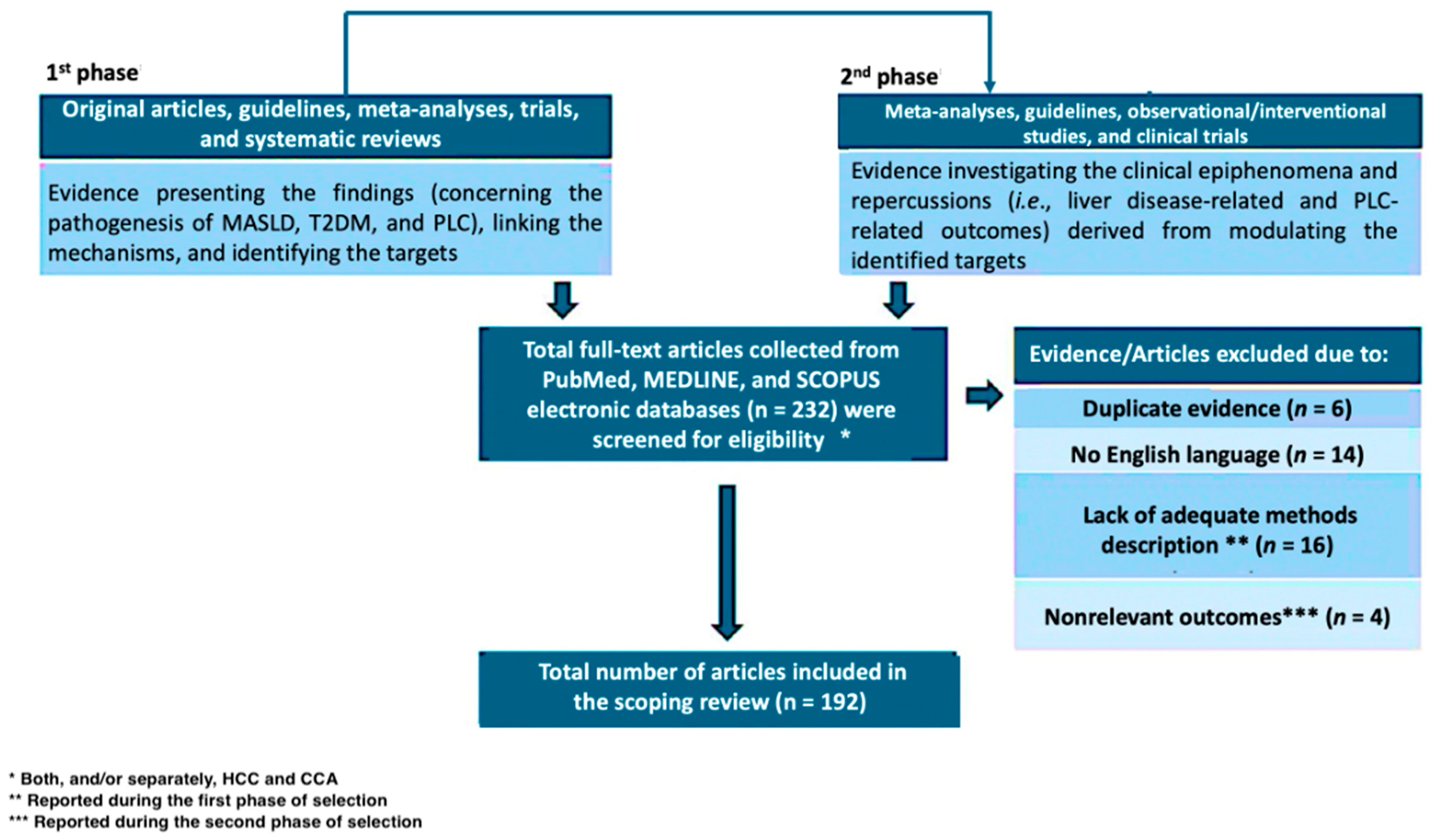
Figure 1. Flow diagram of the evidence selection process. MASLD: Metabolic dysfunction-associated Steatotic Liver Disease; T2DM: Type 2 diabetes mellitus; PLC: primary liver cancer; HCC: hepatocellular carcinoma; CCA: Cholangiocarcinoma.
Diabetology 06 00079 g001
Figure 2. Principal IR-related effects influencing hepatic cancerogenesis in MASLD-T2DM. IR: insulin resistance; MASLD: Metabolic dysfunction-associated Steatotic Liver Disease; T2DM: Type 2 diabetes mellitus; LSECs: liver sinusoidal endothelial cells; FAPB4: Fatty acid binding protein 4; HGF: Hepatocyte growth factor; IGF-1: insulin-like growth factor 1.
Diabetology 06 00079 g002
Figure 3. Mechanisms of insulin resistance-related inflammation influencing hepatocarcinogenesis. IR: insulin resistance; FFAs: Free fatty acids; ROS: reactive oxygen species; IL: interleukin; TNF-alpha: Tumor Necrosis Factor-alpha.
Diabetology 06 00079 g003
Table 1. Gut microbes associated with HCC in MD and relative pathogenic mechanisms.
Bacterial SpeciesKey CharacteristicsRole in HCC PathogenesisReferences
Escherichia coliGram-negative; enriched in MD-related gut dysbiosisActivates TLR4 signaling via LPS; promotes chronic hepatic inflammation and oxidative stress[107]
Klebsiella pneumoniaeGram-negative; enriched in MD-related gut dysbiosisPromoting bacterial translocation; activates Kupffer cells and hepatic immune response via LPS[113]
Bacteroides fragilisCommensal; enriched in MD-related gut dysbiosis contributing to the deterioration of glucose metabolismActivates TLR4 signaling via LPS; induction of senescence-associated secretory phenotypes in hepatic stellate cells; promotion of chronic hepatic inflammation[112]
Fusobacterium nucleatumAnaerobic opportunist; associated with T2DM and various GI malignanciesPromotes inflammation and may activate TLR4-dependent oncogenic pathways in tumor-prone environments.[110]
HCC: hepatocellular carcinoma; GI: gastrointestinal tract; MD: metabolic dysfunction; LPS: lipopolysaccharide; T2DM: Type 2 diabetes mellitus; MASLD: Metabolic dysfunction-associated steatotic liver disease; TLR-4: Toll-like receptor 4.
Table 2. Gut microbes associated with CCA in MD and relative pathogenic mechanisms.
Bacterial SpeciesKey CharacteristicsRole in CCA PathogenesisReferences
Escherichia coliGram-negative facultative anaerobe; enriched in MD-related gut dysbiosisPromotes chronic inflammation and oxidative stress in biliary epithelium via LPS[119]
Clostridium spp.Anaerobic Firmicutes; capable of 7α-dehydroxylation of primary bile acids; enriched in MD-related gut dysbiosisProduction of secondary bile acids (e.g., deoxycholic acid, DCA) with pro-carcinogenic properties[112,120]
Helicobacter spp.Bile-resistant; colonizes biliary tract; includes H. hepaticus and H. bilis; enriched in MD-related gut dysbiosisChronic inflammation and DNA damage; potential carcinogenic role[121,122]
Bacteroides spp.Dominant gut anaerobes; key in bile acid and carbohydrate metabolism; enriched in MD-related gut dysbiosisReduced abundance disrupts bile acid homeostasis and favors dysbiosis[123,124]
Veillonella spp.Gram-negative anaerobes; lactate fermenters; enriched in MD-related gut dysbiosisPromoting local inflammation via LPS/TLR4-dependent pathways[125]
MD: Metabolic dysfunction; DCA: deoxycholic acid; CCA: cholangiocarcinoma; LPS: lipopolysaccharide; TLR-4: Toll-like receptor 4.
Table 3. Antidiabetic agents and relative potential therapeutic implications in primary liver cancer: status of the art.
Drug ClassMechanisms of Action
in Hepatic Cancerogenesis
Potential Therapeutic Implications
(Type of Supporting Evidence)
Ref.
MetforminImproving IR and IR-related effects; modulation of gut-liver axis (regulates BAs metabolism)HCC: reduction of HCC risk and mortality in T2DM patients (metanalysis)
CCA: reduction of malignant cells proliferation and invasion potential (preclinical models)
[143]
[36]
SGLT2-IsBlocking renal glucose reabsorption thus reducing glucose levels and relative pro-cancerogenic effects; activates NAD+ salvage pathways.HCC: combination with metformin reduces incidence (retrospective in human evidence)
CCA: high doses suppress CCA cell viability (dose-dependent effects in vitro).
[148]
[150]
DPP4-IsInhibition of DPP4/CD26-regulated pathways; modulation of immune response and oxidative stressHCC: Promotion of apoptosis in HCC cells via suppressing of p62/Keap1/Nrf2 axis (in vitro); reduction of HCC incidence in T2DM-Hepatitis C Virus (HCV) infected patients (in human)
CCA: lack of solid evidence
[155]
[156]
GLP-1 RAsEnhancing insulin secretion and reducing glucagon levels; promotion of anti-inflammatory effectsHCC: Reduction of HCC incidence in T2DM patients (emerging in vivo evidence)
CCA: Limited role in tumor suppression (in vitro & in vivo evidence)
[161]
[36]
SGLT2-Is: sodium–glucose cotransporter-2 inhibitors; DPP4-Is: Dipeptidyl peptidase-4 inhibitors; GLP-1 RAs: Glucagon-like peptide-1 receptor agonists (GLP-1 RAs); IR: insulin resistance; T2DM: types 2 diabetes mellitus; BA: bile acids.
Disclaimer/Publisher’s Note: The statements, opinions and data contained in all publications are solely those of the individual author(s) and contributor(s) and not of MDPI and/or the editor(s). MDPI and/or the editor(s) disclaim responsibility for any injury to people or property resulting from any ideas, methods, instructions or products referred to in the content.