Article

Bone Substitute in Diabetic Foot Osteomyelitis Treatment

1
Diabetology and Diabetic Foot Treatment Center, ASUGI Monfalcone Hospital, 34074 Monfalcone, Italy
2
Clinic of Endocrinology and Metabolism Diseases ASFO Pordenone Hospital, 33170 Pordenone, Italy
*
Author to whom correspondence should be addressed.
Diabetology 2025, 6(4), 30; https://doi.org/10.3390/diabetology6040030
Submission received: 27 January 2025 / Revised: 25 March 2025 / Accepted: 11 April 2025 / Published: 14 April 2025

Abstract

:
Background: Diabetic foot osteomyelitis (DFO) constitutes a severe and prevalent complication of diabetes mellitus (DM). Individuals afflicted with DM exhibit an elevated risk for DFO development, attributable to a confluence of factors, including peripheral neuropathy, compromised circulation, and impaired immune function. Timely diagnosis and appropriate therapeutic intervention are paramount. In recent years, alongside the ablative approach, the feasibility of substituting compromised bone with a bone substitute has emerged. Methods: We retrospectively analyzed resorbable bone grafting procedures performed at our third-level center for the care of people with diabetes between 2019 and 2024. Forty-nine patients were included in this. The median follow-up period was 13 months (Q1 7, Q3 20). Results: At follow-up, 34 patients (69%) had achieved healing, with a median healing time of 2.3 months (Q1 1.5, Q3 5). Lesion location significantly influenced healing outcomes, with forefoot and midfoot lesions demonstrating an 86% healing rate compared to 50% for hindfoot lesions. Eleven patients (22%) experienced infectious relapse after a median of 1 month (Q1 0.7, Q3 2.9). An analysis of different bone substitutes did not reveal significant differences in terms of healing among the various products and between the presence or absence of a local antibiotic. Conclusions: Bone substitute implantation offers an additional conservative strategy for managing DFO. Healing rates are significantly higher for forefoot and midfoot lesions, suggesting that further research is needed to improve outcomes in hindfoot osteomyelitis. Selection of the most effective bone substitute requires further studies.

Graphical Abstract

1. Background

Osteomyelitis is a serious and frequent complication of DM, particularly affecting the foot [1]. Diabetes-related osteomyelitis of the foot (DFO) complicates 10 to 20% of moderate diabetic foot infections (DFIs) and 40 to 60% of severe DFIs [1].
DFO is defined by the International Working Group on the Diabetic Foot (IWGDF) [2] as an infection of the bone involving the marrow.
Individuals with DM are at increased risk of developing foot osteomyelitis due to a combination of factors, including peripheral neuropathy, impaired circulation, and compromised immune function [3].
Impaired circulation in people with DM further compromises the body’s ability to combat infections and deliver antibiotics to the affected area, exacerbating the risk of osteomyelitis [4].
Diabetic foot ulcers, characterized by slow healing, provide a common portal of entry for bacteria to invade bone and cause osteomyelitis [5]. The standard treatment for osteomyelitis typically necessitates a multidisciplinary strategy, integrating antimicrobial therapy, surgical intervention, and the management of underlying diabetic comorbidities [5,6,7,8,9]. Optimal management of diabetic foot osteomyelitis (DFO) remains a subject of ongoing debate. Although surgical resection of infected bone is widely regarded as the gold standard [8], numerous studies have demonstrated the efficacy of antibiotic monotherapy [6]. Traditionally, surgical excision was favored for DFO, reserving antibiotic therapy for cases without advanced osseous involvement [9]. However, the escalating prevalence of antibiotic resistance [8] and the limited bone penetration of antibiotics frequently prioritize surgical intervention, particularly when conservative surgical approaches, involving minimal bone resection, are feasible. Such approaches have shown promising outcomes, including osteomyelitis resolution, reduced antibiotic therapy duration, and minimized biomechanical alterations in the foot [8,9].
Bone grafting, a surgical technique utilizing artificial bone substitutes to repair or replace damaged or infected osseous tissue, is a viable option in the management of osteomyelitis [10]. In this context, bone grafts are frequently employed to obliterate bone defects resulting from infection, thereby promoting osteogenesis and restoring structural integrity [11]. Given that bone resection can compromise normal foot biomechanics, the utilization of synthetic substitutes to replace lost bone facilitates greater preservation of foot anatomy and function. In specific anatomical locations, such as the calcaneus or midfoot, bone reconstruction can obviate the necessity for amputation. Although data regarding the efficacy of local antibiotics in this setting remain preliminary [12], the primary objectives of surgical treatment and systemic antibiotic therapy are infection eradication. Bone substitutes, irrespective of antibiotic loading, contribute to infection control [13].
The aim of this study is to evaluate the outcomes of conservatively managed diabetic foot osteomyelitis using bone substitutes.

2. Materials and Methods

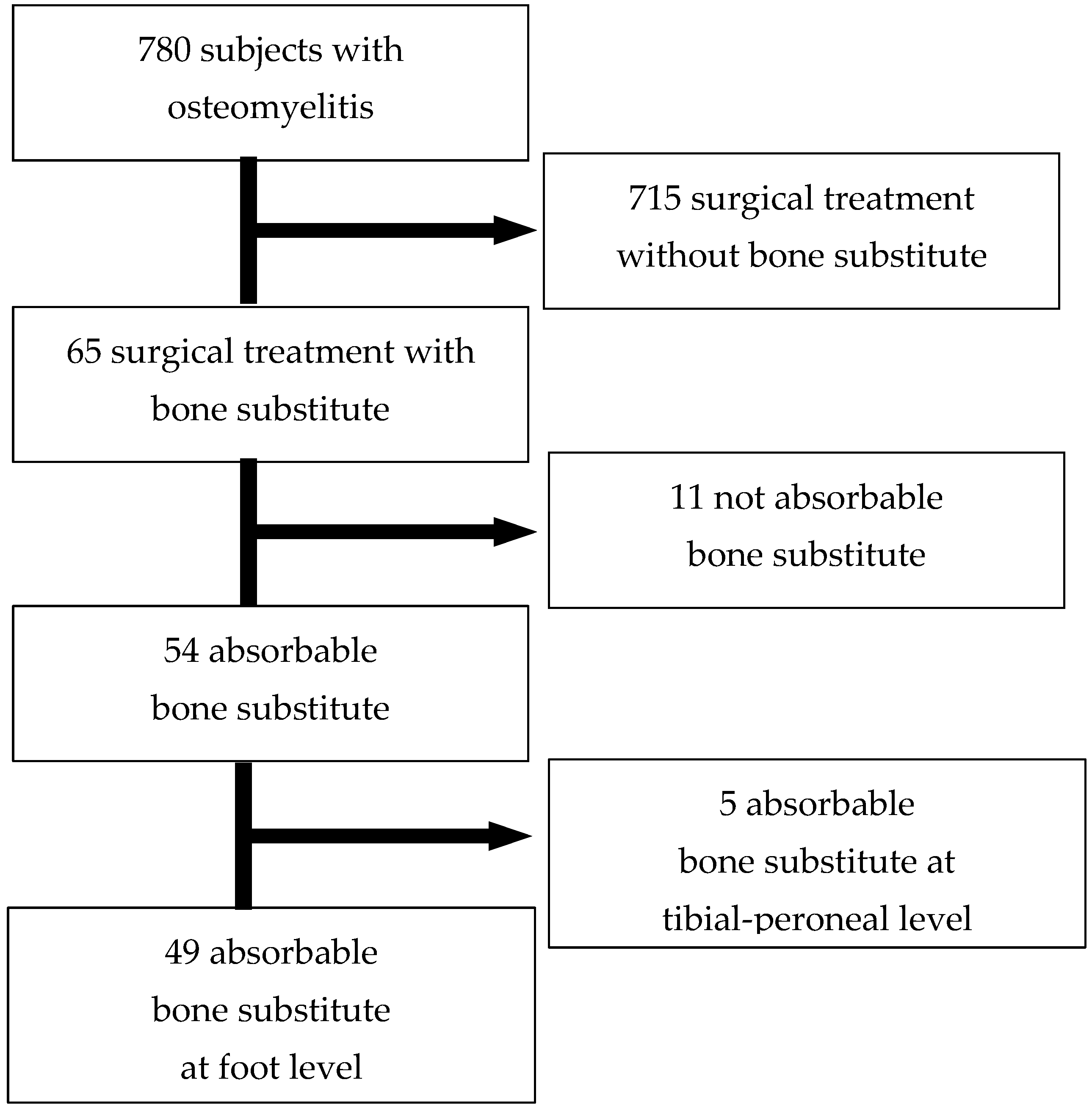
2.1. Study Design and Participants

A single-center retrospective study was conducted at a third-level center for the care of the diabetic foot from January 2019 to June 2024. This study analyzed data from the administrative databases of Monfalcone Hospital. We analyzed patients presenting with new-onset osteomyelitis and treated with surgical intervention, including bone substitutes, at our Foot Clinic.
Exclusion criteria included an inability to provide informed consent for surgical treatment, use of non-resorbable bone substitutes, and participation in other studies.
Inclusion criteria included patients aged 18 years or older, diagnosis of diabetes mellitus, presence of University of Texas grade 3 ulcers, confirmed diagnosis of osteomyelitis, osteomyelitis located in any region of the foot, and an ability to provide written informed consent.
Exclusion criteria included peripheral ischemia without prior revascularization, Charcot neuroarthropathy, osteomyelitis located proximal to the ankle, use of non-resorbable bone substitutes, and severe cognitive impairment.
Out of 780 individuals with DM treated for foot osteomyelitis, 65 received a bone substitute. A total of 11 patients who received non-absorbable bone substitutes were excluded from the analysis because non-absorbable grafts often require removal. A total of five patients who received absorbable bone substitutes implanted in the tibia and peroneal bones were excluded Figure 1.

2.2. Baseline Data Collection

Demographic and clinical data were collected from patient records, including duration of DM, diabetes complications, concomitant medical conditions, and other relevant medical history.
At the initial visit, all patients underwent a standardized physical examination as per the Clinic’s protocol. This included measurements of weight, height, and blood pressure. Laboratory results, specifically HbA1c levels, obtained within three months prior to the initial visit were also recorded.
Diabetic neuropathy is defined as the presence of signs and/or symptoms of peripheral nerve dysfunction in individuals with DM, with no other identifiable cause than DM itself [14].
To diagnose diabetic neuropathy, we assessed both of the following:
-
sensitivity to a 10 g monofilament: patients were considered to have neuropathy if they did not feel the monofilament at one or more designated test points.
-
vibration perception threshold (VPT): This was measured using a biothesiometer (METEDA, San Benedetto del Tronto, Italy). Patients were diagnosed with neuropathy if their VPT was 25 volts or higher at the big toe (hallux) or ankle bone (malleolus) [14].
Peripheral arterial disease (PAD) is defined as any atherosclerotic disease that obstructs arteries below the groin, leading to reduced blood flow to the lower limbs. Diagnosis of PAD was established through two methods:
Ankle-brachial index (ABI): patients were considered to have PAD if their ABI was less than 0.9 [15].
Arterial duplex ultrasound: Lower limb arteries were examined using an Acuson Sequoia (Siemens) ultrasound system. The presence of monophasic waveforms in the tibial arteries (measured at the medial malleolus, dorsalis pedis, and mid-calf for the peroneal artery) was also indicative of PAD [15].

2.3. Clinical Diagnosis of DFO

Osteomyelitis diagnosis was confirmed by a positive probe-to-bone test and radiographic evidence. The probe-to-bone test, performed using sterile metal forceps, was considered positive if a gritty or hard surface was palpated. Standard two-view radiographs were obtained. The presence of cortical disruption, periosteal reaction, sequestrum or involucrum formation, or gross bone destruction was considered indicative of DFO. Both tests were required for a positive diagnosis.

2.4. Treatment of DFO

Following clinical and vascular evaluation, the lesions were classified using the Texas scale [16]. The University of Texas classification system categorizes wounds as Grade 0 (healed pre- or post-ulcer site), Grade 1 (superficial, not involving tendon, capsule, or bone), Grade 2 (penetrating tendon or capsule), and Grade 3 (penetrating bone or joint). Within each grade, four stages are defined: A (clean), B (non-ischemic infected), C (ischemic non-infected), and D (ischemic infected) [16]. A treatment plan was then developed, which involved the removal of infected bone tissue. Patients with ischemia underwent prior endovascular revascularization before surgical intervention.

2.4.1. Antibiotic Treatment

Upon patient presentation with clinical signs of infection, empirical antibiotic therapy was initiated to address soft tissue inflammation. Subsequently, based on intraoperative bone culture results, antibiotic therapy was de-escalated and targeted to the causative organism.

2.4.2. Surgical Intervention

Following local anesthesia, a surgeon debrided infected and non-viable soft tissues, isolating the affected bone segment and assessing its clinical appearance. Infected bone exhibits an altered consistency and appearance compared to healthy bone, with differentiation based on five parameters: density (hard vs. soft), anatomical structure (cortical integrity vs. erosion/fracture), color (white vs. black/gray), vascular thrombosis (patent vessel vs. clot), and presence of a draining sinus (purulence) [17]. Infected and necrotic tissue were meticulously debrided. Resected bone specimens were submitted to microbiology for analysis. After the complete removal of infected bone and thorough irrigation of the wound, a bone substitute was implanted in the resulting defect to replace the bone lost due to infection. The surgical procedure concluded with remodeling of the residual bone and wound closure.

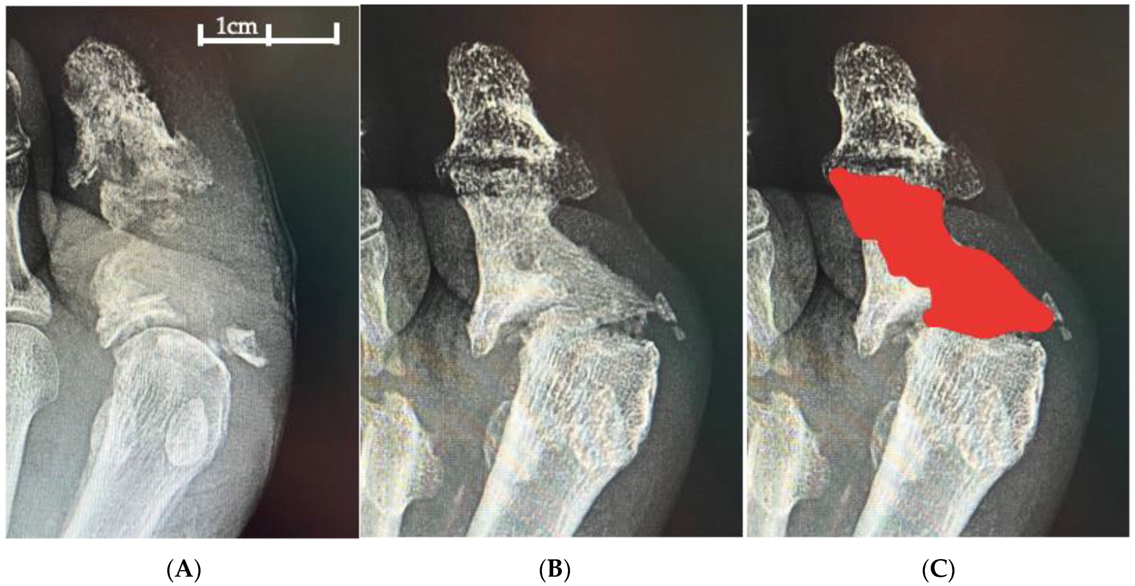
2.4.3. Bone Substitute

In this study, we exclusively utilized resorbable bone grafts. These products exhibit the following properties: osteointegration capacity, osteoconductive ability and promotion of bone growth, and antibiotic delivery capability; and furthermore, they eliminate the necessity for additional surgical interventions for graft removal as opposed to non-resorbable compounds.
The intervention with resorbable bone grafts, therefore, in contrast to non-resorbable ones, represents a curative approach, owing to its osteoconductive and bone growth-promoting capacities, and obviates the need for further surgical steps. The compositions of the bone substitutes employed are detailed in Table 1. Hydroxyapatite demonstrates highly osteoconductive properties and fosters bone growth. Calcium sulfate exhibits resorbability but lacks osteoconductivity and bone growth promotion. Bioactive glass, conversely, is a biocompatible and bioactive bone substitute with osteoconductive and osteointegrative properties, composed of soluble ions that form a silica gel layer on the granule surface, onto which calcium phosphate precipitates and crystallizes over time, forming hydroxyapatite [18]. It promotes osteogenesis by activating osteoblasts and possesses the ability to inhibit bacterial growth [18].
In the absence of literature guidance regarding the optimal bone substitute type, the material available at the time was applied.

2.4.4. Post-Operative Management

For topical wound care, povidone-iodine antiseptic was used. Following the surgical procedure, non-weight-bearing casts were applied for 20 days until suture removal. Subsequently, wound offloading was achieved using removable casts, removable knee braces, or therapeutic footwear, depending on the characteristics of the lesion, its location, and the patient’s ambulatory capacity. All patients were provided with custom-made or standard orthopedic footwear upon wound healing.

2.5. Statistical Analysis

Statistical analysis was performed using the SPSS statistical package, version 12.0.2 (SPSS, Chicago, IL, USA). We use median and standard deviation (SD), and median, quartile 1, and quartile 3 (Q1, Q3) for descriptive purposes in cases of normally and non-normally distributed continuous variables, respectively, whereas categorical variables are reported as the percentage. A Student’s t-test was performed for quantitative variables distributed normally, and a Mann–Whitney U test was used for abnormally distributed quantitative parameters. p-values < 0.05 were considered significant with a 95% confidence interval. The statistical analysis will examine the parameters influencing wound healing and healing time. The various intervention sites on the foot will be compared, as well as the presence or absence of antibiotics in the bone substitute.

2.6. Ethical Approval

The study was conducted in accordance with the Helsinki Declaration of 1964 and its later amendments. We used a general consent system signed by patients at their first visit, in which they agree that clinical data can be used for clinical research, epidemiology, disease study, and training, with the aim of improving knowledge, treatment, and prevention.

3. Results

Our study included 49 patients with DM treated with bone substitutes. Of these, 47 had type 2 diabetes; DM duration was 18 ± 13 years; the mean age was 66 ± 10 years (mean ± SD); and six (12%) were female. The mean HbA1c was 7.9 ± 1.7%. Regarding lower limb complications, peripheral neuropathy was present in 46 patients (94%), while 24 (49%) had peripheral artery disease, all of whom required pre-operative endovascular revascularization. End-stage renal disease was present in two participants, and coronary artery disease (CAD) in 13 (Table 1).
All lesions were classified as Texas grade 3 (17 grade 3A, 8 grade 3B, 14 grade 3C, and 10 grade 3D). Osteomyelitis was confirmed in all patients by the presence of bone exposure and positive radiographic findings, including cortical erosion, cortical lysis, osteolysis, and bone sequestration. The bones affected by osteomyelitis were the phalanx (n = 2), metatarsal (n = 18), cuneiform or cuboid (n = 7), and calcaneus or talus (n = 22) bones.

3.1. Microbiological Results and Antibiotic Treatment

Antibiotic therapy: 18 patients (37%) presented with acute infection prior to surgery and received empirical antibiotic therapy: 39% with amoxicillin/clavulanate, 17% with quinolones, 22% with piperacillin/tazobactam, and 22% with other agents (trimethoprim, cefalothin/tazobactam, colistin, and ceftazidime).
During 44 sequestrectomies (90%), a sample of infected bone was obtained for microbiological analysis: 13 (30%) cultures were negative, 3 yielded commensal flora, 25 were positive for a single microorganism, and 3 were positive for two microorganisms. The most frequently isolated bacteria were as follows: 13 Staphylococcus spp. (13 S. aureus, 1 S. epidermidis), 6 Pseudomonas aeruginosa, 2 Citrobacter freundii, 2 Proteus mirabilis, 2 Enterococcus faecalis, and 2 Enterobacter cloacae. Among the Staphylococcus isolates, 12 (92%) were methicillin-resistant. The presence of Gram-positive or Gram-negative bacteria did not significantly impact outcomes.
Following microbiological analysis, targeted antibiotic therapy was initiated: 47% with amoxicillin/clavulanate, 7% with amoxicillin/clavulanate plus a quinolone, 18% with quinolones, 4% with a quinolone plus rifampicin, 8% with trimethoprim, and 16% with other agents (linezolid, dalbavancin, cefalothin/tazobactam, and piperacillin/tazobactam). In particular, among patients who initiated empirical therapy with amoxicillin/clavulanate (72% of cases), 28% switched to a quinolone and 28% to trimethoprim. Patients who initially received a quinolone continued this therapy in 100% of cases, while those who began with piperacillin/tazobactam switched to amoxicillin/clavulanate in 50% of cases and to a quinolone in the remaining 50%.
Antibiotic therapy was administered for a mean duration of 19 ± 9 days.

3.2. Outcome

Complete wound healing was achieved in 34 patients (69%), with a median healing time of 2.3 months (Q1 1.5, Q3 5). Following the initial surgical procedure, 15 patients (31%) did not achieve healing. Nine patients subsequently healed after a second intervention. These included four Syme amputations, one Pirogoff amputation, one transmetatarsal amputation, one surgical revision, and two patients who healed following a second bone substitute procedure. The remaining six patients had persistent open wounds at the time of follow-up. At a median follow-up of 13 months (Q1 7, Q3 20), 11 (22%) infectious relapses were observed. In the healed group, five patients (15%) experienced relapse after a median of 5.3 months (Q1 1, Q3 9) compared to six patients (40%) in the non-healed group, with a median time to relapse of 1 month (Q1 0.6, Q3 1.3). The difference in both time to and incidence of infectious relapse between the healed and non-healed groups was statistically significant (p < 0.03). Analysis of the two groups (healed and non-healed) revealed no significant differences in age, DM duration, HbA1c levels, dialysis status, or presence of vascular disease.

3.2.1. Lesion Site and Outcome

The anatomical location of bone grafting was a key determinant of healing rates (Table 2). Similar healing rates were observed in the forefoot and midfoot.
The study population was, therefore, divided into two groups: forefoot/midfoot and hindfoot, and the parameters were analyzed. A significant difference in healing rates was observed between the groups (p < 0.01). The remaining parameters did not reach statistical significance (Table 2) (Figure 2).

3.2.2. Bone Substitute Results

The types of bone substitutes used were bioactive glass (BioSphere® Putty, Bio Tis.) in 19 patients (39%), nanocrystalline hydroxyapatite/calcium sulfate (PerOssal® Osartis) in 24 patients (50%), absorbable, gentamicin-loaded calcium sulfate/hydroxyapatite biocomposite (Cerament® G, Bonesupport) in 4 patients (7%), antibiotic-loaded calcium sulfate (Stimulan®, Biocomposites) in 1 patient (2%), and bone chips in 1 patient (2%) (Table 1). Local antibiotics were administered to 28 patients (57%): vancomycin and gentamicin in 19 (39%) and gentamicin alone in 9 (18%). Comparison of patients with and without local antibiotic administration revealed similar baseline characteristics, with the exception of PAD, which was significantly different between the two groups (71% in the no-local-antibiotic group vs. 32% in the local-antibiotic group). Healing rates and healing times were comparable between the groups. No significant difference in infectious recurrence rates was observed (Table 3).

4. Discussion

The results of this study include a large series of people with diabetes-related foot osteomyelitis treated with surgical removal of infected bone, targeted antibiotic therapy, and bone substitute implantation to replace the resected bone. We achieved a 69% healing rate, which was a significant outcome considering the challenging nature of our patient population, which included 45% with hindfoot osteomyelitis and 49% with peripheral arterial disease—both high-risk factors for limb loss. Furthermore, replacing bone segments compromised by osteomyelitis with bone substitutes allows for the preservation of foot anatomy and biomechanics, potentially avoiding amputation and restoring foot functionality. This innovative approach has the potential to shift the management of diabetic foot osteomyelitis from a predominantly destructive paradigm to a conservative one, offering significant advantages for patient quality of life. The healing rate achieved in this study is consistent with that observed in other studies using resorbable bone grafts, all single-product, which range between 50 and 90% [12,13,19,20,21,22,23,24]. The overall result obtained with various bone grafts presents an added value regarding the efficacy of the procedure.
The primary objective in osteomyelitis treatment remains infection eradication. As previously established, the combination of conservative surgery and targeted antibiotic therapy is the gold standard for resolving bone infection [9]. This study investigated the adjunctive use of bone tissue restoration with an artificial bone substitute in osteomyelitis treatment. Significantly, high healing rates were observed, especially in forefoot and midfoot lesions, where resolution exceeded 85%. Treatment of hindfoot lesions was associated with a low success rate (50%) and an elevated risk of recurrence. These location-dependent variations in outcomes have been previously documented [19,20]. Hindfoot osteomyelitis carries a particularly high risk of major amputation [19] and is characterized by low healing rates [19,20,25]. Several factors may contribute to these challenges, including variations in bone size, weight-bearing distribution across foot regions, the specific bone involved, and an inherent difficulty in achieving complete infection eradication. Limited vascularity in the calcaneus may also play a role [26]. Furthermore, complete infection eradication at the hindfoot is often unattainable, and the remaining bone stock may be insufficient for functional recovery. In our study, four (7%) major amputations were performed in patients with calcaneal osteomyelitis, confirming the elevated risk of major amputation associated with hindfoot osteomyelitis. This major amputation rate is consistent with that reported by other studies [21,22] but with the advantage of all being distal amputations. Notably, all four major amputations were Syme amputations, which are the most distal major amputation that preserves the entire leg. This outcome reflects a conservative, multidisciplinary approach that effectively minimized the amputation level [27]. Furthermore, hindfoot lesion healing times, typically prolonged [19,20], were comparable to those observed in other locations in our study, likely due to our surgical approach combining debridement, targeted antibiotic therapy, and bone substitute implantation. However, autologous bone graft availability can be limited in patients with diabetes-related osteomyelitis, and healing potential may be compromised by underlying systemic conditions [28]. Bone graft substitutes, such as demineralized bone matrix and calcium-based materials, provide structural support for new bone formation and can promote bone healing in these patients. The use of synthetic bone substitutes offers several advantages over autologous bone grafts: reduced patient invasiveness, the ability to reconstruct large bone defects, adaptability to various anatomical locations through paste or moldable chip formulations, and the potential for local antibiotic delivery. The efficacy of local antibiotic delivery remains a subject of ongoing debate. In our study, local antibiotics were used as an adjunct to, not a replacement for, systemic antibiotics. Our analysis provides a novel comparison between antibiotic-loaded and antibiotic-free bone substitutes. This is the first study that compares bone substitutes with and without antibiotics in diabetic foot osteomyelitis. Although the preliminary literature suggests potential benefits for bacterial growth inhibition [29] and accelerated healing [30,31], definitive clinical evidence regarding local antibiotic efficacy remains limited [32]. Our analysis did not demonstrate statistically significant differences between groups concerning local antibiotic use; however, healing speed, healing times, and infection recurrence exhibited a favorable trend with local antibiotic delivery. Notably, bioglass, recognized for its antiseptic properties due to the creation of an alkaline environment at the graft site, was employed in the antibiotic-free group [18]. Given the absence of significant differences between the graft materials, we infer comparable efficacy. Nevertheless, we underscore the importance of adhering to the standard osteomyelitis treatment: conservative surgery combined with targeted antibiotic therapy. The selection of bone substitute and the inclusion of local antibiotics remain at the surgeon’s discretion. In this study, the average duration of systemic antibiotic therapy was 19 days. International guidelines recommend up to three weeks of antibiotic therapy following minor amputation for diabetes-related foot osteomyelitis (DFO) with positive bone margin cultures, and one to two weeks if complete removal of infected bone is achieved. However, other studies report longer antibiotic durations in clinical practice [33]. The effectiveness of our approach is supported by the low recurrence rate (15%) observed in the healed group during a prolonged follow-up period exceeding 13 months, enabling a robust assessment of osteomyelitis eradication. Recurrences, consistent with findings from other studies [34], were observed several months post-healing. This underscores the importance of extended follow-up in confirming osteomyelitis eradication. In patients experiencing treatment failure, the recurrence rate increased significantly (40%) and occurred earlier (within one month), likely due to persistent open lesions.
These results highlight the necessity of close surveillance in cases where lesions do not demonstrate a positive healing trajectory and confirm the importance of frequent follow-up for relapse prevention [35]. Furthermore, continuous monitoring of healed patients by multidisciplinary teams is essential to minimize the risk of recurrent ulceration [36,37].
To our knowledge, this is the first retrospective comparative study evaluating outcomes of antibiotic-impregnated versus antibiotic-free bone substitutes in DFO treatment. Furthermore, our analysis encompasses all anatomical locations of foot osteomyelitis.
Certainly, our study has several limitations. The first limitation is that our study is retrospective. The second limitation is the small sample size. The third limitation is the absence of a control group. The final limitation of our study is that we included a white European population without racial/ethnic diversity and, consequently, our findings would benefit from testing in different populations.

5. Conclusions

Integrating bone substitutes into the standard treatment paradigm offers a novel, conservative strategy for managing diabetic foot osteomyelitis. While this approach yields encouraging results, especially in forefoot and midfoot lesions, further research is required to enhance healing outcomes in cases of calcaneal osteomyelitis. The role of local antibiotic delivery continues to be a subject of investigation, and additional studies are needed to definitively establish its clinical efficacy.

Author Contributions

Conceptualization, R.D.R. and C.M.; methodology, C.M.; software, R.D.R.; validation, R.A., B.B. and E.B.; formal analysis, A.M.; investigation, R.A.; resources, R.D.R.; data curation, R.D.R.; writing—original draft preparation, R.D.R.; writing—review and editing, C.M.; visualization, E.B.; supervision, R.A.; project administration, R.D.R. All authors have read and agreed to the published version of the manuscript.

Funding

This research received no external funding.

Institutional Review Board Statement

This study was conducted in accordance with the Helsinki Declaration of 1964 and its later amendments. According to the Italian Medicines Agency det. 20/03/2008 on retrospective observational studies on anonymous data, preemptive approval by an ethics committee was not mandatory, and given that the study collected anonymous data, not referable to specific individuals, approval by one or more ethical committee(s) was not requested.

Informed Consent Statement

We used a general consent system that was signed by patients on their first visit, in which they agreed to their clinical data being used for clinical research, epidemiology, disease study, and training.

Data Availability Statement

The original contributions presented in the study are included in the article; further inquiries can be directed to the corresponding author.

Conflicts of Interest

The authors declare no conflicts of interest.

References

  1. Lipsky, B.A.; Uçkay, I. Treating Diabetic Foot Osteomyelitis: A Practical State-of-the-Art Update. Medicina 2021, 57, 339. [Google Scholar] [CrossRef] [PubMed]
  2. van Netten, J.J.; Bus, S.A. Definitions and criteria for diabetes-related foot disease (IWGDF 2023 update). Diabetes Metab. Res. Rev. 2023, 15, e3654. [Google Scholar] [CrossRef] [PubMed]
  3. Lavery, L.A.; Armstrong, D.G. Diabetic foot syndrome: Evaluating the prevalence and incidence of foot pathology in Mexican Americans and non-Hispanic whites from a diabetes disease management cohort. Diabetes Care 2003, 26, 1435–1438. [Google Scholar] [CrossRef] [PubMed]
  4. Burgess, J.L.; Wyant, W.A. Diabetic Wound-Healing Science. Medicina 2021, 57, 1072. [Google Scholar] [CrossRef]
  5. Lipsky, B.A.; Berendt, A.R. Infectious Diseases Society of America Clinical Practice Guideline for the Diagnosis and Treatment of Diabetic Foot Infections. Clin. Infect. Dis. 2012, 54, e132–e173. [Google Scholar] [CrossRef]
  6. Game, F.L.; Jeffcoate, W.J. Primarily non-surgical management of osteomyelitis of the foot in diabetes. Diabetologia 2008, 51, 962–967. [Google Scholar] [CrossRef]
  7. Da Ros, R.; Assaloni, R. Burden of Infected Diabetic Foot Ulcers on Hospital Admissions and Costs in a Third-Level Center. Diabetology 2024, 5, 141–150. [Google Scholar] [CrossRef]
  8. Da Ros, R.; Assaloni, R.; Michelli, A.; Brunato, B.; Miranda, C. Antibiotic and Surgical Treatment of Diabetic Foot Osteomyelitis: The Histopathological Evidence. Antibiotics 2024, 13, 1142. [Google Scholar] [CrossRef]
  9. Lázaro Martínez, J.L.; García Álvarez, Y. Optimal management of diabetic foot osteomyelitis: Challenges and solutions. Diabetes Metab. Syndr. Obes. 2019, 12, 947–959. [Google Scholar] [CrossRef]
  10. Raghuram, A.; Singh, A.; Chang, D.K.; Nunez, M.; Reece, E.M. Bone Grafts, Bone Substitutes, and Orthobiologics: Applications in Plastic Surgery. Semin. Plast. Surg. 2019, 33, 190–199. [Google Scholar] [CrossRef]
  11. Schade, V.L.; Roukis, T.S. The role of bone graft and bone graft substitutes in the treatment of infection in the diabetic foot. Podiatry Today 2008, 21, 32–38. [Google Scholar]
  12. Chatzipapas, C.; Karaglani, M.; Papanas, N.; Tilkeridis, K.; Drosos, G.I. Local Antibiotic Delivery Systems in Diabetic Foot Osteomyelitis: A Brief Review. Rev. Diabet. Stud. 2021, 17, 75–81. [Google Scholar] [CrossRef] [PubMed]
  13. Patil, P.; Singh, R.; Agarwal, A.; Wadhwa, R.; Bal, A.; Vaidya, S. Diabetic Foot Ulcers and Osteomyelitis: Use of Biodegradable Calcium Sulfate Beads Impregnated with Antibiotics for Treatment of Multidrug-Resistant Organisms. Wounds 2021, 33, 70–76. [Google Scholar]
  14. Armstrong, D.G.; Tan, T.W. Diabetic Foot Ulcers: A Review. JAMA 2023, 330, 62–75. [Google Scholar] [CrossRef]
  15. Hinchliffe, R.J.; Forsythe, R.O.; Apelqvist, J.; Boyko, E.J.; Fitridge, R.; Hong, J.P.; Katsanos, K.; Mills, J.L.; Nikol, S.; Reekers, J.; et al. Guidelines on diagnosis, prognosis, and management of peripheral artery disease in patients with foot ulcers and diabetes (IWGDF 2019 update). Diabetes/Metab. Res. Rev. 2020, 36, e3276. [Google Scholar] [CrossRef]
  16. Monteiro-Soares, M.; Hamilton, E.J.; Russell, D.A.; Srisawasdi, G.; Boyko, E.J.; Mills, J.L.; Jeffcoate, W.; Game, F. Guidelines on the classification of foot ulcers in people with diabetes (IWGDF 2023 update). Diabetes/Metab. Res. Rev. 2024, 40, e3648. [Google Scholar] [CrossRef]
  17. Schmidt, B.M.; Jarocki, C. Making the equivocal unequivocal: Standardization of clean margins in diabetic foot osteomyelitis. Clin. Diabetes Endocrinol. 2020, 6, 8. [Google Scholar] [CrossRef]
  18. Hench, L.L. The story of Bioglass. J. Mater. Sci. Mater. Med. 2006, 17, 967–978. [Google Scholar] [CrossRef]
  19. Winkler, E.; Schöni, M.; Krähenbühl, N.; Uçkay, I.; Waibel, F.W.A. Foot Osteomyelitis Location and Rates of Primary or Secondary Major Amputations in Patients with Diabetes. Foot Ankle Int. 2022, 43, 957–967. [Google Scholar] [CrossRef]
  20. Whisstock, C.; Volpe, A.; Ninkovic, S.; Marin, M.; Meloni, M.; Bruseghin, M.; Boschetti, G.; Brocco, E. Multidisciplinary Approach for the Management and Treatment of Diabetic Foot Infections with a Resorbable, Gentamicin-Loaded Bone Graft Substitute. J. Clin. Med. 2020, 9, 3586. [Google Scholar] [CrossRef]
  21. Niazi, N.S.; Drampalos, E.; Morrissey, N.; Jahangir, N.; Wee, A.; Pillai, A. Adjuvant antibiotic loaded bio composite in the management of diabetic foot osteomyelitis—A multicentre study. Foot 2019, 39, 22–27. [Google Scholar] [CrossRef] [PubMed]
  22. Hutting, K.H.; Aan de Stegge, W.B. Surgical Treatment of Diabetic Foot Ulcers Complicated by Osteomyelitis with Gentamicin-Loaded Calcium Sulphate-Hydroxyapatite Biocomposite. J. Clin. Med. 2021, 19, 371. [Google Scholar] [CrossRef] [PubMed]
  23. Monami, M.; Bordoni, L. Efficacy and safety of a bio-absorbable antibiotic delivery in calcium sulphate granules for the treatment of osteomyelitis in patients with diabetic foot: A randomized, double blinded, controlled clinical study The BIG D-FOOT study. Diabetes Obes. Metab. 2025, 27, 2552–2560. [Google Scholar] [CrossRef] [PubMed]
  24. De Giglio, R.; Di Vieste, G. Efficacy and Safety of Bioactive Glass S53P4 as a Treatment for Diabetic Foot Osteomyelitis. J. Foot Ankle Surg. 2021, 60, 292–296. [Google Scholar] [CrossRef]
  25. Pickwell, K.M.; Siersma, V.D.; Kars, M.; Holstein, P.E.; Schaper, N.C.; on behalf of the Eurodiale consortium. Diabetic foot disease: Impact of ulcer location on ulcer healing. Diabetes/Metab. Res. Rev. 2013, 29, 377–383. [Google Scholar] [CrossRef]
  26. Khoo, R.; Jansen, S. Slow to heel: A literature review on the management of diabetic calcaneal ulceration. Int. Wound J. 2018, 15, 205–211. [Google Scholar] [CrossRef]
  27. Meloni, M.; Giurato, L.; Monge, L.; Miranda, C.; Scatena, A.; Ragghianti, B.; Silverii, G.A.; Vermigli, C.; De Cassai, A.; Volpe, A.; et al. Effect of a multidisciplinary team approach in patients with diabetic foot ulcers on major adverse limb events (MALEs): Systematic review and meta-analysis for the development of the Italian guidelines for the treatment of diabetic foot syndrome. Acta Diabetol. 2024, 61, 543–553. [Google Scholar] [CrossRef]
  28. Thabit, A.K.; Fatani, D.F. Bone graft in diabetic foot osteomyelitis: A review. Int. Surg. J. 2017, 4, 2601–2606. [Google Scholar]
  29. Schade, V.L.; Roukis, T.S. The role of polymethylmethacrylate antibiotic–loaded cement in addition to debridement for the treatment of soft tissue and osseous infections of the foot and ankle. J. Foot Ankle Surg. 2010, 49, 55–62. [Google Scholar] [CrossRef]
  30. Uddin, A.; Russell, D.; Game, F.; Santos, D.; Siddle, H.J. The effectiveness of systemic antibiotics for osteomyelitis of the foot in adults with diabetes mellitus: A systematic review protocol. J. Foot Ankle Res. 2022, 15, 48. [Google Scholar] [CrossRef]
  31. Soldevila-Boixader, L.; Fernández, A.P.; Laguna, J.M.; Uçkay, I. Local Antibiotics in the Treatment of Diabetic Foot Infections: A Narrative Review. Antibiotics 2023, 12, 124. [Google Scholar] [CrossRef] [PubMed]
  32. Patel, N.; Hatch, D.; Wells, C.; Ahn, D.; Harris, M.; Jennings, J.; Haggard, W.; Armstrong, D. Characteristics and clinical assessment of antibiotic delivery by chitosan sponge in the high-risk diabetic foot: A case series. J. Wound Care 2017, 26, S32–S38. [Google Scholar] [CrossRef] [PubMed]
  33. Lavery, L.A.; Tarricone, A.N. Does complete resection of infected bone improve clinical outcomes in patients with diabetic foot osteomyelitis? Int. Wound J. 2024, 21, e70072. [Google Scholar] [CrossRef] [PubMed]
  34. McNally, M.A.; Ferguson, J.Y.; Lau, A.C.; Diefenbeck, M.; Scarborough, M.; Ramsden, A.J.; Atkins, B.L. Single-stage treatment of chronic osteomyelitis with a new absorbable, gentamicin-loaded, calcium sulphate/hydroxyapatite biocomposite: A prospective series of 100 cases. Bone Jt. J. 2016, 98, 1289–1296. [Google Scholar] [CrossRef]
  35. Miranda, C.; Da Ros, R. Prevention of Diabetic Foot Ulcer: A Neglected Opportunity. Transl. Med. UniSa 2020, 22, 50–51. [Google Scholar]
  36. Da Ros, R.; Volpe, A.; Bordieri, C.; Tramonta, R.; Bernetti, A.; Scatena, A.; Monge, L.; Ragghianti, B.; Silverii, A.; Uccioli, L.; et al. Prevention of foot ulcers recurrence in patients with diabetes: A systematic review and meta-analysis of randomized controlled trials for the development of the italian guidelines for the treatment of diabetic foot syndrome. Acta Diabetol. 2024, 61, 1363–1373. [Google Scholar] [CrossRef]
  37. Uivaraseanu, B.; Bungau, S. Clinical, Pathological and Microbiological Evaluation of Diabetic Foot Syndrome. Medicina 2020, 56, 380. [Google Scholar] [CrossRef]
Figure 1. Flowchart.
Diabetology 06 00030 g001
Figure 2. Example of bone reconstruction of a proximal phalanx of the first toe subtotally involved by osteomyelitis. (A). Osteomyelitis of first toe proximal phalanx with subtotal destruction of the bone and presence of bone sequestrum. (B). One year after the bone graft with complete reconstruction of the bone. (C). Red color: evidence of reconstructed bone.
Diabetology 06 00030 g002
Table 1. Bone substitute characteristics.
Commercial ProductCompositionAbsorbableOsteoinductionAntibiotic
(n. Treated)
Subject Treated
Cerament® G
Bonesupport, Lund, Schweden
Calcium sulphate (60%) Hydroxyapatite (40%)YesYesGEN4 (7%)
Stimulan®
Biocomposite Ltd.
Staffordshire, UK
Calcium sulphate (100%)YesYesGEN1 (2%)
BioSphere® Putty
Synergy Biomedical,
Wayne, PA, USA
Bimodal bioactive glass spheres in a resorbable phospholipid carrierYesYesNo19 (39%)
PerOssal®
OSARTIS, Münster,
Germany
51.5% nanocrystalline hydroxyapatite
48.5% calcium sulfate
YesYesGEN (4)
GEN-VAN (18)
24 (50%)
Bone Chips
Bioteck, Arcugnano, Italy
Bone chips 100%NoYesGEN-VAN1 (2%)
Abbreviations: GEN: gentamicin, VAN: vancomicin.
Table 2. Patient characteristics and a comparative analysis of lesion locations.
CharacteristicsAll
49 Subjects
Mean ± SD
Forefoot/Midfoot
27 Subjects (55%)
Mean ± SD
Rearfoot
22 Subjects (45%)
Mean ± SD
P
(Forefoot/Midfoot vs. Rearfoot)
Age (years)66 ± 10 years66 ± 12 66 ± 90.89
DM history (years)18 ± 13 17 ± 1321 ± 130.14
Male/female n.43/624/319/30.79
HbA1c (%)7.9 ± 1.77.7 ± 1.98.1 ± 1.70.45
Peripheral neuropathy n (%)46 (94%)25 (93%)21 (95%)0.68
Peripheral vascular disease n (%)24 (49%)13 (48%)11 (50%)0.9
Revascularization 24 (49)13 (48%)11 (50%)0.9
Dyalisis2 (4%)2 (7%)00.2
Lesion Texas 3A n (%)17 (34%)10 (37%)7 (32%)0.7
Lesion Texas 3B n (%)8 (16%)4 (15%)4 (18%)0.8
Lesion Texas 3C n (%)14 (28%)9 (33%)5 (23%)0.4
Lesion Texas 3D n (%)10 (20%)4 (15%)6 (27%)0.3
Healed n. 34 23 11 <0.01
Healing rate69%85%50%<0.01
Healing time (months)2.3 (Q1 1.5, Q3 5)2.7 (Q1 1.7, Q3 5)2.2 (Q1 1.2, Q3 3.7)0.49
Infective relapse n (%)11 (22%)4 (15%)7 (32%)0.2
Table 3. Comparison between the presence or absence of local antibiotic at the level of the bone substitute used.
CharacteristicsNo Antibiotic
21 Subjects (43%)
Mean ± SD
Local Antibiotic
28 Subjects (57%)
Mean ± SD
P
Age (years)69 ± 13 65 ± 100.16
DM history (years)19 ± 1316 ± 120.2
Male/female n.18/325/30.71
HbA1c (%)8.2 ± 1.97.8 ± 1.70.43
Peripheral neuropathy n (%)21 (100)25 (91%)0.12
Peripheral vascular disease n (%)15 (71%)9 (32%)0.005
Revascularization n (%)15 (71%)9 (32%)0.005
Dyalisis n (%)2 (9%)00.1
Healed n (%)13 (62%)21 (75%)0.33
Healing time (months)2.7 (Q1 1.5, Q3 6)2.3 (Q1 1.7, Q3 4)0.14
Infective relapse n (%)7 (33)4 (14)0.06
Disclaimer/Publisher’s Note: The statements, opinions and data contained in all publications are solely those of the individual author(s) and contributor(s) and not of MDPI and/or the editor(s). MDPI and/or the editor(s) disclaim responsibility for any injury to people or property resulting from any ideas, methods, instructions or products referred to in the content.

