


Abstract
[18F]-PSMA PET/CT is a high-impact modality for the staging and restaging of prostate cancer, but its wide anatomic coverage and tracer biology generate frequent incidental findings on both PET and the accompanying low-dose CT (LDCT). This narrative review is restricted in scope to fluorine-18 PSMA tracers because tracer-specific biodistribution and pitfall profiles shape what is perceived as incidentaloma: how confidently lesions can be categorized, and how often borderline findings trigger downstream testing, particularly for skeletal foci with [18F]-PSMA-1007. Specifically, [18F]-PSMA-1007 shows substantially higher rates of focal unspecific bone uptake than [68Ga]-PSMA-11—reported in multicenter studies as affecting up to 40–50% of patients—which directly inflates the pool of potential incidentalomas and creates a tracer-specific false-positive problem with no parallel in gallium-68 practice. Additionally, [18F]-DCFPyL has different urinary clearance kinetics that affect bladder and ureteral uptake patterns, altering what qualifies as physiologic versus incidental in the pelvis. These differences mean that the threshold for Category B versus C classification—and the appropriate cascade-resistant language—must be tuned to the specific tracer in use. A framework built on [68Ga]-PSMA-11 data would systematically underestimate bone pitfall frequency in [18F]-PSMA-1007 practice and could therefore paradoxically increase rather than reduce cascades if applied uncritically across tracers. These biodistribution differences have direct and concrete consequences for reporting behaviour and downstream management. In [18F]-PSMA-1007 practice, a focal bone uptake without a CT correlate in a mechanically plausible location—such as an anterior rib or vertebral endplate—should trigger Category B language in the report conclusion: the finding is documented in the body with explicit safety netting (“most consistent with unspecific uptake; no routine workup unless interval growth, new pain, or aggressive CT morphology”), and no referral to bone scintigraphy or MRI is generated. Without tracer-specific awareness, the same finding would typically prompt a reflex bone scan or whole-body MRI referral, delaying definitive prostate cancer management by weeks and adding imaging costs without diagnostic gain. By contrast, in [68Ga]-PSMA-11 practice, an equivalent focal bone uptake without a CT correlate carries a higher prior probability of true metastatic disease given the lower background rate of unspecific uptake and should more often be reported at Category B with a lower threshold for escalation or more cautious language. For [18F]-DCFPyL, the higher urinary activity in the pelvis means that ureteral segments can mimic lymph node disease; recognizing this as a physiologic variant (Category C) rather than an equivocal nodal finding (Category B) avoids unnecessary pelvic MRI referrals that would otherwise be triggered by an uncontextualized report. In practical terms, the tracer-specific calibration of the overlay therefore changes not only the category assigned but also the specific safety-netting language and the escalation trigger, which directly modifies the downstream management pathway for each affected finding type. The scanned population—predominantly older men with a high prevalence of degenerative, inflammatory, and vascular abnormalities—creates substantial background noise that can drive low-value diagnostic cascades if incidental findings are communicated without actionability context. We integrate society-endorsed frameworks (EANM/SNMMI procedure guideline 2.0; E-PSMA; PSMA-RADS; and PROMISE/miTNM with miPSMA score) and propose a cascade-aware overlay for incidental findings that can be appended to existing PSMA reporting standards rather than replacing them. The A/B/C actionability overlay is a structured expert-consensus framework informed by existing evidence-based guidelines for specific finding types and by tracer-specific cohort data; it has not yet been prospectively validated as a standalone tool, and its current level of evidence is therefore analogous to a structured expert recommendation rather than an evidence-based clinical guideline. We operationalize a three-tier actionability scheme across PET- and CT-dominant findings, provide cascade-resistant language for conclusions, and clarify why SUVmax-only “probability scales” for lymph nodes are not recommended in routine reports. Three practical tables summarize PET incidental findings, lymph node reporting frameworks, and LDCT incidental findings, and two structured report templates are provided (concise and extended), with the extended version explicitly labelling actionability tiers and escalation triggers. Finally, we outline concrete AI use cases for standardization and triage while emphasizing governance to avoid the amplification of false positives and paradoxical growth of cascades.
1. Introduction
An incidental finding is an unexpected observation detected during an examination performed for a different primary clinical question; it is “incidental” not because it is clinically trivial, but because it was not the target of the diagnostic pathway that motivated the test. In [18F]-PSMA PET/CT, incidental findings are common because acquisition spans large anatomic territories and because PSMA ligands accumulate in multiple benign tissues and in non-prostate pathologies, including some inflammatory conditions and non-prostatic malignancies [1,2]. Incidental findings matter clinically because they can alter care when they represent previously unrecognized serious disease, but they also matter systemically because they can trigger cascades of downstream testing that offer little value, expose patients to harm, and delay time-sensitive oncologic decisions [3,4]. The same sentence in a report can either improve patient outcomes or generate low-value diagnostic activity, depending on how it is framed, which makes this a practical reporting problem rather than a purely conceptual one.
For the purpose of this review, incidental findings in [18F]-PSMA PET/CT are grouped into three pragmatic categories that align with workflow and reporting decisions. The first group is PET-driven incidental uptake outside the typical prostate cancer pattern, such as an atypical visceral focus, focal thyroid uptake, or focal gastrointestinal uptake with a suspicious wall correlate. The second group is LDCT-driven incidental findings detected on CT acquired for attenuation correction and anatomic localization (CTAC/LDCT), including nodules, cysts, vascular abnormalities, or diffuse parenchymal changes. The third group comprises pitfalls and mimics, namely physiologic variants or benign processes that can imitate prostate cancer spread and lead to overstaging or unnecessary follow-up if they are not explicitly recognized as such [1,5,6,7,8,9,10]. Lymph nodes and bone findings are excluded from the incidentaloma category unless they are atypical, indeterminate, or specifically prone to triggering low-value cascades—for example, an equivocal pelvic node below the PSMA-RADS confidence threshold, or a focal bone uptake in [18F]-PSMA-1007 lacking a CT correlate. When these findings are part of the primary oncologic question with sufficient confidence, they belong to the staging report not the incidentaloma discussion. This boundary is defined by cascade risk rather than anatomy: any finding—nodal, osseous, or visceral—qualifies as an incidentaloma for the purpose of this review if it cannot be confidently attributed to prostate cancer disease and has the potential to trigger further testing independent of the primary diagnostic question.
Existing PSMA reporting frameworks already standardize the interpretation of prostate cancer disease burden and confidence, with PSMA-RADS supporting lesion-level likelihood assessment, PROMISE/miTNM providing molecular staging with miPSMA scoring, and E-PSMA offering standardized reporting language [5,6,7]. Professional guidance also exists for indications and appropriate interpretation across PSMA radioligands [1,11]. It is important to distinguish what this review synthesizes from existing guidance versus what it contributes as genuinely novel. The synthesis component draws on PSMA-RADS, E-PSMA, PROMISE/miTNM, and the EANM/SNMMI procedure guideline to provide an integrated view of how incidental findings intersect with established staging workflows [1,5,6,7,11]. The novel contribution consists of three elements not addressed collectively in the existing guidance: (1) a cascade-aware A/B/C actionability overlay applicable to all incidental findings regardless of their modality driver (PET versus CTAC/LDCT); (2) the explicit integration of this overlay with existing PSMA frameworks as an addendum layer rather than a replacement; and (3) reporting language templates specifically engineered to reduce low-value downstream diagnostic cascades while preserving conditional safety netting. The novelty of the present review is not a new staging system but a cascade-aware overlay: a structured approach to incidental findings across PET and CTAC/LDCT that links what is seen to what should, or should not, happen next, using report language designed to minimize low-value cascades while preserving patient safety. To be operationally precise: PSMA-RADS and E-PSMA address lesion-level confidence for prostate cancer findings and provide standardized reporting language for the primary oncologic question, while PROMISE/miTNM translates those findings into molecular staging categories. None of these frameworks systematically addresses what to do with incidental non-prostate findings, how to triage them for actionability, or how to phrase report conclusions to prevent unnecessary downstream testing. The cascade-aware overlay precisely fills this gap: it provides (a) an explicit three-tier (A/B/C) actionability classifier for every incidental finding regardless of its modality driver, (b) conclusion-language templates designed to prevent “just-in-case” cascades while preserving patient safety, and (c) tracer-specific guidance for [18F]-PSMA-1007 bone pitfalls not covered by existing frameworks. In measurable terms, the expected benefits are a reduction in reflex downstream imaging triggered by Category B/C findings, improved inter-reader reproducibility in incidental-finding communication, and faster time to definitive prostate cancer treatment by reducing incidental-finding-driven pathway interruptions—outcomes that the research agenda section outlines as prospective study endpoints.
Key innovations (new to this review): (1) a cascade-aware A/B/C actionability overlay that applies to all incidental findings in [18F]-PSMA PET/CT regardless of whether they are PET-driven, CTAC/LDCT-driven, or represent pitfalls—an integration not provided in any existing PSMA reporting guideline; (2) explicit embedding of this overlay within established frameworks (PSMA-RADS, E-PSMA, and PROMISE/miTNM) as a complementary layer, preserving compatibility with current reporting standards while adding actionability context; (3) ready-to-use reporting templates with cascade-resistant conclusion language that operationalize the overlay in daily nuclear medicine practice, including tracer-specific guidance for [18F]-PSMA-1007 bone pitfalls.
Methods (narrative review): This manuscript was developed as a narrative review focused on actionable thresholds for incidental findings in [18F]-PSMA PET/CT and on structured reporting strategies that reduce low-value diagnostic cascades. Searches were performed in PubMed/MEDLINE and supplemented by citation snowballing from key guidelines and consensus statements, covering 1 January 2016, through 10 February 2026, with the last update on 10 February 2026. Search terms combined tracer and modality concepts with incidentaloma and reporting terms, including “PSMA” AND “18F” AND “PET/CT” AND incidental OR incidentaloma OR pitfall OR uptake OR “low-dose CT” OR “attenuation correction” OR “structured reporting” OR “PSMA-RADS” OR “PROMISE” OR “miTNM” OR “E-PSMA”. Eligible sources included guidelines and consensus documents, cohort and multicenter studies, and high-quality reviews addressing [18F]-labelled PSMA PET/CT interpretation, pitfalls, incidental findings, or management frameworks [1,5,6,7,8,9,10,11]. Case reports were not used to justify action thresholds but were considered selectively as illustrative examples when consistent with higher-level evidence. Records were screened iteratively because terminology for incidental findings is inconsistent across imaging disciplines, which complicates straightforward categorization in clinical practice. Evidence was synthesized thematically with emphasis on operational reporting decisions rather than exhaustive prevalence estimates.
Why incidental findings in [18F]-PSMA PET/CT are cascade-prone: A diagnostic cascade is defined here as a chain of additional tests or consultations initiated by an incidental finding that was not the primary clinical question of the examination, where each step may generate further steps, often with diminishing clinical return and increasing risk of harm from overdiagnosis, procedure complications, or delay to the original diagnostic pathway [3,4]. Incidental findings in PSMA PET/CT are not merely “extra findings”; they intersect with clinical decisions that are often time-sensitive, including the selection and timing of surgery, radiotherapy, systemic therapy, and salvage approaches [11]. A cascade triggered by an incidental lung nodule or an equivocal bone focus can delay definitive prostate cancer management, and such delays are rarely visible in outcome datasets, even when they matter to patients and clinicians [3,4]. PSMA tracer uptake is also not cancer-specific; non-prostate malignancies and benign conditions can demonstrate uptake, and fluorine-18 tracers have pitfall profiles that differ from 68Ga-labelled compounds, with [18F]-PSMA-1007 showing a higher frequency of focal unspecific bone uptake that can be misread as metastasis if interpreted without PET–CT correlation and pattern awareness [8,9,10].
CTAC/LDCT adds another layer of risk: CTs acquired for attenuation correction and localization are typically non-contrast, often with lower mAs, sometimes thicker slices, and optimized for PET attenuation rather than diagnostic characterization, yet CT findings can be compelling to clinicians, and the report may be interpreted as if it were a screening CT if limitations and recommended next steps are not clearly communicated in the report. A practical principle is therefore to treat CTAC/LDCT as a detection and localization tool; if escalation is warranted, characterization should be deferred to diagnostic-quality CT or MRI rather than be attempted using low-dose non-contrast CT [12,13,14,15,16].
A practical actionability framework (A/B/C) and its reporting consequences: To make incidental findings actionable without over-triggering follow-up, we propose a three-tier actionability scheme that applies across PET- and CT-dominant findings and functions as an overlay that coexists with the PSMA-RADS, E-PSMA, and PROMISE/miTNM reporting of prostate cancer disease [5,6,7]. The translation from narrative evidence to these operational categories followed an explicit logic: for each finding type, published prevalence and malignancy-risk data informed the pretest probability component, while guideline-defined escalation criteria (Fleischner, ACR organ-specific algorithms, and PSMA-RADS) provided the actionability thresholds. Where evidence was absent or inconsistent, the category assignment was deliberately conservative, and the corresponding report language was designed to preserve safety netting. The result is that Category A assignments are predominantly evidence-anchored, Category B assignments are a mixture of evidence and expert extrapolation, and Category C assignments reflect well-established physiologic patterns documented in existing PSMA guidelines [1,5,6,7] Table 1. Category A comprises findings with a reasonable probability of clinically meaningful non-prostate disease or urgent comorbidity, where delayed follow-up could plausibly harm the patient; these belong in the report conclusion with a targeted recommendation. Category B includes findings likely benign or indeterminate with a low pretest probability of harm, where routine escalation would generate low-value cascades; these should be documented in the body of the report with explicit safety netting and escalated to Category A only if predefined red flags are present, such as symptoms, interval growth, a strong morphologic correlate, or discordance with expected benign patterns. Category C captures physiologic biodistribution and well-known benign mimics that frequently cause overstaging or unnecessary follow-up if not contextualized; these should be explicitly framed as non-actionable, and the report should discourage further workup. The thresholds that separate these categories are derived from three overlapping sources: (1) published guideline criteria for specific finding types—Fleischner Society thresholds for pulmonary nodules [12], ACR criteria for adrenal, renal, hepatic, and pancreatic incidentalomas [13,14,15,16], and ACR thyroid incidentaloma guidance [17]—which provide evidence-anchored cut-points for CT-dominant findings; (2) tracer-specific evidence on unspecific bone uptake frequency and imaging characteristics for [18F]-PSMA-1007, derived from multicenter cohort and validation studies [8,9,10], which inform PET-specific Category B/C boundaries for skeletal findings; and (3) expert consensus and structured reasoning extrapolated from adjacent imaging disciplines for finding types where PSMA-specific prevalence data are absent. Where thresholds rest on extrapolation rather than direct evidence, the framework is explicitly conservative—defaulting to Category B (document with safety netting) rather than C (suppress entirely)—to preserve a safety margin pending prospective validation. To make the relative weighting between evidence-based criteria and expert extrapolation explicit, the following hierarchy applies across the framework. The strongest evidence level applies to Category A and Category B assignments that are directly governed by published society guidelines: pulmonary nodule thresholds from Fleischner 2017 [12], adrenal management from ACR 2017 [13], renal lesion classification via Bosniak 2019 [14], thyroid nodule guidance from ACR 2015 [17], and hepatic and pancreatic incidentaloma management from ACR 2017 [15,16]. In these cases, the overlay does not introduce new thresholds but translates existing evidence-based criteria into the PSMA PET/CT reporting context, with the addition of an explicit statement that CTAC/LDCT is inadequate for characterization when those criteria require diagnostic-quality imaging. An intermediate evidence level applies to Category B/C boundary decisions for [18F]-PSMA-1007 unspecific bone uptake, which are anchored in multicenter cohort and validation studies [8,9,10] but require extrapolation because the studies were not designed as threshold-derivation analyses. The weakest evidence level—explicit expert extrapolation—applies to finding types for which no PSMA-specific prevalence data exist, such as incidental small pancreatic cysts or incidental adrenal adenomas in this specific population; these receive conservative Category B assignments by default. Readers should treat Category A assignments based on published guidelines as the most transferable component of the framework, Category B bone-uptake assignments as moderately transferable pending tracer-specific validation, and Category B assignments derived from expert extrapolation as the most provisional and most in need of local calibration before clinical deployment.
Table 1.
PET incidental findings in [18F]-PSMA PET/CT: actionability-based triage.
In practice, the most cascade-sensitive element is not the detection itself but phrasing, particularly in the conclusion. For Category B findings, recommended wording explicitly anchors the interpretation as benign or indeterminate and states that additional workup is not routinely recommended in the absence of symptoms or interval change; this reduces “just in case” cascades while still preserving conditional safety netting [3,4]. For CTAC/LDCT-related recommendations, a parallel sentence should clarify that the CT component is low-dose and non-contrast, acquired for attenuation correction and localization, and that dedicated diagnostic imaging may be required for definitive characterization; such explicit language prevents guideline misapplication to LDCT and improves clinician expectations about what the CT component can and cannot provide [12,13,14,15,16].
Lymph nodes and the limits of SUVmax-only probability language: Lymph node interpretation is a classic area where well-intended quantification becomes misleading. SUVmax can support confidence in a given lesion, but it should not be presented as a standalone probability scale, because uptake depends on scanner technology, reconstruction, timing, lesion size with partial volume effects, and biologic heterogeneity, and because non-malignant nodes may show uptake in inflammatory settings [1,5,6,7] Table 2. Structured frameworks such as PSMA-RADS, E-PSMA, and PROMISE/miTNM integrate anatomic plausibility and correlation with CT morphology, which is precisely what SUVmax alone cannot do [5,6,7]. The cascade-prone phrase is “SUV suggests malignancy,” when the real discriminators are pattern, plausibility, and morphology rather than a single number, so reports should preferentially use framework-based likelihood statements and reserve quantification for a supportive context.
Table 2.
Lymph nodes in [18F]-PSMA PET/CT: Recommended likelihood/intensity frameworks and the role of SUVmax.
Bone findings and [18F]-PSMA-1007: A mini-algorithm to reduce unnecessary escalation: Unspecific bone uptake is particularly frequent in [18F]-PSMA-1007 PET/CT and is a well-described pitfall that can drive overstaging and cascade imaging if every equivocal focus triggers MRI or bone scintigraphy [8,9,10]. Evidence from multicenter evaluations and validation approaches indicates that many focal bone uptakes lack malignant correlates, especially when CT does not show a convincing lesion and when uptake intensity is mild-to-moderate in mechanically plausible locations [8,9,10]. A cascade-aware approach treats bone interpretation as a combined PET + CT decision: if there is a concordant morphologic correlate on CT that is typical for metastasis together with PSMA uptake, the finding should be reported as prostate cancer disease with high confidence using the local standard framework [1,5,6,7]. If there is no CT correlate or if there is a benign-mechanical correlate such as degenerative change, enthesopathy, or a fracture pattern with mild-to-moderate uptake, the finding should usually be classified as Category B or C, documented in the body, and not escalated routinely. Escalation should be conditional and based on red flags, including interval growth, atypical morphology or location, uptake out of proportion to benign correlates, focal pain, or discordant clinical course, in which case targeted MRI, diagnostic CT, or short-interval follow-up may be reasonable [8,9,10]. This approach will not resolve every equivocal osseous focus; however, it is expected to eliminate a substantial proportion of reflex follow-up investigations that are unlikely to alter clinical management Table 3.
Table 3.
LDCT (CTAC/low-dose CT) incidental findings in [18F]-PSMA PET/CT: whole-body, cascade-aware triage.
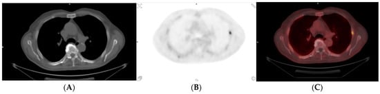
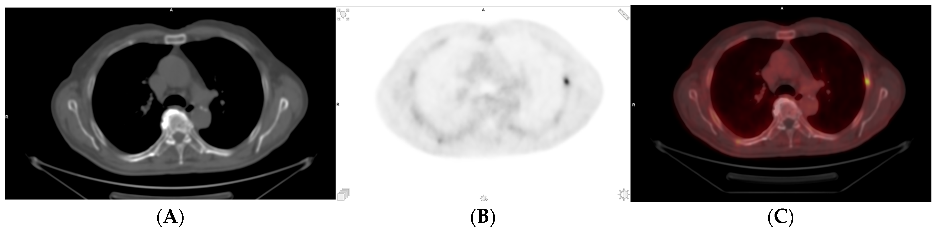
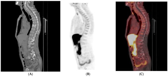
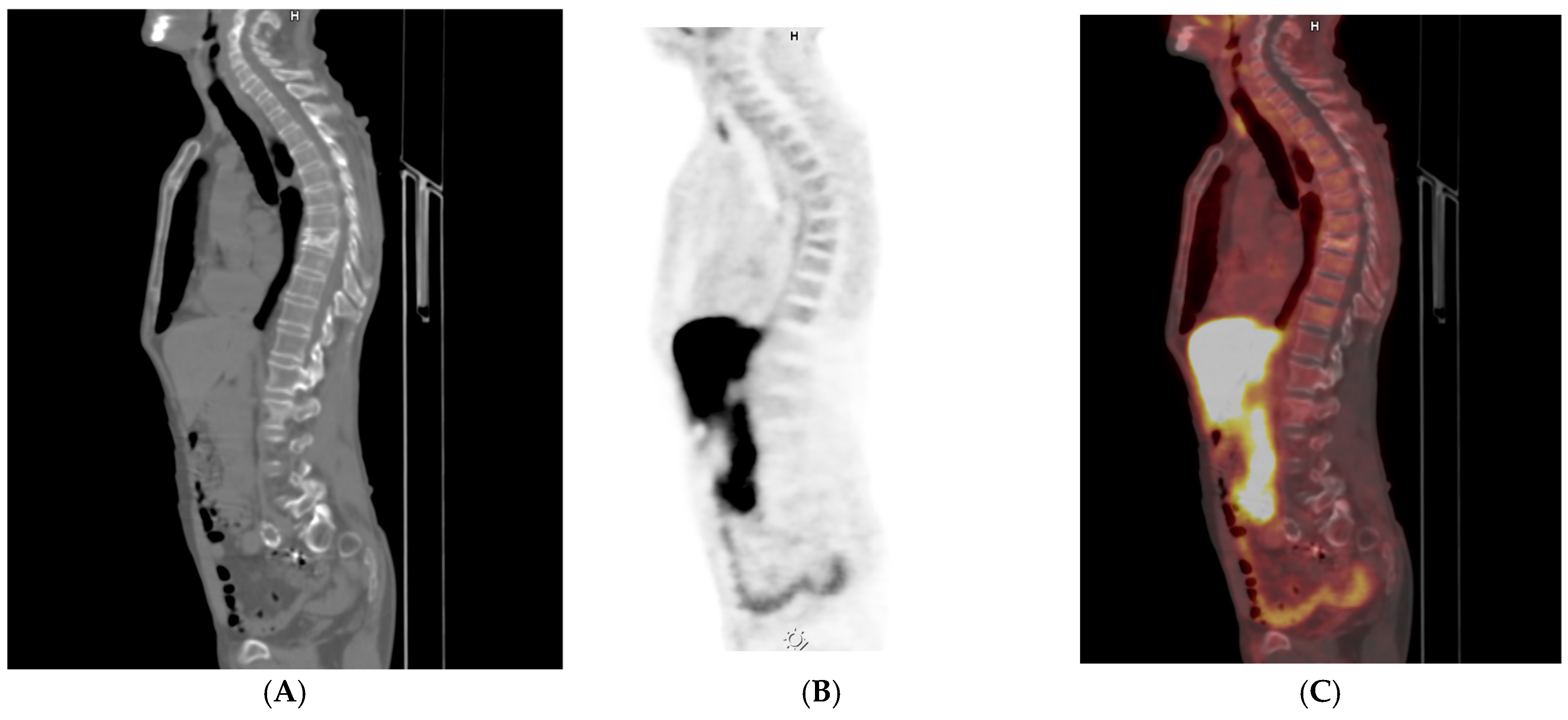
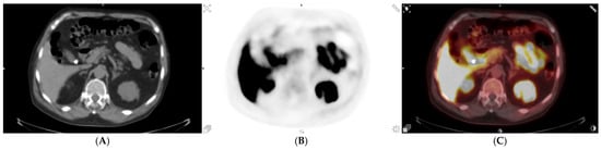
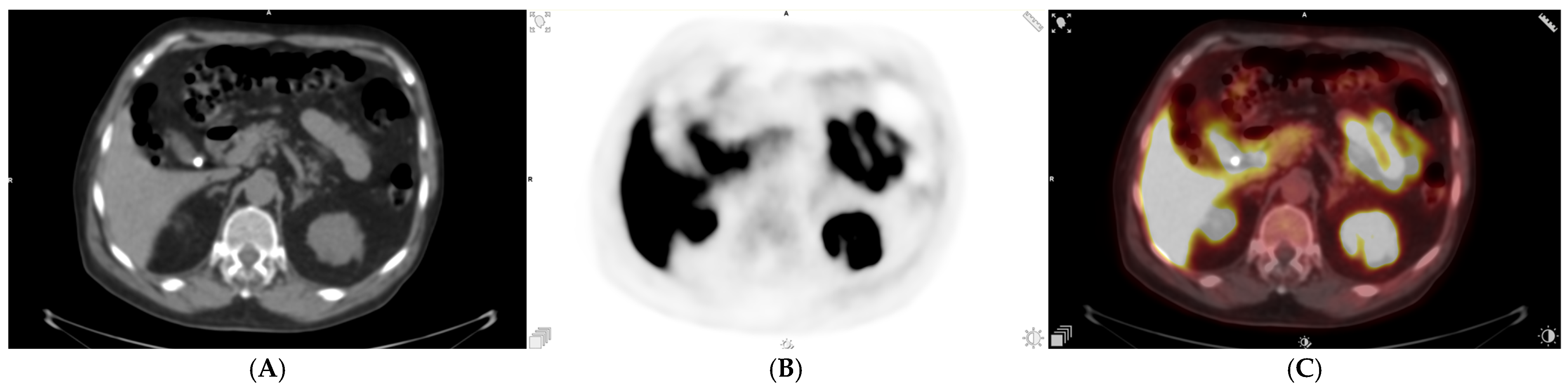
Representative osseous pitfalls and fracture-related mimics discussed in this section are illustrated in Figure 1 and Figure 2.
Figure 1.
(A–C) [18F]-PSMA PET/CT images demonstrating post-traumatic changes related to a healed rib fracture without imaging features suggestive of active malignant involvement.
Figure 2.
(A–C) [18F]-PSMA PET/CT demonstrating a compression fracture of the Th8 vertebral body, identified as an incidental osseous finding. Take-home message: Vertebral compression fractures are common in older men with prostate cancer; escalation should be reserved for cases with PET uptake, aggressive CT morphology, or clinical concern rather than as a default reflex.
Take-home message: Mechanically patterned bone uptake without a CT correlate is a Category B or C finding; conditional safety-netting language should replace reflex MRI referral.
LDCT incidental findings and appropriate use of incidentaloma guidelines: CTAC/LDCT-dominant findings are common in older men, and a structured approach helps prevent both over-calling and under-calling. High-risk pulmonary nodules, suspicious adrenal masses, complex renal lesions, and urgent vascular abnormalities are Category A patterns, where a single targeted next step is appropriate, typically a diagnostic-quality dedicated imaging study guided by established recommendations [12,13,14]. Small low-risk pulmonary nodules, simple renal cysts, uncomplicated small pancreatic cysts in appropriate contexts, benign degenerative disease, and benign calcifications are typically Category B or C depending on context and should not populate the conclusion, because, otherwise, the conclusion becomes a list of low-yield follow-ups rather than a decision-support tool [12,16]. Hepatic steatosis is frequent in this population and should be acknowledged as such, yet it rarely warrants an imaging cascade in the absence of focal lesions or clinical suspicion of significant liver disease; it is best handled as a brief body-only note with escalation anchored in clinical context rather than imaging reflex [15]. A key bridge statement should explicitly note that Fleischner, Bosniak, and ACR incidental findings algorithms were developed for diagnostic-quality CT and MRI and that, in PSMA PET/CT, the CTAC/LDCT should be treated as a detection map; if escalation criteria are met, characterization should move to dedicated diagnostic imaging rather than be forced on low-dose non-contrast CT [12,13,14,15,16].
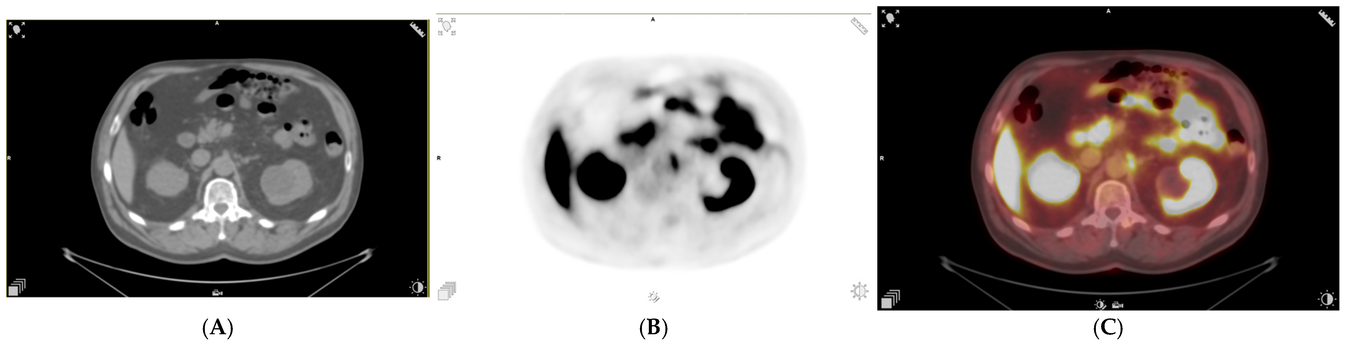
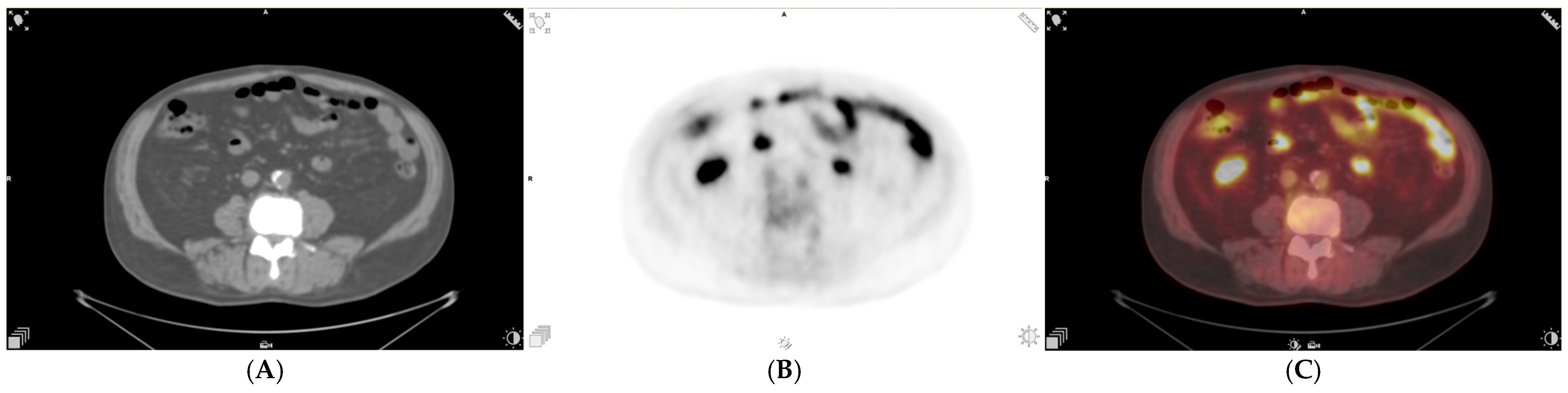
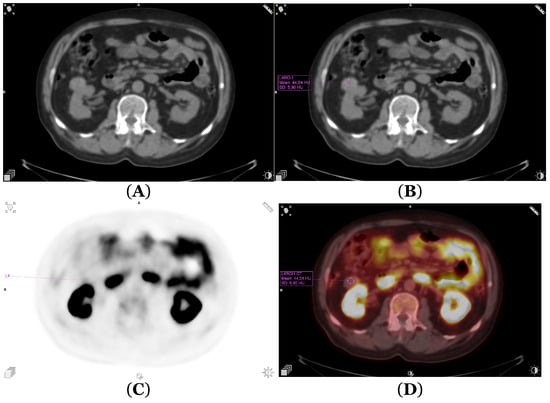
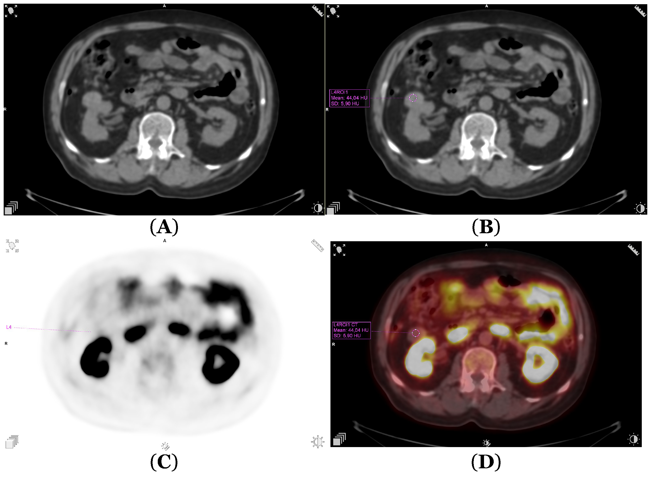
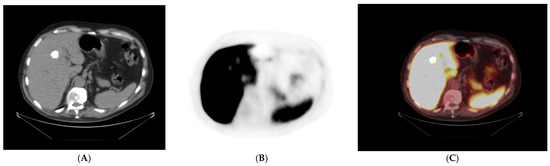
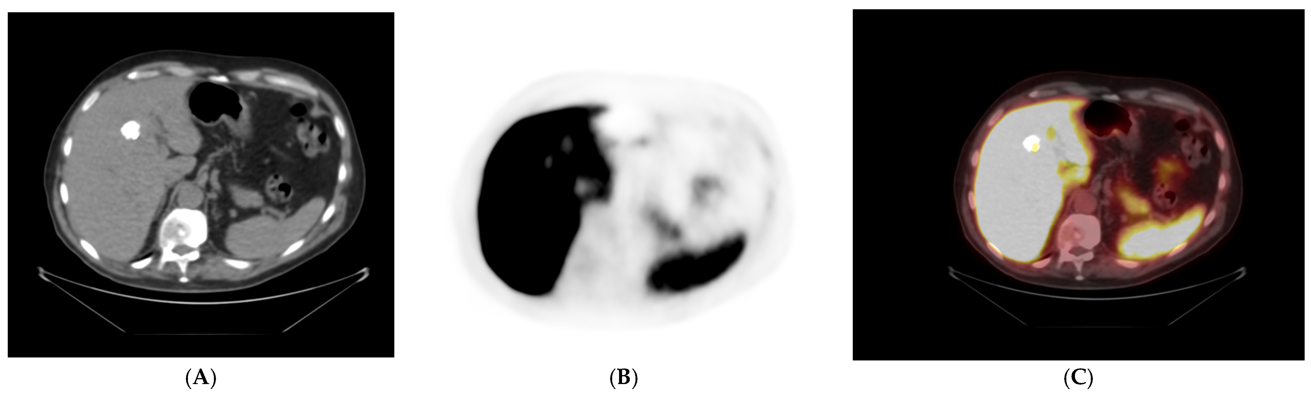
The representative CTAC/LDCT incidental findings discussed in this section are illustrated in Figure 3, Figure 4, Figure 5, Figure 6, Figure 7, Figure 8 and Figure 9.
Figure 3.
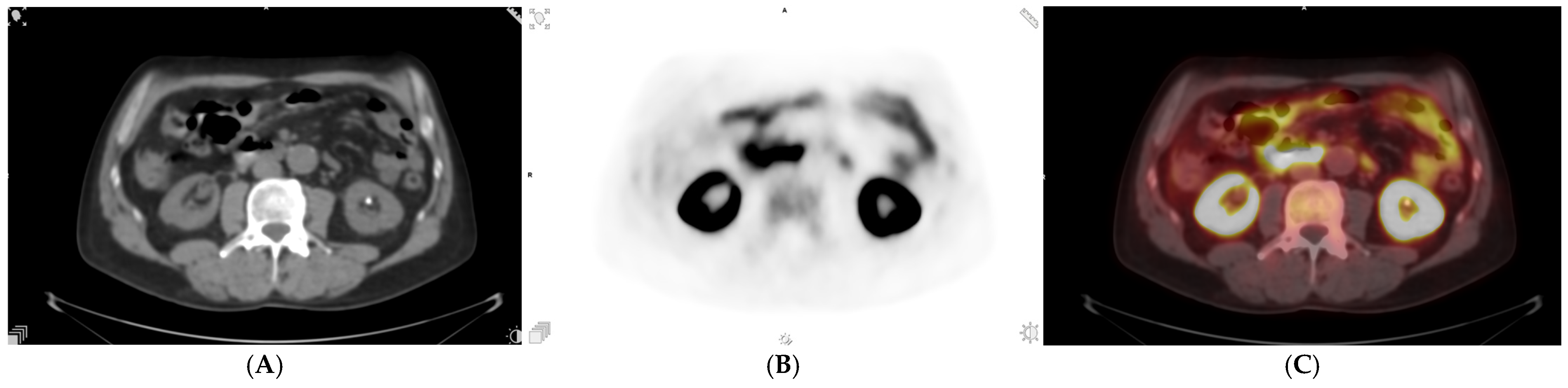
(A–C) [18F]-PSMA PET/CT showing a cystic lesion in the left kidney, consistent with a simple renal cyst identified as an incidental finding. Take-home message: Simple renal cysts on CTAC/LDCT are Category C findings requiring only a brief body-only note; no imaging cascade is warranted unless atypical features appear on dedicated diagnostic imaging.
Figure 4.
(A–C) [18F]-PSMA PET/CT demonstrating atherosclerotic calcifications within the aortic wall, incidentally detected on the CT component of the examination. Take-home message: Vascular calcifications are Category C findings in older men; they should not populate the report conclusion and require no imaging cascade.
Figure 5.
(A–C) [18F]-PSMA PET/CT revealing aneurysmal dilatation of the aorta as an incidental non-oncologic finding. Take-home message: Large aortic aneurysm is a Category A finding; the conclusion should contain a single targeted recommendation (vascular referral or dedicated CTA) with an explicit CTAC/LDCT limitation statement.
Figure 6.
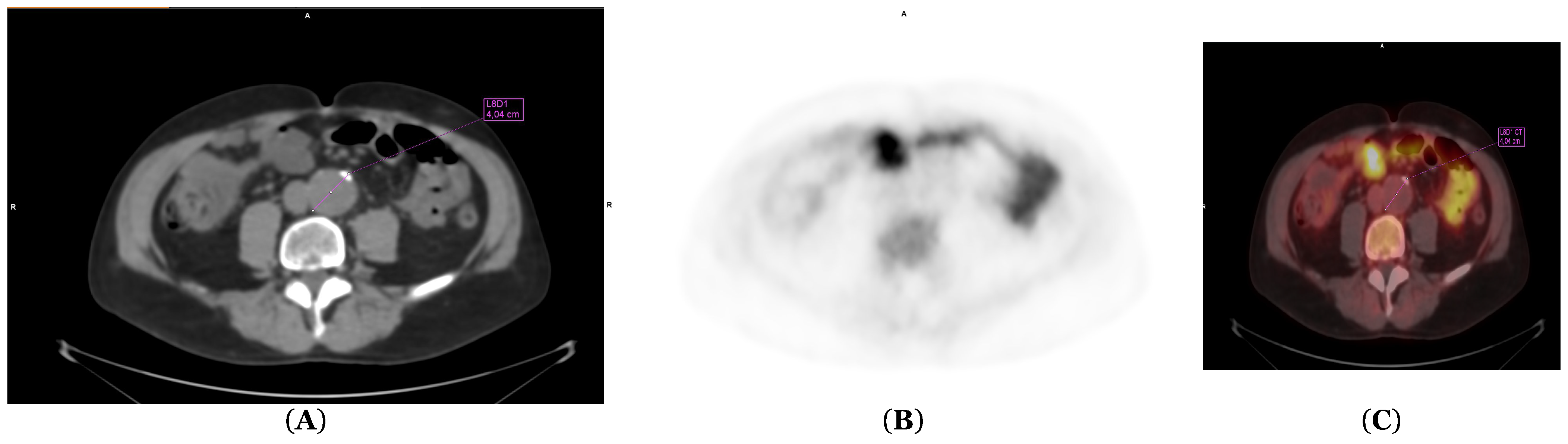
(A–C) [18F]-PSMA PET/CT demonstrating renal calculi incidentally identified on the CT component of the study. Take-home message: Renal calculi are Category B or C findings in the oncology context; escalation should be guided by clinical symptoms rather than imaging reflex.
Figure 7.
(A–D) [18F]-PSMA PET/CT showing a focal renal lesion incidentally detected during oncologic imaging and requiring further diagnostic characterization. Take-home message: A solid or complex renal lesion is a Category A finding; recommend dedicated renal CT or MRI, noting that Bosniak classification requires diagnostic-quality imaging rather than CTAC/LDCT.
Figure 8.
(A–C) [18F]-PSMA PET/CT demonstrating a gallbladder calculus as an incidental finding on the CT component of the examination. Take-home message: Gallstones are Category B or C findings in asymptomatic oncology patients; document this in the report body without populating the conclusion unless the patient is symptomatic.
Figure 9.
(A–C) [18F]-PSMA PET/CT showing focal calcification within the liver parenchyma, incidentally detected on the CT component of the study. Take-home message: Hepatic calcifications are Category C findings consistent with granulomatous disease; no imaging cascade is required in the absence of focal hepatic lesions or clinical liver disease.
Structured reporting templates: In this setting, templates are not meant to increase verbosity; they are meant to keep the conclusion uncluttered, ensure Category A items are actionable with a single next step, ensure Category B items are documented with conditional safety netting rather than automatic workup, and ensure Category C pitfalls are explicitly neutralized in language. Template A is intended for routine high-throughput practice, particularly when the primary question is clear and incidental findings are limited or straightforward; it is the default for most staging and restaging examinations because it minimizes cognitive noise while preserving traceability. Template B is intended for complex cases, tumour board discussions, equivocal patterns (notably bone foci with [18F]-PSMA-1007), or when prior imaging correlation materially changes actionability; it adds explicit A/B/C labelling and trigger rules to make downstream intent unambiguous to all readers, including those reviewing the report under time pressures.
Template A (concise, cascade-aware)
Clinical context: [staging/restaging; PSA; prior therapy; and key clinical question].
Radiopharmaceutical and uptake time: [18F]-PSMA ligand, [MBq], and uptake time [min].
Acquisition: PET/CT from [vertex] to [mid-thigh/knees].
CT component: Low-dose non-contrast CT (CTAC/LDCT) for attenuation correction and localization; limited for definitive characterization.
Physiologic distribution: [expected biodistribution; mention only if atypical] [1].
Head/neck: [no suspicious PSMA-avid lesions/describe relevant lesion(s); only note common pitfalls if needed] [1,5].
Chest: [primary prostate cancer-related lesions outside nodes if present; otherwise, negative].
Lymph nodes (N category): Pelvic stations (obturator, internal/external iliac, common iliac, and presacral): [side; station; short-axis if measurable; uptake pattern; CT correlate if present; and PSMA-RADS if used]. Retroperitoneal (para-aortic/paracaval): [same fields]. Other nodal regions (mediastinal/supraclavicular): [same fields; note atypical distribution when relevant]. Summarize using PROMISE/miTNM (miN1 vs. miM1a) and/or E-PSMA wording [5,6,7].
Abdomen/pelvis: Prostate/prostatic bed: [local recurrence yes/no; site; uptake; and correlate]. Visceral organs: [metastatic/indeterminate/negative]. Incidental findings: Category A described with one targeted action; Category B documented in the body using “This finding is most consistent with a benign/indeterminate process; in the absence of symptoms or interval change, additional workup is not routinely recommended.” If LDCT-driven escalation is suggested, include “CT component is low-dose and non-contrast (CTAC/LDCT) acquired for attenuation correction and localization; dedicated diagnostic imaging may be required for definitive characterization.”
Skeleton (M category): [metastatic/indeterminate/negative; CT correlate yes/no; and conditional escalation only if red flags, especially for [18F]-PSMA-1007 patterns] [8,9,10].
Conclusion: Prostate cancer summary using PROMISE/miTNM ± miPSMA and/or E-PSMA, explicitly including nodal status [5,6,7]. Category A incidental actionable item(s) with a single next step. CTAC/LDCT limitation statement if any CT-driven recommendation is made.
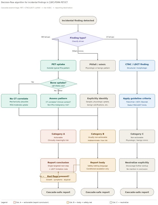
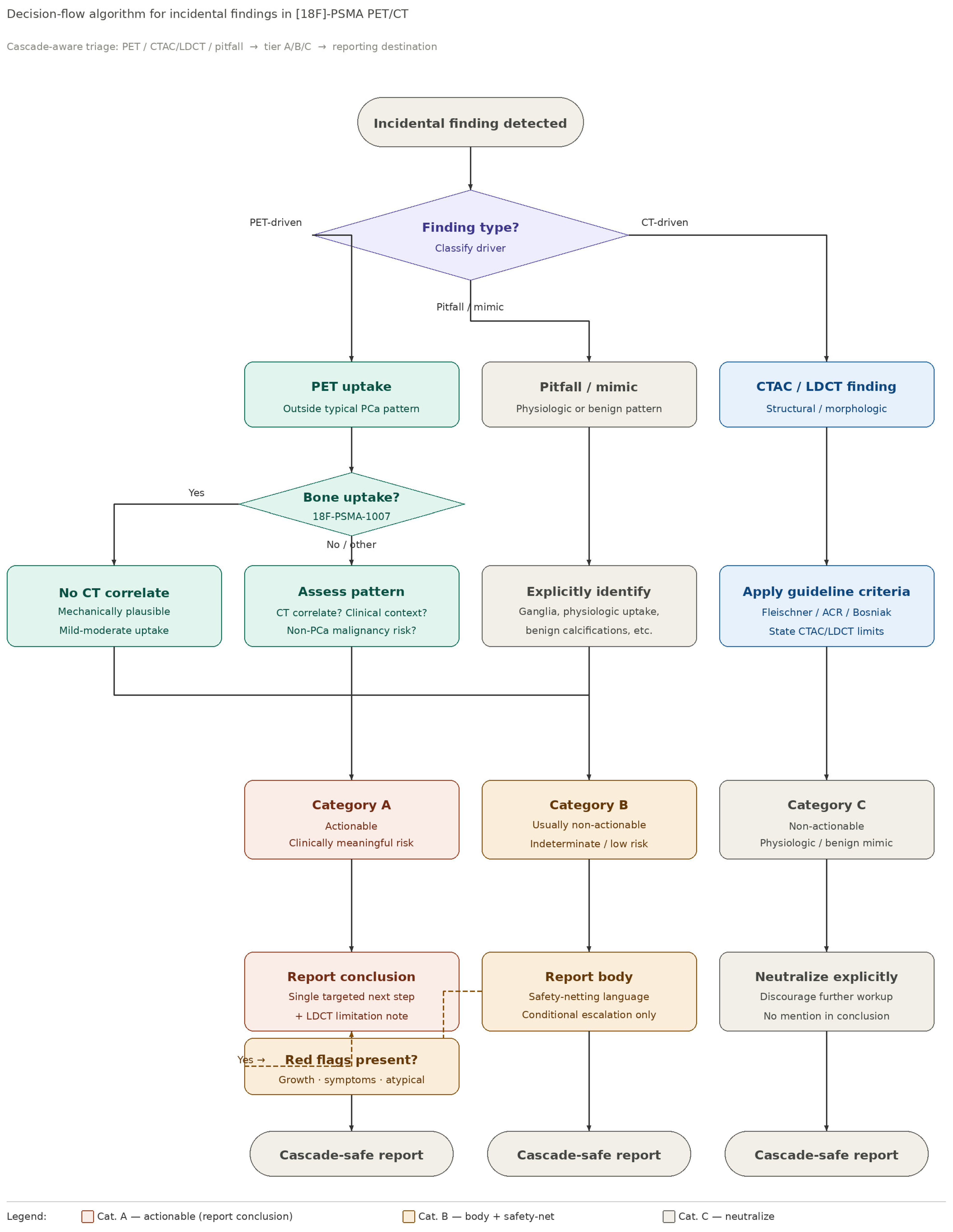
Template B (extended, cascade-aware) [The abbreviated version retained here summarizes the structural elements; the complete field-by-field template is provided in Supplementary File S1. A summary decision-flow algorithm is shown in Figure 10].
Figure 10.
Summary decision-flow algorithm for incidental findings in [18F]-PSMA PET/CT. The algorithm guides the reporting radiologist from detection through category assignment (PET-driven uptake/CTAC/LDCT-driven finding/pitfall or mimic) to actionability tier (A: actionable, report in conclusion with single next step; B: document in body with conditional safety netting; and C: explicitly neutralize as non-actionable) and final reporting destination. Tracer-specific guidance for [18F]-PSMA-1007 unspecific bone uptake is incorporated as a branch within the PET-driven pathway. Take-home message: A single structured decision flow applied consistently across all incidental findings reduces variability in reporting language and protects the report conclusion from low-value cascade-triggering content.
A summary decision-flow algorithm for cascade-aware triage is provided in Figure 10.
Template B (full version): See Supplementary File S1. Summary of structural elements: Clinical context: [staging/restaging; PSA; prior therapy; and key clinical question]. Prior imaging correlation: [date/modality; stability/progression; and key comparisons]. Radiopharmaceutical and uptake time: [18F]-PSMA ligand, [MBq], and uptake time [min]. Acquisition: PET/CT from [vertex] to [mid-thigh/knees]. CT component: Low-dose non-contrast CT (CTAC/LDCT) for attenuation correction and localization; limited for definitive characterization; and if escalation is proposed, characterization is deferred to diagnostic-quality CT/MRI, and incidentaloma algorithms apply in the diagnostic imaging context [12,13,14,15,16].
Physiologic distribution and pitfalls: [expected biodistribution; explicitly identify potential mimics when relevant, e.g., sympathetic ganglia, ureteral activity] [1,5].
Head/neck: [lesions if present; specify site, uptake pattern, and CT correlate; and add PSMA-RADS when helpful]. Thyroid (if focal uptake): [finding] (A/B/C); [conditional trigger and escalation framing per ACR thyroid incidentaloma guidance] [17].
Chest (non-nodal): [pulmonary/pleural/mediastinal organ findings; PET and CT correlate]. Lung nodule(s) on CTAC/LDCT: [location, size, and morphology] (A/B/C); include LDCT limitation sentence; and single next-step rule linked to diagnostic chest CT when A [12].
Lymph nodes (explicit station-by-station; likelihood language): Pelvic stations: For each PSMA-avid node record [side; station; short-axis; uptake relative to reference; CT correlate; and PSMA-RADS if used]. Retroperitoneal: [same fields]. Mediastinal/supraclavicular/other: [same fields; explicitly note “atypical for PCa” when appropriate and consider alternative pathology only when pattern supports it]. Overall nodal classification: PROMISE/miTNM (miN1 vs. miM1a) with miPSMA score and/or E-PSMA wording; avoid SUVmax-only probability statements [5,6,7]. Indeterminate nodal findings: [statement with conditional triggers rather than reflex escalation].
Abdomen/pelvis: Prostate/prostatic bed: [site; uptake; correlate; and likelihood statement]. Visceral organs (liver, adrenals, kidneys, bowel, and pancreas): each notable finding written as “[finding, location, size/morphology if CT-based] (A/B/C)” with one next-step rule. For renal/adrenal/liver/pancreas items that prompt escalation, include an LDCT limitation sentence and brief justification that Bosniak/ACR algorithms require diagnostic-quality imaging [13,14,15,16]. Hepatic steatosis: [present/absent; brief note] (B) with no imaging cascade unless there is clinical indication [15].
Skeleton: Bone findings reported with PET–CT correlation; each indeterminate focus written as “[site; uptake; CT correlate yes/no] (A/B/C)” with explicit trigger criteria. For [18F]-PSMA-1007-type unspecific uptake patterns, state “mechanically plausible distribution with no CT correlate” and embed the non-routine workup sentence unless red flags exist (pain, interval growth, and atypical morphology) [8,9,10].
Conclusion: Prostate cancer summary using PROMISE/miTNM ± miPSMA and/or E-PSMA, explicitly including nodal distribution and stage [5,6,7]. Category A incidental actionable item(s) with a single next step each. Cascade guardrail sentence for B/C items. CTAC/LDCT limitation statement if any CT-driven recommendation is made.
2. Mini-Case Illustrating How the Framework Reads
A 71-year-old man with biochemical recurrence after prostatectomy undergoes [18F]-PSMA PET/CT for localization of recurrence. PET shows one PSMA-avid left pelvic nodal focus consistent with metastatic disease, reported using the PROMISE miTNM and miPSMA score [7]. In the skeleton, there is mild focal uptake in the right seventh rib without a CT correlate, with a mechanically plausible location and no reported pain, which is documented as Category B with conditional escalation only if interval change or symptoms develop, consistent with evidence on unspecific bone uptake in [18F]-PSMA-1007 [8,9,10]. On CTAC/LDCT, a 9 mm solid pulmonary nodule is detected in the right upper lobe, which is handled as Category A with a targeted recommendation for dedicated diagnostic chest CT and explicit CTAC/LDCT limitation language, aligning with pulmonary nodule management guidance while acknowledging the oncology context [12]. The conclusion contains the pelvic nodal metastasis and the actionable lung nodule with a single next step, while the rib focus remains in the body with cascade-resistant phrasing, so the clinical pathway is supported rather than distracted.
3. Practical Implementation Considerations
Three dimensions of practical adoption merit explicit discussion. First, reporting time: Structured templates, once integrated into speech-recognition macros or PACS/RIS text fields, typically add an estimated 1–3 min per examination, based on analogous implementations of structured reporting in CT colonography and prostate MRI reporting programmes; this initial time investment is offset by reduced iterative communication with referring clinicians who receive a self-contained, actionable conclusion rather than a list of findings requiring telephone clarification [5]. Second, training requirements: Initial departmental adoption should include a calibration session using retrospective cases to align A/B/C classification thresholds among all readers in the group; experience from structured reporting programmes suggests that a single half-day workshop with 15–20 calibration cases is sufficient to achieve acceptable initial agreement (kappa ≥ 0.65) and that inter-reader drift can be managed through a quarterly audit of a random sample of reports. Third, PACS and RIS integration: The tiered language blocks in Templates A and B are modular and can be embedded as macros, auto-text entries, or structured fields in most modern reporting platforms (e.g., PowerScribe, Nuance, Sectra, and Carestream); the published PSMA-RADS reporting experience provides a precedent for this type of integration, and the A/B/C tier labels can be mapped to structured data fields for downstream audit without requiring free-text parsing.
4. Artificial Intelligence: Concrete Use Cases and a Governance Warning
AI can be useful in PSMA PET/CT incidental findings when applied to governed, task-specific problems rather than generic “automation.” Practical use cases include pitfall recognition, such as differentiating ganglia uptake from nodal disease and recognizing physiologic patterns that commonly generate false positives, and structured report enforcement, such as template completion and standardized lexicon insertion that keeps conclusions protected and consistent [5,18,19]. Another high-yield use case is triaging CTAC/LDCT findings, where AI can only flag those meeting predefined escalation criteria, thereby reducing random follow-up recommendations and keeping attention on clinically meaningful abnormalities [18,19]. However, sensitivity-biassed algorithms can increase cascades if they generate excessive alerts, so governance, audit, and threshold calibration toward value-based endpoints should be considered part of the clinical deployment rather than an optional add-on [18,19]. Three concrete workflows illustrate the scope of AI applicability. First, a pitfall-recognition pipeline can be trained to distinguish celiac and stellate ganglia uptake from adjacent nodal disease using multi-parametric PET–CT feature inputs (location, uptake pattern, and CT morphology), thereby reducing the rate of false-positive nodal reports without requiring reader retraining [18,19]. Second, a CTAC/LDCT triage algorithm can be configured to flag only findings that meet predefined Fleischner or ACR escalation criteria—for example, pulmonary nodules above the 6 mm threshold with solid morphology—and suppress alerts for findings below actionability thresholds; published experience with automated lung nodule detection in oncology CT workflows suggests that this approach can reduce random follow-up recommendations by 30–50% when thresholds are calibrated to guideline criteria rather than sensitivity alone [18,19]. Third, a structured-report enforcement tool can check for mandatory A/B/C tier labelling before report sign-off, ensuring that every conclusion contains at least one actionable item without Category B or C content populating the impression. For each of these applications, failure modes must be anticipated: sensitivity-biassed models trained on enriched datasets may generate excessive low-confidence alerts that paradoxically increase cascade volume; demographic or scanner-type bias in training data may produce differential false-positive rates across sites; and automated language generation may introduce non-standard phrasing that undermines the standardization goal these tools are meant to serve. Governance therefore requires prospective performance monitoring against value-based endpoints—cascade rate, time to definitive prostate cancer treatment initiation, and downstream imaging utilization—rather than sensitivity-only benchmarks, and audit cycles should be planned before clinical deployment rather than deferred as optional add-ons [18,19].
5. Limitations and Research Agenda
This is a narrative review and does not provide pooled prevalence estimates for all incidental findings; reported frequencies vary across tracers, scanners, patient populations, and definitions of what constitutes an incidental finding. The proposed A/B/C overlay prioritizes actionability and cascade reduction, but prospective validation would strengthen confidence in its safety and clinical impact. Future work should quantify the rates and types of downstream cascades attributable to incidental findings in PSMA PET/CT, measure time-to-treatment effects in prostate cancer pathways, and evaluate inter-reader agreement and downstream imaging utilization before and after the implementation of cascade-aware templates; without such data, the framework remains a structured expert recommendation rather than an evidence-validated clinical tool. Dedicated quantitative data on cascade rates specifically attributable to incidental findings in [18F]-PSMA PET/CT are currently unavailable, and this absence reflects structural rather than incidental limitations. First, the term “cascade” is not a coded outcome in most radiology information systems or electronic health records, making retrospective extraction labour-intensive and dependent on manual chart review. Second, the causal link between a specific incidental finding and a subsequent investigation is rarely captured prospectively; downstream tests may be ordered by different clinicians, in different departments, or across institutional boundaries, fragmenting the data trail. Third, inconsistent nomenclature for incidental findings across institutions and imaging disciplines complicates uniform capture and cross-study comparison. Fourth, published PSMA PET/CT cohorts were predominantly designed to evaluate oncological endpoints rather than incidental-finding management and therefore lack the granularity required to quantify cascade rates by finding type or actionability tier. Collectively, these factors explain why the evidence base for the proposed framework draws on cascade epidemiology from adjacent domains rather than PSMA-specific datasets and underscore why prospective, registry-based studies with pre-specified cascade endpoints represent the most direct path towards filling this evidence gap [3,4]. The proposed A/B/C framework reflects the authors’ clinical experience and interpretive practice in [18F]-PSMA PET/CT reporting, and this expert-driven origin constitutes a potential bias that should be explicitly acknowledged: category assignments and report-language recommendations may reflect individual or institutional preferences that are not universally shared, and readers should interpret the framework as a structured starting point for local adaptation rather than a prescriptive standard. Formal consensus methodology (e.g., Delphi process across multiple centres and specialties) would be the appropriate next step to move from expert recommendation to endorsed guidance. Interobserver variability in assigning A/B/C categories is a specific and clinically important concern that warrants explicit discussion, particularly for borderline or equivocal findings. Category A and Category C assignments are expected to show relatively high inter-reader agreement because they either correspond to findings that clearly meet published guideline escalation thresholds (Category A) or to well-documented physiologic patterns (Category C): both types benefit from explicit published criteria that constrain individual judgement. Category B assignments, however, are inherently more susceptible to interobserver variability because they occupy the uncertain middle ground between clearly actionable and clearly dismissible—the very findings for which expert extrapolation is required and where clinical context, scanner quality, and reader experience interact. The scenarios most prone to disagreement include: a focal bone uptake in [18F]-PSMA-1007 with a mild-to-moderate CT correlate that is ambiguous between degenerative change and early metastasis; a small solid pulmonary nodule at or near the Fleischner-size threshold; and a borderline-size adrenal lesion without clear lipid-rich features on LDCT. For these borderline cases, the framework provides conditional escalation triggers (symptoms, interval growth, and atypical morphology) rather than binary rules, which limits but does not eliminate interobserver variability. The calibration session proposed in the Practical Implementation section—a half-day workshop using 15–20 retrospective cases—is specifically designed to align threshold application for these boundary scenarios within a reporting team, and the proposed kappa target of ≥0.70 is intended to benchmark acceptable reproducibility before departmental deployment. Until prospective inter-reader studies are performed, users of the framework should treat Category B assignments for equivocal findings as the most variable component and plan for the calibration session accordingly. The transferability of the recommendations to different practice contexts has not been evaluated and constitutes a significant limitation. Low-volume centres may lack the case density to build reader familiarity with tracer-specific pitfall patterns, making the A/B/C classification more difficult to apply reliably without dedicated training support. Centres using older PET scanners with lower spatial resolution may encounter higher rates of equivocal findings that the framework does not resolve. International variation in reporting culture, medicolegal environment, and downstream specialist availability may influence which Category B findings are escalated in practice regardless of recommended language. These contextual factors should be considered when adapting the templates and thresholds to local conditions, and future implementation studies should stratify outcomes by centre volume and equipment type. The potential impact of implementing cascade-aware reporting in [18F]-PSMA PET/CT can be hypothesized as follows, pending prospective data. If approximately one-third of incidental findings currently generate at least one downstream test—consistent with the general incidental-finding cascade literature [3,4]—and if the A/B/C overlay successfully reclassifies the majority of currently escalated Category B and C findings as non-actionable through explicit report language, the expected reduction in reflex imaging could be clinically meaningful at the population level given the growing volume of [18F]-PSMA PET/CT examinations. The impact on therapeutic delay is harder to quantify but potentially important: a single unnecessary follow-up CT or bone scan triggered by an incidental finding can delay multidisciplinary tumour board decisions by weeks in systems with a limited scheduling capacity. These hypotheses are testable in a before–after audit design and should be the primary outcome targets of prospective implementation studies. To be explicit about the evidence base: the A/B/C framework is consensus-driven and conceptual in its current form. No prospective cascade-reduction data exist for this specific overlay at the time of writing. The quantitative context for the problem comes from adjacent literature—for example, Ganguli et al. found that 36.5% of incidental findings in a US national survey generated at least one additional test within 90 days [4], and Perry et al. identified non-prostate malignancies in approximately 1–2% of patients undergoing [18F]-DCFPyL PET/CT [2]—but these figures describe the magnitude of the cascade problem rather than validating the proposed solution. We frame the framework as hypothesis-generating and present it as a structured basis for future prospective studies. The minimum evidence required to move from consensus to validated practice would include a before–after study measuring cascade rates following structured template implementation, with pre-specified outcome measures of downstream imaging utilization, time from PET/CT to the initiation of definitive prostate cancer treatment, and inter-reader agreement on A/B/C classification (for which a kappa target of ≥0.70 would represent acceptable reproducibility). Regarding tracer generalizability: the framework was deliberately developed for [18F]-labelled PSMA tracers because their biodistribution and pitfall profile—in particular, the higher frequency of unspecific bone uptake with [18F]-PSMA-1007 compared with [68Ga]-PSMA-11—shape what is perceived as incidental and which findings are most cascade-prone [1,8,9,10]. Extrapolation to [68Ga]-labelled compounds should therefore be done cautiously; [68Ga]-PSMA-11 has a lower rate of unspecific bone uptake and somewhat different renal clearance kinetics, which may reduce the frequency of Category C pitfall findings while leaving the PET- and LDCT-driven A/B framework broadly applicable. Prospective inter-reader variability studies using [68Ga] tracers alongside [18F] tracers are needed before cross-tracer generalizability can be assumed.
6. Conclusions
Incidental findings are inevitable in [18F]-PSMA PET/CT, but unnecessary diagnostic cascades are not. A structured approach that distinguishes PET-driven uptake, LDCT-driven findings, and pitfalls and mimics, and then applies an actionability-based overlay that protects the report conclusion from non-actionable clutter, may improve patient safety while reducing low-value downstream testing [3,4]. Established PSMA reporting frameworks should remain the foundation for prostate cancer assessment, whereas incidental findings should be incorporated through an additional cascade-aware layer that clearly defines reporting intent and thresholds for escalation [5,6,7]. The CTAC/LDCT component should be interpreted within its technical limitations; whenever further characterization is justified, this should be performed with dedicated diagnostic imaging rather than inferred from low-dose non-contrast CT, particularly when management recommendations are being considered [12,13,14,15,16]. Practical reporting templates supported by cautious conditional language may reduce misinterpretation, and AI-based tools may further enhance standardization, provided they are implemented in a way that does not amplify false-positive findings or follow-up inflation [18,19]. Given the growing volume of [18F]-PSMA PET/CT examinations, there is an increasing need for nuclear medicine societies to propose dedicated reporting templates or more detailed guidance on the classification, communication, and follow-up of incidental findings. Such standardization may help limit overdiagnosis and reduce the time spent reporting abnormalities that are unlikely to affect patient management in the context of the original indication for the examination.
Supplementary Materials
The following supporting information can be downloaded at: https://www.mdpi.com/article/10.3390/uro6020017/s1, Supplementary File S1: Template B (Extended, Cascade-Aware Reporting Template for [18F]-PSMA PET/CT Incidental Findings).
Author Contributions
Conceptualisation, K.S. and J.W.; methodology, K.S. and M.M.; literature review and synthesis, K.S., M.K. (Marek Kasprowicz), M.M., B.O., and M.R.; writing—original draft preparation, K.S.; writing—review and editing, K.S., M.K. (Marek Kasprowicz), M.M., B.O., T.B., M.K. (Małgorzata Kobylecka), J.W., and M.R.; supervision, J.W. and M.K. (Małgorzata Kobylecka). All authors have read and agreed to the published version of the manuscript.
Funding
This research received no external funding.
Data Availability Statement
No new data were created or analyzed in this study. Data sharing is not applicable to this article.
Acknowledgments
Generative AI was used exclusively to assist with spelling and language correction during manuscript preparation. No AI tools were used to generate scientific content, interpret data, or draw conclusions. All outputs were critically reviewed by the authors, who take full responsibility for the final manuscript.
Conflicts of Interest
The authors declare no conflicts of interest.
References
- Fendler, W.P.; Eiber, M.; Beheshti, M.; Bomanji, J.; Calais, J.; Ceci, F.; Cho, S.Y.; Fanti, S.; Giesel, F.L.; Goffin, K.; et al. PSMA PET/CT: Joint EANM Procedure Guideline/SNMMI Procedure Standard for Prostate Cancer Imaging 2.0. Eur. J. Nucl. Med. Mol. Imaging 2023, 50, 1466–1486. [Google Scholar] [CrossRef] [Scilit] [PubMed]
- Perry, E.; Talwar, A.; Sharma, S.; O’connor, D.; Wong, L.-M.; Taubman, K.; Sutherland, T.R. Non-prostate cancer tumours: Incidence on [18F]-DCFPyL PSMA PET/CT and uptake characteristics in 1445 patients. Eur. J. Nucl. Med. Mol. Imaging 2022, 49, 3277–3288. [Google Scholar] [CrossRef] [Scilit] [PubMed]
- Davenport, M.S. Incidental Findings and Low-Value Care. AJR Am. J. Roentgenol. 2023, 221, 117–123. [Google Scholar] [CrossRef] [Scilit] [PubMed]
- Ganguli, I.; Simpkin, A.L.; Lupo, C.; Weissman, A.; Mainor, A.J.; Orav, E.J.; Rosenthal, M.B.; Colla, C.H.; Sequist, T.D. Cascades of Care After Incidental Findings in a US National Survey of Physicians. JAMA Netw. Open. 2019, 2, e1913325. [Google Scholar] [CrossRef] [Scilit] [PubMed]
- Ceci, F.; Oprea-Lager, D.E.; Emmett, L.; Adam, J.A.; Bomanji, J.; Czernin, J.; Eiber, M.; Haberkorn, U.; Hofman, M.S.; Hope, T.A.; et al. E-PSMA: The EANM standardized reporting guidelines v1.0 for PSMA-PET. Eur. J. Nucl. Med. Mol. Imaging 2021, 48, 1626–1638. [Google Scholar] [CrossRef] [Scilit] [PubMed]
- Rowe, S.P.; Pienta, K.J.; Pomper, M.G.; Gorin, M.A. PSMA-RADS Version 1.0: A Step Towards Standardizing the Interpretation and Reporting of PSMA-Targeted PET Imaging Studies. Eur. Urol. 2018, 73, 485–487. [Google Scholar] [CrossRef] [Scilit] [PubMed]
- Eiber, M.; Herrmann, K.; Calais, J.; Hadaschik, B.; Giesel, F.L.; Hartenbach, M.; Hope, T.A.; Reiter, R.; Maurer, T.; Weber, W.A.; et al. Prostate Cancer Molecular Imaging Standardized Evaluation (PROMISE): Proposed miTNM Classification for the Interpretation of PSMA-Ligand PET/CT. J. Nucl. Med. 2018, 59, 469–478. [Google Scholar] [CrossRef] [Scilit] [PubMed]
- Grünig, H.; Maurer, A.; Thali, Y.; Kovacs, Z.; Strobel, K.; Burger, I.A.; Müller, J. Focal Unspecific Bone Uptake on [18F]-PSMA-1007 PET: A Multicenter Retrospective Evaluation of the Distribution, Frequency, and Quantitative Parameters of a Potential Pitfall in Prostate Cancer Imaging. Eur. J. Nucl. Med. Mol. Imaging 2021, 48, 4483–4494. [Google Scholar] [CrossRef] [Scilit] [PubMed]
- Seifert, R.; Telli, T.A.; Opitz, M.; Barbato, F.; Berliner, C.; Nader, M.; Umutlu, L.; Stuschke, M.; Hadaschik, B.; Herrmann, K.; et al. Unspecific [18F]-PSMA-1007 Bone Uptake Evaluated Through PSMA-11 PET, Bone Scanning, and MRI Triple Validation in Patients with Biochemical Recurrence of Prostate Cancer. J. Nucl. Med. 2023, 64, 738–743. [Google Scholar] [CrossRef] [Scilit] [PubMed]
- Janssen, J.; Noordzij, W.; Velleman, T.; de Jong, I.J.; Langendijk, J.A.; Verzijlbergen, J.F.; Stormezand, G.N.; Aluwini, S. Skeletal [18F]-PSMA-1007 Uptake in Prostate Cancer Patients. Ther. Adv. Med. Oncol. 2023, 15, 17588359231179311. [Google Scholar] [CrossRef] [Scilit] [PubMed]
- Jadvar, H.; Calais, J.; Fanti, S.; Feng, F.; Greene, K.L.; Gulley, J.L.; Hofman, M.; Koontz, B.F.; Lin, D.W.; Morris, M.J.; et al. Appropriate Use Criteria for Prostate-Specific Membrane Antigen PET Imaging. J. Nucl. Med. 2022, 63, 59–68. [Google Scholar] [CrossRef] [Scilit] [PubMed]
- MacMahon, H.; Naidich, D.P.; Goo, J.M.; Lee, K.S.; Leung, A.N.C.; Mayo, J.R.; Mehta, A.C.; Ohno, Y.; Powell, C.A.; Prokop, M.; et al. Guidelines for Management of Incidental Pulmonary Nodules Detected on CT Images: From the Fleischner Society 2017. Radiology 2017, 284, 228–243. [Google Scholar] [CrossRef] [Scilit] [PubMed]
- Mayo-Smith, W.W.; Song, J.H.; Boland, G.L.; Francis, I.R.; Israel, G.M.; Mazzaglia, P.J.; Berland, L.L.; Pandharipande, P.V. Management of Incidental Adrenal Masses: A White Paper of the ACR Incidental Findings Committee. J. Am. Coll. Radiol. 2017, 14, 1038–1044. [Google Scholar] [CrossRef] [Scilit] [PubMed]
- Silverman, S.G.; Pedrosa, I.; Ellis, J.H.; Hindman, N.M.; Schieda, N.; Smith, A.D.; Remer, E.M.; Shinagare, A.B.; Curci, N.E.; Raman, S.S.; et al. Bosniak Classification of Cystic Renal Masses, Version 2019. Radiology 2019, 292, 475–488. [Google Scholar] [CrossRef] [Scilit] [PubMed]
- Gore, R.M.; Pickhardt, P.J.; Mortele, K.J.; Fishman, E.K.; Horowitz, J.M.; Fimmel, C.J.; Talamonti, M.S.; Berland, L.L.; Pandharipande, P.V. Management of Incidental Liver Lesions on CT: A White Paper of the ACR Incidental Findings Committee. J. Am. Coll. Radiol. 2017, 14, 1429–1437. [Google Scholar] [CrossRef] [Scilit] [PubMed]
- Megibow, A.J.; Baker, M.E.; Morgan, D.E.; Kamel, I.R.; Sahani, D.V.; Newman, E.; Brugge, W.R.; Berland, L.L.; Pandharipande, P.V. Management of Incidental Pancreatic Cysts: A White Paper of the ACR Incidental Findings Committee. J. Am. Coll. Radiol. 2017, 14, 911–923. [Google Scholar] [CrossRef] [Scilit] [PubMed]
- Hoang, J.K.; Langer, J.E.; Middleton, W.D.; Wu, C.C.; Hammers, L.W.; Cronan, J.J.; Tessler, F.N.; Grant, E.G.; Berland, L.L. Managing Incidental Thyroid Nodules Detected on Imaging: White Paper of the ACR Incidental Thyroid Findings Committee. J. Am. Coll. Radiol. 2015, 12, 143–150. [Google Scholar] [CrossRef] [Scilit] [PubMed]
- Belal, S.L.; Frantz, S.; Minarik, D.; Enqvist, O.; Wikström, E.; Edenbrandt, L.; Trägårdh, E. Applications of Artificial Intelligence in PSMA PET/CT for Prostate Cancer Imaging. Semin. Nucl. Med. 2024, 54, 141–149. [Google Scholar] [CrossRef] [Scilit] [PubMed]
- Liu, J.; Cundy, T.P.; Woon, D.T.S.; Lawrentschuk, N. A Systematic Review on Artificial Intelligence Evaluating Metastatic Prostatic Cancer and Lymph Nodes on PSMA PET Scans. Cancers 2024, 16, 486. [Google Scholar] [CrossRef] [Scilit] [PubMed]

Disclaimer/Publisher’s Note: The statements, opinions and data contained in all publications are solely those of the individual author(s) and contributor(s) and not of MDPI and/or the editor(s). MDPI and/or the editor(s) disclaim responsibility for any injury to people or property resulting from any ideas, methods, instructions or products referred to in the content. |
© 2026 by the authors. Licensee MDPI, Basel, Switzerland. This article is an open access article distributed under the terms and conditions of the Creative Commons Attribution (CC BY) license.