Article

Transplantation of the Horseshoe Kidneys: A Model for Dual Adult Kidney Transplantation

Department of Surgery, Division of Transplantation, Allegheny General Hospital, Pittsburgh, PA 15212, USA
Uro 2022, 2(3), 157-165; https://doi.org/10.3390/uro2030019
Submission received: 7 June 2022 / Revised: 23 June 2022 / Accepted: 28 June 2022 / Published: 5 July 2022
(This article belongs to the Special Issue Current Concepts in Transplantation)

Abstract

:
Background: The shortage of organs has called for the use of two marginal adult kidneys (MAKs) with a low nephron mass as dual adult kidneys transplanted to a single recipient. The operative techniques of the transplantation of these kidneys are still debated. Since the horseshoe kidneys have been transplanted as early as 1975, it is theorized that the technique of the en bloc transplantation of the horseshoe kidney may be applied to the MAKs. Material and Methods: The world literature search during the period 1975–2021 on the use of deceased-donor horseshoe kidneys was reviewed. The selection of the donors, the anatomy of the kidneys, the principles of organ recovery, the transplantation procedure, and the results were discussed. Finally, this technique of en bloc transplantation was applied successfully to seven pairs of MAKs and is described herein. The dual adult kidneys were simultaneously vascularized by the donor aorta and vena cava, which were anastomosed, respectively, to the recipient iliac artery and vein. Results: A total of 131 case reports of deceased horseshoe kidney donors were reviewed, of which 53 en bloc kidneys were transplanted successfully to a single recipient, and the remaining 78 were divided and transplanted as single units to 131 recipients. Twenty-five single kidneys were discarded. At the time of publication, all horseshoe kidneys had a good renal function. In the series of seven pairs of MAKs transplanted en bloc, the operative time was 3 h. There were no primary nonfunctions, no vascular thromboses, no urinary leakages, and no wound infections. Only two patients required temporary dialysis despite an average of 28.4 h of cold ischemia time. No hydronephrosis and lymphocele was experienced. Both patient and graft survival were 100%. At the time of follow-up at 36 months, serum creatinine levels averaged 1.8 mg/dL (range 1.4–1.9). Conclusions: This technique of en bloc renal transplantation using the donor aorta and vena cava for revascularization can be applied to both the horseshoe kidneys and the MAK, and improve organ utilization.

1. Introduction

Renal transplantation is considered the treatment of choice for patients with end-stage renal disease by providing improved quality and extended life expectancy, with 75% of deceased-donor kidney recipients surviving at 5 years compared to 40% of patients staying on dialysis. As of December, 2021, there were 61,277 patients active on the waiting list receiving only 26,670 transplanted kidneys, with 18,699 from deceased donors and 5971 from living donors [1]. As a result of the shortage of organs, the patients had to wait a median of 3.9 years to receive a kidney, by which time approximately 46% of transplant candidates had died or had been removed from the waiting list because of aging, which increases their comorbidities. Those of blood group B or O may experience a significantly longer waiting period [2]. To solve the organ shortage, the transplant community in 1996 introduced the concept of transplanting both kidneys from marginal donors with suboptimal functions to a single age-matched adult recipient, so-called dual transplants that would have been discarded as single units since “nobody wanted them“ [3]. However, the techniques for such transplants, described as sequential unilateral [4,5,6], sequential bilateral [7,8], and en bloc unilateral [9,10,11,12], are still controversial, hence the use of this dual-kidney transplant model was very restricted, and 6643 kidneys with a kidney donor profile index from 88% to 92% or with a donor age > 66 years of age recovered during the 2010–2015 period were discarded [13]. Since en bloc horseshoe kidneys have been transplanted since 1975 [14], it is theorized that this model can be applied to the transplantation of the MAK; hence, the global experience of horseshoe kidney transplantation is reviewed with particular attention to the donor selection [15,16,17,18,19,20,21,22,23,24,25,26], the anatomy of the kidneys [27,28,29,30,31,32,33,34,35,36,37,38], the technique of transplantation [25,26,27,28,39,40,41,42,43,44,45,46,47,48,49], and the results [22,23]. Since no review has been published since 2002, and the use of horseshoe kidneys is neither tracked by the United Network for Organ Sharing (UNOS) nor by the Scientific Registry of Transplant Recipients (SRTR) databases, it is hoped that this review can provide an analysis of all cases of horseshoe kidneys reported to date and derive a surgical approach that can be applied to both the horseshoe kidneys and the MAK.

2. Material and Methods

Electronic databases were searched for case reports of transplantation of the horseshoe kidneys from 1975 to April 2022. There were 131 pairs of horseshoe kidneys, which were procured worldwide [15,16,17,18,19,20,21,22,23,24,25,26,27,28,29,30,31,32,33,34,35,36,37,44,45,46,47,48,49]. Fifty-three pairs of horseshoe kidneys were transplanted en bloc to a single recipient, and 78 pairs were divided at the isthmus with their blood supply separated and transplanted as single units to 131 recipients. Twenty-five kidneys were discarded because of complex vascular anatomy, vascular injuries, and injuries to the collecting system nonamenable to repair. For instance, one kidney was not used after thrombosis of the contralateral half after graft reperfusion [22]. In most cases, at the time of transplantation of the en bloc horseshoe kidneys, the distal donor aorta and the distal vena cava were anastomosed to the external iliac artery and vein, respectively. In one instance where the renal vessels arise behind the isthmus, the donor common iliac artery was used as inflow instead. In another case where the low-sitting isthmus hid the aortic bifurcation, the donor common iliac arteries were spatulated medially and sutured together to lengthen the donor aorta [16]. In three patients, the “huge kidneys” required an intra-abdominal approach with vascular anastomoses to the recipient’s distal aorta and vena cava using the donor’s proximal aorta and vena cava [17]. In general, a right iliac extraperitoneal approach was performed in a standard transplant procedure, and the kidneys were vascularized through the distal aortic inflow and the distal caval outflow [20,21,22,23,24,25,26,27]. The ureters were reimplanted into the bladder using the Gregoire–Lich extravesical approach in the majority of cases.
Based on this model of en bloc transplantation of the horseshoe kidneys, a series of seven en bloc dual marginal adult kidney transplants were performed in our transplant center. In four instances, the kidneys were already separated at the donor procurement site and sent to us packed separately. They were reconstructed to make an en bloc graft by capping the proximal aspect of the aorta with an aortic patch bearing the renal artery/or arteries, and the proximal vena cava was closed with a patch carrying the left renal veins. The back table surgery was performed in a 4 °C ice bath. In 3 other cases, the kidneys were shipped to us en bloc with the proximal aorta and vena cava stapled and did not require any further bench surgery. Thus, the seven en bloc kidneys had the donor aorta as a common arterial inflow and the vena cava as a common venous outflow, which were then anastomosed, respectively, to the recipient iliac artery and vein, similar to the horseshoe kidneys transplant model. The similarity between the horseshoe kidneys and the en bloc dual kidneys is shown in Figure 1.
The transplantation was performed via a right Gibson iliac incision, with dissection of both external iliac artery and vein as in a standard transplant procedure. The right iliac quadrant was chosen because there is more room and the vessels are more superficial than the contralateral side. The en bloc kidneys were inverted 180 degrees to position the vena cava on the medial site, concordant with the right iliac vein of the patient, thus preventing scissoring leading to venous thrombosis. The position of the en bloc adult kidneys prior to the vascular anastomosis is shown in Figure 2A. If the right iliac quadrant was excluded due to a preexisting transplant or the presence of very severe arteriosclerotic iliac vessels, the left side could be used, but the anatomic inversion of the kidneys was not required since there was already concordance of the donor vena cava and the recipient’s left iliac vein (Figure 2B). Once the donor distal aorta and vena cava have been anastomosed end-to-side to the external iliac artery and vein, respectively, with the venous anastomosis positioned more distally, and after thorough hemostasis had been completed, the medial kidney was flipped for the second time laterally and stacked in front of the lateral kidney. This maneuver wedged the kidney between the lateral kidney and the iliac wing without vascular kinking and stabilized it. The ureters were connected then to the bladder through a common tunnel in the extravesical fashion over a small 22 cm-long double-J ureteral stent folded at the middle [25]. The incision was closed in layers in the regular fashion. There has not been any problem with lack of space after the kidneys had been folded as a “close book”, or compartment syndrome following the closure. The lateral kidney was “pexed” in place when the peritoneum fell back, preventing postoperative shifting and venous thrombosis.

3. Results

In the horseshoe kidneys en bloc group versus the split group, the median cold ischemia time was 24 h (range from 9 to 48) versus 22 h (range from 2 to 61), the median recipient age was 29 years (range 1 to 65) versus 30 years, and the median donor age was 29 (range from 1 to 65) versus 30, with a range of 12 to 59 [23,24]. There was a 13% complication rate in the en bloc recipients, including ureteral necrosis in two and ureteral lithiasis in one. In the split organs group, an 11.3% complications rate occurred from urinary leaks with sepsis in two patients and ureteral stenoses in two others. A delayed graft function occurred in 43% of all cases. Primary nonfunction was recorded as 4.3% in the en bloc group and 13.4% in the other. Although the operative times were not recorded, it is assumed that the split group technique required a much longer rewarming time necessary to perform multiple vascular anastomoses, to which was added the presence of ischemic areas from overlooked severed small-end arteries, hence the higher rate of primary nonfunction. Post-operative hemorrhage from the symphysiotomy was recorded as 5%. The median follow-up time for the en bloc group was 14 months (range from 1 to 26) versus 24 months (range from 0 to 166) for the split group. Graft function was similar in both groups, with a serum creatinine of 90 micromoles/L (range from 53 to 256) and 150 micromoles/L (range from 65 to 400), respectively. The overall graft survival for both groups was 87% after a mean follow-up of 22 months, equivalent to that of a control group of kidney transplants [24]. The transplant characteristics are summarized in Table A1.

4. Discussion

The horseshoe kidney was described first by the Italian physician Jacopo Berengario da Carpi from the University of Bologna in 1552 in the book “Isagoge Breves” or “A Short Introduction to Anatomy”. The credit for its use in transplantation went to Politano who, in 1963, successfully transplanted the right half of a horseshoe kidney to a monozygotic twin (personal communication). Nelson first officially reported the successful use of split horseshoe kidneys in two recipients in 1975 [15].
The horseshoe kidney is the most common congenital fusion abnormality of the kidneys, with an estimated incidence of 1/400 to 1/800 from childhood autopsy series, with a male preponderance of 2:1. It can be associated with other chromosome disorders such as Edward syndrome (67%), Turner syndrome (14–20%), and Down syndrome (1%). It occurs during the 4th to 8th week of gestation from the fusion of the metanephrogenic caps across the midline, usually at the lower poles (15%) and rarely in the upper poles (5%), through the low-seated isthmus crossing the midline, which is more lateralized toward the dominant left side, hence the U shape of the en bloc kidneys and the name of horseshoe kidney given to the partially fused kidneys. Since the fusion occurs before the rotation, the urinary collecting system is anterior and prominent and can be often mistaken for hydronephrosis. The short ureters cross the anterior aspect of the isthmus, with duplication present in 10% of the cases. These ureteral anomalies may explain the multiple episodes of urinary tract infection during childhood. From the lack of developmental ascension due to the presence of the inferior mesenteric artery, the isthmus becomes “stuck” at the level of L1–L4, and the true renal arteries, instead of numbering one or two as in normal kidneys, become multiple, “anomalous”, and segmental, and can take their “aberrant” origins from the aorta and the anterior aspect of the distal aorta and/or the common iliac arteries. Thus, 70% of horseshoe kidneys have multiple anomalous renal arteries to each kidney [27]. The same holds true for the renal veins, which can drain separately into the inferior vena cava or the iliac veins. The fleshy isthmus may contain some excretory systems and receive aberrant vessels from the common iliac arteries, but may be replaced by fibrous tissue in 15%. The horseshoe kidneys have an increased incidence of stone disease, squamous cells, and transitional cell carcinoma [27]. For these reasons, the horseshoe kidney donors, which are not different from any other deceased kidney donors, should be scrutinized during screening for past histories of obscure abdominal pain, recurrent urinary tract infection episodes, and a history of lithiasis. These findings should lead to further radiologic workup, such as a plain abdominal X-ray showing renal stones, a low-seating kidney shadow, or a sonogram showing the isthmus crossing the midline, with or without the excretory system. Computed abdominal tomograms with contrasting materials and 3D reconstructions, used frequently in living donors, give the best evaluation of the renal arteries, as well as the blood supply to the isthmus [17,18]. Despite a completely normal workup, the kidneys should still be thoroughly examined for tumors during the organ procurement exploratory laparotomy. Normal renal function stratified for age and a calculated eGFR exceeding or equal to 40 mL/min, not mentioned in the horseshoe literature, should make the horseshoe kidneys transplantable to age-matched recipients, since a calculated creatinine clearance over 90 mL/min is an accepted criterion for living donor kidney donation and dual-kidney allocation [18,19]. Such kidneys should be doing as well as, if not better than, any other MAKs described herein.
Since injuries occurring during organ procurement were common due to the presence of multiple anomalous arteries and veins, it was concluded that the kidneys should be procured en bloc, and the decision to divide the kidneys was best left to the receiving transplant center during bench surgery [23,24]. They should be explanted en bloc with as much aorta and vena cava with distal common iliac arteries and distal vena cava as possible, and the ureters should be divided at the bladder junction to provide maximal length. Extra vessels such as carotid and iliac arteries, jugular veins, and saphenous veins should be procured and packed with the kidneys in anticipation of isthmus and vascular splitting, with reconstruction at the recipient center. In all instances, the cannula used for cold visceral perfusion during organ procurement should be inserted in the external iliac artery instead of the standard distal aorta. All dissection should be performed carefully at the receiving transplant center in a 4 °C ice bath, during which time the anatomy of the kidney can be thoroughly reassessed. If the uretero–pelvic junction is too prominent, suggesting uretero–pelvic junction obstruction, one can flush the ureters retrogradely with University of Wisconsin renal perfusate and observe their emptying instead of using the cumbersome retrograde injection with a radiopaque contrast. The visualization and the control of the bleeders from the kidneys can be facilitated by flushing the distal aorta with renal perfusate stained in blue with indigo–carmine dye, using a low-pressure bulb syringe. The use of methylene blue is not advised because of the risk of post-transplant methemoglobinuria. This simple “indigo–carmine angiogram” is more practical to use than the pulsatile perfusion advocated [29,30]. It also helps delineate the size, the location, and the number of the ischemic areas, which may lead to an organ discard because of the risk of pyelo-calyceal necrosis. It may finally identify the severed vessels and prevent the large eventual intraoperative blood loss amounting to 1100 mL from overlooked bleeders, as in a case report [46]. Since the operative procedure is already complicated, it does not seem that further pyelo-ureteral reconstruction for uretero–pelvic junction obstruction is warranted due to potential dreadful complications. The splitting of the kidneys depends on the size of the isthmus and the number of arteries and veins. When the isthmus is fibrotic or small, there is a low risk of damage to the urinary collecting system or of post-transplant hemorrhage despite the precautions taken at the time of the division of the isthmus. Safer division can be performed using the harmonic scalpel or the stapler, as reported by the living donor experience [31,32]. Division of the aorta and the vena cava was very controversial due to the complexity of the vascular system, both arterial and venous, and required challenging and extensive vascular reconstruction. All of these decisions were summarized by Uzzo, who put into account the number of anomalous arteries and veins, the size of the isthmus, the length of the ureters, the results of renal biopsy, and the characteristics of the pulsatile perfusion, although the latter two criteria were not confirmed by additional literature [21,22,23,24,26,27,28,29,30,31,32,33]. However, bearing in mind the very complex vascular anatomy of the horseshoe kidney, it seems advisable to transplant the horseshoe kidneys en bloc, instead of transplanting them as single units. The discard of 25 kidneys (18% of the collected series) lost after renal splitting because of the injuries to the complex vascular and excretory systems nonamenable to repair seems to further support this concept of the en bloc transplantation of the horseshoe kidneys, whereby the proximal aorta and vena cava ends are oversewn and the kidneys are vascularized through the distal aorta and the vena cava [20,21,24,25,27,28,29,30,31,32,33,34,35,36,37,38].
During the transplantation of the horseshoe kidneys, the anastomoses of the distal aorta and vena cava to the recipient iliac artery and vein have never been reported to be of a technical problem, even with the elderly donor aorta [24]. Their use in the dual adult kidney transplant reduces the warm ischemia time and shortens the operative time by half, since they required only two anastomoses between the aorta and the vena cava to the respective iliac artery and vein, instead of the four arterial and venous anastomoses required by the sequential transplantation of two kidneys with a single artery and vein. Gaber took 297 +/− 32 min to perform a dual transplantation, while single-kidney transplantation took significantly less time at 219 +/− 26 min p < 0.001 [5]. Ekser reported a significantly longer operative time of 260 +/− 36 min for a dual transplant compared to 157 +/− 25 min for a single-kidney transplant, with p < 0.001 [6]. De Serres recorded 275 +/− 80 min vs. 167 +/− 47 min for the two groups, respectively [39]. Stratta recorded 1.4 h of cold ischemia time difference between the two transplants [13]. On the contrary, Salehipour reported 180 min (range from 185 to 210 min) for the en bloc procedure [38], while Tran et al., reported an operative time of 206 +/− 57 min [11]. Our operative time averaged only 180 min, independently of the number of renal arteries and veins [9,22]. The use of the aorta and vena cava is universally accepted for the transplantation of en bloc pediatric kidneys because of their small size [43].
The use of a small folded single long ureteral stent left in for a short time had been shown to sufficiently protect our seven cases of dual-kidney transplantation from urinary complications and reduce leakage rates in multiple series [42,43]. Larger 6F stents fitting the ureters snuggly were avoided, since there have been reports on the disruption of the uretero–ureteral anastomosis at the time of their removal. The use of a single stent also facilitates its removal by requiring only one cystoscopy pass [26].
It is worth mentioning a major difference in the anatomy and the techniques of transplantation that makes both models free of vascular thrombosis. In the horseshoe kidney model, the isthmus prevents the rotation of the kidneys and stabilizes the vessels; therefore, thrombosis of the en bloc horseshoe kidney had not been reported in the literature. On the contrary, the medial kidney of the MAKs model, lying on the pelvic floor, is more susceptible to renal vein shifting, twisting, stretching, and hence thrombosis of the medial vein than the contralateral kidney sitting on the psoas muscle. Thus, the final maneuver of flipping the medial kidney 180 degrees and stacking it between the iliac wing and the lateral kidney stabilized the medial kidney and prevented venous thrombosis.
The only limitation of this study is the small number of the MAKs available. This results from the implementation of the Kidney Donor Profile Index in 2013, which unintentionally increased the discard rate from 10% in 1998 to 18.7% in 2015. Particularly for kidneys from donors > 66 years old who suit the MAKs criteria, the discard rate exceeds 60% of all kidneys, with a KDPI between 88% and 92% recovered during the period 2010–2015 [13]. The allocation for dual adult kidneys in the US is still subject to conjecture.

5. Conclusions

It is hoped that: (1) this study will lead to fostering the use of horseshoe kidneys. Considering the unfortunate death of 100,000 people from the opioid epidemic, reported as of 25 October 2021 [50], one may theoretically expect 100,000: 800 = 125 (incidence of 1/800 peoples) to 250 potential donors (incidence of 1/400 peoples) with horseshoe kidneys that can be made available for transplantation after fulfilling the guidelines for donor risk identification, the use of the nucleic acid testing, the implementation of recipient informed consent, and in some cases, after the treatment of the donor infections with antiviral or antibiotic therapy; (2) the donor aorta and vena cava will be used universally as a single vascular inflow and outflow for all adult en bloc kidneys, irrespective of the number of vessels, as practiced in the transplantation of en bloc pediatric kidneys; and (3) the strategies reviewed herein will help maximize the use of both the horseshoe kidneys and the MAKs, and expand the marginal adult kidney donor pool.

Funding

The author has received no funding for this study.

Institutional Review Board Statement

Not applicable.

Informed Consent Statement

Not applicable.

Data Availability Statement

Not available.

Conflicts of Interest

The author declares no conflict of interest.

Abbreviations

MAKmarginal adult kidneys.
UNOSUnited Network for Organ Sharing.
SRTRScientific Registry of Transplant Recipients.
eGFRestimated glomerular filtration rate.

Appendix A

Table A1. Results of horseshoe kidney transplants. En bloc vs. split [23,24].
En Bloc Group (n = 53)Split Group (n = 131)
Donor age (years)29 (1–65)30 (12–59)
Recipient age (years)44 (3–59)44 (7–69)
Cold ischemia time (h)24 (9–48)22 (2–61)
Primary nonfunction (%)4.313.4
Delayed graft function (%)4343
Follow-up (months)14 (1–26)24 (0–166)
Complications rate (%)1311.3
Serum creatinine (millimole/L)90 (53–256)150 (65–400)
Graft survival at 22 months (%)8787

References

  1. United Network of Organ Sharing Reports (UNOS). 2021 Data Registry. Available online: https://unos.org/data/ (accessed on 1 April 2022).
  2. Cooper, D.K.C.; Hara, H.; Iwase, H.; Yamamoto, T.; Jagdale, A.; Kumar, V.; Mannon, R.B.; Hanaway, M.J.; Anderson, D.J.; Eckhoff, D.E. Clinical Pig Xenotransplantation: How Close Are We? J. Am. Soc. Nephrol. 2020, 31, 12–21. [Google Scholar] [CrossRef] [PubMed]
  3. Lee, C.M.; Scandling, J.D.; Shen, G.K.; Salvatierra, O.; Dafoe, D.C.; Alfrey, E.J. The Kidneys that nobody wanted: Support for utilization of expanded criteria donors. Transplantation 1996, 62, 1832–1841. [Google Scholar] [CrossRef]
  4. Masson, D.; Hefty, T. A technique for transplantation of two adult cadaver kidneys into one recipient. J. Urol. 1998, 160, 1779–1780. [Google Scholar] [CrossRef]
  5. Gaber, A.O.; Shokouh-Amiri, H.; Nezakatgoo, N.; Gaber, L.W.; Saharia, A.; Shimizu, A.; Mehrazin, R.; Moore, L.W. Ipsilateral placement in double-kidney transplantation. Transplantation 2007, 84, 929–931. [Google Scholar] [CrossRef]
  6. Ekser, B.; Baldan, N.; Margani, G.; Furian, L.; Frison, L.; Valente, M.; Rigotti, P. Monolateral placement of both kidneys in dual kidney transplantation: Low surgical complications rate and short operating time. Transplant. Int. 2006, 19, 485–491. [Google Scholar] [CrossRef] [PubMed]
  7. Kuo, P.C.; Dela Torre, A.; Johnson, L.B.; Schweitzer, E.J.; Bartlett, S.T. Bilateral retroperitoneal allografts: Technique for placement through a midline incision. J. Am. Coll. Surg. 1996, 183, 529–530. [Google Scholar]
  8. Johnson, L.B.; Kuo, P.C.; Dafoe, D.C.; Drachenberg, C.B.; Schweitzer, E.J.; Alfrey, E.J.; Ridge, L.A.; Salvatierra, P.; Papadimitriou, J.C.; Mergner, W.J.; et al. The use of bilateral adult renal allografts—A method to optimize function from donor kidneys with suboptimal nephron mass. Transplantation 1996, 61, 1261–1263. [Google Scholar] [CrossRef]
  9. Nghiem, D.D. Transplantation of double adult kidneys to single recipients. In Proceedings of the Movie Session of the American College of Surgeons, New Orleans, LA, USA, 7–12 October 2001. [Google Scholar]
  10. Veroux, P.; Giuffrida, G.; Cappellani, A.; Caglià, P.; Palmucci, S.; Sorbello, M.; Puzzo, L.; Veroux, M. Two-as-one monolateral dual kidney transplantation. Urology 2011, 77, 227–230. [Google Scholar] [CrossRef]
  11. Tran, K.C.; Li, D.; Taqi, A.; Sener, A.; Mc Allister, V.; Luke, P.P. Dual en bloc technique for adult renal transplantation. Clin. Transplant. 2017, 31, e13017. [Google Scholar] [CrossRef]
  12. Seth, A.; Sharma, A.; Singh, S.; Pandey, G.H.; Kenwar, D.B. A novel technique of dual kidney transplantation (DKT) from adult donors. Urology 2019, 130, 201–204. [Google Scholar] [CrossRef]
  13. Stratta, R.J.; Farney, A.C.; Orlando, G.; Farooq, U.; Al-Shraideh, Y.; Palanisamy, A.; Reeves-Daniel, A.; Doares, W.; Kaczmarski, S.; Gautreaux, M.D. Dual kidneys from Adult Marginal Donors successfully expand the limited deceased donor organ pool. Clin. Transplant. 2016, 30, 380–392. [Google Scholar] [CrossRef] [PubMed]
  14. Nelson, R.P.; Palmer, J.M. Use of horseshoe kidney in renal transplantation. Urology 1975, 6, 357–359. [Google Scholar] [CrossRef]
  15. Hefty, T.R.; Olson, L.C.; Latchamsetty, K. Aortic extension for en bloc transplantation of horseshoe kidneys. Urology 2007, 69, 375–376. [Google Scholar] [CrossRef] [PubMed]
  16. Bang, J.B.; Lee, J.M.; Oh, C.K.; Lee, K.W.; Park, J.B.; Kim, S.J.; Lee, S.H. En bloc transplantation of horseshoe kidney in Korea. Ann. Surg. Treat. Res. 2017, 92, 168–172. [Google Scholar] [CrossRef]
  17. Rodriguez, S.A.; Palacios, V.H.; dos Santos, V.G.; Nicolas, V.D.; Canet, F.D.A.D.; Gordaliza, C.G.; Revilla, F.J.B. Horseshoe Kidney Transplantation: An Underpowered Source for Kidney Allografts, Review and Case Report. Nephro. Urol. Mon. 2017, 9, e61422. [Google Scholar]
  18. Gaillard, F.; Flamant, M.; Lemoine, S.; Baron, S.; Timsit, M.; Eladari, M.; Fournier, C.; Prot-Bertoye, C.; Bertocchio, J.; Vidal-Petiot, E.; et al. Estimated or Measured GFR in Living Kidney Donors Work-up. Am. J. Transplant. 2016, 16, 3024–3032. [Google Scholar] [CrossRef]
  19. Snanoudj, R.; Rabant, M.; Timsit, M.O.; Karras, A.; Savoye, E.; Tricot, L.; Loupy, A.; Hiesse, C.; Zuber, J.; Kreis, H.; et al. Donor-Estimated GFR as an Appropriate Criterion for Allocation of ECD Kidneys into Single or Dual Kidney Transplantation. Am. J. Transplant. 2009, 9, 2542–2651. [Google Scholar] [CrossRef] [PubMed]
  20. Butler-Peres, K.; Scalea, J.R. Horseshoe kidney in a deceased organ donor: A rare glimpse at an uncommon finding. Lancet 2018, 391, 10134. [Google Scholar] [CrossRef]
  21. Sengupta, B.; Khan, I.; Saighi, A.; Gaw, E.A.; Taufeeq, M.; Al Qahtani, M.S.; Obeid, M. En Bloc Transplantation of Horseshoe Kidney from Deceased Donor: An Unusual Transplantation Utilizing Kidneys with Congenital Fusion Abnormality. Case Rep. Transplant. 2021, 2021, 1–4. [Google Scholar] [CrossRef]
  22. Nghiem, D.D. Simultaneous double adult kidney transplantation using single arterial and venous anastomosis. Urology 2006, 67, 1076–1078. [Google Scholar] [CrossRef]
  23. Stroosma, O.B.; Wilhelmus, G.; Schurink, H.; Koostra, G. Current opinions in horseshoe kidney transplantation. Transplant. Int. 2002, 15, 196–199. [Google Scholar] [CrossRef]
  24. Stroosma, O.B.; Shurink, G.W.; Smits, J.M.; Koostra, G. Transplanting horseshoe kidneys: A worldwide survey. J. Urol. 2001, 166, 2039–2042. [Google Scholar] [CrossRef]
  25. Nghiem, D.D. Use of a single stent for double ureter support in transplantation. Transplant. Int. 2005, 8, 55–56. [Google Scholar] [CrossRef]
  26. Nemes, B.; Kanyari, Z.; Zadorri, G.; Zsom, L.; Berhes, M.; Hamar, M.; Kobor, K.; Peter, A. Horseshoe kidney transplantation. Interv. Med. Appl. Sci. 2015, 7, 85–89. [Google Scholar] [CrossRef]
  27. Bauer, S.B. Anomalies of the upper urinary tract. In Campbell-Walsh Urology, 9th ed.; Wein, A.J., Kavoussi, L.R., Novick, A.C., Partin, A.W., Peters, C.A., Eds.; Elsevier: Philadelphia, PA, USA, 2007; pp. 3287–3291. [Google Scholar]
  28. Uzzo, R.G.; Hsu, H.S.; Goldfarb, D.A.; Taylor, R.J.; Novick, A.C.; Gill, I.S. Strategies for transplantation of cadaveric kidneys with congenital fusion anomalies. J. Urol. 2001, 165, 761–765. [Google Scholar] [CrossRef]
  29. Galganski, L.A.; Perez, R.V.; Troppmann, C.; McVicar, J.; Santhanakrishnan, C.; Mortimer, B.; Kelly, B.; Sageshima, J. Use of Hypothermic Machine Perfusion to Identify Anatomic Variation Before Transplantation of a Pancake Kidney: A Case Report. Transplant. Direct 2019, 5, e445. [Google Scholar] [CrossRef]
  30. Justo-Janeiro, J.M.; Orozco, E.P.; Reyes, F.I.R.E.; de la Paredes, R.; de Lara Espinosa, A.L.; Naylor, J.M. Transplantation of a horseshoe kidney from a living donor: Case report, long term outcome and donor safety. Int. J. Surg. Case Rep. 2015, 15, 21–25. [Google Scholar] [CrossRef]
  31. Sozener, U. Transplantation of a horseshoe kidney from a living donor using stapler for transection. J. Surg. Case Rep. 2019, 11, rjz299. [Google Scholar] [CrossRef]
  32. Tan, H.; Samaniego, M.D.; Montgomery, R.A.; Burdick, J.F.; Maley, W.R.; Kraus, E.S.; Ratner, L.E. Donor Horseshoe Kidneys for Transplantation. Transplantation 2001, 72, 869–873. [Google Scholar] [CrossRef]
  33. Zipitis, C.S.; Augustine, T.; Tavakoli, A.; Surange, A.; Agrawal, H.N. Horseshoe kidney transplantation. Surgeon 2003, 1, 160–163. [Google Scholar] [CrossRef]
  34. Martinez-Mier, G.; Rayhilloye, S.C.; Katz, D.A. Cadaveric kidney transplant using horseshoe kidney: Report of two cases. Cir. Cir. 2005, 73, 211–216. [Google Scholar] [PubMed]
  35. Pedro, R.N.; Leitao, V.A.; Miyaoka, A.; Fregonesa, A.; Villas Boas, C.C.; Reges, R.; Matheus, W.E.; Netto, N.R. Split techniques in horseshoe kidney transplantation. Case Rep. Urol. Int. 2006, 77, 6–8. [Google Scholar] [CrossRef] [PubMed]
  36. Corcoran, A.T.; Shapiro, R.; Kayler, L.K. Transplantation of a Horseshoe Kidney. Transplantation 2007, 83, 828–829. [Google Scholar] [CrossRef] [PubMed]
  37. Salehipoor, M.; Bahador, A.; Salahi, H.; Nikeghbali, S.; Jalaeian, H.; Davari, H.R.; Hossein, S.A.M. Transplantation of a horseshoe kidney. Case Rep. Arch. Iran. Med. 2007, 10, 239–241. [Google Scholar]
  38. De Serres, S.A.; Caumartin, Y.; Noel, R.; Lachance, J.G.; Cote, I.; Naud, A.; Fradet, Y.; Mfarrej, B.G.; Agharazii, M.; Houde, I. Dual Kidney Transplantation as an Alternative for Very Marginal Donors: Long-Term Follow-Up in 63 patients. Transplantation 2010, 90, 1125–1130. [Google Scholar] [CrossRef]
  39. Ekser, B.; Furian, L.; Broggiato, A.; Silvestre, C.; Pierobon, E.S.; Baldan, N.; Rigotti, P. Technical Aspects of Unilateral Dual Kidney Transplantation from Expanded Criteria Donors: Experience of 100 Patients. Am. J. Transplant. 2010, 10, 2000–2007. [Google Scholar] [CrossRef]
  40. Troppmann, C.; Santhanakrishnan, C.; Fananazapir, G.; Troppmann, K.; Perez, R. Pediatric en bloc kidney transplantation from very small (</=10 kg) donation after circulatory death (versus brain death) donors: Single-center matched-pair analysis of 130 transplants. Am. J. Transplant. 2018, 11, 2811–2817. [Google Scholar] [CrossRef]
  41. Mangus, R.S.; Hoag, B.W. Stented vs. non stented extravesical Uretero-neo-cystostomy in Renal Transplantation, A Meta-analysis. Am. J. Transplant. 2004, 4, 1889–1896. [Google Scholar] [CrossRef]
  42. Nghiem, D.D. En bloc transplantation of kidneys from donors under 15 kg into adult recipients. J. Urol. 1991, 145, 14–16. [Google Scholar] [CrossRef]
  43. Ojo, P.; Ranga, K.V.; Brown, M.; Hull, D.; Charpentier, K.P. Transplantation of a unilateral fused kidney with inferior ectopia: Revascularization utilizing donor aorta and vena-cava. Case Rep. Conn. Med. 2008, 72, 585–588. [Google Scholar]
  44. Pontinen, T.; Khanmoradi, K.; Kumar, A.; Kudsi, H.; Kung, S.C.; Chewaproug, D.; Zaki, R.; Ortiz, J. Horseshoe kidneys: An underutilized resource in kidney transplant. Case Rep. Exp. Clin. Transplant. 2010, 8, 74–78. [Google Scholar]
  45. Hau, H.M.; Morgul, H.M.; Uhlman, D.; Thelen, A.; Fellmer, P.; Benckert, C.; Tautenhaln, H.M.; Bartels, M.; Jonas, S. Horseshoe kidney for transplantation: Technical considerations. Cases Rep. Scand. J. Urol. 2013, 47, 76–79. [Google Scholar] [CrossRef] [PubMed]
  46. Ordones, F.V.; Yamamoto, H.; Paiolli, P.I.; Meduna, R.R.; Filho, F.F.G.; Guerra, R.; Amaro, J.L.; Kawano, P.R. Transplanting a horseshoe kidney: A case report and review of surgical strategies. Urol. Cases Rep. 2018, 21, 119–121. [Google Scholar] [CrossRef] [PubMed]
  47. Kaabas, M.M.; Babenko, N.N.; Zokoey, A.K.; Khovrin, V.V.; Galyan, T.N. Renal Transplantation from a Living Donor with a Horseshoe Kidney. Transplant. Direct 2016, 1, e53. [Google Scholar]
  48. Zarrabi, A.D.; Wessels, S.G.; Viok, L.; van der Merwe, A. Successful en bloc transplantation of a horseshoe kidney without division of the isthmus: First case reported in South Africa. S. Afr. J. Surg. 2018, 56, 43–45. [Google Scholar] [CrossRef]
  49. Tadros, N.N. Horseshoe Kidney Treatment & Management Medscape 25 February 2020. Available online: https://emedicine.medscape.com/article/441510-treatment (accessed on 31 March 2022).
  50. Drug Overdose Deaths in the U.S. Top 100,000 Annually. Center for Disease Control and Prevention, as of 17 November 2021. Available online: https://www.cdc.gov/nchs/pressroom/nchs_press_releases/2021/20211117.htm (accessed on 31 March 2022).
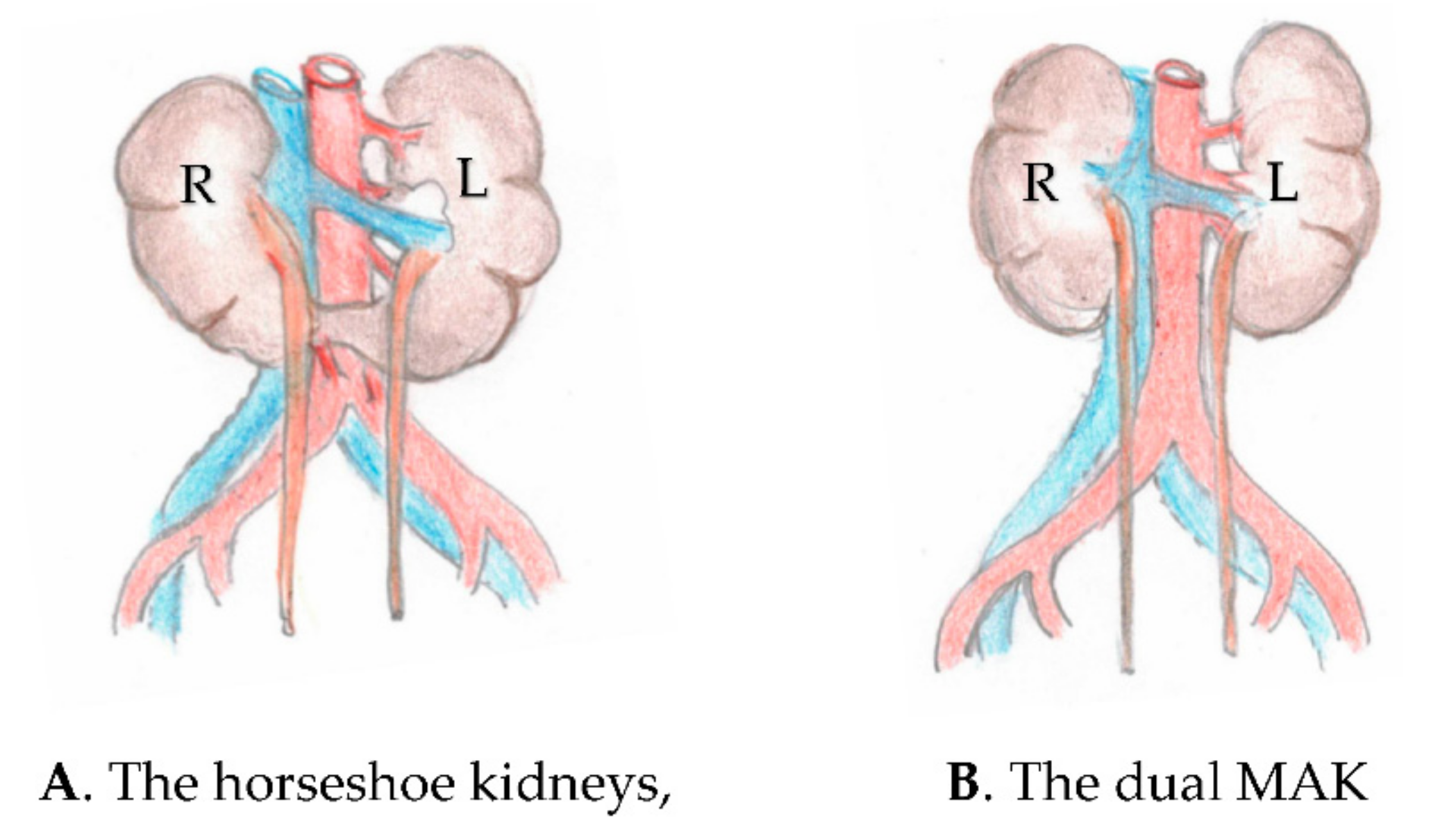
Figure 1. Schematic drawing showing the similarity of (A) the horseshoe kidneys with the transverse isthmus slightly above the level of the aortic bifurcation, and (B) the en bloc dual adult kidneys. The horseshoe kidneys are located 1–2 vertebras below the regular kidneys. Note the multiple segmental renal arteries and the arteries to the isthmus arising from the iliac arteries. In both drawings, the vena cava is on the left side of the aorta. R = right kidney, L = left kidney.
Uro 02 00019 g001
Figure 2. (A) The inverted reconstructed dual kidneys ready to be transplanted to the right iliac fossa showing the backwall of the aorta with the spinal arteries suture ligated. The vena is on the right side of the aorta. The left kidney is figured with the aortic cap bearing the 2 renal arteries and a cava patch carrying the left renal vein hidden by the artery. The kidneys represented herein would require 3 separate arterial anastomoses and 5 venous anastomoses had the aorta and the vena cava not been used as a single arterial inflow and venous inflow, respectively. (B) The pelvic vascular anatomy is shown. Taking consideration of Figure 2A, one can note the concordance of the donor vena cava with the recipient’s right iliac vein, facilitating the venous anastomosis (single arrow) and avoiding the arterio–venous scissoring leading to renal vein thrombosis. Taking consideration of Figure 1B when the left iliac quadrant is used, the anatomic inversion of the en bloc kidneys is not required, since there is already anatomic concordance of the donor vena cava and the left iliac vein. The double arrow shows the site of the left iliac anastomosis.
Uro 02 00019 g002
Publisher’s Note: MDPI stays neutral with regard to jurisdictional claims in published maps and institutional affiliations.