Review

Approach to Biliary Complications After Liver Transplantation: Current Evidence and the Emerging Role of Therapeutic Endoscopic Ultrasound


by
Gabriele Rancatore
1,2,*,
Carmelo Marco Giacchetto
1,2,
Maria Vittoria Grassini
1,2,
Dario Quintini
1,2,3,
Margherita Pizzicannella
1,2,
Dario Ligresti
1,2 and
Ilaria Tarantino
1,2
1
Gastroenterology and Hepatology, IRCCS-ISMETT (Mediterranean Institute for Transplantation and Advanced Therapies), 90127 Palermo, Italy
2
UPMC Italy (University of Pittsburgh Medical Center Italy), 90133 Palermo, Italy
3
Surgical and Critical Area (MePreCC), Department of Precision Medicine in Medical, University of Palermo, 90133 Palermo, Italy
*
Author to whom correspondence should be addressed.
Livers 2026, 6(5), 97; https://doi.org/10.3390/livers6050097
Submission received: 2 May 2026 / Revised: 16 July 2026 / Accepted: 28 August 2026 / Published: 16 September 2026
(This article belongs to the Special Issue Endohepatology: Expanding the Frontiers of Hepatobiliary Care)

Abstract

Background: Biliary complications represent the most frequent and clinically relevant adverse events after liver transplantation, contributing substantially to morbidity, graft dysfunction, and reduced patient survival. Methods: We performed a review of current evidence to explore the prevalence, etiology, diagnosis and treatment of biliary complications after liver transplantation, focusing on the emerging role of therapeutic endoscopic ultrasound. Results: The most common biliary complications include anastomotic and non-anastomotic biliary strictures and bile leaks, while gallstones, casts, and sphincter of Oddi dysfunction are less common. Diagnosis is based on a combination of clinical evaluation, laboratory tests, ultrasound and magnetic resonance cholangiopancreatography. Endoscopic therapy, in most cases, is the first therapeutic approach, followed by interventional radiology and, ultimately, surgery. In recent years, therapeutic endoscopic ultrasound has emerged as a promising, minimally invasive salvage option, particularly when conventional access is unfeasible or has failed. Conclusions: Early recognition and appropriate treatment of these complications are essential for preserving graft integrity and improving long-term outcomes. Although current evidence is still limited, therapeutic EUS could expand the therapeutic arsenal at highly specialized centers. Overall, a multidisciplinary approach is essential for the optimal diagnosis and management of biliary complications after liver transplantation.

1. Introduction

Liver transplantation (LT) is a lifesaving procedure for patients with end-stage liver disease, fulminant hepatitis and liver cancer [1]. In recent years, thanks to the improved management of these patients, indications for LT have increasingly expanded. This has resulted in a growing demand for organs and the need for an increase in living-donor liver transplants (LDLTs). In Europe, survival rates have improved over time, reaching 86% at one year and 74% at five years after LT [2]. Biliary complications (BCs) are the most common complications after LT and, despite improvements in organ preservation, surgical techniques and immunosuppressive therapies, they represent a significant source of morbidity and mortality both in the short and long term after LT. The early identification and, consequently, early treatment of BCs play a crucial role in preserving graft integrity and improving overall patient survival [3,4]. The most common BCs are biliary strictures and biliary leaks. Hemobilia, sphincter of Oddi dysfunction (SOD), bile stones, and casts are observed less frequently [5].
We performed a review of current evidence to explore prevalence, etiology, diagnosis and treatment of biliary complications after LT, focusing on the emerging role of therapeutic endoscopic ultrasound (EUS).

2. Risk Factors

Several risk factors have been identified, which can be classified as: immunological factors, type of biliary reconstruction and donor characteristics.

2.1. Immunological Factors

Cytomegalovirus (CMV), already considered a possible trigger of vanishing bile duct syndrome because it binds to human leukocyte antigens (HLA), may increase the risk of developing BCs. Furthermore, BCs after LT occur more often in patients with previous or concomitant CMV viremia and are especially common in association with primary CMV infection [6]. For these reasons, CMV prophylaxis is recommended for CMV-seronegative recipients.

2.2. Type of Biliary Reconstruction

The two strategies for biliary reconstruction are duct-to-duct anastomosis and bilioenteric anastomosis (choledochojejunostomy or hepaticojejunostomy). Duct-to-duct anastomosis is the method of choice in most transplants, both whole and split liver transplants from living or deceased donors [7,8]. Hepaticojejunostomy is currently used in select cases, such as previous biliary–digestive surgeries or primary sclerosing cholangitis (PSC), due to concerns about possible cholangiocarcinoma arising from the duct remnant [9,10]. Biliary duct-to-duct anastomosis is preferred because it offers several advantages: preservation of SOD with a lower risk of cholangitis, a reduction in the number of surgical anastomoses during LT, and easier endoscopic access to the biliary tract in case of potential complications [5,11,12]. T-tube placement has been largely abandoned since two systematic reviews and meta-analyses (SRMAs) have shown a higher incidence of biliary strictures with T-tube use but no difference in bile leakage rates [13,14].

2.3. Graft Characteristics

Many complications are related to the reduced blood supply to the bile ducts from the hepatic artery (HA). Indeed, damage to the HA, either during explant or LT, can disrupt the blood supply and result in irreversible damage to the intra- and extrahepatic bile ducts, leading to complications such as the formation of complex stenosis [15]. Overall, the rate of BCs is 2–3 times higher in LDLT than in deceased donor liver transplant (DDLT). In fact, several factors related to LDLT techniques may contribute to the increased incidence of biliary complications [8]. Since LDLT is a partial liver resection, the need to dissect the recipient’s left or right hepatic duct may prolong ischemic time, and thereby lead to an increase in the incidence of biliary stenosis. Post-LT biliary strictures occur in up to 15% of patients undergoing DDLT and 40% of patients undergoing LDLT, significantly impacting morbidity and graft survival [16]. In the same way, a recent SRMA showed that LDLT was associated with a higher incidence of bile leaks (OR: 3.38, 95% CI: 2.52–4.53) [17]. The resection of the liver parenchyma is associated with an increased risk of bile leakage from the intrahepatic ducts that run along the resection plane. Furthermore, as a consequence of surgical dissection, nervous or vascular structures of the biliary tree could be injured, possibly leading to duct dehiscence and subsequent bile leakage [12,18,19,20]. Among deceased donor grafts, donation after circulatory death (DCD) has consistently been associated with a higher incidence of biliary complications than donation after brain death (DBD). The unavoidable period of donor warm ischemia preceding organ procurement renders DCD grafts more susceptible to ischemia–reperfusion injury, increasing the risk of ischemic cholangiopathy and non-anastomotic strictures (NAS). A recent comparative study reported NAS rates of 15.2% in DCD recipients compared with 1.4% in DBD recipients, while DCD grafts were associated with a 1.7-fold higher adjusted risk of graft loss. Similarly, a meta-analysis demonstrated significantly increased odds of biliary strictures (OR 1.58, 95% CI 1.21–2.06) and biliary leaks or stones (OR 1.69, 95% CI 1.22–2.34) following DCD transplantation [21,22]. Ischemia represents one of the principal determinants of biliary injury after liver transplantation. Both prolonged donor warm ischemia and cold ischemia contribute to ATP depletion, oxidative stress, mitochondrial dysfunction, and cholangiocyte injury, ultimately impairing biliary regeneration. These mechanisms are particularly relevant in DCD grafts, where limiting warm and cold ischemia times is essential to reduce the risk of post-transplant biliary complications [23]. The method of graft preservation also influences biliary outcomes. Although static cold storage remains the standard preservation strategy, machine perfusion techniques have emerged as promising approaches to reduce ischemia–reperfusion injury. Normothermic machine perfusion (NMP) allows functional assessment of the graft before implantation, whereas hypothermic oxygenated perfusion (HOPE) and dual hypothermic oxygenated perfusion (D-HOPE) have shown encouraging results in reducing ischemic cholangiopthy and non-anastomotic strictures, particularly in DCD grafts, through improved mitchodrial protection and preservation of the peribiliary vascular plexus [23].

3. Classification

BCs can be classified according to time of onset and location of the anastomosis [24,25].
According to the time of onset, BCs can be divided into early (<3 months) and late (>3 months) [26]. Depending on their location, BCs can be classified as anastomotic or non-anastomotic [27,28,29]. Anastomotic BCs are often caused by technical issues during LT, while non-anastomotic BCs result from immune reactions or local ischemia [30].
Biliary strictures can be classified into two main categories: anastomotic biliary strictures (ABSs) and non-anastomotic biliary strictures (N-ABSs).
Based on the localization of injury, biliary leaks can be classified, according to a system described by Nagano et al., as Type A, B, C and D [31] (Table 1).

4. Biliary Strictures

Biliary strictures are the most common complications after LT and concern about 15% of DDLTs and up to 40% of LDLTs [16]. They lead to major morbidity and decreased graft survival. A systematic review comprising a total of 14,359 LTs revealed an overall incidence of biliary strictures of 13%, with more cases in the LDLT group than in the DDLT group [5]. Biliary strictures can be classified into two main categories: anastomotic biliary strictures (ABSs) and non-anastomotic biliary strictures (N-ABSs).

4.1. Anastomotic Biliary Strictures

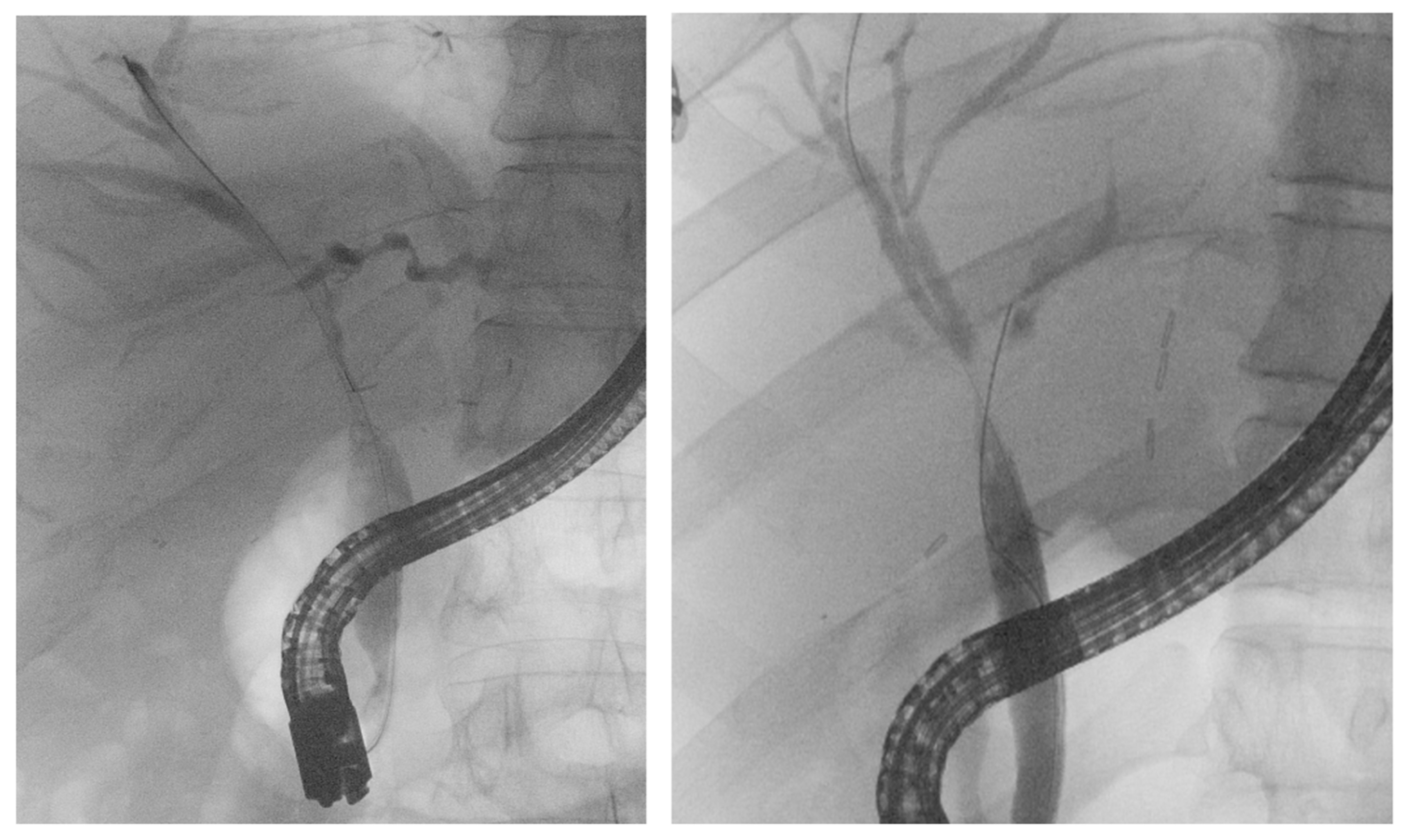
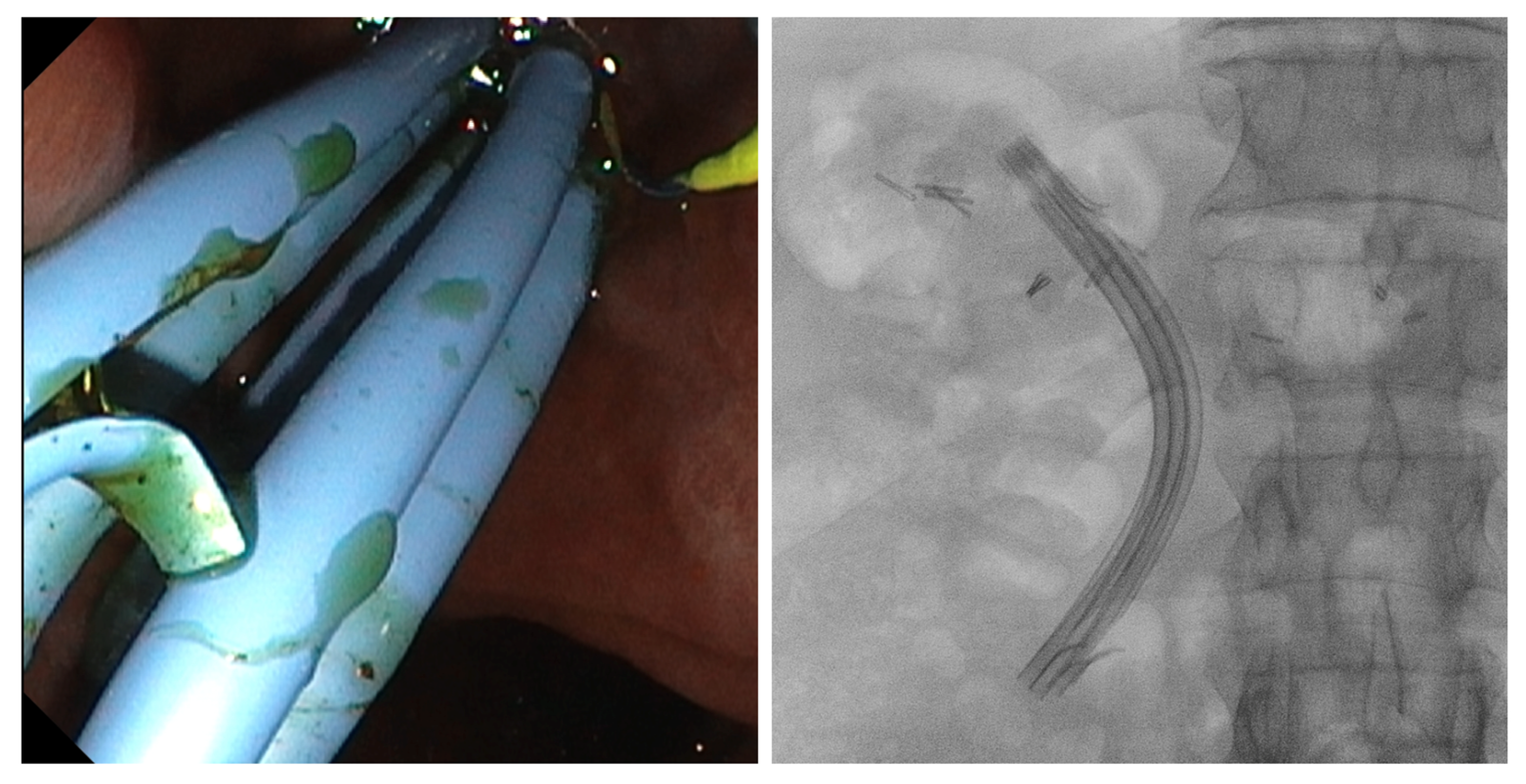
ABSs are defined as strictures that involve the site of surgical anastomosis (duct-to-duct, hepaticojejunostomy): they are usually short and single, and can be an early or late complication depending on when they occur after LT. Early ABS develops within the first three months after LT, and late ABS develops after three months (Figure 1).
A 2024 multicenter retrospective study including 3633 adult patients from 18 centers showed a 20.6% ABS incidence [32]. In a 2015 meta-analysis including patients with PSC, there was no difference in biliary stricture incidence between the duct-to-duct reconstruction group and the Roux-en-Y hepaticojejunostomy group [33]. An SRMA confirmed these data: fifteen retrospective studies including 1770 patients who underwent duct-to-duct anastomosis versus Roux-en-Y hepaticojejunostomy revealed no difference in rates of biliary strictures [34]. About the suture technique in performing the biliary anastomosis, a recent metanalysis including 1617 patients (of whom 1186 patients underwent Roux-en-Y hepaticojejunostomy and 431 patients underwent duct-to-duct choledochochole-dochostomy) comparing interrupted versus continuous suturing for Roux-en-Y hepati-cojejunostomy and duct-to-duct choledochocholedochostomy showed no significant difference in overall biliary complications (OR: 1.34, p = 0.11), particularly in biliary stricture (OR: 0.84, p = 0.65), but also in bile leak (OR: 1.64, p = 0.14), cholangitis (OR: 1.54, p = 0.35), or liver abscess (OR: 0.58, p = 0.40) between the two groups [35].

4.1.1. Etiology

The etiology varies between early and late ABSs. Early ABSs mainly arise due to technical/surgical reasons, such as a discrepancy in bile duct caliber between the donor and recipient, excessive use of electrocauterization for bleeding control, and irregular anastomosis reconstruction. Conversely, late ABSs are related to ischemia and fibrosis events [16]. A retrospective cohort study of 717 patients identified biliary leaks, HA thrombosis and acute rejection as independent risk factors for the development of ABS [36].

4.1.2. Clinical Presentation

The clinical presentation of ABS is often heterogeneous, so it can be challenging to obtain a quick diagnosis. Frequently, patients are asymptomatic but with abnormal liver tests, particularly in a cholestatic pattern, while others present with jaundice, chills or cholangitis due to the biliary obstruction [37]. On the other hand, sometimes, the signs and symptoms of ABS are misunderstood for other post-LT complications, such as acute cellular rejection.

4.1.3. Diagnosis

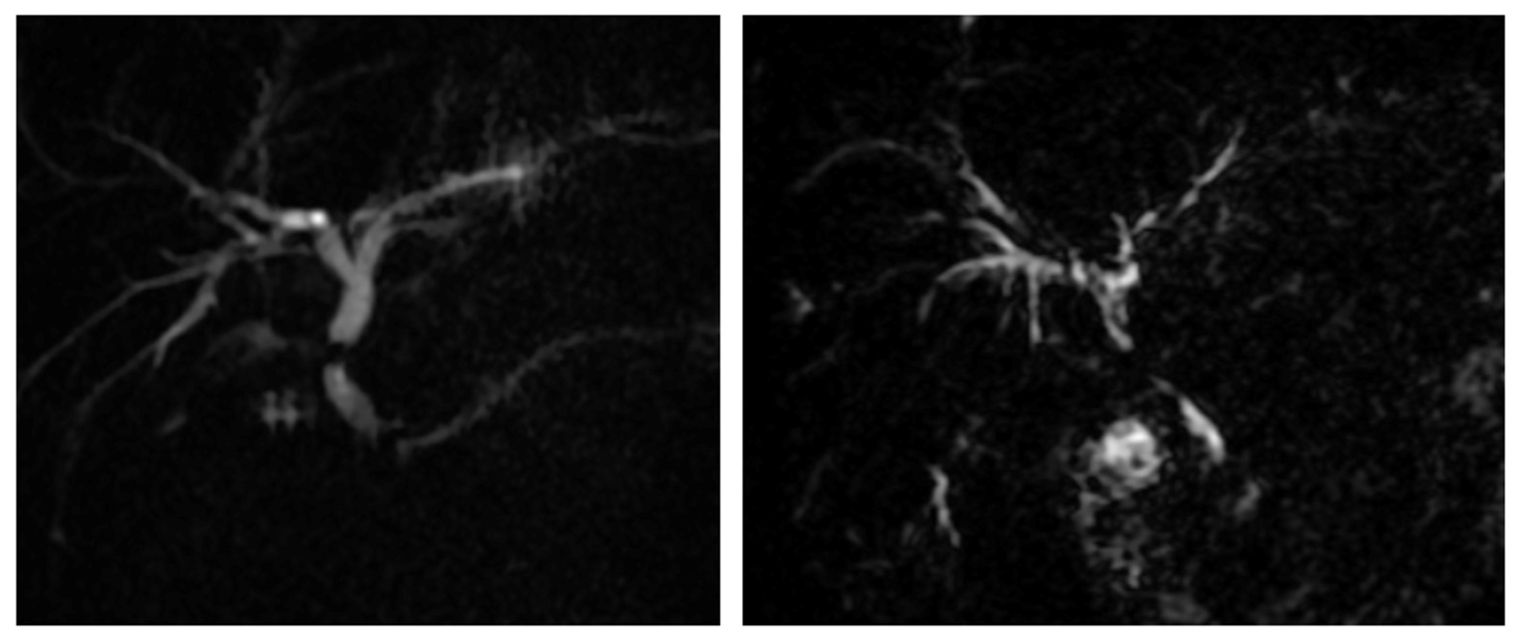
The first steps for diagnosis are generally made by abdominal ultrasound (US) and computed tomography (CT) scans: US can exclude HA flow alterations and may show bile duct dilation or intrahepatic collections due to bile leakage. However, magnetic resonance cholangiopancreatography (MRCP) is the most accurate non-invasive method, with a 94–96% sensitivity and a 94–95% specificity in detecting biliary tree alterations such as strictures [16]. In MRCP, a biliary stricture appears as a sharp, focal narrowing of the bile duct at the anastomosis, often associated with the dilation of the upstream bile ducts (Figure 2).

4.1.4. Treatment

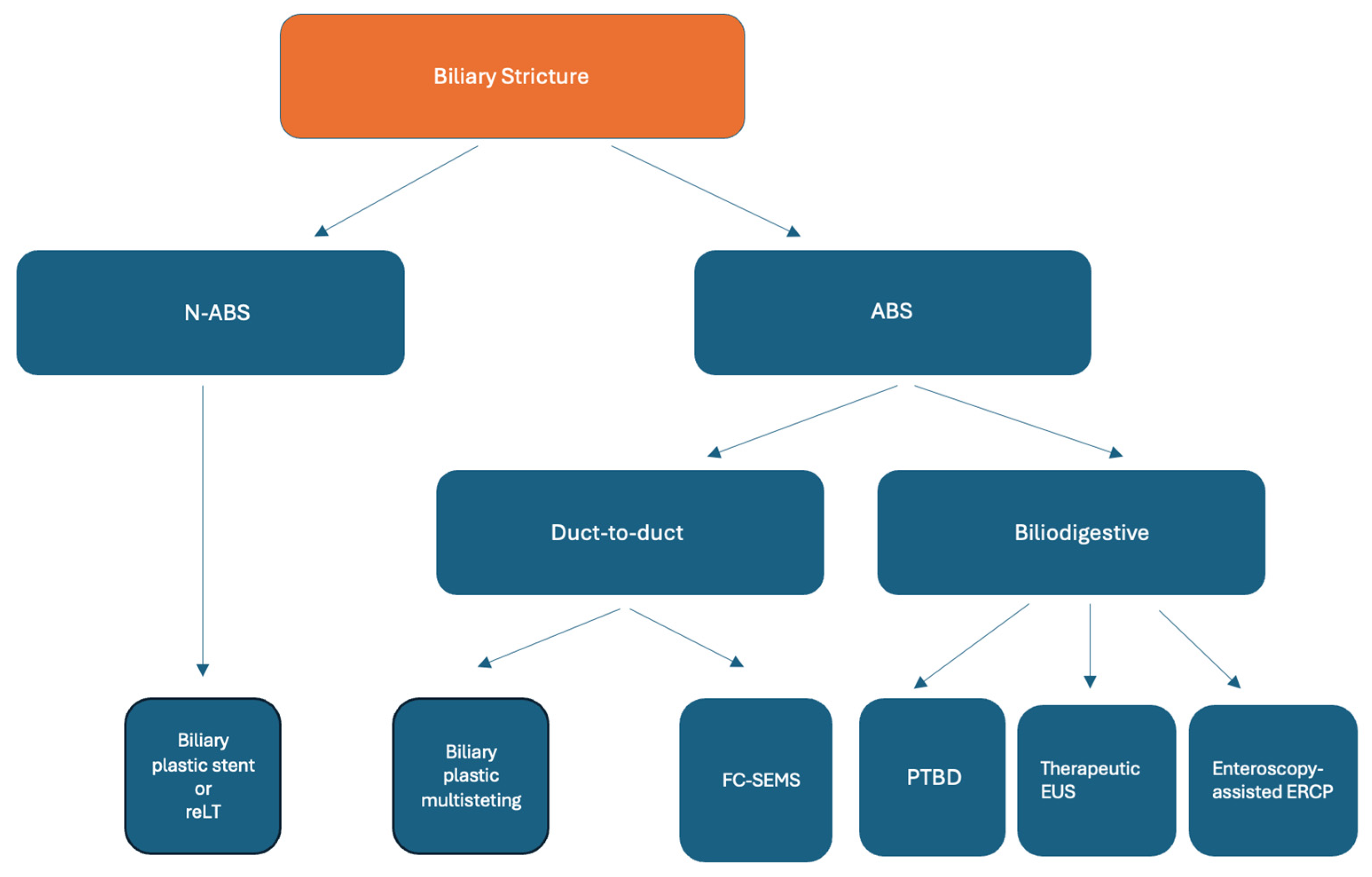
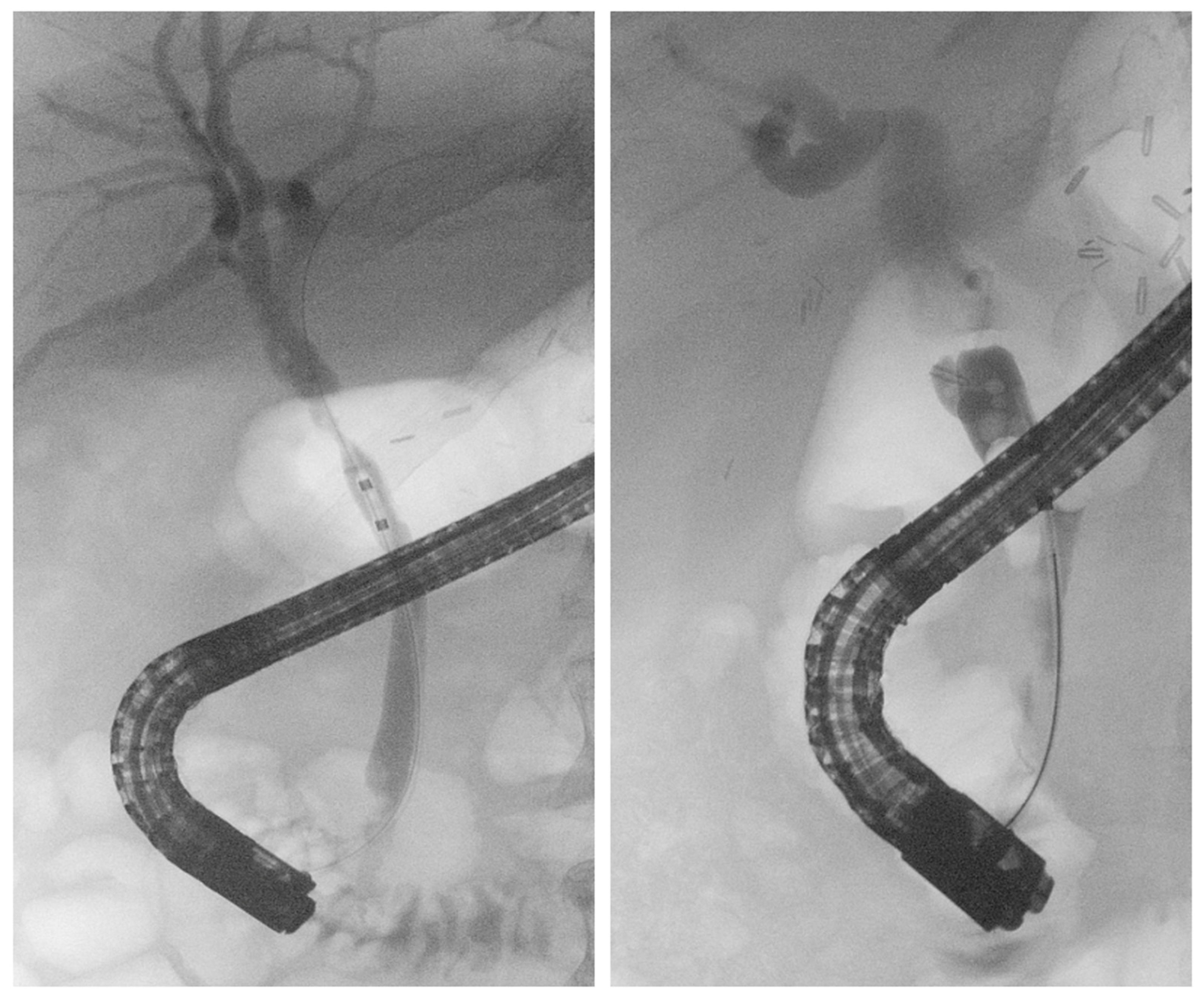
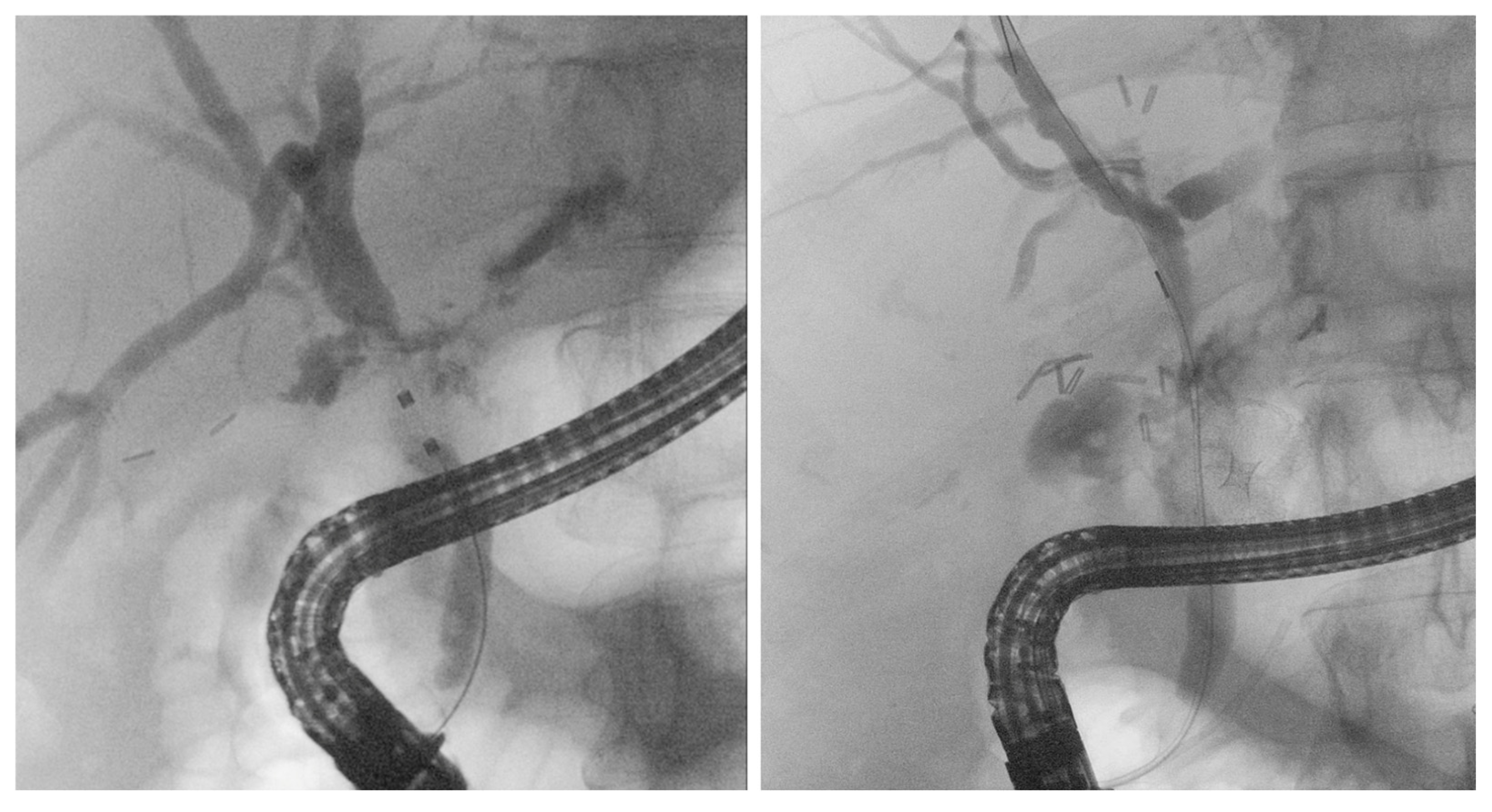
Regarding treatment, there are different steps in duct-to-duct anastomosis and Roux-en-Y anastomosis. In the first case, the gold standard is endoscopic treatment through endoscopic retrograde cholangiopancreatography (ERCP): sphincterotomy, balloon dilation and stent placement are the first therapeutic choices [36]. In biliary stenting, the current approach consists of placing plastic stents at about three-month intervals in multiple endoscopic interventions (generally 4 or 5 procedures) to resolve stenosis [37] (Figure 3). Another approach involves placing a fully covered self-expandable metal stent (SEMS) for almost six months. Two recent SRMAs showed no difference in terms of ABS resolution or recurrence when comparing plastic multistenting to SEMS; however, multistenting requires more sessions [38,39].
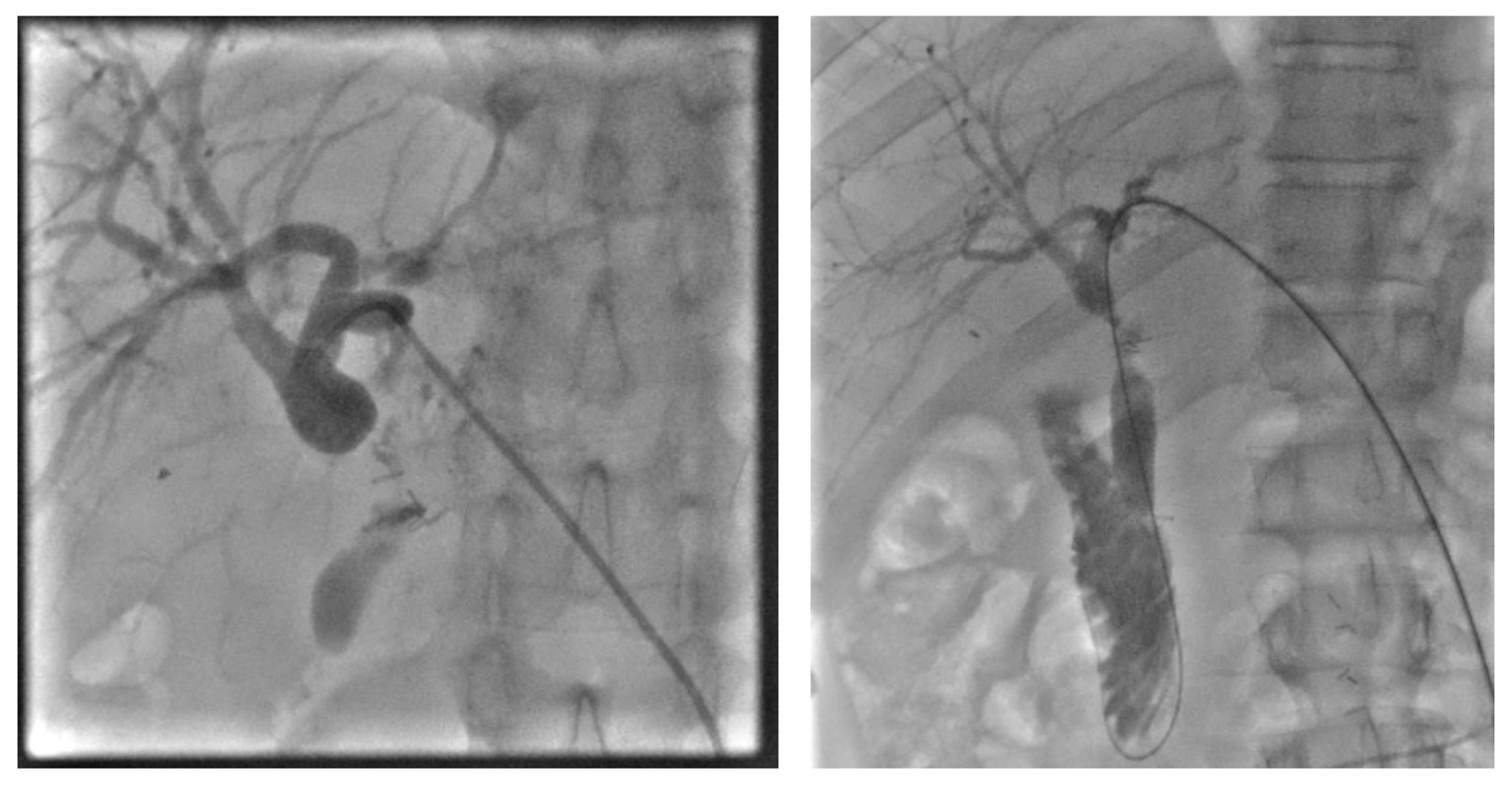
When we are faced with a Roux-en-Y anastomosis stricture, endoscopic access to the biliary tree is difficult due to the altered anatomy: it may require deep ERCP techniques, like enteroscopy-assisted ERCP. However, in these cases, the preferred route for dealing with the stenosis is the percutaneous transhepatic drainage (PTBD) [37] (Figure 4). This access is the first choice in other conditions, like complex strictures in which ERCP has already failed. There are some case reports that cite anastomotic dilation through enteroscopic placement of a biliary prosthesis or balloon-assisted dilation as possible alternative treatments [40,41], but data in this field are poor.
Some studies suggest magnetic compression for the treatment of anastomotic strictures, both in biliobiliary and bilioenteric anastomosis. This technique involves delivering magnets to the site of anastomosis via a percutaneous or peroral bile duct route to form a track and achieve magnet approximation, which will lead to fistula formation due to ischemic necrosis. The magnets are then removed, and a catheter is left in place for about 6 months to prevent restenosis of the fistulous track [42,43,44]. Even if magnetic compression were an alternative to surgery, nowadays its use is very limited.

4.2. Non-Anastomotic Biliary Strictures

N-ABSs are defined as strictures that can involve many segments of the biliary tree, extending up to 0.5 cm proximal to the anastomosis, but they typically affect the hilum or intrahepatic bile ducts. They are often long, multiple and complex, and they are sometimes associated with the formation of biliary stones in the intrahepatic ducts (Figure 5). An SRMA based on 19 international studies including 8269 adult LT patients indicated an 8% overall incidence of N-ABSs, making them less frequent than ABSs.

4.2.1. Etiology

A number of prognostic factors for the development of N-ABS following LT have been identified, such as donation after cardiac death (DCD) donors compared to donation after brain death (DBD) donors, PSC as an indication for LT, Roux-en-Y bile duct reconstruction compared to duct-to-duct reconstruction, HA thrombosis and consequent ischemic cholangiopathy, longer cold or warm ischemia times, and total operative times [45].

4.2.2. Clinical Presentation, Diagnosis and Treatment

The clinical presentation and the diagnostic pathways are analogous to those of ABS. An endoscopic approach is the first choice of treatment, but these kinds of stenosis are often resistant or refractory to this approach and to percutaneous therapies. Moreover, frequent re-interventions are needed, and successful endoscopic therapy requires multiple sessions. Early N-ABSs, which evolve within the first three months, are easier to treat and result in fewer complications than late N-ABSs [46]. A single-center retrospective study involving 35 N-ABS patients treated with ERCP demonstrated successful endoscopic treatment without requiring an alternative treatment approach in only 41% of patients [47]. Surgical revision is a last-resort option, as these patients tend to develop multiple complications and may require re-LT [48]. Figure 6 shows a therapeutic algorithm for ABSs and N-ABSs.

4.3. Intraoperative and Postoperative Preventive Approaches

There are various intra- and post-operative preventive approaches to minimize the risk of developing anastomotic and non-anastomotic biliary strictures. About the intra-operative approaches, since most anastomotic strictures result from local ischemia and subsequent fibrosis of the suture line, making a tension-free and well-vascularized biliary anastomosis is one of the most important cornerstones [49]. Another important factor is that the biliary epithelium is particularly sensitive to ischemia: reductions in cold and warm ischemia times, preservation of the hepatic artery and accurate graft perfusion are mandatory to prevent or reduce the incidence of strictures, as well as avoiding the mismatch of caliber of the bile duct between donor and recipient [50]. About the post-operative approach, the most important approaches are early surveillance of the hepatic function, Doppler ultrasound of the hepatic artery, timely treatment of biliary fistulas, since an untreated biliary fistula could cause inflammation, fibrosis and secondary stenosis and optimization of immunosuppression, since rejection episodes cause inflammation and fibrosis [51].

5. Biliary Leaks After Liver Transplantation

Biliary leakage represents one of the most common early BCs following LT and remains an important source of postoperative morbidity and graft dysfunction. It is defined as the escape of bile from the biliary tree into the abdominal cavity due to the disruption of biliary continuity after LT (Figure 7) and can be classified according to a system described by Nagano et al. [31] [Table 1]. The reported incidence of biliary leakage ranges from approximately 2% to 10% of transplant recipients, and it typically occurs during the early postoperative period, most often within the first weeks after surgery [46]. Meta-analytic data suggest that bile leaks occur in approximately 7.8% of DDLT and 9.5% of LDLT procedures, reflecting the greater technical complexity of biliary reconstruction in living donor transplantation [5]. The expanding use of extended-criteria donors has also influenced the epidemiology of biliary complications. In particular, grafts from DCD donors are associated with a higher risk of biliary injury due to prolonged warm ischemia and ischemia–reperfusion damage affecting the biliary epithelium [52]. From an anatomical perspective, bile leaks are typically classified as anastomotic or non-anastomotic [53,54]. Anastomotic leaks arise at the site of biliary reconstruction and are usually related to surgical factors, such as excessive tension at the anastomosis, inadequate suturing, or duct size mismatch [55]. Non-anastomotic leaks are less common and may originate from the cystic duct remnant or from the cut surface of the graft, particularly in living donor transplantation.

5.1. Etiology

Overall, the development of biliary leakage is multifactorial and reflects the interaction of donor-, recipient-, vascular-, and surgical-related factors [56]. Donor age, graft steatosis, and prolonged ischemia times may increase susceptibility to biliary injury, while recipient conditions [57], such as PSC, CMV infection, or acute cellular rejection, may impair biliary healing. Technical aspects of the surgical procedure—including excessive periductal dissection, electrocautery-related injury, duct size mismatch, and the use of T-tubes—have also been associated with an increased risk of postoperative bile leaks [53]. More recently, increasing attention has been directed toward the potential role of the gut-liver axis and microbial factors in post-transplant BCs [58]. Patients undergoing LT frequently present with gut dysbiosis related to advanced liver disease and are exposed to repeated antibiotic therapies and biliary interventions. These factors may alter both intestinal and biliary microbiota, promoting bacterial colonization of the biliary tract and potentially contributing to local inflammation and impaired epithelial healing.

5.2. Clinical Presentation

Clinical presentation may vary widely, ranging from asymptomatic cases detected incidentally to more severe manifestations associated with infection or peritonitis. The most common clinical findings include abdominal pain, fever, leukocytosis, and persistent elevation of liver function tests, particularly cholestatic enzymes. In many patients, however, the earliest and most suggestive sign is the presence of bilious output from surgical drains placed during transplantation [59].

5.3. Diagnosis

Imaging plays a crucial role in confirming the diagnosis and defining the extent of biliary leakage. Ultrasound is usually the first-line imaging modality in the postoperative evaluation because of its widespread availability and ability to rapidly detect perihepatic fluid collections or ascites. However, US findings are often nonspecific and might not reliably differentiate bile collections from other postoperative fluid collections such as hematomas, seromas, or abscesses. Computed tomography (CT) is frequently used for further assessment and can better characterize the location and extent of fluid collections, typically demonstrating hypoattenuating perihepatic or subhepatic fluid collections consistent with biloma formation [60]. Magnetic resonance imaging (MRI), particularly MRCP, is the most accurate non-invasive imaging technique for evaluating BCs after LT. MRCP provides a detailed visualization of the biliary tree and may help identify the site of leakage, as well as associated biliary abnormalities [60]. On MRI, bile collections typically appear hypointense on T1-weighted images and hyperintense on T2-weighted sequences, while MRCP can demonstrate abnormalities of the biliary tree and associated collections [61,62]. Despite the high diagnostic performance of advanced imaging techniques, small bile leaks may be difficult to detect because imaging findings can overlap with other postoperative collections. For this reason, the diagnosis of biliary leakage often relies on the integration of clinical findings, biochemical analysis of drainage fluid, and imaging results in order to achieve an early and accurate diagnosis.

5.4. Treatment

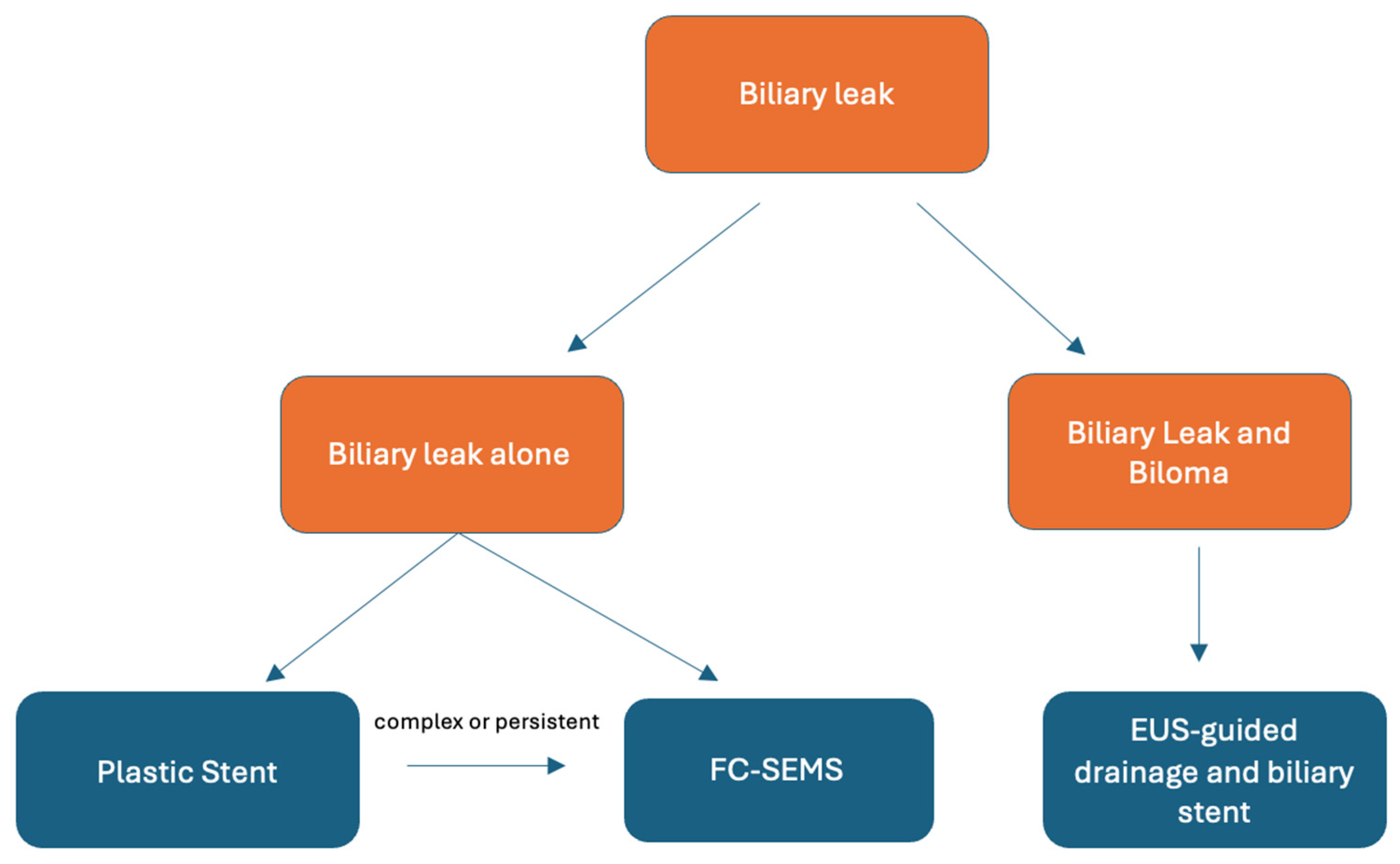
The management of biliary leakage after LT depends largely on the type of biliary reconstruction and the severity of the leak. In patients with duct-to-duct biliary reconstruction, endoscopic therapy represents the cornerstone of treatment [59]. ERCP aims to reduce the pressure gradient between the biliary system and the duodenum, thereby diverting bile flow away from the leak site and facilitating healing [20]. Several endoscopic strategies have been described, including sphincterotomy, nasobiliary drainage, and biliary stent placement, with overall success rates approaching 80–90% in most series [63]. Among these options, biliary stent placement has emerged as the most effective strategy. Plastic stents remain the standard first-line approach in most centers. Several studies evaluating ERCP-based management of post-transplant bile leaks have reported high rates of leak resolution with plastic stent placement, significantly higher than sphincterotomy alone [64,65,66,67,68,69,70]. In a multicenter analysis, Sendino et al. [71] reported the complete resolution of bile leakage in approximately 94% of patients treated with plastic stents, compared with lower success rates in patients undergoing sphincterotomy alone. Plastic stents are generally well tolerated and are associated with a favorable safety profile, although they may require repeated endoscopic procedures for exchange or removal. FC-SEMS have also been increasingly used, particularly in patients with persistent or complex leaks [72]. These stents provide a larger diameter and greater radial force, which may enhance biliary decompression and promote faster closure of the leak. However, compared with plastic stents, some studies have reported a higher rate of adverse events [73]. In addition, current European guidelines do not provide clear recommendations for their routine upfront use in the management of biliary leaks, and randomized controlled trials (RCTs) comparing the two approaches are lacking. A recent study by Stegagnini et al. [74] reported no significant differences in adverse events between plastic stents and FC-SEMS, suggesting that FC-SEMS may represent not only a rescue therapy for refractory cases but also a potentially effective and safe primary treatment option. The management strategy differs in patients who undergo Roux-en-Y hepaticojejunostomy, in whom endoscopic access to the biliary tree is often technically challenging or impossible using standard ERCP techniques [75]. In these cases, percutaneous transhepatic approaches play a central role. Percutaneous transhepatic cholangiography allows both the diagnostic evaluation and therapeutic drainage of the biliary system through the placement of internal–external biliary catheters, which reduce intrabiliary pressure and facilitate leak closure. Clinical success rates of percutaneous approaches are reported to be approximately 70–80%, although treatment may require prolonged catheter drainage [76]. When endoscopic access is difficult or unsuccessful, combined percutaneous–endoscopic approaches may be required. The rendezvous technique represents an effective strategy in these situations, allowing guidewire access to the biliary tree through a percutaneous transhepatic route followed by endoscopic retrieval and completion of the therapeutic procedure. Alternative endoscopic approaches have also been described when conventional ERCP is not feasible or has failed. Endoscopic ultrasound-guided (EUS) biliary drainage techniques have emerged as potential salvage strategies and will be discussed further below.
Overall, the management of biliary leakage after LT requires a multidisciplinary approach involving transplant surgeons, hepatologists, interventional radiologists, and therapeutic endoscopists.
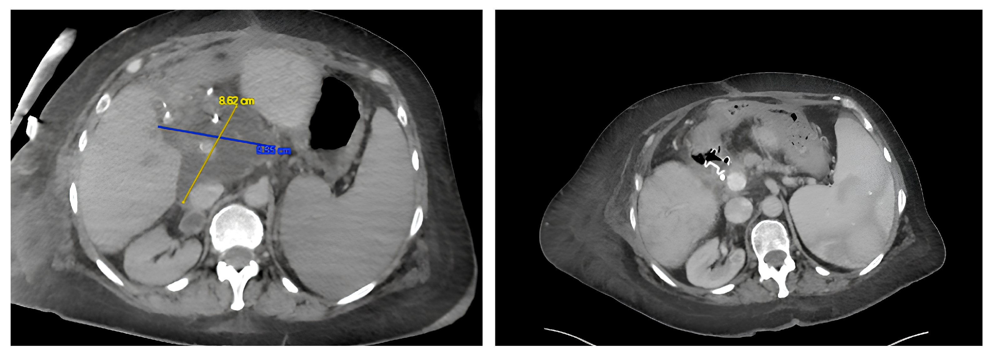
Biliary leaks are frequently associated with the development of bilomas, defined as well-circumscribed collections of bile located outside the biliary tree resulting from persistent leakage of bile into the peritoneal or intrahepatic space. When symptomatic or large, bilomas may present with abdominal pain, fever, leukocytosis, or signs of sepsis and therefore require prompt management. The most serious complication is the erosion of the HA. The traditional therapeutic approach consists of percutaneous drainage under US or CT guidance, which allows the effective evacuation of the collection and control of infection while definitive treatment of the underlying bile leak is pursued [77,78]. In recent years, EUS-guided drainage has emerged as an alternative, minimally invasive strategy, particularly for large or mature collections. The development of lumen-apposing metal stents (LAMSs) has further expanded this approach, enabling direct transluminal drainage of bilomas with rapid decompression and high technical success rates [79] (Figure 8). Importantly, drainage of the biloma alone does not address the underlying bile leak, which should be treated concurrently through biliary decompression using endoscopic or percutaneous techniques. Figure 9 shows a therapeutic algorithm for biliary leak and biloma.

5.5. SOD, Stones and Casts

In addition to bile leaks and strictures, other BCs after LT include SOD, biliary stones, sludge, and biliary casts. These conditions may contribute to biliary obstruction, cholestasis, and graft dysfunction and often occur in association with biliary strictures or ischemic injury of the bile ducts [5,51]. SOD is considered an uncommon complication after LT and should be suspected in patients presenting with cholestasis or biliary-type pain in the absence of a clear mechanical obstruction. The underlying mechanism is not completely understood but may involve denervation of the sphincter during transplantation or altered biliary motility. In select cases, endoscopic sphincterotomy may improve biliary drainage and symptoms [53,54]. Biliary stones and sludge can occur at any time after transplantation, with reported incidences of approximately 5–10%, and are frequently associated with biliary strictures. Their pathogenesis is multifactorial and includes biliary stasis, ischemic injury, and alterations in bile composition related to immunosuppressive therapy [5,51,54]. A distinct condition is biliary cast syndrome, which is characterized by the formation of hard intraductal casts that may cause biliary obstruction, cholangitis, and graft dysfunction. The condition has been reported in up to 2.5–18% of transplant recipients and is usually related to ischemia, rejection, or prolonged biliary stasis [5]. ERCP remains the primary diagnostic and therapeutic modality. In patients with duct-to-duct reconstruction, sphincterotomy followed by balloon or basket extraction is usually effective for stone and sludge removal, with high rates of duct clearance [54,63]. More complex cases, particularly those involving intrahepatic stones or casts, may require advanced techniques such as cholangioscopy-guided lithotripsy [80].

6. Clinical Presentation and Diagnostic Approach

Due to immunosuppression, altered anatomy, and biliary denervation, the clinical presentation of BCs after LT can differ widely from that of non-LT patients. Presentation can vary widely from asymptomatic elevations of hepatic cholestasis enzymes, such as gamma-glutamyl transferase (GGT) and/or alkaline phosphatase (ALP), to nonspecific symptoms such as anorexia, pruritus, abdominal distension, and fever, and, in the most severe cases, cholangitis, peritonitis, and septic shock [49,63]. The main differential diagnoses are: rejection, sepsis and HA occlusion.
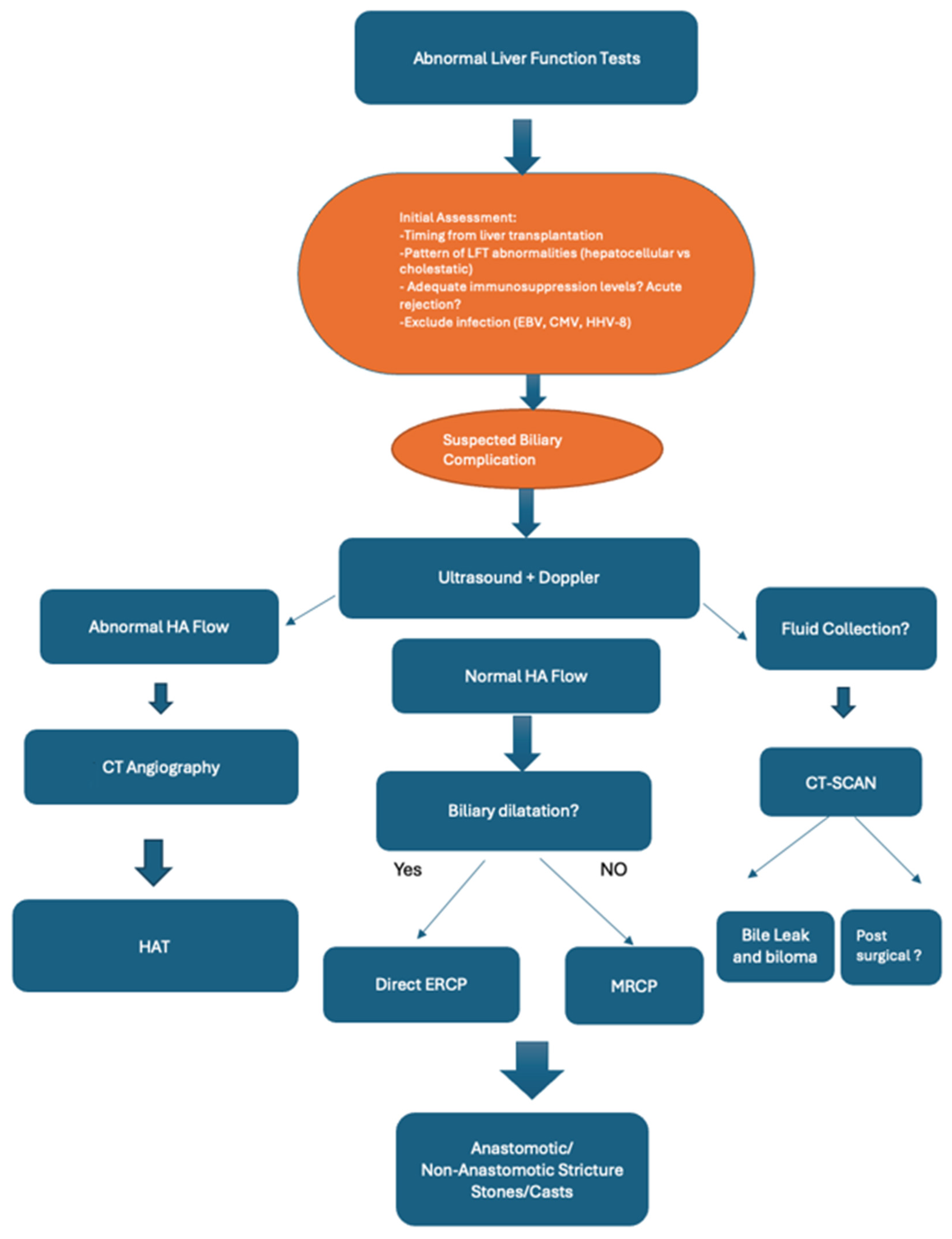
First of all, in the presence of asymptomatic elevation of liver enzymes, it is necessary to check whether immunosuppression levels are low in order to evaluate possible liver rejection. A liver biopsy may be required to dispel any doubts. Very often, the diagnosis of biliary leak is performed immediately after LT with evidence of bile in the abdominal drainage, which may require an MRCP to confirm the diagnosis. In the remaining cases, patients with suspected BCs after LT should undergo abdominal US as the first step, as it is a non-invasive modality that allows for the evaluation of the presence of fluid collections and bile duct dilation, and the exclusion of vascular disorders such as HA occlusion through the use of Doppler (Figure 10). In the case of altered flow in the HA, CT-angiography should be performed to exclude vascular impairment. In the presence of dilated bile ducts, given the high positive predictive value of US [81,82], ERCP should be performed for a strong suspicion of stenosis without proceeding to MRCP [36]. Conversely, in the presence of non-dilated bile ducts, the sensitivity of US is reduced, so if the suspicion of post-LT BCs remains high, MRCP should be performed [83]. MRCP is a non-invasive diagnostic modality that does not require an intravenous contrast agent and allows the entire biliary tree to be studied with great accuracy. It has a sensitivity and specificity of 80% and 100%, respectively, for biliary stones [84] and a high sensitivity and specificity of 90% and 95%, respectively, for post-LT biliary stricture, as shown in a recent meta-analysis [85]. For these reasons, MRCP is recommended to guide therapeutic interventions, and the use of ERCP or PTC should be restricted to therapeutic use only when MRCP is equivocal. In recent years, EUS has become established as an additional diagnostic modality due to its minimally invasive nature and proximity of the probe to the liver, combining elastography and liver biopsy. Recently, EUS was evaluated as a diagnostic tool for the diagnosis of post-LT BCs in a cohort of 32 patients. In this study, EUS achieved an overall sensitivity and accuracy of 94.6%, surpassing ERCP in identifying bile casts and ischemic cholangiopathy, with potential implications for clinical management [86]. In another study evaluating BCs after LT, EUS demonstrated an overall accuracy, specificity, and sensitivity of 94.6%, 100%, and 94.6%, respectively [87]. Based on the above, diagnostic EUS could be considered as a single-session strategy with ERCP in cases of elevated liver enzymes when rejection or anastomotic strictures are suspected and when MRCP is unclear. In such a setting, the combination of the two procedures could lead to EUS-guided liver biopsy to exclude liver rejection and ERCP to ascertain the stricture.

7. Role of Endoscopic Ultrasound (EUS)-Guided Therapeutic Interventions

Alternative endoscopic approaches have also been described for cases where conventional ERCP is not feasible or has failed. Although therapeutic EUS has been established as a potential salvage strategy, its use in the post-LT setting is currently limited to select cases, and supporting evidence remains limited to small case series or single case reports.
Patients with altered anatomy, such as those with PSC undergoing LT with bilioenteric anastomosis and Roux-en-Y reconstruction, may not be optimal candidates for standard ERCP with a conventional lateral-view duodenoscope [88]. In such cases, the current therapeutic options are enteroscopy-assisted ERCP or percutaneous transhepatic interventions. However, these procedures often require multiple sessions or are difficult to carry out. In this context, therapeutic EUS could be used to gain biliary access directly (EUS-guided biliary drainage) or indirectly after EUS-guided gastroenteroanastomosis (EUS-GEA) or after EUS-guided enteroenterostomy (EDEE). Bukhari et al. [89] described the case of a 41-year-old woman who underwent LT with Roux-en-Y reconstruction for PSC complicated by recurrent cholangitis due to biliary cast syndrome, and who underwent multiple ineffective enteroscopy-assisted ERCPs. In this case, a gastrojejunal fistula was created using a 15 mm LAMS with an electrocautery-enhanced delivery system (Axios; Boston Scientific, Quincy, MA, USA), followed by ERCP with electrohydraulic lithotripsy (EHL). Pizzicannella et al. [90] described the case of a 71-year-old man who underwent LT with Roux-en-Y reconstruction complicated by recurrent non-stenotic cholangitis. In this case, EUS-guided choledochoduodenostomy (EUS-CDS) was successfully performed by placing an 8 × 8 mm LAMS.
Traditionally, abdominal collections were treated with percutaneous drainage performed by an interventional radiologist or surgeon. Therapeutic EUS has emerged as a minimally invasive alternative to the percutaneous approach for drainage of intra-abdominal abscesses post-LT when conventional approaches fail or are unsuitable [91], as well as for postoperative fluid collections. The technical and clinical success rates of therapeutic EUS exceed 90%, while adverse events remain infrequent (<10%) and rarely require surgical intervention [92,93,94,95]. Similarly, therapeutic EUS could be a valid alternative for the drainage of liver bilomas after LT. However, this hypothesis has been explored in a limited number of cases, necessitating a case-by-case approach for decision-making. Uchida et al. reported that six patients with symptomatic or growing intra-abdominal fluid collections after LT underwent successful EUS-guided drainage, including a biloma [96]. Similarly, Cassis et al. [97] described the case of a 65-year-old man affected by a large biloma after cholecystectomy who was successfully treated by placing a 15 mm × 10 mm LAMS under EUS guidance, followed by ERCP with a biliary plastic stent. Finally, regarding bile leaks in patients with surgically altered anatomy or inaccessible papilla, EUS-guided antegrade stenting (EUS-AGS) can allow access to the biliary system via a transhepatic route under EUS guidance, followed by guidewire manipulation through the biliary tract and antegrade stent placement to restore bile flow [98].

8. Conclusions

BCs are among the most important, clinically relevant adverse events after LT, despite significant advances in surgical techniques, graft preservation, and perioperative care. They remain a significant challenge after LT, not only due to their frequency but also due to their often subtle, nonspecific, and easily overlooked clinical presentation. This diagnostic ambiguity makes BCs particularly insidious, as they can initially be confused with rejection, infection, or vascular complications, thus delaying definitive treatment. Early recognition is crucial, as delayed diagnosis and treatment can significantly compromise transplanted liver function and negatively impact both graft and patient survival. Given the complexity of diagnosis and management, the optimal care of these patients requires a multidisciplinary approach involving hepatologists, endoscopists, interventional radiologists, and transplant surgeons. This collaboration is essential in order to personalize treatment based on the type of BC, biliary tract reconstruction, and the anatomy and clinical condition of the individual patient. Although ERCP and percutaneous transhepatic approaches remain the mainstay of treatment in most cases, therapeutic EUS may represent an evolving adjunctive or rescue approach that may be particularly useful in highly selected patients with altered anatomy or after the failure of conventional access. However, the current evidence supporting therapeutic EUS in the post-transplant setting is still limited and is largely derived from isolated case reports and small case series, without robust comparative studies or pooled clinical outcome data. Therefore, its role in routine practice should be considered exploratory rather than definitive, and its use should be restricted to expert centers with substantial endoscopic experience. As experience increases and prospective data become available, therapeutic EUS may assume a greater role in the treatment algorithm for post-transplant BCs, particularly in cases where conventional strategies are limited by anatomy or previous failures.

9. Future Research Priorities

Although EUS represents a potential addition to the therapeutic armamentarium, more studies with a larger number of patients are needed for it to play a more defined role. Future studies should focus on prospective, multicenter designs evaluating therapeutic EUS as a possible alternative treatment in patients with altered anatomy, refractory biliary strictures, biliary fistulas, and bilomas, where current treatment decisions are still primarily guided by local experience and anatomical feasibility. Specifically, such studies should focus on technical success, clinical success, adverse events, the need for reoperation, and related outcomes to define the true clinical value of these approaches. Only in this way can we identify those patients who can best benefit from this type of approach.

Author Contributions

G.R.: manuscript writing and design; C.M.G.: manuscript writing and comments. M.V.G.: manuscript writing and comments; D.Q.: comments; M.P.: comments; D.L.: Comments. I.T.: Supervision and comments. All authors have read and agreed to the published version of the manuscript.

Funding

Italian Health Ministry, Ricerca Corrente.

Institutional Review Board Statement

Not applicable.

Informed Consent Statement

Not applicable.

Data Availability Statement

No new data were created or analyzed in this study. Data sharing is not applicable to this article.

Conflicts of Interest

The authors declare no conflicts of interest.

Abbreviations

The following abbreviations are used in this manuscript:
LTLiver transplantation
EUSEndoscopic ultrasound
LDLTLiving-donor liver transplants
BCsBiliary complications
SODSphincter of Oddi dysfunction
CMVCytomegalovirus
HLAHuman leukocyte antigens
SRMASystematic reviews and meta-analyses
HAHepatic artery
PSCPrimary sclerosing cholangitis
DDLTDeceased-donor liver transplants
ABSAnastomotic biliary stricture
N-ABSNon anastomotic biliary stricture
USAbdominal ultrasound
CTComputed tomography
MRCPMagnetic resonance cholangiopancreatography
ERCPEndoscopic retrograde cholangiopancreatography
FC-SEMSFully covered self-expandable metal stent
PTDBPercutaneous transhepatic drainage
DCDDonation after cardiac death
DBDDonation after brain death
ESBDLExcluded segmental bile duct leakage
MRIMagnetic resonance imaging
RCTRandomized controlled trials
LAMSLumen apposing metal stent
GGTGamma-glutamyl transferase
ALPAlkaline phosphatase
EUS-GEAEndoscopic ultrasound-guided gastroenterostomy
EUS-CDSEndoscopic ultrasound-guided choledochoduodenostomy
EUS-AGSEndoscopic ultrasound-guided antegrade stenting

References

  1. European Association for the Study of the Liver. EASL Clinical Practice Guidelines on liver transplantation. J. Hepatol. 2024, 81, 1040–1086. [Google Scholar] [CrossRef] [Scilit] [PubMed]
  2. Adam, R.; Karam, V.; Cailliez, V.; Grady, J.G.O.; Mirza, D.; Cherqui, D.; Klempnauer, J.; Salizzoni, M.; Pratschke, J.; Jamieson, N.; et al. 2018 Annual Report of the European Liver Transplant Registry (ELTR)—50-year evolution of liver transplantation. Transpl. Int. 2018, 31, 1293–1317. [Google Scholar] [CrossRef] [Scilit] [PubMed]
  3. Girotra, M.; Soota, K.; Klair, J.S.; Dang, S.M.; Aduli, F. Endoscopic management of post-liver transplant biliary complications. World J. Gastrointest. Endosc. 2015, 7, 446–459. [Google Scholar] [CrossRef] [Scilit] [PubMed]
  4. Moy, B.T.; Birk, J.W. A Review on the Management of Biliary Complications after Orthotopic Liver Transplantation. J. Clin. Transl. Hepatol. 2019, 7, 61–71. [Google Scholar] [CrossRef] [Scilit] [PubMed]
  5. Akamatsu, N.; Sugawara, Y.; Hashimoto, D. Biliary reconstruction, its complications and management of biliary complications after adult liver transplantation: A systematic review of the incidence, risk factors and outcome. Transpl. Int. 2011, 24, 379–392. [Google Scholar] [CrossRef] [Scilit] [PubMed]
  6. Halme, L.; Hockerstedt, K.; Lautenschlager, I. Cytomegalovirus infection and development of biliary complications after liver transplantation1. Transplantation 2003, 75, 1853–1858. [Google Scholar] [CrossRef] [Scilit] [PubMed]
  7. Greif, F.; Bronsther, O.L.; Van Thiel, D.H.; Casavilla, A.; Iwatsuki, S.; Tzakis, A.; Todo, S.; Fung, J.J.; Starzl, T.E. The incidence, timing, and management of biliary tract complications after orthotopic liver transplantation. Ann. Surg. 1994, 219, 40–45. [Google Scholar] [CrossRef] [Scilit] [PubMed]
  8. Graziadei, I.W.; Schwaighofer, H.; Koch, R.; Nachbaur, K.; Koenigsrainer, A.; Margreiter, R.; Vogel, W. Long-term outcome of endoscopic treatment of biliary strictures after liver transplantation. Liver Transpl. 2006, 12, 718–725. [Google Scholar] [CrossRef] [Scilit] [PubMed]
  9. Wells, M.M.; Croome, K.P.; Boyce, E.; Chandok, N. Roux-en-Y choledochojejunostomy versus duct-to-duct biliary anastomosis in liver transplantation for primary sclerosing cholangitis: A meta-analysis. Transpl. Proc. 2013, 45, 2263–2271. [Google Scholar] [CrossRef] [Scilit] [PubMed]
  10. Graziadei, I.W. Recurrence of primary sclerosing cholangitis after liver transplantation. Liver Transpl. 2002, 8, 575–581. [Google Scholar] [CrossRef] [Scilit] [PubMed]
  11. Pascher, A.; Neuhaus, P. Biliary complications after deceased-donor orthotopic liver transplantation. J. Hepatobiliary Pancreat. Surg. 2006, 13, 487–496. [Google Scholar] [CrossRef] [Scilit] [PubMed]
  12. Welling, T.H.; Heidt, D.G.; Englesbe, M.J.; Magee, J.C.; Sung, R.S.; Campbell, D.A.; Punch, J.D.; Pelletier, S.J. Biliary complications following liver transplantation in the model for end-stage liver disease era: Effect of donor, recipient, and technical factors. Liver Transpl. 2008, 14, 73–80. [Google Scholar] [CrossRef] [Scilit] [PubMed]
  13. Sotiropoulos, G.C.; Sgourakis, G.; Radtke, A.; Molmenti, E.P.; Goumas, K.; Mylona, S.; Fouzas, I.; Karaliotas, C.; Lang, H. Orthotopic Liver Transplantation: T-Tube or Not T-Tube? Systematic Review and Meta-Analysis of Results. Transplantation 2009, 87, 1672–1680. [Google Scholar] [CrossRef] [Scilit] [PubMed]
  14. Riediger, C.; Müller, M.W.; Michalski, C.W.; Hüser, N.; Schuster, T.; Kleeff, J.; Friess, H. T-tube or no T-tube in reconstruction of the biliary tract during orthotopic liver transplantation—Systematic review and meta-analysis. Liver Transpl. 2010, 16, 705–717. [Google Scholar] [CrossRef] [Scilit] [PubMed]
  15. Abouljoud, M.S.; Kim, D.Y.; Yoshida, A.; Arenas, J.; Jerius, J.; Malinzak, L.; Raoufi, M.; Brown, K.A.; Moonka, D.K. Impact of aberrant arterial anatomy and location of anastomosis on technical outcomes after liver transplantation. J. Gastrointest. Surg. 2005, 9, 672–678. [Google Scholar] [CrossRef] [Scilit] [PubMed]
  16. Keane, M.G.; Devlin, J.; Harrison, P.; Masadeh, M.; Arain, M.A.; Joshi, D. Diagnosis and management of benign biliary strictures post liver transplantation in adults. Transpl. Rev. 2021, 35, 100593. [Google Scholar] [CrossRef] [Scilit] [PubMed]
  17. Giri, S.; Sahu, S.K.; Mohapatra, V.; Chaudhary, M.; Panigrahi, M.; Nath, P.; Mallick, B.; Praharaj, D.L. Comparison of Biliary Complications Between Living and Deceased Donor Liver Transplantations: A Systematic Review and Meta-analysis. Cureus 2024, 16, e69019. [Google Scholar] [CrossRef] [Scilit] [PubMed] [PubMed Central]
  18. Kasahara, M.; Egawa, H.; Takada, Y.; Oike, F.; Sakamoto, S.; Kiuchi, T.; Yazumi, S.; Shibata, T.; Tanaka, K. Biliary reconstruction in right lobe living-donor liver transplantation: Comparison of different techniques in 321 recipients. Ann. Surg. 2006, 243, 559–566. [Google Scholar] [CrossRef] [Scilit] [PubMed]
  19. Johnston, T.D.; Gates, R.; Reddy, K.S.; Nickl, N.J.; Ranjan, D. Nonoperative management of bile leaks following liver transplantation. Clin. Transpl. 2000, 14, 365–369. [Google Scholar] [CrossRef] [Scilit] [PubMed]
  20. Oh, D.W.; Lee, S.K.; Song, T.J.; Park, D.H.; Lee, S.S.; Seo, D.W.; Kim, M.H. Endoscopic management of bile leakage after liver transplantation. Gut Liver 2015, 9, 417–423. [Google Scholar] [CrossRef] [Scilit] [PubMed]
  21. Meier, R.P.H.; Kelly, Y.; Braun, H.; Maluf, D.; Freise, C.; Ascher, N.; Roberts, J.; Roll, G. Comparison of Biliary Complications Rates After Brain Death, Donation After Circulatory Death, and Living-Donor Liver Transplantation: A Single-Center Cohort Study. Transpl. Int. 2022, 35, 10855. [Google Scholar] [CrossRef] [Scilit] [PubMed] [PubMed Central]
  22. Vivalda, S.; Zhengbin, H.; Xiong, Y.; Liu, Z.; Wang, Z.; Ye, Q. Vascular and Biliary Complications Following Deceased Donor Liver Transplantation: A Meta-analysis. Transpl. Proc. 2019, 51, 823–832. [Google Scholar] [CrossRef] [Scilit] [PubMed]
  23. Schlegel, A.; Dutkowski, P. Impact of Machine Perfusion on Biliary Complications after Liver Transplantation. Int. J. Mol. Sci. 2018, 19, 3567. [Google Scholar] [CrossRef] [Scilit] [PubMed] [PubMed Central]
  24. Manay, P.; Seth, A.; Jackson, K.; Lentine, K.L.; Schnitzler, M.A.; Xiao, H.; Segev, D.L.; Axelrod, D.A.M. Biliary complications after liver transplantation in the United States: Changing trends and economic implications. Transplantation 2023, 107, e127–38. [Google Scholar] [CrossRef] [Scilit] [PubMed]
  25. Kohli, D.R.; Desai, M.V.; Kennedy, K.F.; Pandya, P.; Sharma, P. Patients with post-transplant biliary stric- tures have significantly higher rates of liver transplant failure and rejection: A nationwide inpatient analysis. J. Gastroenterol. Hepatol. 2021, 36, 2008–2014. [Google Scholar] [CrossRef] [Scilit] [PubMed]
  26. Gastaca, M. Biliary complications after orthotopic liver transplantation: A review of incidence and risk factors. Transpl. Proc. 2012, 44, 1545–1549. [Google Scholar] [CrossRef] [Scilit] [PubMed]
  27. Deen, F.Z.L.; Lee, C.S.; Lee, W.C. Endoscopic Management of Biliary Complications after Liver Transplantation. In Therapeutic Gastrointestinal Endoscopy; IntechOpen: London, UK, 2011; pp. 93–114. [Google Scholar] [CrossRef] [Scilit] [PubMed]
  28. Wojcicki, M.; Milkiewicz, P.; Silva, M. Biliary tract complications after liver transplantation: A review. Dig. Surg. 2008, 25, 245–257. [Google Scholar] [CrossRef] [Scilit] [PubMed]
  29. Williams, E.D.; Draganov, P.V. Endoscopic management of biliary strictures after liver transplantation. World J. Gastroenterol. 2009, 15, 3725–3733. [Google Scholar] [CrossRef] [Scilit] [PubMed]
  30. Tashiro, H.; Itamoto, T.; Sasaki, T.; Ohdan, H.; Fudaba, Y.; Amano, H.; Fukuda, S.; Nakahara, H.; Ishiyama, K.; Ohshita, A.; et al. Biliary complications after duct-to-duct biliary reconstruction in living-donor liver transplantation: Causes and treatment. World J. Surg. 2007, 31, 2222–2229. [Google Scholar] [CrossRef] [Scilit] [PubMed]
  31. Nagano, Y.; Togo, S.; Tanaka, K.; Masui, H.; Endo, I.; Sekido, H.; Nagahori, K.; Shimada, H. Risk factors and management of bile leakage after hepatic resection. World J. Surg. 2003, 27, 695–698. [Google Scholar] [CrossRef] [Scilit] [PubMed]
  32. Li, Z.; Rammohan, A.; Gunasekaran, V.; Hong, S.; Chih-Yi Chen, I.; Kim, J.; Hervera Marquez, K.A.; Hsu, S.C.; Kirimker, E.O.; Akamatsu, N.; et al. Biliary complications after adult-to-adult living-donor liver transplantation: An international multicenter study of 3633 cases. Am. J. Transplant. 2024, 24, 1233–1246. [Google Scholar] [CrossRef] [Scilit] [PubMed]
  33. Pandanaboyana, S.; Bell, R.; Bartlett, A.J.; McCall, J.; Hidalgo, E. Meta-analysis of Duct-to-duct versus Roux-en-Y biliary reconstruction following liver transplantation for primary sclerosing cholangitis. Transpl. Int. 2015, 28, 485–491. [Google Scholar] [CrossRef] [Scilit] [PubMed]
  34. Cardoso, S.A.; Cardoso, R.P.; Bhangui, P.; Dasari, B.V.M. Duct to duct vs. hepaticojejunostomy for biliary reconstruction in adult living donor right lobe liver transplantation—A systematic review and meta-analysis. HPB 2026, 28, 470–479. [Google Scholar] [CrossRef] [Scilit] [PubMed]
  35. Hajibandeh, S.; Hajibandeh, S.; Parente, A.; Bartlett, D.; Chatzizacharias, N.; Dasari, B.V.M.; Hartog, H.; Perera, M.T.P.R.; Marudanayagam, R.; Sutcliffe, R.P.; et al. Meta-analysis of interrupted versus continuous suturing for Roux-en-Y hepaticojejunostomy and duct-to-duct choledochocholedochostomy. Langenbecks Arch. Surg. 2022, 407, 1817–1829. [Google Scholar] [CrossRef] [Scilit] [PubMed]
  36. Eslami, O.; Moazzami, B.; Zabala, Z.E.; Roushan, N.; Dashti, H.; Fakhar, N.; Saberi, H.; Jafarian, A.; Toosi, M.N. Anastomotic biliary stricture following liver transplantation and management analysis: 15 years of experience at a high-volume transplant center. Indian J. Gastroenterol. 2022, 41, 231–239. [Google Scholar] [CrossRef] [Scilit] [PubMed]
  37. Te, H.S.; Agopian, V.G.; Demetris, A.J.; Kwo, P.Y.; McGuire, B.M.; Russo, M.W.; Selzner, N.; Washburn, W.K.; Winder, G.S.; Schiano, T.D. AASLD AST Practice Guideline on adult liver transplantation: Diagnosis and management of graft-related complications. Liver Transpl. 2026, 32, 444–490. [Google Scholar] [CrossRef] [Scilit] [PubMed]
  38. Landi, F.; De’Angelis, N.; Sepulveda, A.; Martínez-Pérez, A.; Sobhani, I.; Laurent, A.; Soubrane, O. Endoscopic treatment of anastomotic biliary stricture after adult deceased donor liver transplantation with multiple plastic stents versus self-expandable metal stents: A systematic review and meta-analysis. Transpl. Int. 2018, 31, 131–151. [Google Scholar] [CrossRef] [Scilit] [PubMed]
  39. Papaefthymiou, A.; Ramai, D.; Maida, M.; Tziatzios, G.; Facciorusso, A.; Triantafyllou, K.; Arvanitakis, M.; Johnson, G.; Phillpotts, S.; Webster, G.; et al. Efficacy of different stent types in post-liver-transplant anastomotic biliary strictures: A systematic review and meta-analysis. Ann. Gastroenterol. 2024, 37, 485–492. [Google Scholar] [CrossRef] [Scilit] [PubMed] [PubMed Central]
  40. Costa-Genzini, A.; Takahashi, W.; Dos Santos, R.G.; Gaboardi, M.T.; Noujaim, H.M.; Yamashita, E.T.; Perosa, M.; Genzini, T. Single-balloon enteroscopy for treating Roux-en-Y choledochojejunostomy stenosis after liver transplantation: A case report. Transpl. Proc. 2012, 44, 2503–2504. [Google Scholar] [CrossRef] [Scilit] [PubMed]
  41. Haruta, H.; Yamamoto, H.; Mizuta, K.; Kita, Y.; Uno, T.; Egami, S.; Hishikawa, S.; Sugano, K.; Kawarasaki, H. A case of successful enteroscopic balloon dilation for late anastomotic stricture of choledochojejunostomy after living donor liver transplantation. Liver Transpl. 2005, 11, 1608–1610. [Google Scholar] [CrossRef] [Scilit] [PubMed]
  42. Lambe, T.; Ríordáin, M.G.; Cahill, R.A.; Cantillon-Murphy, P. Magnetic compression in gastrointestinal and bilioenteric anastomosis: How much force? Surg. Innov. 2014, 21, 65–73. [Google Scholar] [CrossRef] [Scilit] [PubMed]
  43. Jang, S.I.; Rhee, K.; Kim, H.; Kim, Y.H.; Yun, J.; Lee, K.H.; Bang, S.; Chung, J.B.; Lee, D.K. Recanalization of refractory benign biliary stricture using magnetic compression anastomosis. Endoscopy 2014, 46, 70–74. [Google Scholar] [CrossRef] [Scilit] [PubMed]
  44. Itoi, T.; Yamanouchi, E.; Ikeda, T.; Sofuni, A.; Kurihara, T.; Tsuchiya, T.; Tsuchida, A.; Kasuya, K.; Moriyasu, F. Magnetic compression anastomosis: A novel technique for canalization of severe hilar bile duct strictures. Endoscopy 2005, 37, 1248–1251. [Google Scholar] [CrossRef] [Scilit] [PubMed]
  45. Fasullo, M.; Ghazaleh, S.; Sayeh, W.; Vachhani, R.; Chkhikvadze, T.; Gonda, T.; Janec, E.; Khanna, L.; Haber, G.; Shah, T. Prognostic Factors for Non-anastomotic Biliary Strictures Following Adult Liver Transplantation: A Systematic Review and Meta-Analysis. Dig. Dis. Sci. 2023, 68, 2683–2694. [Google Scholar] [CrossRef] [Scilit] [PubMed]
  46. Pascher, A.; Neuhaus, P. Bile duct complications after liver transplantation. Transpl. Int. 2005, 18, 627–642. [Google Scholar] [CrossRef] [Scilit] [PubMed]
  47. Michael, F.A.; Friedrich-Rust, M.; Erasmus, H.P.; Graf, C.; Ballo, O.; Knabe, M.; Walter, D.; Steup, C.D.; Mücke, M.M.; Mücke, V.T.; et al. Treatment of Non-Anastomotic Biliary Strictures after Liver Transplantation: How Effective Is Our Current Treatment Strategy? J. Clin. Med. 2023, 12, 3491. [Google Scholar] [CrossRef] [Scilit] [PubMed] [PubMed Central]
  48. Fujiki, M.; Hashimoto, K.; Palaios, E.; Quintini, C.; Aucejo, F.N.; Uso, T.D.; Eghtesad, B.; Miller, C.M. Probability, management, and long-term outcomes of biliary complications after hepatic artery thrombosis in liver transplant recipients. Surgery 2017, 162, 1101–1111. [Google Scholar] [CrossRef] [Scilit] [PubMed]
  49. Ryu, C.H.; Lee, S.K. Biliary strictures after liver transplantation. Gut Liver 2011, 5, 133–142. [Google Scholar] [CrossRef] [Scilit] [PubMed] [PubMed Central]
  50. Kochhar, G.; Parungao, J.M.; Hanouneh, I.A.; Parsi, M. Biliary complications following liver transplantation. World J. Gastroenterol. 2013, 19, 2841–2846. [Google Scholar] [CrossRef] [Scilit] [PubMed]
  51. Macías-Gómez, C.; Dumonceau, J.M. Endoscopic management of biliary complications after liver transplantation: An evidence-based review. World J. Gastrointest. Endosc. 2015, 7, 606–616. [Google Scholar] [CrossRef] [Scilit] [PubMed] [PubMed Central]
  52. Ly, M.; Crawford, M.; Verran, D. Biliary complications in donation after circulatory death liver transplantation: The Australian National Liver Transplantation Unit’s experience. ANZ J. Surg. 2021, 91, 445–450. [Google Scholar] [CrossRef] [Scilit] [PubMed]
  53. Jagannath, S.; Kalloo, A.N. Biliary Complications After Liver Transplantation. Curr. Treat. Options Gastroenterol. 2002, 5, 101–112. [Google Scholar] [CrossRef] [Scilit] [PubMed]
  54. Thuluvath, P.J.; Atassi, T.; Lee, J. An endoscopic approach to biliary complications following orthotopic liver transplantation. Liver Int. 2003, 23, 156–162. [Google Scholar] [CrossRef] [Scilit] [PubMed]
  55. Moser, M.A.; Wall, W.J. Management of biliary problems after liver transplantation. Liver Transpl. 2001, 7, S46–S52. [Google Scholar] [CrossRef] [Scilit] [PubMed]
  56. Forde, J.J.; Bhamidimarri, K.R. Management of Biliary Complications in Liver Transplant Recipients. Clin. Liver Dis. 2022, 26, 81–99. [Google Scholar] [CrossRef] [Scilit] [PubMed]
  57. Nakamura, T.; Iida, T.; Ushigome, H.; Osaka, M.; Masuda, K.; Matsuyama, T.; Harada, S.; Nobori, S.; Yoshimura, N. Risk Factors and Management for Biliary Complications Following Adult Living-Donor Liver Transplantation. Ann. Transplant. 2017, 22, 671–676. [Google Scholar] [CrossRef] [Scilit] [PubMed]
  58. Wirth, U.; Jiang, T.; Schardey, J.; Kratz, K.; Li, M.; Schirren, M.; Kühn, F.; Bazhin, A.; Werner, J.; Guba, M.; et al. The Role of Microbiota in Liver Transplantation and Liver Transplantation-Related Biliary Complications. Int. J. Mol. Sci. 2023, 24, 4841. [Google Scholar] [CrossRef] [Scilit] [PubMed]
  59. Xiao, Y.; Cai, H.-Q. Clinical management and therapeutic strategies for biliary leakage after liver transplantation. World J. Gastrointest. Surg. 2025, 17, 108275. [Google Scholar] [CrossRef] [Scilit] [PubMed]
  60. Vernuccio, F.; Mercante, I.; Tong, X.-X.; Crimì, F.; Cillo, U.; Quaia, E. Biliary complications after liver transplantation: A computed tomography and magnetic resonance imaging pictorial review. World J. Gastroenterol. 2023, 29, 3257–3268. [Google Scholar] [CrossRef] [Scilit] [PubMed]
  61. Ratcliffe, G.E.; Kirkpatrick, I.D.; Sahni, V.A.; Greenberg, H.M.; Henderson, B.; Radulovic, D.; Mottola, J.C. Detection and localization of bile duct leaks after cholecystectomy using Gd-EOB-DTPA-enhanced MR cholangiography: Retrospective study of 16 patients. J. Comput. Assist. Tomogr. 2014, 38, 518–525. [Google Scholar] [CrossRef] [Scilit] [PubMed]
  62. Castellanos, A.; Granados, J.F.M.; Fernandez, J.E.; Muñoz, I.G.; Tarradas, F.d.A.T. Early phase detection of bile leak after hepatobiliary surgery: Value of Gd-EOB-DTPA-enhanced MR cholangiography. Abdom. Imaging 2012, 37, 795–802. [Google Scholar] [CrossRef] [Scilit] [PubMed]
  63. Thuluvath, P.J.; Pfau, P.R.; Kimmey, M.B.; Ginsberg, G.G. Biliary complications after liver transplantation: The role of endoscopy. Endoscopy 2005, 37, 857–863. [Google Scholar] [CrossRef] [Scilit] [PubMed]
  64. Llach, J.; Bordas, J.M.; Elizalde, J.I.; Enrico, C.; Ginès, A.; Pellisé, M.; Mondelo, F.; Piqué, J.M. Sphincterotomy in the treatment of biliary leakage. Hepatogastroenterology 2002, 49, 1496–1498. [Google Scholar] [PubMed]
  65. Ward, E.M.; Wiesner, R.H.; Hughes, R.W.; Krom, R.A. Persistent bile leak after liver transplantation: Biloma drainage and endoscopic retrograde cholangiopancreatographic sphincterotomy. Radiology 1991, 179, 719–720. [Google Scholar] [CrossRef] [Scilit] [PubMed]
  66. Davids, P.H.; Rauws, E.A.; Tytgat, G.N.; Huibregtse, K. Postoperative bile leakage: Endoscopic management. Gut 1992, 33, 1118–1122. [Google Scholar] [CrossRef] [Scilit] [PubMed]
  67. Saraswat, V.A.; Choudhuri, G.; Sharma, B.; Agarwal, D.; Gupta, R.; Baijal, S.; Sikora, S.; Saxena, R.; Kapoor, V. Endoscopic management of postoperative bile leak. J. Gastroenterol. Hepatol. 1996, 11, 148–151. [Google Scholar] [CrossRef] [Scilit] [PubMed]
  68. Tewani, S.K.; Turner, B.G.; Chuttani, R.; Pleskow, D.K.; Sawhney, M.S. Location of bile leak predicts the success of ERCP performed for postoperative bile leaks. Gastrointest. Endosc. 2013, 77, 601–608. [Google Scholar] [CrossRef] [Scilit] [PubMed]
  69. Morelli, J.; Mulcahy, H.E.; Willner, I.R.; Baliga, P.; Chavin, K.D.; Patel, R.; Payne, M.; Cotton, P.B.; Hawes, R.; Reuben, A.; et al. Endoscopic treatment of post-liver transplantation biliary leaks with stent placement across the leak site. Gastrointest. Endosc. 2001, 54, 471–475. [Google Scholar] [CrossRef] [Scilit] [PubMed]
  70. Quintini, D.; Rizzo, G.E.M.; Tarantino, I.; Sarzo, G.; Fantin, A.; Miraglia, R.; Maruzzelli, L.; Ligresti, D.; Carrozza, L.; Rancatore, G.; et al. Endoscopic or combined management of post-surgical biliary leaks: A two-center recent experience. Surg. Endosc. 2024, 38, 7233–7242. [Google Scholar] [CrossRef] [Scilit] [PubMed] [PubMed Central]
  71. Sendino, O.; Fernández-Simon, A.; Law, R.; Abu Dayyeh, B.; Leise, M.; Chavez-Rivera, K.; Cordova, H.; Colmenero, J.; Crespo, G.; de Miguel, C.R.; et al. Endoscopic management of bile leaks after liver transplantation: An analysis of two high-volume transplant centers. United Eur. Gastroenterol. J. 2018, 6, 89–96. [Google Scholar] [CrossRef] [Scilit] [PubMed]
  72. Tringali, A.; Costa, D.; Ramai, D. Endoscopic management of biliary leaks: Where are we now? World J. Gastrointest. Endosc. 2025, 17, 107587. [Google Scholar] [CrossRef] [Scilit] [PubMed]
  73. Phillips, M.S.; Bonatti, H.; Sauer, B.; Smith, L.; Javaid, M.; Kahaleh, M.; Schmitt, T. Elevated stricture rate following the use of fully covered self-expandable metal biliary stents for biliary leaks following liver transplantation. Endoscopy 2011, 43, 512–517. [Google Scholar] [CrossRef] [Scilit] [PubMed]
  74. Stegagnini, M.; Dioscoridi, L.; Forti, E.; Pugliese, F.; Cintolo, M.; Bonato, G.; Bravo, M.; Palermo, A.; Gallo, C.; Mutignani, M. Adverse Events Rate in Plastic versus Metal Stenting for Bile Leaks Management after Liver Transplantation. Endoscopy 2025, 57, S387. [Google Scholar]
  75. Muniraj, T. Post-liver transplantation bile leaks and strictures. Gastrointest. Endosc. 2022, 95, 1233–1237. [Google Scholar] [CrossRef] [Scilit] [PubMed]
  76. Mosconi, C.; Calandri, M.; Mirarchi, M.; Vara, G.; Breatta, A.D.; Cappelli, A.; Brandi, N.; Paccapelo, A.; De Benedittis, C.; Ricci, C.; et al. Percutaneous management of postoperative Bile leak after hepato-pancreato-biliary surgery: A multi-center experience. HPB Off. J. Int. Hepato Pancreato Biliary Assoc. 2021, 23, 1518–1524. [Google Scholar] [CrossRef] [Scilit] [PubMed]
  77. Vadvala, H.V.; Arellano, R.S. Imaging and Intervention of Biliary Leaks and Bilomas. Dig. Dis. Interv. 2017, 1, 014–021. [Google Scholar] [CrossRef] [Scilit]
  78. Copelan, A.; Bahoura, L.; Tardy, F.; Kirsch, M.; Sokhandon, F.; Kapoor, B. Etiology, Diagnosis, and Management of Bilomas: A Current Update. Tech. Vasc. Interv. Radiol. 2015, 18, 236–243. [Google Scholar] [CrossRef] [Scilit] [PubMed]
  79. Sakamoto, J.; Ogura, T.; Ueno, S.; Okuda, A.; Nishioka, N.; Hakoda, A.; Uba, Y.; Tomita, M.; Hattori, N.; Nakamura, J.; et al. Evaluation of exclusive internal endoscopic drainage for complex biloma with transluminal and transpapillary stenting. Endosc. Int. Open 2024, 12, E262–E268. [Google Scholar] [CrossRef] [Scilit] [PubMed]
  80. Lee, H.W.; Shah, N.H.; Lee, S.K. An Update on Endoscopic Management of Post-Liver Transplant Biliary Complications. Clin. Endosc. 2017, 50, 451–463. [Google Scholar] [CrossRef] [Scilit] [PubMed]
  81. Potthoff, A.; Hahn, A.; Kubicka, S.; Schneider, A.; Wedemeyer, J.; Klempnauer, J.; Manns, M.; Gebel, M.; Boozari, B. Diagnostic value of ultrasound in detection of biliary tract complications after liver transplantation. Hepat. Mon. 2013, 13, e6003. [Google Scholar] [CrossRef] [Scilit] [PubMed]
  82. Sharma, S.; Gurakar, A.; Jabbour, N. Biliary strictures following liver transplantation: Past, present and preventive strategies. Liver Transpl. 2008, 14, 759–769. [Google Scholar] [CrossRef] [Scilit] [PubMed]
  83. Rerknimitr, R.; Sherman, S.; Fogel, E.L.; Kalayci, C.; Lumeng, L.; Chalasani, N.; Kwo, P.; Lehman, G.A. Biliary tract complications after orthotopic liver transplantation with choledochocholedochostomy anastomosis: Endoscopic findings and results of therapy. Gastrointest. Endosc. 2002, 55, 224–231. [Google Scholar] [CrossRef] [Scilit] [PubMed]
  84. Garg, B.; Rastogi, R.; Gupta, S.; Rastogi, H.; Garg, H.; Chowdhury, V. Evaluation of biliary complications on magnetic resonance cholangiopancreatography and comparison with direct cholangiography after living-donor liver transplantation. Clin. Radiol. 2017, 72, 518.e9–518.e15. [Google Scholar] [CrossRef] [Scilit] [PubMed]
  85. Kohli, D.R.; Amateau, S.K.; Desai, M.; Chinnakotla, S.; Harrison, M.E.; Chalhoub, J.M.; Coelho-Prabhu, N.; Elhanafi, S.E.; Forbes, N.; Fujii-Lau, L.L.; et al. American Society for Gastrointestinal Endoscopy guideline on management of post-liver transplant biliary strictures: Summary and recommendations. Gastrointest. Endosc. 2023, 97, 607–614. [Google Scholar] [CrossRef] [Scilit] [PubMed]
  86. Hüsing, A.; Cicinnati, V.R.; Beckebaum, S.; Wilms, C.; Schmidt, H.H.; Kabar, I. Endoscopic ultrasound: Valuable tool for diagnosis of biliary complications in liver transplant recipients? Surg. Endosc. 2015, 29, 1433–1438. [Google Scholar] [CrossRef] [Scilit] [PubMed]
  87. Gadour, E.; Hassan, Z. Post-orthotopic liver transplant cholangiopathy assessment and surveillance with endoscopic ultrasonography: The way forward. Int. J. Innov. Res. Med. Sci. 2023, 8, 269–278. [Google Scholar] [CrossRef] [Scilit]
  88. Kohli, D.R.; Aqel, B.A.; Segaran, N.L.; Harrison, M.E.; Fukami, N.; Faigel, D.O.; Moss, A.; Mathur, A.; Hewitt, W.; Katariya, N.; et al. Outcomes of endoscopic retrograde cholangiography and percutaneous transhepatic biliary drainage in liver transplant recipients with a Roux-en-Y biliary-enteric anastomosis. Ann. Hepatobiliary Pancreat. Surg. 2023, 27, 49–55. [Google Scholar] [CrossRef] [Scilit] [PubMed]
  89. Bukhari, M.A.; Brewer, O.; Chen, Y.I.; Ngamruengphong, S.; Sanaei, O.; Saxena, P.; Khashab, M.A. Endosonography-guided alteration of upper surgical anatomy to facilitate endoscopic management of biliary cast syndrome post-liver transplantation. Endoscopy 2019, 51, E374–E375. [Google Scholar] [CrossRef] [Scilit] [PubMed]
  90. Pizzicannella, M.; Ligresti, D.; Rancatore, G.; Quintini, D.; Grassini, M.V.; Rizzo, G.E.M.; Tarantino, I. Endoscopic ultrasound-guided treatment for non-stenotic cholangitis in transplant recipient with hepaticojejunal anastomosis. Endoscopy 2026, 58, E394–E395. [Google Scholar] [CrossRef] [Scilit] [PubMed] [PubMed Central]
  91. Decker, C.; Varadarajulu, S. EUS-guided drainage of an intra-abdominal abscess after liver transplantation. Gastrointest. Endosc. 2011, 73, 1056–1058. [Google Scholar] [CrossRef] [Scilit] [PubMed]
  92. Terrin, M.; D’Errico, F.; Rotkopf, H.; Tuszynski, T.; Dumont, J.-L.; Dehry, S.; Maselli, R.; Fugazza, A.; Tranchart, H.; Gaujoux, S.; et al. First-intention EUS-guided transluminal drainage with LAMS: An effective and safe method for management of fluid collections after any kind of surgery. Surg. Endosc. 2025, 39, 2415–2424. [Google Scholar] [CrossRef] [Scilit] [PubMed]
  93. Storm, A.C.; Levy, M.J.; Kaura, K.; Abu Dayyeh, B.K.; Cleary, S.P.; Kendrick, M.L.; Truty, M.J.; Vargas, E.J.; Topazian, M.; Chandrasekhara, V. Acute and early EUS-guided transmural drainage of symptomatic postoperative fluid collections. Gastrointest. Endosc. 2020, 91, 1085–1091.e1. [Google Scholar] [CrossRef] [Scilit] [PubMed]
  94. Oh, D.; Lee, H.; Song, T.J.; Hyun Park, D.; Lee, S.K.; Kim, M.-H.; Song, K.B.; Lee, J.H.; Hwang, D.W.; Kim, S.C.; et al. Effectiveness of early endoscopic ultrasound-guided drainage for postoperative fluid collection. Surg. Endosc. 2022, 36, 135–142. [Google Scholar] [CrossRef] [Scilit] [PubMed]
  95. Takeishi, K.; Yoshizumi, T.; Ikegami, T.; Itoh, S.; Harada, N.; Fujimori, N.; Ohno, T.; Mori, M. Transgastric Endoscopic Lumen-Apposing Metal Stents for Intra-abdominal Fluid Collections After Living Donor Liver Transplantation. Liver Transpl. 2020, 26, 598–601. [Google Scholar] [CrossRef] [Scilit] [PubMed]
  96. Uchida, D.; Tsutsumi, K.; Kato, H.; Okada, H. Endoscopic ultrasonography-guided drainage of intra-abdominal fluid collection after liver transplantation: A case series of six patients. J. Med. Ultrason. 2016, 43, 421–426. [Google Scholar] [CrossRef] [Scilit] [PubMed]
  97. Cassis, P.; Shah-Khan, S.M.; Nasr, J. EUS-guided drainage of a 20-cm biloma by use of a lumen-apposing metal stent. VideoGIE 2019, 5, 20–21. [Google Scholar] [CrossRef] [Scilit] [PubMed] [PubMed Central]
  98. Mukai, S.; Itoi, T. EUS-guided antegrade procedures. Endosc. Ultrasound 2019, 8, S7–S13. [Google Scholar] [CrossRef] [Scilit] [PubMed]
Figure 1. Anastomotic biliary stricture.
Livers 06 00097 g001
Figure 2. Anastomotic biliary stricture on MRCP.
Livers 06 00097 g002
Figure 3. Duct-to-duct anastomotic biliary stricture treated with biliary plastic multistenting.
Livers 06 00097 g003
Figure 4. Anastomotic biliary stricture treated with PTBD.
Livers 06 00097 g004
Figure 5. Non-anastomotic biliary stricture during ERCP.
Livers 06 00097 g005
Figure 6. Therapeutic algorithm for biliary stricture after liver transplantation. ABS: anastomotic biliary stricture; N-ABS: non-anastomotic biliary stricture; FC-SEMS: fully covered self-expandable metallic stent; LT: liver transplantation; PTBD: percutaneous transhepatic drainage; ERCP: endoscopic retrograde cholangiopancreatography; EUS: endoscopic ultrasound.
Livers 06 00097 g006
Figure 7. Bile leak during ERCP.
Livers 06 00097 g007
Figure 8. Biloma on MRI and after endoscopic ultrasound-guided (EUS) drainage.
Livers 06 00097 g008
Figure 9. Therapeutic algorithm for biliary leak and biloma after liver transplantation. FC-SEMS: fully covered self-expandable metal stent; EUS: endoscopic ultrasound.
Livers 06 00097 g009
Figure 10. Diagnostic algorithm for complications after LT. HA: hepatic artery; HAT: hepatic artery thrombosis; CT: computed tomography; ERCP: endoscopic retrograde cholangiopancreatography.
Livers 06 00097 g010
Table 1. Type of classification of biliary leakage.
TypeDescription
Type AMinor, self-limiting leakage originating from small bile radicles on the cut liver surface.
Type BLeakage caused by insufficient closure of major bile duct branches on the cut liver surface.
Type CLeakage resulting from injury to the main bile duct, typically located near the hepatic hilum.
Type DLeakage arising from a transected, isolated, or disconnected bile duct, often referred to as excluded segmental bile duct leakage (ESBDL).
Disclaimer/Publisher’s Note: The statements, opinions and data contained in all publications are solely those of the individual author(s) and contributor(s) and not of MDPI and/or the editor(s). MDPI and/or the editor(s) disclaim responsibility for any injury to people or property resulting from any ideas, methods, instructions or products referred to in the content.