Review

Context-Dependent Roles of NCOA4-Mediated Ferritinophagy in Liver Diseases: From Ferroptosis to Fibrosis Reversal and Therapeutic Targeting


1
Department of Quality Management, Hangzhou Center for Disease Control and Prevention (Hangzhou Health Supervision Institution), Hangzhou 310021, China
2
Institute of Disinfection and Vector Control, Hangzhou Center for Disease Control and Prevention (Hangzhou Health Supervision Institution), Hangzhou 310021, China
*
Authors to whom correspondence should be addressed.
Livers 2026, 6(5), 105; https://doi.org/10.3390/livers6050105
Submission received: 10 August 2026 / Revised: 14 September 2026 / Accepted: 24 September 2026 / Published: 9 October 2026

Abstract

Nuclear receptor coactivator 4 (NCOA4) is the cargo receptor that mediates ferritinophagy, the selective autophagic degradation of ferritin, thereby controlling iron bioavailability. In the liver, this pathway is essential for iron homeostasis, but its dysregulation contributes to ferroptosis and multiple liver diseases. This review aims to summarize the context-dependent roles of NCOA4-mediated ferritinophagy across the pathophysiological spectrum of liver diseases, including drug-induced liver injury, metabolic dysfunction-associated steatotic liver disease, alcoholic liver disease, ischemia–reperfusion injury, liver fibrosis, and hepatocellular carcinoma. We discuss how the pathological versus protective outcomes depend on the cellular redox reserve and the distance from the iron toxicity threshold. We also highlight emerging post-translational modifications that regulate NCOA4 stability and activity, and outline therapeutic strategies for either inhibiting or activating this pathway depending on the disease context. Finally, we discuss priorities for clinical translation, including cell type-specific targeting and biomarker-guided patient stratification.

1. Introduction

Iron is an essential trace element within the human body, involved in vital physiological processes such as oxygen transport, mitochondrial respiration, and DNA synthesis. The liver, serving as the primary organ for iron storage and systemic regulation, plays a crucial and irreplaceable role in maintaining overall body iron homeostasis [1]. Hepatocytes are responsible for the synthesis of hepcidin, the principal hormonal regulator of iron metabolism, and they play a crucial role in maintaining systemic iron homeostasis by modulating both intestinal iron absorption and the release of iron from the reticuloendothelial system. Within these cells, iron is predominantly stored in ferritin, a complex composed of 24 subunits, which include heavy chain (FTH1) and light chain (FTL) subunits, and is capable of sequestering up to 4500 iron atoms [2]. Under physiological conditions, the degradation of ferritin is meticulously regulated to ensure the controlled release of free iron.
Ferritinophagy represents a specialized form of autophagy that targets ferritin for degradation to release stored iron. In 2014, Mancias et al. [3] identified nuclear receptor coactivator 4 (NCOA4) as a pivotal cargo receptor facilitating the selective autophagic degradation of ferritin, utilizing quantitative proteomic analysis. Their study elucidated that NCOA4 directly interacts with the ferritin heavy chain FTH1 via its C-terminal domain and recruits autophagosomal membranes through the N-terminal LC3-interacting region (LIR) motif, thereby directing the ferritin–iron complex to lysosomes for degradation. In the same year, Mancias et al. [4] further substantiated the essential role of NCOA4-dependent ferritinophagy in sustaining in vivo iron homeostasis by employing a selective VPS34 inhibitor. Subsequently, NCOA4 has attracted significant attention as a fundamental regulator of ferritinophagy.
Ferroptosis, a non-apoptotic form of cell death reliant on iron and characterized by the accumulation of lipid peroxides, was initially introduced by Dixon et al. in 2012 [5]. The release of Fe2+ via ferritinophagy facilitates the peroxidation of polyunsaturated fatty acids through the Fenton reaction, culminating in membrane damage and cell death. Consequently, NCOA4-mediated ferritinophagy is regarded as a pivotal upstream event in ferroptosis [6], with the expression levels of NCOA4 directly affecting cellular susceptibility to this process.
The exploration of NCOA4 within the context of liver diseases is rapidly advancing; however, a comprehensive review that systematically integrates the molecular regulatory mechanisms of NCOA4 with its roles across various liver diseases remains absent. This review aims to elucidate the mechanistic role of NCOA4-mediated ferritinophagy in drug-induced liver injury, metabolic dysfunction-associated steatotic liver disease, alcoholic liver disease, ischemia–reperfusion injury, liver fibrosis, and hepatocellular carcinoma, as well as to discuss advancements in drug development targeting this pathway.
Several recent reviews have summarized the role of ferritinophagy in liver diseases [7,8]. This review differentiates itself from the previous literature in four principal aspects. Firstly, it systematically delineates the context-dependent roles of NCOA4 across the pathophysiological spectrum of liver conditions, including acute injury, chronic metabolic liver disease, fibrosis, and malignancy, thereby underscoring the molecule’s divergent functions at various disease stages. Secondly, it provides a comprehensive synthesis of the latest advancements in NCOA4 post-translational modifications, such as ubiquitination, phosphorylation, SUMOylation, and ISGylation. Thirdly, it incorporates newly identified regulatory pathways discovered between 2024 and 2025, including the USP18 deISGylation axis, the SCARA5–FTH1 interaction, and the ERK/ULK1/NCOA4 axis, along with their implications for therapeutic translation. Lastly, it specifically addresses the potential role of NCOA4 in liver regeneration, an area not yet explored in existing reviews.

2. Molecular Features and Regulatory Mechanisms of NCOA4

2.1. NCOA4 Protein Structure and Functional Domains

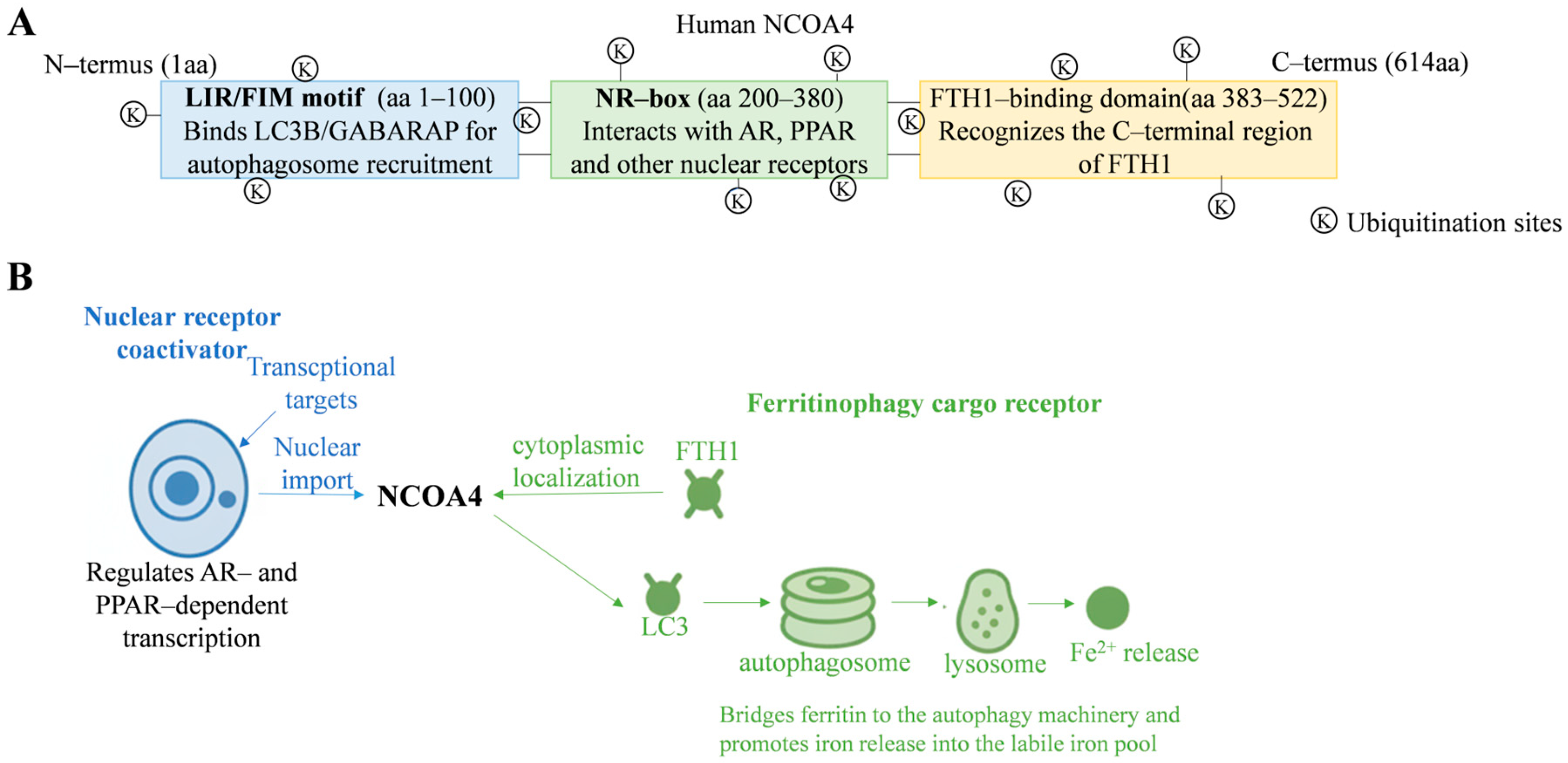
NCOA4, encoded by the NCOA4 gene on chromosome 10q11.22, is a 614-amino-acid protein with key functional domains: an N-terminal LC3-interacting region for binding to ATG8 family members on autophagosomal membranes, a central nuclear receptor-interacting domain for transcriptional coactivation, and a C-terminal ferritin-binding domain for binding to FTH1 [3,9]. It also has multiple ubiquitination sites for post-translational regulation (Figure 1A).

2.2. Transcriptional Regulation of NCOA4

The transcription of NCOA4 is subject to stringent regulation by various factors. Under hypoxic conditions, hypoxia-inducible factors (HIF-1α/2α) directly stimulate the transcription of the NCOA4 gene. Research by Li et al. [10] has demonstrated that the stabilization of HIF-1/2α through the use of hypoxia mimetics, such as deferoxamine and cobalt chloride, leads to a rapid increase in NCOA4 mRNA and protein levels in hepatocytes. Conversely, the knockdown of HIF-1/2α significantly diminishes this upregulation, indicating that the hypoxia–HIF–NCOA4 axis is a crucial adaptive mechanism for mobilizing hepatic iron reserves. This mechanism facilitates the release of stored iron to support erythropoiesis during the recovery period following hemorrhage.
Additionally, the Hippo–YAP signaling pathway is implicated in the transcriptional regulation of NCOA4. Yes-associated protein (YAP) directly enhances NCOA4 transcription in hepatocytes, as reported by Qi et al. [11]. Furthermore, hepatocyte nuclear factor 4 alpha (HNF4A), a liver-enriched nuclear receptor, has been recently identified to suppress NCOA4-mediated ferritinophagy, thereby exerting a protective effect in instances of acute liver failure [12].

2.3. Post-Translational Modifications of NCOA4

The stability of the NCOA4 protein is modulated by a complex array of post-translational modifications, with ubiquitination being particularly pivotal. In conditions of iron sufficiency, the E3 ubiquitin ligase HERC2 binds to NCOA4, facilitating its degradation through the ubiquitin-proteasome pathway, thereby preventing excessive ferritinophagy and subsequent iron toxicity [4]. In contrast, during iron deficiency, the ubiquitination of NCOA4 by HERC2 is reduced, leading to enhanced stability of NCOA4. Recent research has identified additional regulators of ubiquitination that function in a tissue- or disease-specific context. For example, TRIM7, another E3 ubiquitin ligase, enhances the ubiquitination and degradation of NCOA4 in hepatic stellate cells, thereby diminishing ferritinophagy [13]. Furthermore, the deubiquitinating enzyme OTULIN is implicated in inadequate deubiquitination of NCOA4 during drug-induced liver injury, resulting in the accumulation of NCOA4 and persistent degradation of ferritin [14].
In addition to ubiquitination, various other post-translational modifications play a role in modulating NCOA4 function under specific conditions. Phosphorylation by ATM kinase in response to DNA damage facilitates ferritinophagy and ferroptosis [15]. The desumoylation of NCOA4 by SENP2 has been shown to inhibit ferritinophagy and protect cardiomyocytes during myocardial ischemia–reperfusion injury [16], suggesting potential applications for this mechanism in hepatic ischemia–reperfusion injury. Moreover, type I interferon signaling induces the ISGylation of NCOA4, impacting its protein stability and ferritinophagy activity [17]. Additionally, USP18, a member of the deubiquitinating enzyme family, has been demonstrated to regulate NCOA4 stability by mediating its deISGylation in hepatocellular carcinoma (see Table 1).

2.4. Mechanisms of NCOA4–Ferritin–Autophagy Machinery Interactions

NCOA4 exhibits dual functional localization (Figure 1B). Within the nucleus, it functions as a canonical transcriptional coactivator for nuclear receptors, modulating the transcriptional activity of targets such as the androgen receptor (AR) and peroxisome proliferator-activated receptor (PPAR). In the cytoplasm, NCOA4 acts as a cargo receptor for ferritinophagy [3,9]. It specifically recognizes FTH1 through its C-terminal ferritin-binding domain and simultaneously interacts with ATG8 family proteins (e.g., LC3B and GABARAP) on autophagosomal membranes via its N-terminal LIR motif. This interaction facilitates the targeting of the ferritin–iron complex for autophagic encapsulation and subsequent lysosomal degradation, leading to ferritin breakdown and iron release [3]. The released Fe2+ enters the labile iron pool (LIP), where a portion is utilized to meet cellular metabolic demands, while excess iron promotes the Fenton reaction and the subsequent lipid peroxidation–ferroptosis cascade. Consequently, NCOA4 is considered a pivotal molecular hub linking iron metabolism with cell fate decisions [6,18].

3. Role of NCOA4 in Liver Physiology and Iron Homeostasis

3.1. Liver Iron Storage and Mobilization

The liver functions as the body’s largest reservoir of iron, containing approximately 300–1000 mg of iron in healthy adults, predominantly stored as ferritin within hepatocytes and Kupffer cells [1]. In response to increased physiological demand for iron, such as during blood loss, pregnancy, or enhanced erythropoiesis, the liver swiftly mobilizes stored iron through NCOA4-mediated ferritinophagy to replenish the circulating iron pool. The phenotype observed in NCOA4 knockout mice highlights the critical physiological role of this pathway: NCOA4−/− mice demonstrate significant ferritin accumulation in the liver and spleen, reduced bioavailability of free iron, and increased sensitivity to exogenous iron overload [19].

3.2. The Core Role of NCOA4 in Hepatocyte Iron Recycling

NCOA4 precisely regulates iron homeostasis through a hierarchical mechanism: (1) it senses intracellular iron levels, where elevated iron levels promote HERC2-mediated ubiquitination and subsequent degradation of NCOA4, while reduced iron levels allow for its accumulation [4]; (2) it responds to systemic iron demand. HIF-1α/HIF-2α stabilization increases NCOA4 mRNA and protein in hepatic cells [10], whereas hepcidin indirectly regulates NCOA4 by altering intracellular iron levels, which in turn affect NCOA4 stability and ferritinophagy [20]; and (3) it executes selective autophagy by targeting ferritin for lysosomal degradation, thereby modulating the rate of iron release. Notably, the NCOA4–ferritinophagy pathway and the classical iron regulatory protein (IRP1/IRP2)–iron-responsive element (IRE) system form complementary regulatory networks in iron sensing. The IRE–IRP system primarily influences the synthesis of transferrin receptor and ferritin at the translational level in response to transient iron fluctuations, whereas NCOA4-mediated ferritinophagy offers a “stock-release” mechanism at the level of ferritin degradation to address sustained iron demands [3,4]. The coordination and potential crosstalk between these two pathways require further investigation. The expression and activity of NCOA4 must be precisely regulated to maintain a balance between “iron sufficiency” and “iron toxicity.” Dysregulation of this equilibrium predisposes cells to iron-dependent oxidative damage.

3.3. NCOA4 and the Balance of Hepatic Oxidative Stress

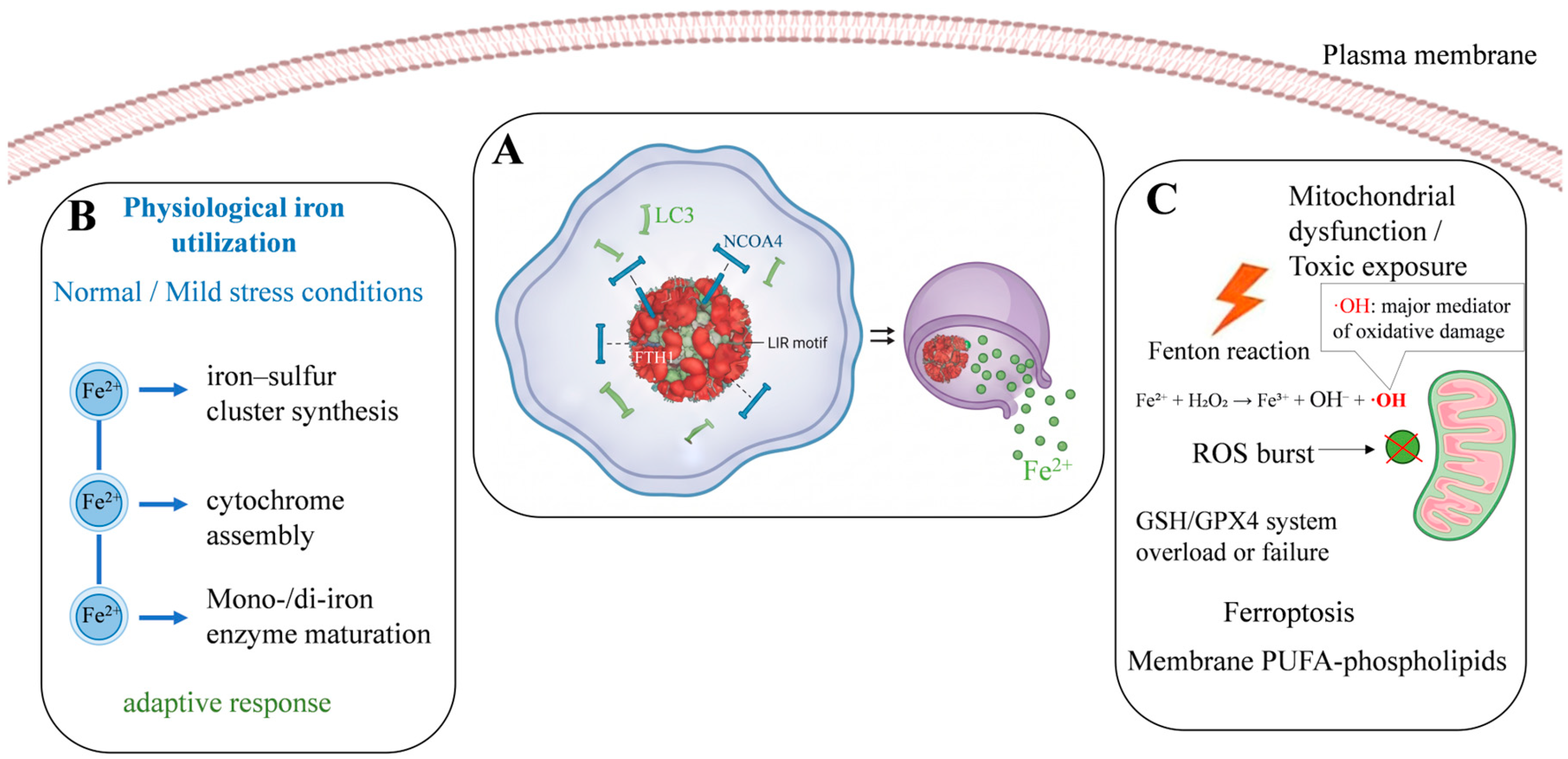
Under physiological conditions, iron released through NCOA4 is predominantly utilized for the biosynthesis of iron–sulfur clusters within mitochondria and the assembly of other vital iron-containing proteins, such as cytochromes. However, under conditions of elevated oxidative stress—arising from factors such as mitochondrial dysfunction or exposure to exogenous toxicants—excessive activation of NCOA4 results in the release of free iron, which subsequently generates significant quantities of hydroxyl radicals via the Fenton reaction. When the production of these radicals surpasses the cellular antioxidant defense capacity, such as the glutathione/GPX4 system, it initiates ferroptosis [5,6]. Consequently, the activity level of NCOA4 plays a critical role in determining the threshold at which hepatocytes transition from an adaptive response to ferroptosis under oxidative stress conditions (Figure 2).
NCOA4 is notably implicated in a positive feedback loop that connects inflammation and ferroptosis, a mechanism crucial for amplifying inflammatory responses in various liver diseases. The innate immune sensor STING (stimulator of interferon genes) has been demonstrated to directly interact with NCOA4 in macrophages. Upon activation, STING not only initiates type I interferon-mediated inflammatory responses but also sequesters NCOA4 in the cytoplasm through direct interaction. This process enhances ferritinophagy and ferroptosis while concurrently diminishing the nuclear coactivator function of NCOA4 [22]. Ferroptosis releases damage-associated molecular patterns (DAMPs), such as mitochondrial DNA, which can further activate the cGAS–STING pathway, thereby establishing a self-amplifying injury–inflammation loop. It is important to note that the STING–NCOA4 interaction has been directly demonstrated in macrophages during sepsis [22] and in acute kidney injury models [23]. Whether this axis operates in liver diseases remains to be established. Therefore, its potential involvement in alcoholic hepatitis, hepatic ischemia–reperfusion injury, and other liver diseases is discussed below as a hypothesis that warrants further investigation.

3.4. The Concept of Iron Toxicity Threshold: Why Iron Overload Is Not Always Injurious

Hepatocytes are not merely passive reservoirs for iron storage; rather, they have evolved as specialized iron-handling cells, equipped with sophisticated, multi-layered regulatory mechanisms to manage iron uptake, buffering, and trafficking. These cells express a precisely regulated array of iron transporters, such as transferrin receptor 1 and divalent metal transporter 1, storage proteins like ferritin, and efflux pumps such as ferroportin. These components function under the coordinated regulation of the hepcidin–ferroportin axis and the iron regulatory protein (IRP)/iron-responsive element (IRE) system [24,25]. This complex network ensures that, under physiological conditions, hepatocytes can accommodate significant fluctuations in systemic iron supply while maintaining the cytosolic labile iron pool (LIP) within a safe, homeostatic range [25].
Nevertheless, even the most resilient homeostatic mechanisms possess a limited buffering capacity. The shift from “iron sufficiency” to “iron toxicity” is not solely dictated by the absolute amount of iron released through ferritinophagy. Instead, it is governed by the dynamic balance between iron availability and the cell’s ability to safely utilize or detoxify it—a concept we define as the iron toxicity threshold. Below this threshold, Fe2+ released via NCOA4-mediated ferritinophagy is effectively integrated into critical metabolic processes, such as mitochondrial iron–sulfur cluster formation, heme synthesis [26], and DNA replication, thereby sustaining normal cellular functions and proliferation. Pathological damage due to iron occurs only when this threshold is surpassed, typically necessitating one or more “second hits” that undermine the cellular defense and repair systems. These concurrent stressors may include: (i) significant depletion of glutathione (GSH) and inactivation of GPX4, which impair the primary lipid peroxide clearance mechanism [27]; (ii) mitochondrial dysfunction, which exacerbates the production of endogenous reactive oxygen species (ROS) [28]; (iii) chronic inflammatory signaling, exemplified by the STING–NCOA4 feedback loop, which establishes a self-amplifying cascade of injury [22]; and (iv) genetic polymorphisms in genes regulating iron metabolism or ferroptosis, which reduce the intrinsic cellular tolerance to oxidative stress [29].
This threshold concept is robustly supported by clinical observations in patients with transfusion-dependent thalassemia, as highlighted by the editor. Despite experiencing chronic iron overload due to repeated blood transfusions, resulting in hepatic iron concentrations that significantly exceed those observed in most metabolic liver diseases, a substantial proportion of these patients do not develop progressive liver fibrosis, cirrhosis, or cardiac dysfunction [30]. This notable clinical heterogeneity emphasizes that while iron overload is a necessary condition for tissue injury, it is not sufficient on its own. The ultimate phenotype is influenced by additional modifiers, including the efficacy of iron chelation therapy, the integrity of the hepcidin response, the activity of alternative iron detoxification pathways (such as ferritin sequestration and hemosiderin formation), and inter-individual genetic variations that influence ferroptosis susceptibility [29,30].
When applied to the NCOA4–ferritinophagy axis, the iron toxicity threshold model offers a comprehensive framework that reconciles the seemingly contradictory roles of this pathway across various liver diseases. In conditions where cellular antioxidant capacity is preserved, transient activation of NCOA4 functions as a regulated mechanism for mobilizing iron to satisfy metabolic demands without inducing ferroptotic damage. This is consistent with its vital physiological roles in erythropoiesis and liver regeneration [31]. Conversely, in pathological contexts characterized by chronic inflammation, metabolic lipotoxicity, mitochondrial damage, or drug-induced glutathione (GSH) depletion—such as in advanced metabolic-associated steatohepatitis (MASH), alcoholic hepatitis, and acetaminophen (APAP) overdose—the same ferritinophagy-derived iron load becomes pathogenic due to a pathological reduction in the threshold for safe iron handling [32]. This conceptual framework also elucidates why hepatic stellate cells (HSCs) are particularly susceptible to NCOA4-driven ferroptosis: their inherently low expression of GPX4 and SLC7A11 positions them closer to the toxicity threshold, rendering iron release selectively lethal for these fibrogenic cells [33].
Therefore, we propose that the pathological versus protective outcomes of NCOA4 activation should not be solely attributed to ferritinophagy; instead, they should be considered in the context of the cellular environment, which determines the proximity to the iron toxicity threshold. This perspective not only reconciles the divergent roles of NCOA4 discussed in subsequent sections but also has significant therapeutic implications. Specifically, interventions aimed at modulating ferritinophagy should be accompanied by strategies that assess or restore the cellular redox reserve to ensure predictable and safe clinical outcomes. Biomarker panels that combine ferritinophagy markers (e.g., NCOA4/FTH1 interaction) with indicators of redox capacity (e.g., GSH/GSSG ratio, GPX4 activity, and 4-HNE adducts) may be crucial for patient stratification [34]. Such panels would enable clinicians to predict whether activating or inhibiting NCOA4 will shift the balance toward therapeutic benefit or unintended harm.

4. Role of NCOA4-Mediated Ferritinophagy in Various Liver Diseases

4.1. Drug-Induced Liver Injury

Drug-induced liver injury (DILI), such as from acetaminophen (APAP) overdose, is a classic example of acute liver damage. APAP is metabolized by CYP2E1 into NAPQI, which depletes glutathione and causes oxidative stress and mitochondrial dysfunction. Recent studies highlight ferroptosis as a crucial factor in APAP toxicity [35]. APAP disrupts iron balance in liver cells, leading to increased ferritinophagy, iron buildup, and ferroptotic cell death [3]. In mouse models, APAP intoxication significantly raises liver iron levels, lipid peroxidation, and ferroptosis markers [36] (Table 2).
The deubiquitinating enzyme OTULIN has been demonstrated to play a protective role in acetaminophen (APAP)-induced liver injury by modulating the ubiquitination status of NCOA4 [14]. Mechanistically, OTULIN expression decreases following APAP intoxication, leading to inadequate deubiquitination of NCOA4, increased stability of NCOA4, and continuous degradation of ferritin accompanied by iron release. Overexpression of OTULIN facilitates the degradation of NCOA4, restores levels of ferritin heavy chain 1 (FTH1), reduces the release of free iron, and consequently mitigates ferroptosis and markers of liver injury [4]. These findings indicate that modulating the homeostasis of NCOA4 ubiquitination may effectively alleviate APAP-induced liver injury. Furthermore, targeting the protein–protein interaction interface between NCOA4 and FTH1 presents an additional potential intervention strategy. The small-molecule compound 9a has been reported to directly inhibit the interaction between NCOA4 and FTH1, thereby reducing ferritin degradation, limiting iron release, and suppressing ferroptosis [37].
The pathogenesis of acetaminophen (APAP)-induced liver injury is notably intricate. While iron released from ferritinophagy is implicated in oxidative damage processes, such as protein nitration during APAP intoxication, this injury may not entirely conform to the biochemical criteria of canonical ferroptosis [36]. This suggests that ferritinophagy may exacerbate liver injury through both ferroptosis-dependent and ferroptosis-independent pathways within the context of drug-induced liver injury (DILI). Despite this complexity, substantial evidence indicates that NCOA4-mediated ferritinophagy primarily contributes to APAP hepatotoxicity via ferroptosis-dependent mechanisms.
Overall, current research suggests that NCOA4 plays a predominantly pathogenic role in DILI by facilitating ferroptosis and necrosis. Targeted inhibition of NCOA4-mediated ferritinophagy—through strategies such as enhancing OTULIN function, disrupting the NCOA4–FTH1 interaction, or administering iron chelators—emerges as a promising therapeutic approach for the prevention and treatment of DILI [35].

4.2. Metabolic Dysfunction-Associated Steatotic Liver Disease

In contrast to the acute toxic profile associated with drug-induced liver injury, metabolic dysfunction-associated steatotic liver disease (MASLD) and its progressive variant, metabolic dysfunction-associated steatohepatitis (MASH), are characterized by hepatic steatosis, inflammation, and fibrosis, and are recognized as the most prevalent chronic liver diseases globally. Dysregulation of iron metabolism significantly contributes to the progression of MASLD, with NCOA4-mediated ferritinophagy serving as a fundamental mechanism that links iron overload to hepatocyte injury [7] (Table 2).
A considerable proportion of patients with MASLD present with mild to moderate hepatic iron deposition, a condition referred to as dysmetabolic iron overload, which is often associated with elevated serum ferritin levels [38,39]. The metabolically inflamed microenvironment results in inappropriately elevated hepcidin levels, which may inhibit NCOA4 function and disrupt ferritinophagy, consequently leading to iron retention within hepatocyte ferritin stores—a phenomenon known as “hepatic iron trapping” [40]. This hypothesis posits a connection between iron overload and diminished NCOA4 activity, suggesting that iron accumulates passively rather than being actively released. The chronic iron-rich environment exacerbates oxidative stress and steatosis. These contrasting scenarios likely represent different stages of disease progression: hepcidin-mediated suppression of NCOA4 in early steatosis versus oxidative stress-induced overactivation in advanced disease. In hepatocytes that have already developed steatosis, lipid peroxidation and mitochondrial dysfunction may lead to the overactivation of NCOA4, resulting in the release of substantial amounts of free iron. This free iron reacts with accumulated lipids through peroxidation, further promoting ferroptosis and inflammatory responses. Animal studies have shown that markers of ferroptosis are significantly elevated in high-fat diet models, and treatment with iron chelators or ferroptosis inhibitors reduces hepatic inflammation and cellular injury [41]. Research utilizing zebrafish models has elucidated that insulin-like growth factor binding protein 7 (IGFBP7) facilitates NCOA4-mediated ferritinophagy, which results in hepatic iron accumulation and lipid peroxidation. Conversely, the knockdown of IGFBP7 has been shown to mitigate hepatic iron deposition and lipid peroxidative damage, thereby delaying the transition from Metabolic-Associated Steatotic Liver Disease (MASLD) to Metabolic-Associated Steatohepatitis (MASH) [42].
Furthermore, recent investigations have identified several novel regulators of ferroptosis in MASLD. Notably, the ferroptosis-associated gene EFEMP1 has been recognized as a positive regulator of NCOA4-mediated ferritinophagy. Additionally, FOT1, a multi-target iron chelator that also inhibits c-Myc/ACSL4-driven ferroptosis, has demonstrated efficacy in MASH models by reducing hepatic iron accumulation and ACSL4-dependent lipid peroxidation [43,44]. This effect is NCOA4-independent and does not involve direct disruption of the NCOA4–FTH1 interaction.
Iron overload and lipotoxic stress are implicated in the premature senescence of hepatocytes, a process recognized as a contributing factor to fibrotic progression in Metabolic-Associated Steatohepatitis (MASH). Empirical evidence indicates that the activation of ferritinophagy and the upregulation of cellular senescence markers are simultaneously observed in high-fat diet models and lipid-laden hepatocytes. Furthermore, curcumol has been shown to mitigate YAP-driven NCOA4 expression, thereby reducing hepatocyte senescence and lipid accumulation in Metabolic-Associated Steatotic Liver Disease (MASLD) models [11].
Collectively, the function of NCOA4 in MASLD demonstrates stage-dependent characteristics. In the simple steatosis phase, moderate activation of NCOA4 may support the maintenance of iron metabolic flexibility. However, as the condition progresses to MASH, excessive NCOA4 activation exacerbates hepatic injury through mechanisms involving both ferroptosis and cellular senescence. Consequently, the inhibition of NCOA4-mediated ferritinophagy presents a potential therapeutic strategy in the management of MASH, although the timing and extent of such intervention are critically important.

4.3. Alcoholic Liver Disease

In contrast to Metabolic-Associated Steatotic Liver Disease (MASLD), which is predominantly driven by metabolic disturbances, the pathogenesis of Alcoholic Liver Disease (ALD) is primarily centered on the direct hepatotoxic effects of ethanol and its metabolites. This involves mechanisms such as oxidative stress, increased intestinal permeability, and the activation of inflammatory cascades. Chronic alcohol consumption results in an elevated hepatic iron burden, mainly due to the suppression of hepcidin synthesis, which enhances intestinal iron absorption and subsequent hepatic accumulation [45]. Studies utilizing murine models have demonstrated that prolonged ethanol exposure induces a hepatic ferroptosis phenotype, characterized by an increase in lipid peroxidation products, such as malondialdehyde, a reduction in GPX4 activity, and the upregulation of genes associated with ferroptosis [46] (see Table 2).
NCOA4-mediated ferritinophagy has been identified as a critical pro-injurious factor in alcoholic liver disease (ALD). Zhao et al. [47] demonstrated that both pharmacological and genetic inhibition of the NCOA4–ferritinophagy pathway—achieved by reducing NCOA4 activity or silencing NCOA4 expression—results in a significant increase in intracellular ferritin levels, a decrease in free iron, a reduction in ferroptosis marker accumulation, and an alleviation of hepatic necrosis and inflammation in alcohol-fed mice and ethanol-treated hepatocytes. Mechanistically, chronic alcohol exposure appears to activate NCOA4 through two distinct pathways: excessive reactive oxygen species (ROS) generated from alcohol metabolism stabilize hypoxia-inducible factor (HIF) signaling, thereby upregulating NCOA4 transcription, while alcohol-induced endoplasmic reticulum stress and inflammatory responses enhance NCOA4 protein stability. The downstream effects follow a clear cascade: elevated NCOA4 levels lead to ferritin degradation, resulting in substantial iron release; the liberated iron subsequently promotes lipid peroxidation and ferroptosis in hepatocytes, leading to the release of damage-associated molecular patterns (DAMPs) and immune cell infiltration, ultimately contributing to liver injury [48].
In the fibrotic stage of alcoholic liver disease (ALD), NCOA4-mediated ferroptosis may have dual, cell type-dependent effects: it exacerbates hepatocyte loss and inflammation in hepatocytes, while its induction in activated hepatic stellate cells (HSCs) aids in reducing collagen production (refer to Section 4.5 for further details). Additionally, the STING–NCOA4 positive feedback loop, which links inflammation and ferroptosis as discussed in Section 3.3, may amplify the inflammatory response in alcoholic hepatitis.
In conclusion, NCOA4-mediated ferritinophagy plays a pivotal role in driving hepatocyte ferroptosis and inflammation in ALD. Both animal and cellular studies have conclusively shown that inhibiting this pathway significantly mitigates alcohol-induced liver injury [47], presenting a promising therapeutic approach targeting ferroptosis for ALD.

4.4. Ischemia–Reperfusion Liver Injury

In contrast to the chronic or subacute pathologies that characterize drug-induced liver injury (DILI), metabolic-associated steatotic liver disease (MASLD), and alcoholic liver disease (ALD), hepatic ischemia–reperfusion injury (IRI) constitutes a distinct form of damage arising from acute hemodynamic stress, as observed in clinical contexts such as liver transplantation and resuscitation following shock. The abrupt restoration of oxygen during the reperfusion phase induces substantial production of reactive oxygen species (ROS), leading to pronounced oxidative stress and cellular damage [49]. Recent investigations have identified ferroptosis as a principal mechanism of cell death in IRI, with nuclear receptor coactivator 4 (NCOA4) potentially acting as a critical mediator [50] (Table 2).
During the ischemic phase, tissue hypoxia stabilizes hypoxia-inducible factor 1α/2α (HIF 1α/2α) signaling, which upregulates NCOA4 transcription [10], thereby priming the system for subsequent iron release. Upon reperfusion, the rapid influx of oxygen reacts with free Fe2+ released via NCOA4 through the Fenton reaction, generating abundant hydroxyl radicals that initiate a ferroptotic cascade. Consequently, NCOA4 accumulation during ischemia may serve as a “priming” factor for ferroptosis upon reperfusion.
The involvement of the STING–NCOA4 positive feedback loop in ischemia–reperfusion injury (IRI) is further elucidated in Section 3.3, highlighting its role in linking inflammation and ferroptosis. Damage-associated molecular patterns (DAMPs) released from injured hepatocytes activate the cGAS–STING pathway, which in turn interacts with NCOA4 to enhance ferritinophagy and ferroptosis, thereby establishing a self-reinforcing injury loop [22,23].
Recent studies using myocardial ischemia models provide insights that may be applicable to hepatic IRI. Specifically, SENP2-mediated desumoylation of NCOA4 has been shown to inhibit ferritinophagy and protect cardiomyocytes during myocardial IRI [16]. Similar strategies, such as upregulating SENP2 or administering NCOA4 inhibitors, could potentially mitigate hepatocyte loss during hepatic reperfusion. Nevertheless, the subsequent regenerative phase of the liver requires increased iron availability, and NCOA4 may facilitate cell proliferation by supplying iron (refer to Section 5). Consequently, perioperative modulation of NCOA4 must carefully balance the need for acute injury protection with the requirement for effective regeneration.

4.5. Liver Fibrosis and Cirrhosis

Liver fibrosis represents a prevalent pathological consequence of various chronic hepatic injuries, characterized by the activation of hepatic stellate cells (HSCs) and excessive collagen deposition. The interplay between dysregulated iron metabolism and liver fibrosis is bidirectionally complex, with NCOA4 demonstrating opposing effects depending on the cell type involved (refer to Table 2). Insufficient NCOA4 function in hepatocytes and Kupffer cells leads to significant ferritin accumulation and impaired iron mobilization. This persistent iron overload induces sustained oxidative stress, fosters a chronic inflammatory microenvironment, and accelerates fibrotic progression. In murine models subjected to a high-iron diet, the onset of liver fibrosis is closely linked to iron overload, and the administration of the ferroptosis inhibitor Fer-1 mitigates fibrosis [51], thereby supporting the hypothesis that iron-induced cell death facilitates the initiation of fibrosis.
In the context of established fibrosis, hepatic stellate cells (HSCs) exhibit a heightened vulnerability to ferroptosis, primarily due to glutathione depletion and the downregulation of SLC7A11 [52]. Consequently, the targeted activation of NCOA4-mediated ferritinophagy within HSCs holds potential for reducing collagen synthesis and facilitating the reversal of fibrosis. A pertinent example of this approach is demonstrated by blueberry anthocyanins, which, in a carbon tetrachloride (CCl4)-induced mouse model of liver fibrosis, preserve NCOA4 protein levels by inhibiting TRIM7. TRIM7 is an E3 ubiquitin ligase that is abundantly expressed in HSCs and promotes the ubiquitination and degradation of NCOA4. This inhibition enhances ferritinophagy and ferroptosis in HSCs, ultimately mitigating hepatic collagen deposition [13]. Similarly, the clinical agent sorafenib induces ferroptosis in HSCs via the HIF-1α/SLC7A11 axis [53], while artesunate operates through the ROCK1/ATF3 axis [54], and berberine triggers reactive oxygen species (ROS)-mediated ferroptosis in HSCs via iron redox cycling [55]. Collectively, these interventions exemplify the therapeutic strategy of selectively inducing ferroptosis in HSCs.
The aforementioned dual effects constitute the central challenge of NCOA4-targeted therapy in the context of liver fibrosis: specifically, the challenge lies in moderately inhibiting NCOA4 in hepatocytes to mitigate ferroptosis-induced fibrotic triggers, while concurrently enhancing NCOA4 activity selectively in hepatic stellate cells (HSCs) to facilitate their clearance. Attaining such cell type-specific regulation of NCOA4 is a pivotal focus for future research endeavors.

4.6. Hepatocellular Carcinoma

Hepatocellular carcinoma (HCC) commonly develops against a backdrop of chronic liver injury and inflammation. The nuclear receptor coactivator 4 (NCOA4) exhibits a dual function in HCC: it facilitates the provision of essential iron for rapid tumor proliferation while simultaneously increasing the susceptibility of HCC cells to ferroptosis-inducing therapies. This is due to the elevated expression of NCOA4, which predisposes these cells to a ferroptosis-prone state (Table 2).

4.6.1. NCOA4 Upregulation in HCC and Its Implications for Iron Metabolism

In comparison to adjacent non-tumorous liver tissues, hepatocellular carcinoma (HCC) tissues typically exhibit significantly elevated levels of NCOA4 mRNA and protein [56]. This upregulation indicates the dependency of tumor cells on increased ferritinophagy to meet their substantial iron requirements, driven by rapid cellular proliferation. Nevertheless, elevated NCOA4 expression concurrently predisposes HCC cells to ferroptosis, necessitating the upregulation of antioxidant defense mechanisms, such as GPX4 and SLC7A11, to mitigate iron-induced toxicity [57].

4.6.2. NCOA4 and Sensitivity to Ferroptosis-Inducing Therapy in HCC

Sorafenib, a first-line therapeutic agent for advanced HCC, partially exerts its antitumor effects by inducing ferroptosis through the inhibition of the System Xc−/GPX4 axis [58]. NCOA4 plays a pivotal “pro-synergistic” role in this process: the knockdown of NCOA4 diminishes the sensitivity of HCC cells to ferroptosis inducers like sorafenib, as intracellular free iron is reduced and lipid peroxidation levels decrease. Conversely, augmenting NCOA4 expression or increasing iron loading enhances the ferroptotic efficacy of these agents [59]. This suggests that NCOA4 levels may serve as a predictive biomarker for response to ferroptosis-inducing therapy in HCC.
Recent studies have explored combination strategies to overcome sorafenib resistance by targeting NCOA4-mediated ferritinophagy. Huaier (HUA), a traditional Chinese medicine used as an adjuvant therapy for advanced HCC, has been shown to reverse sorafenib resistance and exhibit synergistic effects with sorafenib by activating NCOA4-mediated ferritinophagy. In sorafenib-resistant Huh7R cells and mouse xenograft models, Huaier alone or combined with sorafenib upregulated NCOA4, increased the LC3B-II/I ratio, and promoted FTH1 degradation, leading to Fe2+ accumulation, ROS generation via the Fenton reaction, and lipid peroxidation. Notably, Huaier also upregulated ACSL4 and downregulated SCD1 and GPX4, collectively inducing ferroptosis that was rescued by ferrostatin-1 or deferoxamine. NCOA4 silencing substantially attenuated Huaier’s efficacy in overcoming sorafenib resistance, confirming that NCOA4-mediated ferritinophagy is a key driver of this combination strategy [60]. These findings provide a rationale for the clinical development of Huaier plus sorafenib combination therapy in HCC.
Recent research has elucidated several novel regulatory pathways of NCOA4 in hepatocellular carcinoma (HCC). These pathways can be broadly classified into those that facilitate NCOA4-mediated ferroptosis, such as SCARA5, PTBP1, and ATM, and those that inhibit it, including ERK/ULK1 and USP18. This classification underscores the complex equilibrium cancer cells must maintain to avoid ferroptosis while ensuring an adequate iron supply. Among the pathways that inhibit ferroptosis, the ERK/ULK1/NCOA4 pathway is characterized by hyperactivation of ERK1/2 in HCC tissues, which phosphorylates and inhibits ULK1. This inhibition obstructs NCOA4-mediated ferritinophagy, allowing tumor cells to evade ferroptosis. The intervention of Epimedium extract in this pathway alleviates ERK-mediated inhibition of the ULK1–NCOA4 axis, thereby reactivating ferritinophagy, inducing ferroptosis in HCC, and suppressing tumor growth [61]. Another inhibitory mechanism is the USP18 ISGylation NCOA4 pathway. Ye et al. [17] demonstrated that sorafenib upregulates USP18 through the STING/IRF3/ISG15 pathway. Importantly, this regulation is indirect and involves STING/IRF3/ISG15-mediated USP18 upregulation rather than a direct STING–NCOA4 interaction. The existence of a direct STING–NCOA4 axis in HCC remains to be determined. USP18 subsequently catalyzes the deISGylation and degradation of NCOA4, diminishing ferritinophagy and establishing a feedback loop that contributes to acquired resistance in HCC. The aforementioned study additionally identified hyperoside as a novel inhibitor of USP18, which restores the function of NCOA4 and re-sensitizes hepatocellular carcinoma (HCC) cells to the effects of sorafenib and regorafenib.
In contrast, several pathways facilitate ferroptosis by enhancing NCOA4 activity. SCARA5 promotes ferritin degradation and iron release by strengthening the interaction between NCOA4 and FTH1; the absence of SCARA5 renders HCC cells resistant to ferroptosis induced by erastin, RSL3, and sorafenib [62]. PTBP1 binds to NCOA4 mRNA, promoting its translation, which increases NCOA4 protein levels and heightens cellular susceptibility to ferroptosis [59]. Furthermore, ATM directly phosphorylates NCOA4 in response to DNA damage, thereby promoting ferritinophagy and ferroptosis [15], suggesting that genotoxic stress may be linked to the ferroptotic pathway through the ATM–NCOA4 axis.

4.6.3. NCOA4 and the HCC Immune Microenvironment

The expression levels of NCOA4 are also correlated with the immune microenvironment status in hepatocellular carcinoma (HCC). Comprehensive pan-cancer bioinformatics analyses have demonstrated that reduced expression of NCOA4 is frequently linked to an immunosuppressive tumor microenvironment and a poorer survival prognosis [63]. Potential underlying mechanisms include the following: NCOA4 deficiency protects tumor cells from ferroptosis, while diminished iron release restricts the availability of functional iron for tumor-infiltrating immune cells. However, it remains to be determined whether NCOA4-mediated iron release, upon uptake by tumor-associated macrophages, facilitates their polarization towards an immunosuppressive (M2) phenotype.
In conclusion, NCOA4 emerges as a highly promising target for therapeutic intervention in HCC. Strategies aimed at promoting the NCOA4–ferritinophagy axis—such as the combination of USP18 inhibitors with sorafenib or the use of Epimedium extract—show potential for enhancing ferroptosis-inducing efficacy and overcoming drug resistance. Conversely, inhibiting this pathway during the chronic liver disease stage may reduce the risk of HCC development.

4.7. Viral Hepatitis-Associated Liver Injury

Research specifically examining NCOA4-mediated ferritinophagy in the context of chronic hepatitis B (HBV) and hepatitis C (HCV) remains scarce, highlighting a significant research gap in this area. Considering that HBV and HCV infections are the predominant etiological factors for hepatocellular carcinoma (HCC) globally, responsible for over 80% of HCC cases, understanding the involvement of the NCOA4–ferritinophagy axis in viral hepatitis is of considerable clinical importance (see Table 2).
Chronic HCV infection is often associated with hepatic iron accumulation and elevated serum ferritin levels. Iron overload is believed to enhance viral replication, expedite fibrotic progression, and compromise the virological response to interferon-based therapies. HCV infection is associated with dysregulated hepcidin expression and hepatic iron accumulation. Notably, the HCV non-structural protein NS5A suppresses hepcidin expression, potentially leading to increased intestinal iron absorption, whereas the core protein has been reported to stimulate hepcidin [64]. In addition, virus-induced oxidative stress may influence ferritinophagy activity in hepatocytes. In this scenario, hepatocyte ferritin levels are elevated, yet the regulation of iron release may be impaired. The potential contribution of the NCOA4–ferritinophagy pathway to iron metabolic disturbances associated with HCV and the efficacy of iron chelation as an adjunctive therapy in enhancing hepatic histological outcomes in HCV-infected patients have yet to be thoroughly investigated.
The modifications in iron metabolism linked to chronic HBV infection warrant significant attention. The HBV X protein (HBx) is known to induce oxidative stress and mitochondrial dysfunction, both of which are established activators of ferritinophagy. Moreover, observational clinical studies have demonstrated an independent association between elevated serum ferritin levels and an increased risk of HBV-related hepatocellular carcinoma (HCC). However, the intermediary role of NCOA4 in this association remains unexplored.
Recent findings by Yu et al. [12] indicate that HNF4A exerts a protective effect in acute liver failure by inhibiting NCOA4-mediated ferritinophagy. Given that HNF4A is also a crucial regulator of HBV transcription, replication, and hepatocyte differentiation, the functional implications of the HNF4A–NCOA4 axis in HBV-associated liver disease merit further investigation. In conclusion, the role of the NCOA4–ferritinophagy–ferroptosis axis in viral hepatitis represents a significant research gap that urgently needs to be addressed. Research in this domain holds potential for the development of novel therapeutic strategies for HBV/HCV-related fibrosis and HCC.

5. Potential Role of NCOA4 in Liver Regeneration and Repair

The liver exhibits an extraordinary capacity for regeneration, with cellular proliferation necessitating a substantial iron supply. This is essential for both DNA synthesis, which depends on iron-dependent ribonucleotide reductase, and the assembly of the mitochondrial respiratory chain. Although direct investigations into the mechanistic role of NCOA4 in liver regeneration are limited, various indirect pieces of evidence suggest its potential function as a critical “iron donor”.
Li et al. [10] demonstrated that following blood loss, the mouse liver rapidly upregulates NCOA4 through HIF signaling to mobilize iron stores, thereby supporting erythropoiesis. This indicates that the NCOA4–HIF axis constitutes an important adaptive mechanism in response to increased iron demand. In the context of partial hepatectomy, the remaining liver tissue undergoes transient hypoxia and HIF activation, which could theoretically induce the same NCOA4-mediated iron release to facilitate hepatocyte proliferation. Moreover, NCOA4 may contribute to liver regeneration not only as an iron donor via ferritinophagy but also as a nuclear receptor coactivator. NCOA4 was originally identified as an androgen receptor (AR)-associated coactivator (ARA70) that interacts with the AR ligand-binding domain and enhances AR-mediated transcription [9,65,66]. Androgen/AR signaling promotes hepatocyte proliferation and liver regeneration after partial hepatectomy, at least in part by upregulating cell cycle-related genes and growth factor signaling pathways [65,66]. Therefore, NCOA4 may amplify AR-dependent transcriptional programs in hepatocytes and thereby support regenerative proliferation. However, direct evidence linking NCOA4-AR coactivation to hepatocyte proliferation in vivo remains limited, and whether NCOA4’s nuclear coactivator function is required for liver regeneration warrants further investigation.
It is conceivable that a deficiency or loss of function in NCOA4 could result in impaired or delayed liver regeneration following hepatectomy. Conversely, a short-term, moderate enhancement of NCOA4 activity, potentially in conjunction with iron supplementation, might enhance regenerative capacity post-hepatectomy. Nonetheless, the principal challenge of this hypothesis lies in promoting regeneration while mitigating the risk of ferroptosis induced by excessive iron release. Future research should aim to elucidate the spatiotemporal dynamics of NCOA4 expression in liver tissue after partial hepatectomy, the equilibrium between NCOA4-mediated iron supply and the risk of ferroptosis during regeneration, and whether the nuclear receptor coactivator function of NCOA4 directly influences hepatocyte proliferation. Investigating the “balance point” between liver regeneration and ferroptosis offers a promising new perspective in this field.

6. Clinical Translation Prospects of NCOA4 as a Therapeutic Target

6.1. Strategies for Inhibiting NCOA4–Ferritinophagy

These strategies target conditions like acute liver injury (including DILI and early-stage IRI), advanced MASLD, active ALD, and active autoimmune hepatitis, where excessive ferritinophagy leads to hepatocyte damage. One method involves inhibiting the NCOA4–FTH1 interaction; compound 9a disrupts this binding and reduces iron release [37], suggesting the need for inhibitors with better affinity and selectivity. Another strategy focuses on enhancing NCOA4 degradation, such as by upregulating OTULIN, which facilitates NCOA4 ubiquitination and proteasomal degradation, shown to improve APAP-induced acute liver injury [14]. Developing TRIM7 agonists or OTULIN activators could offer additional therapeutic options. Iron chelation is a third approach, with deferoxamine (DFO) and the novel agent FerroTerminator 1 (FOT1) showing hepatoprotective effects in MASH models [44]. Unlike compound 9a, FOT1 does not directly inhibit NCOA4–FTH1 interaction; it reduces the labile iron pool and ACSL4-dependent lipid peroxidation in an NCOA4-independent manner. In conclusion, the temporary suppression of HIF signaling during the ischemic phase of ischemia–reperfusion injury (IRI) may inhibit the initial accumulation of NCOA4, thereby mitigating reperfusion-associated ferroptosis. Taken together, these four complementary strategies provide a range of options for reducing NCOA4-driven ferritinophagy in scenarios where the protection of hepatocytes is the primary therapeutic objective.

6.2. Strategies for Activating NCOA4–Ferritinophagy and Their Indications

These strategies are predominantly applicable to liver fibrosis, specifically targeting hepatic stellate cells, and hepatocellular carcinoma, where the selective elimination of pathogenic cells, such as activated hepatic stellate cells or tumor cells, constitutes the therapeutic objective. One approach involves the suppression of negative regulators of NCOA4. For example, USP18 inhibitors like hyperoside have been shown to restore NCOA4 function and enhance sensitivity to ferroptosis in hepatocellular carcinoma [17]. Additionally, intervention in the ERK pathway, as demonstrated by the use of Epimedium extract, alleviates the inhibition of the ULK1–NCOA4 axis [61]. A second strategy focuses on inhibiting E3 ubiquitin ligases that target NCOA4 for degradation. In hepatic stellate cells, the targeted suppression of TRIM7 preserves NCOA4 protein levels, thereby promoting ferroptosis in these cells and facilitating the reversal of fibrosis [13]. Lastly, naturally derived NCOA4 modulators, including blueberry anthocyanins acting through the TRIM7–NCOA4 axis [13]; curcumol via the YAP–NCOA4 axis [11]; Epimedium extract via the ERK/ULK1/NCOA4 axis [61]; and Huaier, which activates NCOA4-mediated ferritinophagy and reverses sorafenib resistance in HCC [60], merit further exploration as promising therapeutic candidates. Together, these approaches highlight the potential of utilizing NCOA4 activation for the selective eradication of pathogenic cell populations.

6.3. Drug Development Progress and Challenges

The compounds examined in this section are primarily at the preclinical stage, with the exception of deferoxamine, which has been clinically approved for uses other than liver disease.
Among the synthetic or design-based agents, compound 9a represents the first small-molecule inhibitor that directly disrupts the NCOA4–FTH1 protein–protein interaction (PPI). By inhibiting ferritin recruitment to autophagosomes, it reduces the free iron burden and has demonstrated protective effects against ferroptosis in models of drug-induced liver injury (DILI), ischemia–reperfusion injury (IRI), and orthodontic root resorption. Currently, it is undergoing lead optimization. Nonetheless, the inherently flat PPI interface, which lacks a conventional binding pocket, complicates the development of high-affinity selective inhibitors. Additionally, systemic inhibition of NCOA4 may disrupt whole-body iron homeostasis, with long-term safety risks yet to be fully assessed [37]. FOT1 (FerroTerminator1, formerly CN128) is a next-generation iron chelator identified through drug screening. Mechanistically, unbiased multi-omics analyses in multiple MASH models revealed that hepatic iron accumulation aggravates MASH primarily through c-Myc–ACSL4-driven ferroptosis, and FOT1 concurrently inhibits both hepatic iron accumulation and this ferroptotic pathway. Unlike compound 9a, FOT1 does not directly disrupt the NCOA4–FTH1 interaction; instead, it acts upstream of NCOA4 by reducing the labile iron pool that drives ACSL4-dependent lipid peroxidation. Importantly, FOT1 does not disrupt systemic iron homeostasis and shows superior safety compared with clinically approved chelators such as deferoxamine (DFO) and deferasirox (DFX) in preclinical models. Serum ferritin levels have been proposed as a predictive biomarker for FOT1-based therapy. These findings support FOT1 as a promising NCOA4-independent therapeutic option for MASH, although its clinical evaluation remains in the early stages [44].
Several naturally derived compounds have been explored as modulators of NCOA4. Curcumol has been shown to downregulate NCOA4 expression by inhibiting YAP transcriptional activity, thereby mitigating hepatocyte senescence and lipid accumulation in MASLD models [11]. Hyperoside stabilizes NCOA4 protein by inhibiting USP18 deISGylation activity, which enhances ferritinophagy and increases the sensitivity of HCC cells to sorafenib and regorafenib [17]. Blueberry anthocyanins reduce NCOA4 ubiquitination and degradation by inhibiting TRIM7, thereby promoting ferroptosis in hepatic stellate cells and exerting antifibrotic effects in CCl4-induced fibrosis models [13]. Furthermore, Epimedium extract alleviates the inhibition of NCOA4 through the ERK/ULK1 pathway, demonstrating tumor-suppressive potential in HCC xenograft models [61]. Despite the diverse mechanistic pathways offered by these natural products, they share common limitations: the primary active constituents remain largely unidentified; achieving batch-to-batch consistency and quality control is challenging; oral bioavailability is generally poor; and robust prospective clinical evidence is required to substantiate their therapeutic efficacy.
Deferoxamine (DFO), an iron chelator approved for the treatment of iron overload disorders, has demonstrated efficacy in mitigating injury across various liver damage models by scavenging iron released through ferritinophagy, thereby serving as a crucial tool compound for validating the pathogenic role of this pathway [67]. Nonetheless, the hepatoprotective evidence for DFO is exclusively derived from preclinical studies. Its limited oral bioavailability, short half-life, and the necessity for frequent parenteral administration significantly constrain its potential for repurposing in the treatment of liver diseases.
The aforementioned agents collectively encounter four primary challenges that must be addressed to facilitate clinical translation. Firstly, the bidirectional nature of the pathway—encompassing both inhibition and activation—necessitates precise alignment with the disease context to prevent contradictory effects. Secondly, many current targets are upstream regulators (such as YAP, USP18, TRIM7, etc.) that exhibit limited selectivity and a tendency to disrupt multiple physiological processes. Thirdly, the translation from animal models to human applications is impeded by the absence of reliable biomarkers and a standardized basis for dose conversion. Lastly, the lack of tissue-specific delivery systems results in systemic interventions that may disrupt overall iron homeostasis. Future advancements should prioritize the optimization of highly selective protein–protein interaction inhibitors, the development of targeted delivery systems, and the implementation of biomarker-based patient stratification strategies (refer to Table 3).

6.4. Biomarkers and Personalized Therapy

The integration of NCOA4 and associated ferroptosis markers offers significant potential for molecular subtyping and informing treatment decisions in patients with liver diseases. Specifically, the colocalization rate of NCOA4 with FTH1 in liver biopsies, when evaluated alongside GPX4 and 4-HNE levels, may serve as an indicator of the “ferroptosis activity” within a lesion, thereby informing whether NCOA4 should be inhibited or activated. Concurrently, non-invasive evaluation tools could be developed by incorporating iron homeostasis factors, such as serum ferritin and GDF15, with circulating ferroptosis markers, including malondialdehyde (MDA) and 4-HNE adducts, to enhance disease monitoring and assess treatment response. A critical step in translating basic research into clinical practice involves the standardization of assays for evaluating NCOA4 pathway activity, which can be informed by established guidelines for autophagy monitoring [68].

7. Conclusions and Future Directions

7.1. Conclusions

NCOA4, the key receptor for ferritinophagy, is crucial for liver iron balance and influences liver disease development. The effects of NCOA4-mediated ferritinophagy depend not just on iron release but on three factors: the level of iron flux, the cell’s antioxidant capacity, and additional stressors lowering iron toxicity thresholds. Normally, hepatocytes manage iron well, integrating it into metabolic processes. However, when overwhelmed by factors like glutathione depletion or chronic inflammation, this iron flux can lead to ferroptosis and tissue damage. This conditional framework, which is strongly supported by clinical observations of variable organ damage among iron-overloaded thalassemia patients, reconciles the apparently contradictory roles of NCOA4 across different liver diseases.
NCOA4’s role in the liver is highly context-dependent. In acute liver injuries and chronic metabolic liver diseases, excessive NCOA4 activity, combined with weak antioxidant defenses, promotes hepatocyte ferroptosis, leading to harmful effects. However, in liver fibrosis, NCOA4 triggers ferroptosis in hepatic stellate cells, offering antifibrotic benefits due to their low GPX4 and SLC7A11 levels. In hepatocellular carcinoma, NCOA4 supplies iron for tumor growth but also presents a target for ferroptosis-inducing treatments, affecting the tumor’s response to these therapies. The modulation of NCOA4 and the ferroptosis pathway shows promise for treating liver disease, supported by various preclinical studies on natural products and small molecules. However, the effectiveness of these interventions will rely on accurately assessing each patient’s redox reserve and iron toxicity threshold, rather than using a universal approach to NCOA4 modulation.

7.2. Future Directions

To standardize immunofluorescence scoring, future research should progress along three interconnected dimensions: precision regulation, clinical translation, and mechanistic elucidation. Regarding precision regulation, the contrasting pathological roles of NCOA4 in hepatocytes and hepatic stellate cells necessitate the development of cell type-specific intervention technologies. For hepatocytes, GalNAc-conjugated siRNA could be utilized to achieve targeted NCOA4 knockdown, whereas for hepatic stellate cells, RGD peptide-modified nanocarriers could be engineered for the selective delivery of NCOA4 activators. The strategic application of these two approaches, tailored to different liver disease contexts, holds potential for resolving the therapeutic challenges posed by the opposing effects of NCOA4.
In the realm of clinical translation, efforts should focus on advancing the NCOA4–ferritin interaction inhibitor FOT1 from preclinical studies to clinical trials. This compound has exhibited significant efficacy and a favorable safety profile in various MASLD models [44], providing a robust data foundation that supports its progression to Phase I clinical trials. The repurposing of deferoxamine (DFO), an iron chelator already approved for clinical use, for the treatment of liver diseases represents a viable avenue for drug repurposing. Concurrently, it is imperative to establish a clinical assay system to evaluate the activity of the NCOA4 pathway. This involves the standardization of immunofluorescence scoring for NCOA4/FTH1 colocalization and LC3B/LAMP2 colocalization in liver biopsy specimens, serving as indicators of ferritinophagy flux [68]. These measures should be integrated with non-invasive markers, including serum ferritin, GDF15, and 4-HNE adducts, to develop a comprehensive biomarker panel for patient stratification and treatment monitoring. Notably, retrospective studies have indicated a correlation between serum ferritin levels and hepatic NCOA4 expression [39]; however, prospective cohort studies are required to validate its predictive value.
At a mechanistic level, future research should elucidate the division of labor between the nuclear and cytoplasmic functions of NCOA4 in liver diseases. Specifically, it remains to be determined whether its role as a nuclear receptor coactivator and its involvement in cytoplasmic ferritinophagy function independently or synergistically. This question could be investigated using molecular tools such as NLS mutants, which enforce cytoplasmic localization, and NES fusion proteins, which enforce nuclear localization, to functionally segregate these two activities. Furthermore, considering the critical role of the STING–NCOA4 interaction in the inflammation–ferroptosis loop [22], combining the inhibition of NCOA4-driven ferroptosis with immune checkpoint blockade may yield synergistic effects in hepatocellular carcinoma (HCC) immunotherapy. This approach has the potential to reduce the release of immunosuppressive damage-associated molecular patterns (DAMPs) while concurrently enhancing T cell-mediated antitumor immunity.
In conclusion, NCOA4 is increasingly recognized as a pivotal element in hepatology research, functioning as a versatile molecular nexus that integrates iron metabolism, autophagy, cell death, and immuno-inflammation. While the preponderance of existing evidence is based on animal and cellular models, with clinical data still comparatively scarce, a discernible trend has emerged: the targeted modulation of NCOA4 and the ferritinophagy pathway offers significant potential for the development of novel therapeutic strategies for diverse liver diseases across various stages.

Author Contributions

Conceptualization, T.D. and B.J.; methodology, T.D., B.C. and B.J.; validation, T.D., B.J. and X.H.; formal analysis, T.D.; data curation, T.D., B.C. and B.J.; writing—original draft preparation, T.D. and X.H.; writing—review and editing, B.C. and B.J.; visualization, B.J.; supervision, B.J.; project administration, T.D. and B.C.; funding acquisition, T.D. All authors have read and agreed to the published version of the manuscript.

Funding

This research was supported by a combination of funding from the Zhejiang Science and Technology Plan for Disease Prevention and Control (No. 2025JK238).

Institutional Review Board Statement

Not applicable.

Informed Consent Statement

Not applicable.

Data Availability Statement

No new data were created or analyzed in this study. Data sharing is not applicable to this article.

Conflicts of Interest

The authors declare no conflicts of interest.

Abbreviations

The following abbreviations are used in this manuscript:
4-HNE4-Hydroxynonenal
ACSL4Acyl-CoA synthetase long-chain family member 4
ALDAlcoholic liver disease
ALTAlanine aminotransferase
APAPAcetaminophen (N-acetyl-para-aminophenol)
ASTAspartate aminotransferase
ATF3Activating transcription factor 3
ATG8Autophagy-related 8
ATMAtaxia telangiectasia mutated
CCl4Carbon tetrachloride
cGASCyclic GMP-AMP synthase
c-MycMYC proto-oncogene (cellular myelocytomatosis)
CYP2E1Cytochrome P450 2E1
DAMPsDamage-associated molecular patterns
DFODeferoxamine
DILIDrug-induced liver injury
EFEMP1EGF-containing fibulin-like extracellular matrix protein 1
ERKExtracellular signal-regulated kinase
FOT1FerroTerminator 1
FTH1Ferritin heavy chain 1
FTLFerritin light chain
GABARAPGamma-aminobutyric acid type A receptor-associated protein
GalNAcN-acetylgalactosamine
GDF15Growth differentiation factor 15
GPX4Glutathione peroxidase 4
GSHGlutathione (reduced form)
HBVHepatitis B virus
HBxHepatitis B virus X protein
HCCHepatocellular carcinoma
HCVHepatitis C virus
HERC2HECT and RLD domain-containing E3 ubiquitin protein ligase 2
HFDHigh-fat diet
HIFHypoxia-inducible factor
HNF4AHepatocyte nuclear factor 4 alpha
HSCsHepatic stellate cells
IGFBP7Insulin-like growth factor binding protein 7
IREIron-responsive element
IRIIschemia–reperfusion injury
IRP1/IRP2Iron regulatory protein 1/2
ISG15Interferon-stimulated gene 15
LAMP2Lysosomal-associated membrane protein 2
LC3Microtubule-associated protein 1A/1B light chain 3
LC3BMicrotubule-associated protein 1A/1B light chain 3 beta
LIPLabile iron pool
LIRLC3-interacting region
MASLDMetabolic dysfunction-associated steatotic liver disease
MASHMetabolic dysfunction-associated steatohepatitis
MCDMethionine–choline deficient diet
MDAMalondialdehyde
NAPQIN-acetyl-p-benzoquinone imine
NCOA4Nuclear receptor coactivator 4
OTULINOTU deubiquitinase with linear linkage specificity (also OTUD7B)
PTBP1Polypyrimidine tract binding protein 1
RGDArginine–glycine–aspartic acid (peptide motif)
ROCK1Rho-associated coiled-coil-containing protein kinase 1
ROSReactive oxygen species
SCARA5Scavenger receptor class A member 5
SENP2Sentrin-specific protease 2
siRNASmall interfering RNA
SLC7A11Solute carrier family 7 member 11
STINGStimulator of interferon genes
SUMOSmall ubiquitin-like modifier
TRIM7Tripartite motif-containing protein 7
ULK1Unc-51 like autophagy activating kinase 1
USP18Ubiquitin-specific protease 18
VPS34Vacuolar protein sorting 34
YAPYes-associated protein

References

  1. Wang, Z.; Zhang, H. NCOA4: More than a receptor for ferritinophagy. J. Cell Biol. 2022, 221, e202209004. [Google Scholar] [CrossRef] [Scilit]
  2. Dowdle, W.E.; Nyfeler, B.; Nagel, J.; Elling, R.A.; Liu, S.; Triantafellow, E.; Menon, S.; Wang, Z.; Honda, A.; Pardee, G.; et al. Selective VPS34 inhibitor blocks autophagy and uncovers a role for NCOA4 in ferritin degradation and iron homeostasis in vivo. Nat. Cell Biol. 2014, 16, 1069–1079. [Google Scholar] [CrossRef] [Scilit]
  3. Mancias, J.D.; Wang, X.; Gygi, S.P.; Harper, J.W.; Kimmelman, A.C. Quantitative proteomics identifies NCOA4 as the cargo receptor mediating ferritinophagy. Nature 2014, 509, 105–109. [Google Scholar] [CrossRef] [Scilit]
  4. Mancias, J.D.; Pontano Vaites, L.; Nissim, S.; Biancur, D.E.; Kim, A.J.; Wang, X.; Liu, Y.; Goessling, W.; Kimmelman, A.C.; Harper, J.W. Ferritinophagy via NCOA4 is required for erythropoiesis and is regulated by iron dependent HERC2-mediated proteolysis. eLife 2015, 4, e10308. [Google Scholar] [CrossRef] [Scilit]
  5. Dixon, S.J.; Lemberg, K.M.; Lamprecht, M.R.; Skouta, R.; Zaitsev, E.M.; Gleason, C.E.; Patel, D.N.; Bauer, A.J.; Cantley, A.M.; Yang, W.S.; et al. Ferroptosis: An Iron-Dependent Form of Nonapoptotic Cell Death. Cell 2012, 149, 1060–1072. [Google Scholar] [CrossRef] [Scilit]
  6. Gao, M.; Monian, P.; Pan, Q.; Zhang, W.; Xiang, J.; Jiang, X. Ferroptosis is an autophagic cell death process. Cell Res. 2016, 26, 1021–1032. [Google Scholar] [CrossRef] [Scilit]
  7. Zhao, H.; Wang, Z.; Wang, H. The role of NCOA4-mediated ferritinophagy in the ferroptosis of hepatocytes: A mechanistic viewpoint. Pathol.-Res. Pract. 2025, 270, 155996. [Google Scholar] [CrossRef] [Scilit]
  8. Wu, K.; Zhao, W.; Hou, Z.; Zhang, W.; Qin, L.; Qiu, J.; Wang, D.; Zhuang, L.; Xue, X.; Sun, D. Ferritinophagy: Multifaceted roles and potential therapeutic strategies in liver diseases. Front. Cell Dev. Biol. 2025, 13, 1551003. [Google Scholar] [CrossRef] [Scilit]
  9. Santana-Codina, N.; Mancias, J.D. The Role of NCOA4-Mediated Ferritinophagy in Health and Disease. Pharmaceuticals 2018, 11, 114. [Google Scholar] [CrossRef] [Scilit]
  10. Li, X.; Lozovatsky, L.; Sukumaran, A.; Gonzalez, L.; Jain, A.; Liu, D.; Ayala-Lopez, N.; Finberg, K.E. NCOA4 is Regulated by HIF and Mediates Mobilization of Murine Hepatic Iron Stores After Blood Loss. Blood 2020, 136, 2691–2702. [Google Scholar] [CrossRef] [Scilit]
  11. Qi, X.; Song, A.; Ma, M.; Wang, P.; Zhang, X.; Lu, C.; Zhang, J.; Zheng, S.; Jin, H. Curcumol inhibits ferritinophagy to restrain hepatocyte senescence through YAP/NCOA4 in non-alcoholic fatty liver disease. Cell Prolif. 2021, 54, e13107. [Google Scholar] [CrossRef] [Scilit]
  12. Yu, W.; Zhao, C. HNF4A ameliorates acute liver failure by inhibiting NCOA4-mediated ferritinophagy. Sci. Rep. 2025, 15, 29714. [Google Scholar] [CrossRef] [Scilit]
  13. Liu, L.; Du, J.; Fan, H.; Yu, Y.; Luo, Y.; Gu, F.; Yu, H.; Liao, X. Blueberry anthocyanins improve liver fibrosis by regulating NCOA4 ubiquitination through TRIM7 to affect ferroptosis of hepatic stellate cells. Am. J. Physiol.-Gastrointest. Liver Physiol. 2024, 326, G426–G437. [Google Scholar] [CrossRef] [Scilit]
  14. Gao, L.; Chang, X.; Han, Y.; Li, J.; Meng, Y.; Yang, X.; Jiang, Z. OTULIN orchestrates NCOA4-FTH1 complex to alleviate APAP-induced hepatocyte Ferroptosis. Int. Immunopharmacol. 2025, 154, 114490. [Google Scholar] [CrossRef] [Scilit]
  15. Wu, H.; Liu, Q.; Shan, X.; Gao, W.; Chen, Q. ATM orchestrates ferritinophagy and ferroptosis by phosphorylating NCOA4. Autophagy 2023, 19, 2062–2077. [Google Scholar] [CrossRef] [Scilit]
  16. Xue, S.; Zeng, J.; Hao, J.; Cai, W.; Ding, Y.; Chao, Y.; Miao, Z.; Xu, G.; Xu, L.; Jiang, Z. SENP2-mediated deSUMOylation of NCOA4 protects against ferritinophagy-dependent ferroptosis in myocardial ischemia-reperfusion injury. Autophagy 2025, 21, 2367–2384. [Google Scholar] [CrossRef] [Scilit]
  17. Ye, S.; Chen, J.; Zheng, Y.; He, M.; Zhang, Y.; Cheng, Y.; Leng, Y.; Wu, E.; Kong, L.; Zhang, H. Targeting USP18 overcomes acquired resistance in hepatocellular carcinoma by regulating NCOA4 deISGylation and ferroptosis. Cell Death Dis. 2025, 16, 448. [Google Scholar] [CrossRef] [Scilit]
  18. Li, J.Y.; Feng, Y.H.; Li, Y.X.; He, P.Y.; Zhou, Q.Y.; Tian, Y.P.; Yao, R.Q.; Yao, Y.M. Ferritinophagy: A novel insight into the double-edged sword in ferritinophagy–ferroptosis axis and human diseases. Cell Prolif. 2024, 57, e13621. [Google Scholar] [CrossRef] [Scilit]
  19. Bellelli, R.; Federico, G.; Matte’, A.; Colecchia, D.; Iolascon, A.; Chiariello, M.; Santoro, M.; De Franceschi, L.; Carlomagno, F. NCOA4 Deficiency Impairs Systemic Iron Homeostasis. Cell Rep. 2016, 14, 411–421. [Google Scholar] [CrossRef] [Scilit]
  20. Guggisberg, C.A.; Kim, J.; Lee, J.; Chen, X.; Ryu, M.S. NCOA4 Regulates Iron Recycling and Responds to Hepcidin Activity and Lipopolysaccharide in Macrophages. Antioxidants 2022, 11, 1926. [Google Scholar] [CrossRef] [Scilit]
  21. Galy, B.; Conrad, M.; Muckenthaler, M. Mechanisms controlling cellular and systemic iron homeostasis. Nat. Rev. Mol. Cell Biol. 2024, 25, 133–155. [Google Scholar] [CrossRef] [Scilit]
  22. Wu, J.; Liu, Q.; Zhang, X.; Tan, M.; Li, X.; Liu, P.; Wu, L.; Jiao, F.; Lin, Z.; Wu, X.; et al. The interaction between STING and NCOA4 exacerbates lethal sepsis by orchestrating ferroptosis and inflammatory responses in macrophages. Cell Death Dis. 2022, 13, 653. [Google Scholar] [CrossRef] [Scilit]
  23. Jin, L.; Yu, B.; Wang, H.; Shi, L.; Yang, J.; Wu, L.; Gao, C.; Pan, H.; Han, F.; Lin, W.; et al. STING promotes ferroptosis through NCOA4-dependent ferritinophagy in acute kidney injury. Free Radic. Biol. Med. 2023, 208, 348–360. [Google Scholar] [CrossRef] [Scilit]
  24. Katsarou, A.; Pantopoulos, K. Basics and principles of cellular and systemic iron homeostasis. Mol. Asp. Med. 2020, 75, 100866. [Google Scholar] [CrossRef] [Scilit]
  25. Camaschella, C.; Nai, A.; Silvestri, L. Iron metabolism and iron disorders revisited in the hepcidin era. Haematologica 2020, 105, 260–272. [Google Scholar] [CrossRef] [Scilit]
  26. Maio, N.; Zhang, D.L.; Ghosh, M.C.; Jain, A.; SantaMaria, A.M.; Rouault, T.A. Mechanisms of cellular iron sensing, regulation of erythropoiesis and mitochondrial iron utilization. Semin. Hematol. 2021, 58, 161–174. [Google Scholar] [CrossRef] [Scilit]
  27. Stockwell, B.R. Ferroptosis turns 10: Emerging mechanisms, physiological functions, and therapeutic applications. Cell 2022, 185, 2401–2421. [Google Scholar] [CrossRef] [Scilit]
  28. Gan, B. Mitochondrial regulation of ferroptosis. J. Cell Biol. 2021, 220, e202105043. [Google Scholar] [CrossRef] [Scilit]
  29. Teschke, R. Hemochromatosis: Ferroptosis, ROS, Gut Microbiome, and Clinical Challenges with Alcohol as Confounding Variable. Int. J. Mol. Sci. 2024, 25, 2668. [Google Scholar] [CrossRef] [Scilit]
  30. Rohimi, S.; Siswanto, B.B.; Mansyur, M.; Gatot, D.; Sutanto, I.; Pandelaki, J.; Soesanto, A.M.; Ontoseno, T. Factor analysis of hepcidin on cardiac iron overload and fibrosis among thalassemia major children. Egypt. Pediatr. Assoc. Gaz. 2024, 72, 63. [Google Scholar] [CrossRef] [Scilit]
  31. Le, Y.; Liu, Q.; Yang, Y.; Wu, J. The emerging role of nuclear receptor coactivator 4 in health and disease: A novel bridge between iron metabolism and immunity. Cell Death Discov. 2024, 10, 312. [Google Scholar] [CrossRef] [Scilit]
  32. He, Y.; Lin, Y.; Song, J.; Song, M.; Nie, X.; Sun, H.; Xu, C.; Han, Z.; Cai, J. From mechanisms to medicine: Ferroptosis as a Therapeutic target in liver disorders. Cell Commun. Signal. CCS 2025, 23, 125. [Google Scholar] [CrossRef] [Scilit]
  33. Cheng, Z.; Zhang, X.; Chen, P.; Wang, H.; Wang, K.; Shen, Y. Sarcoma protein kinase inhibition alleviates liver fibrosis by promoting hepatic stellate cells ferroptosis. Open Life Sci. 2023, 18, 20220781. [Google Scholar] [CrossRef] [Scilit]
  34. Zheng, J.; Conrad, M. The Metabolic Underpinnings of Ferroptosis. Cell Metab. 2020, 32, 920–937. [Google Scholar] [CrossRef] [Scilit]
  35. Yamada, N.; Karasawa, T.; Kimura, H.; Watanabe, S.; Komada, T.; Kamata, R.; Sampilvanjil, A.; Ito, J.; Nakagawa, K.; Kuwata, H.; et al. Ferroptosis driven by radical oxidation of n-6 polyunsaturated fatty acids mediates acetaminophen-induced acute liver failure. Cell Death Dis. 2020, 11, 144. [Google Scholar] [CrossRef] [Scilit]
  36. Liang, S.-M.; Shen, J.; Ma, R.-T.; Du, N.-D.; Wang, R.; Wu, Z.-M.; Shan, M.; Liang, S.-R.; Hu, W.-R.; Wang, W.; et al. Ferritinophagy-derived iron causes protein nitration and mitochondrial dysfunction in acetaminophen-induced liver injury. Toxicol. Appl. Pharmacol. 2025, 500, 117376. [Google Scholar] [CrossRef] [Scilit]
  37. Fang, Y.; Chen, X.; Tan, Q.; Zhou, H.; Xu, J.; Gu, Q. Inhibiting Ferroptosis through Disrupting the NCOA4–FTH1 Interaction: A New Mechanism of Action. ACS Cent. Sci. 2021, 7, 980–989. [Google Scholar] [CrossRef] [Scilit]
  38. Nelson, J.E.; Wilson, L.; Brunt, E.M.; Yeh, M.M.; Kleiner, D.E.; Unalp-Arida, A.; Kowdley, K.V. Relationship between the pattern of hepatic iron deposition and histological severity in nonalcoholic fatty liver disease. Hepatology 2011, 53, 448–457. [Google Scholar] [CrossRef] [Scilit]
  39. Ryan, J.D.; Armitage, A.E.; Cobbold, J.F.; Banerjee, R.; Borsani, O.; Dongiovanni, P.; Neubauer, S.; Morovat, R.; Wang, L.M.; Pasricha, S.R.; et al. Hepatic iron is the major determinant of serum ferritin in NAFLD patients. Liver Int. 2017, 38, 164–173. [Google Scholar] [CrossRef] [Scilit]
  40. Colucci, S.; Marques, O.; Altamura, S. 20 years of Hepcidin: How far we have come. Semin. Hematol. 2021, 58, 132–144. [Google Scholar] [CrossRef] [Scilit]
  41. Park, J.B.; Ko, K.; Baek, Y.H.; Kwon, W.Y.; Suh, S.; Han, S.-H.; Kim, Y.H.; Kim, H.Y.; Yoo, Y.H. Pharmacological Prevention of Ectopic Erythrophagocytosis by Cilostazol Mitigates Ferroptosis in NASH. Int. J. Mol. Sci. 2023, 24, 12862. [Google Scholar] [CrossRef] [Scilit]
  42. Wang, Y.; Bo, J.; Zhao, Z.; Han, Y.; Zhang, Q.; Liu, L. Depletion of Igfbp7 alleviates zebrafish NAFLD progression through inhibiting hepatic ferroptosis. Life Sci. 2023, 332, 122086. [Google Scholar] [CrossRef] [Scilit]
  43. Ucdal, M.; Buruş, A.; Çeltikçi, B.; Yasemin Balaban, H.J.M.H. A novel brick on the wall of non-alcoholic fatty liver disease (NAFLD): Nuclear receptor coactivator 4 (NCOA4). Med. Hypotheses 2023, 181, 111203. [Google Scholar] [CrossRef] [Scilit]
  44. Tao, L.; Yang, X.; Ge, C.; Zhang, P.; He, W.; Xu, X.; Li, X.; Chen, W.; Yu, Y.; Zhang, H.; et al. Integrative clinical and preclinical studies identify FerroTerminator1 as a potent therapeutic drug for MASH. Cell Metab. 2024, 36, 2190–2206.e5. [Google Scholar] [CrossRef] [Scilit]
  45. Harrison-Findik, D.D.; Schafer, D.; Klein, E.; Timchenko, N.A.; Kulaksiz, H.; Clemens, D.; Fein, E.; Andriopoulos, B.; Pantopoulos, K.; Gollan, J. Alcohol Metabolism-mediated Oxidative Stress Down-regulates Hepcidin Transcription and Leads to Increased Duodenal Iron Transporter Expression. J. Biol. Chem. 2006, 281, 22974–22982. [Google Scholar] [CrossRef] [Scilit]
  46. Luo, J.; Song, G.; Chen, N.; Xie, M.; Niu, X.; Zhou, S.; Ji, Y.; Zhu, X.; Ma, W.; Zhang, Q.; et al. Ferroptosis contributes to ethanol-induced hepatic cell death via labile iron accumulation and GPx4 inactivation. Cell Death Discov. 2023, 9, 311. [Google Scholar] [CrossRef] [Scilit]
  47. Zhao, Y.; Li, B.; Liu, J.; Yu, F.; Chen, L.; Teng, H. Targeted inhibition NCOA4-mediated ferritinophagy limits ferroptosis to ameliorate alcohol-induced liver disease in vivo and in vitro. Food Sci. Hum. Wellness 2025, 14, 9250313. [Google Scholar] [CrossRef] [Scilit]
  48. Lu, Y.; Hu, J.; Chen, L.; Li, S.; Yuan, M.; Tian, X.; Cao, P.; Qiu, Z. Ferroptosis as an emerging therapeutic target in liver diseases. Front. Pharmacol. 2023, 14, 1196287. [Google Scholar] [CrossRef] [Scilit]
  49. Bardallo, R.G.; Panisello-Roselló, A.; Sanchez-Nuno, S.; Alva, N.; Roselló-Catafau, J.; Carbonell, T. Nrf2 and oxidative stress in liver ischemia/reperfusion injury. FEBS J. 2022, 289, 5463–5479. [Google Scholar] [CrossRef] [Scilit]
  50. Luo, L.; Mo, G.; Huang, D. Ferroptosis in hepatic ischemia-reperfusion injury: Regulatory mechanisms and new methods for therapy (Review). Mol. Med. Rep. 2021, 23, 225. [Google Scholar] [CrossRef] [Scilit]
  51. Yu, Y.; Jiang, L.; Wang, H.; Shen, Z.; Cheng, Q.; Zhang, P.; Wang, J.; Wu, Q.; Fang, X.; Duan, L.; et al. Hepatic transferrin plays a role in systemic iron homeostasis and liver ferroptosis. Blood 2020, 136, 726–739. [Google Scholar] [CrossRef] [Scilit]
  52. Wang, L.; Zhang, Z.; Li, M.; Wang, F.; Jia, Y.; Zhang, F.; Shao, J.; Chen, A.; Zheng, S. P53-dependent induction of ferroptosis is required for artemether to alleviate carbon tetrachloride-induced liver fibrosis and hepatic stellate cell activation. IUBMB Life 2018, 71, 45–56. [Google Scholar] [CrossRef] [Scilit]
  53. Yuan, S.; Wei, C.; Liu, G.; Zhang, L.; Li, J.; Li, L.; Cai, S.; Fang, L. Sorafenib attenuates liver fibrosis by triggering hepatic stellate cell ferroptosis via HIF-1α/SLC7A11 pathway. Cell Prolif. 2021, 55, e13158. [Google Scholar] [CrossRef] [Scilit]
  54. Wang, Y.; Li, Y.; Qiu, Y.; Shen, M.; Wang, L.; Shao, J.; Zhang, F.; Xu, X.; Zhang, Z.; Guo, M.; et al. Artesunate Induces Ferroptosis in Hepatic Stellate Cells and Alleviates Liver Fibrosis via the ROCK1/ATF3 Axis. J. Clin. Transl. Hepatol. 2023, 12, 36–51. [Google Scholar] [CrossRef] [Scilit]
  55. Yi, J.; Wu, S.; Tan, S.; Qin, Y.; Wang, X.; Jiang, J.; Liu, H.; Wu, B. Berberine alleviates liver fibrosis through inducing ferrous redox to activate ROS-mediated hepatic stellate cells ferroptosis. Cell Death Discov. 2021, 7, 374. [Google Scholar] [CrossRef] [Scilit]
  56. Xiu, Z.; Zhu, Y.; Han, J.; Li, Y.; Yang, X.; Yang, G.; Song, G.; Li, S.; Li, Y.; Cheng, C.; et al. Caryophyllene Oxide Induces Ferritinophagy by Regulating the NCOA4/FTH1/LC3 Pathway in Hepatocellular Carcinoma. Front. Pharmacol. 2022, 13, 930958. [Google Scholar] [CrossRef] [Scilit]
  57. Kim, D.H.; Kim, W.D.; Kim, S.K.; Moon, D.H.; Lee, S.J. TGF-β1-mediated repression of SLC7A11 drives vulnerability to GPX4 inhibition in hepatocellular carcinoma cells. Cell Death Dis. 2020, 11, 406. [Google Scholar] [CrossRef] [Scilit]
  58. Huang, C.-Y.; Chen, L.-J.; Chen, G.; Chao, T.-I.; Wang, C.-Y. SHP-1/STAT3-Signaling-Axis-Regulated Coupling between BECN1 and SLC7A11 Contributes to Sorafenib-Induced Ferroptosis in Hepatocellular Carcinoma. Int. J. Mol. Sci. 2022, 23, 11092. [Google Scholar] [CrossRef] [Scilit]
  59. Yang, H.; Sun, W.; Bi, T.; Wang, Q.; Wang, W.; Xu, Y.; Liu, Z.; Li, J. The PTBP1-NCOA4 axis promotes ferroptosis in liver cancer cells. Oncol. Rep. 2023, 49, 45. [Google Scholar] [CrossRef] [Scilit]
  60. Zhang, Z.; Feng, Y.; Mu, Z.; Zhang, W.; Zhou, F. Huaier counteracts sorafenib resistance in hepatocellular carcinoma by activating NCOA4-mediated ferritinophagy to induce ferroptosis via modulation of iron and lipid metabolism. Biochim. Biophys. Acta. Mol. Basis Dis. 2026, 1872, 168313. [Google Scholar] [CrossRef] [Scilit]
  61. Han, L.; Tian, X.; Yang, X.; Li, T.; Wang, S.; Bao, Y.; Meng, X. The pathogenesis of hepatocellular carcinoma: ERK/ULK1/NCOA4-mediated inhibition of iron autophagy, and Epimedium extract targeted modulation of this pathway to treat hepatocellular carcinoma. Phytomed. Int. J. Phyther. Phytopharm. 2025, 141, 156666. [Google Scholar] [CrossRef] [Scilit]
  62. Chen, C.; Wang, L.; Cui, X.-F.; Shang, X.-Y.; Bai, S.-H.; Li, L.; Wang, N.; Han, Z.-G. SCARA5 deficiency inhibits ferroptosis via regulating iron homeostasis and results in sorafenib resistance in hepatocellular carcinoma. Cell. Signal. 2025, 129, 111656. [Google Scholar] [CrossRef] [Scilit]
  63. Gu, C.; Chang, W.; Wu, J.; Yao, Y.; Liu, G.; Yuan, Y.; Quan, W.; Sun, Z.; Shang, A.; Li, D.; et al. NCOA4: An Immunomodulation-Related Prognostic Biomarker in Colon Adenocarcinoma and Pan-Cancer. J. Oncol. 2022, 2022, 5242437. [Google Scholar] [CrossRef] [Scilit]
  64. Dimitriadis, A.; Foka, P.; Kyratzopoulou, E.; Karamichali, E.; Petroulia, S.; Tsitoura, P.; Kakkanas, A.; Eliadis, P.; Georgopoulou, U.; Mamalaki, A. The Hepatitis C virus NS5A and core proteins exert antagonistic effects on HAMP gene expression: The hidden interplay with the MTF-1/MRE pathway. FEBS Open Bio 2021, 11, 237–250. [Google Scholar] [CrossRef]
  65. Yeh, S.; Chang, C. Cloning and characterization of a specific coactivator, ARA70, for the androgen receptor in human prostate cells. Proc. Natl. Acad. Sci. USA 1996, 93, 5517–5521. [Google Scholar] [CrossRef] [Scilit]
  66. Heinlein, C.A.; Chang, C. Androgen Receptor (AR) Coregulators: An Overview. Endocr. Rev. 2002, 23, 175–200. [Google Scholar] [CrossRef]
  67. Zhang, H.; Li, H.; Liu, S.; Zheng, J.; Li, P. Deferoxamine attenuates sepsis-induced liver injury by suppressing ferroptosis. Immunobiology 2025, 230, 153125. [Google Scholar] [CrossRef] [Scilit]
  68. Klionsky, D.J.; Abdel-Aziz, A.K.; Abdelfatah, S.; Abdellatif, M.; Abdoli, A.; Abel, S.; Abeliovich, H.; Abildgaard, M.H.; Abudu, Y.P.; Acevedo-Arozena, A.; et al. Guidelines for the use and interpretation of assays for monitoring autophagy (4th edition). Autophagy 2021, 17, 1–382. [Google Scholar] [CrossRef] [Scilit]
Figure 1. Illustrates the NCOA4 protein’s domain structure, post-translational modifications, and dual functional localization. (A) NCOA4, a 614-amino acid protein, includes: the LIR/FIM motif (aa 1–100) for autophagosomal membrane recruitment via LC3B/GABARAP recognition; the NR box (aa 200–380) for nuclear receptor binding and transcriptional coactivation; and the ferritin-binding domain (aa 383–522) for selective ferritin encapsulation in autophagy. Lysine residues throughout the protein are potential ubiquitination sites affecting its stability. (B) NCOA4 has dual roles in the nucleus and cytoplasm. In the nucleus, it acts as a transcriptional coactivator for nuclear receptors, influencing gene expression like AR and PPAR. In the cytoplasm, NCOA4 serves as a cargo receptor for ferritinophagy, linking FTH1 and LC3 to enable ferritin degradation and release of Fe2+. Its movement between the nucleus and cytoplasm underscores its diverse functions in gene regulation, iron metabolism, and stress responses.
Livers 06 00105 g001
Figure 2. Highlights the key role of NCOA4 in controlling liver cell iron balance through ferritinophagy. (A) NCOA4 selectively binds ferritin complexes containing FTH1 and recruits LC3 to form autophagosomes. These fuse with lysosomes, degrading ferritin and releasing Fe2+ into the labile iron pool (LIP). NCOA4’s activity is crucial for maintaining cellular homeostasis or triggering ferroptosis. (B) Under normal or mild stress, released Fe2+ supports the synthesis of diverse iron cofactors, including iron–sulfur clusters, cytochromes, and mono- and di-iron centers (e.g., prolyl hydroxylases, ribonucleotide reductases, and deoxyhypusine hydroxylase) [21], thereby maintaining liver cell balance. (C) During high oxidative stress, excessive NCOA4 activity leads to iron buildup in the LIP. Fe2+ then participates in the Fenton reaction (Fe2+ + H2O2 → Fe3+ + OH− + ·OH). The hydroxyl radical (·OH) is the major mediator of oxidative damage, causing a ROS burst, GSH/GPX4 system overload or failure, and ultimately ferroptosis.
Livers 06 00105 g002
Table 1. NCOA4 Post-Translational Modification Regulatory Network.
Modification TypeModifying Enzyme(s)De-Modifying Enzyme(s)Functional Effects (with Conditions/Cell Types)Associated Liver Diseases
UbiquitinationHERC2 (E3 ligase)OTULIN (deubiquitinase)
  • Under iron-sufficient conditions, HERC2 promotes NCOA4 ubiquitination and degradation.
  • Restricts ferritinophagy activity.
  • Maintains cellular iron homeostasis.
Drug-Induced Liver Injury (DILI)
TRIM7 (E3 ligase)(same OTULIN involved in balance)
  • In hepatic stellate cells (HSCs), TRIM7 promotes NCOA4 degradation.
  • Involved in liver fibrosis progression.
Liver Fibrosis
PhosphorylationATM (kinase)Not reported (—)
  • In the context of DNA damage response (DDR), ATM phosphorylates NCOA4.
  • Enhances NCOA4 activity and promotes ferroptosis.
  • Closely associated with hepatocellular carcinoma progression.
Hepatocellular Carcinoma (HCC)
SUMOylationNot specified (—)SENP2 (deSUMOylase)
  • SENP2 removes SUMO modification from NCOA4.
  • Inhibits NCOA4-mediated ferritinophagy.
  • Reduces cellular damage and exerts protective effects.
  • (Note: extrapolated from cardiac IRI models to hepatic context.)
Ischemia–Reperfusion Injury (IRI)
ISGylationISG15 conjugation system (E1/E2/E3)USP18 (deISGylase)
  • ISG15 system mediates ISGylation of NCOA4.
  • USP18 removes ISG modification, promoting NCOA4 degradation.
  • This degradation is associated with sorafenib resistance in HCC.
HCC (Sorafenib Resistance)
Table 2. Summary of NCOA4 roles in different liver diseases.
Liver DiseaseNCOA4/Ferritinophagy AlterationKey Upstream Regulators/PathwaysDownstream EffectsPathological RoleTargeting StrategyRepresentative Interventional Evidence
Drug-induced liver injury (DILI)↑ Excessive activationOTULIN ↓, APAP → NAPQI → GSH depletionFerritin degradation ↑ → Fe2+ ↑ → Lipid peroxidation → Hepatocyte ferroptosisPathogenicInhibition: Block NCOA4-FTH1 interaction (9a), enhance OTULINMouse APAP model: OTULIN overexpression → NCOA4 ↓ → Liver injury ↓
MASLDEarly ↓ → Late ↑ (stage-dependent)Hepcidin ↑, YAP, IGFBP7, EFEMP1Ferroptosis + lipid peroxidation + hepatocyte senescence → MASH progressionPathogenic (MASH stage)Inhibition: Iron chelation (FOT1), YAP activation (Curcumol)FOT1 reverses MASH across multiple models; Curcumol → YAP → NCOA4 ↓ → Senescence ↓
Alcoholic liver disease (ALD)↑ Sustained activationROS → HIF, Hepcidin ↓, STING (potential; not directly validated in ALD) *Fe2+ ↑ → Ferroptosis → DAMPs release → Inflammatory infiltration → Tissue necrosisPathogenicInhibition: Block NCOA4-FTH1, iron chelatorsAlcohol-fed mice: NCOA4 inhibition → Ferritin ↑ → Ferroptosis ↓ → AST/ALT ↓
Ischemia–reperfusion injury (IRI)↑ Pre-accumulation during ischemiaHIF-1α/2α, STING (potential; not directly validated in hepatic IRI) *, SENP2 ↓Reperfusion Fe2+ + ROS → Fenton → Ferroptosis → DAMPs → STING loopPathogenicInhibition: SENP2 upregulation, HIF inhibitors (ischemic phase), iron chelatorsCardiac IRI model: SENP2 desumoylation → NCOA4 ↓ → Protection (liver validation pending)
Liver fibrosis/cirrhosisHepatocyte ↓/HSC ↓ (cell-type specific)TRIM7 (HSC), GPX4 ↓, SLC7A11 ↓Hepatocytes: Iron retention → Oxidative stress → Pro-fibrotic trigger; HSC: Ferroptosis → Collagen ↓ → Anti-fibroticBidirectionalHSC-selective activation: TRIM7 inhibition (anthocyanins), ferroptosis inductionCCl4 mice: Anthocyanins → TRIM7 ↓ → NCOA4 ↑ (HSC) → Ferroptosis ↑ → Fibrosis ↓
Hepatocellular carcinoma (HCC)↑ Significantly upregulatedUSP18, ERK/ULK1, SCARA5, PTBP1, ATMIron supply for proliferation + Ferroptosis susceptibility (dual nature)BidirectionalActivation: USP18 inhibitors + sorafenib, ERK pathway intervention (Epimedium)Hyperoside → USP18 ↓→NCOA4 restoration → Sorafenib sensitization; Epimedium → ERK/ULK1/NCOA4 → Ferroptosis
Viral hepatitis (HBV/HCV)Unknown (presumably upregulated)HCV core protein → Hepcidin ↓, HBx → Oxidative stress, HNF4AIron accumulation → Oxidative stress → Possible ferroptosis involvementPresumably pathogenicTo be validatedNo direct NCOA4 studies (research gap)
Note: → = NCOA4/ferritinophagy process; ↑ = enhanced NCOA4/ferritinophagy activity; ↓ = inhibited NCOA4/ferritinophagy activity or downstream iron effects. * The direct STING–NCOA4 interaction has been demonstrated in septic macrophages and acute kidney injury models, but has not yet been directly validated in ALD or hepatic IRI. These entries are therefore presented as potential mechanisms requiring further investigation.
Table 3. Progress in drug and natural product development targeting the NCOA4–ferritinophagy pathway.
Drug/CompoundSourceTargeting MechanismNCOA4 EffectIndication ExplorationKey Experimental ModelsDevelopment Stage
NCOA4-FTH1 inhibitor 9aChemically synthesizedNCOA4-FTH1 protein–protein interaction inhibitor↓ Blocks ferritin recruitmentDILI (APAP injury; iron chelation or antioxidant treatment is beneficial), IRIHT-1080 cells, primary hepatocytesPreclinical (lead optimization)
FerroTerminator1 (FOT1)Chemically synthesizedMulti-target iron chelation + c-Myc-ACSL4 ferroptosis inhibition (NCOA4-independent)Indirectly reduces iron substrate availability; does not directly inhibit NCOA4–FTH1 interactionMASHMCD/HFD/CDAHFD mouse models; retrospective clinical cohort validationPreclinical (Phase I data-ready)
HyperosideNatural product (flavonoid glycoside)USP18 inhibitor → restores NCOA4 protein stability↑ Restores ferritinophagyHCC (combined with sorafenib/regorafenib)HepG2/Huh7/Hep3B cell lines; nude mouse xenograft modelsPreclinical
Blueberry anthocyaninsNatural product (polyphenol)TRIM7 inhibition → ↓ NCOA4 ubiquitination and degradation↑ NCOA4 stabilization in HSCsLiver fibrosisCCl4-induced liver fibrosis mouse model; LX-2 HSC linePreclinical
CurcumolTraditional Chinese medicine (from Curcuma/Zingiber)YAP inhibition → ↓ NCOA4 transcription↓ Downregulates NCOA4 expressionMASLD/hepatocyte senescenceHFD-fed mouse/hamster models; primary hepatocytesPreclinical
Epimedium extractTraditional Chinese medicine (Epimedium spp.)ERK/ULK1 pathway intervention → relieves NCOA4 suppression↑ Restores ferritinophagyHCCHepG2/Huh7 cell lines; nude mouse xenograft modelsPreclinical
Deferoxamine (DFO)Microbial fermentation productIron chelation → reduces free iron substrate↓ Reduces iron effects downstream of ferritinophagyMultiple liver injuries (DILI/IRI/MASLD, etc.)FDA-approved for iron overload disorders; hepatoprotective effects mainly in preclinical modelsClinical use (iron overload)/Preclinical (liver disease)
Note: → = NCOA4/ferritinophagy process; ↑ = enhanced NCOA4/ferritinophagy activity; ↓ = inhibited NCOA4/ferritinophagy activity or downstream iron effects.
Disclaimer/Publisher’s Note: The statements, opinions and data contained in all publications are solely those of the individual author(s) and contributor(s) and not of MDPI and/or the editor(s). MDPI and/or the editor(s) disclaim responsibility for any injury to people or property resulting from any ideas, methods, instructions or products referred to in the content.