Skip to Content
  • Review
  • Open Access

26 May 2026

Experimental Rodent Models of Metabolic Dysfunction-Associated Fatty Liver Disease: Present Status and Future Perspective


and
Department of Pathology, The University of Texas Medical Branch, Galveston, TX 77555, USA
*
Author to whom correspondence should be addressed.

Abstract

Background/Objectives: Metabolic dysfunction-associated fatty liver disease (MAFLD), previously known as non-alcoholic fatty liver disease (NAFLD), is the most prevalent chronic liver disease worldwide, affecting approximately 25% of the global population. MAFLD represents a broad disease spectrum ranging from simple steatosis to metabolic dysfunction-associated steatohepatitis (MASH), fibrosis, cirrhosis, and hepatocellular carcinoma (HCC). The availability of experimental models that faithfully reproduce human metabolic and hepatic pathology is essential for elucidating disease mechanisms and advancing therapeutic development. This review aims to critically evaluate commonly used rodent models of MAFLD and provide guidance for model selection based on specific research objectives. Methods: A narrative, semi-systematic literature search was performed using PubMed Central, Ovid MEDLINE, and Google Scholar. Rodent models were classified according to their mode of disease induction, including diet-induced, genetically engineered, chemically or pharmacologically induced, and combination models. Models were assessed based on frequency of use, relevance to different stages of MAFLD progression, metabolic fidelity, and suitability for mechanistic studies and preclinical therapeutic evaluation. Results: Diet-induced models incorporating high fat, fructose, and cholesterol most closely recapitulate human metabolic dysfunction and are highly relevant for translational research and drug screening. Nutrient-deficient diets induce rapid steatohepatitis and fibrosis but lack key features of metabolic syndrome. Genetic models enable the targeted interrogation of specific metabolic and inflammatory pathways, whereas chemical and combination models accelerate fibrosis and HCC development. No single rodent model fully reproduces the entire spectrum of human MAFLD. Conclusions: Rodent models remain indispensable tools for MAFLD research; however, their applicability depends on alignment with the defined experimental goals. Careful selection of models based on disease stage, dominant pathogenic mechanisms, and translational intent is essential for improving reproducibility and clinical relevance. This review provides a practical framework to guide investigators in choosing appropriate preclinical models for mechanistic studies and therapeutic development in MAFLD.

1. Introduction

Metabolic dysfunction-associated fatty liver disease (MAFLD) is a chronic liver disorder characterized by excessive hepatic fat accumulation (steatosis) in the context of metabolic dysfunction, including overweight/obesity and type 2 diabetes mellitus. The term MAFLD was introduced in 2020 [1], endorsed by a panel of experts from 22 countries, to more accurately reflect the central role of systemic metabolic dysregulation in disease pathogenesis [2,3]. MAFLD affects approximately 25% of the global adult population and is strongly associated with the dramatic rise in obesity over the past three to four decades [4]. Key contributors to its increasing prevalence include sedentary lifestyle, physical inactivity, unhealthy dietary patterns, and nutritional imbalance. We have previously discussed the various etiological factors contributing to MAFLD pathogenesis [5].
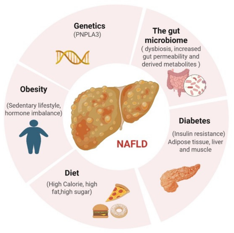
Earlier terminology, non-alcoholic fatty liver disease (NAFLD), described hepatic steatosis in the absence of significant alcohol consumption or other identifiable causes of liver disease. NAFLD represents the most prevalent liver disease worldwide and encompasses a spectrum ranging from simple steatosis to cirrhosis. The major risk factors traditionally associated with NAFLD are illustrated in Figure 1; notably, these risk factors largely overlap with those of MAFLD, with the latter placing greater emphasis on metabolic dysfunction as a defining criterion.
Figure 1. Risk factors for NAFLD.
The global obesity epidemic has been a major driver of the rising incidence and prevalence of fatty liver disease. The adoption of the MAFLD nomenclature reflects a conceptual shift from a diagnosis of exclusion toward one grounded in positive metabolic criteria, with the aim of improving diagnostic precision, enhancing clinical care, legitimizing research practices, and facilitating therapeutic development [2]. Most individuals with MAFLD remain asymptomatic in the early stages, with clinical manifestations often emerging only as the disease progresses to steatohepatitis, fibrosis, cirrhosis, hepatocellular carcinoma (HCC), and ultimately liver failure [6].
In 2023, an alternative terminology, metabolic dysfunction-associated steatotic liver disease (MASLD), was proposed by an international consensus group [7]. MASLD is highly prevalent, particularly in the United States, affecting more than 90% of individuals with obesity, approximately 60% of those with diabetes, and up to 20% of lean individuals [8]. The economic burden of fatty liver disease is substantial: the annual cost in the United States was estimated at $103 billion in 2016, with an additional €35 billion across four major European countries (Germany, France, Italy, and the UK). The projected lifetime cost of care for U.S. patients with NASH alone reached $222 billion in 2017 [9].
According to the World Health Organization, more than 13% of the global population—exceeding 600 million individuals—suffers from obesity, with a particularly high prevalence among children and adolescents [10]. Beyond liver-related morbidity, MAFLD is increasingly recognized as a multisystem disease, with well-documented extrahepatic manifestations including cardiovascular disease (CVD) and chronic kidney disease (CKD) [11,12].
A definitive diagnosis of MAFLD or NASH traditionally requires liver biopsy. However, non-invasive diagnostic modalities, such as magnetic resonance imaging-proton density fat fraction (MRI-PDFF), magnetic resonance spectroscopy (MRS), computed tomography (CT), ultrasound (US) with controlled attenuation parameter (CAP), and elastography, are increasingly being used to assess hepatic steatosis and fibrosis [5]. Despite advances in imaging and biomarker development, accurately distinguishing disease stages and severity remains challenging [13].
NAFLD progresses to NASH when hepatic lipid accumulation triggers hepatocellular injury and inflammation. Although the precise mechanisms underlying disease progression are still being elucidated, established risk factors include obesity, diabetes, hypertension, and dyslipidemia. NASH can ultimately lead to liver fibrosis, cirrhosis, and HCC [14]. The transition from simple steatosis to NASH is typically slow and heterogeneous, often spanning up to 14 years [15]. Approximately 20% of patients exhibit rapid disease progression, likely driven by genetic susceptibility affecting pathways such as intrahepatic lipolysis, triglyceride export, mitochondrial β-oxidation, and glucokinase activity [16].
NASH pathogenesis involves complex intracellular signaling networks mediated by proinflammatory cytokines, immune cell activation, and external influences such as diet, visceral adiposity, and gut microbiota. Activation of hepatic stellate cells and inflammasome signaling pathways promotes fibrogenesis through excessive extracellular matrix deposition [17]. MASLD encompasses a broad disease spectrum, ranging from isolated steatosis to metabolic dysfunction-associated steatohepatitis (MASH), progressive fibrosis, cirrhosis, and HCC [18,19].
To elucidate disease mechanisms and identify diagnostic biomarkers and effective therapeutic strategies, the development and refinement of experimental animal models are of critical importance. It is well-recognized that existing models must be continuously optimized to more faithfully recapitulate the histopathological, metabolic, and pathophysiological features of human disease, thereby enabling progress in mechanistic understanding and therapeutic discovery [20].
Animal models have been instrumental in dissecting the biological processes underlying MAFLD and NASH, including disease initiation, progression, and therapeutic response. Rodents—particularly mice and rats—are widely used due to their genetic tractability, well-characterized metabolism, and experimental flexibility. These models have provided valuable insights into the transition from steatosis to steatohepatitis and fibrosis. Preclinical models that closely mirror human disease are essential for investigating MAFLD phenotypes that cannot be readily studied in humans due to ethical or practical constraints.
In this review, we provide a critical and integrative assessment of commonly used rodent models of MAFLD, with the objective of guiding investigators in selecting appropriate preclinical systems for mechanistic studies and therapeutic evaluation. Broadly, experimental models are categorized as diet-induced, nutrient-deficiency-induced, toxin-induced, genetically engineered, or combination models. Rather than presenting a purely descriptive overview, we emphasize a comparative analysis of model strengths, limitations, and translational relevance across disease stages, thereby enhancing the practical utility of this review for MAFLD research.
Terminology note: The nomenclature surrounding fatty liver disease has evolved substantially over the past decade. Much of the experimental literature referenced in this review predates the adoption of the terms MAFLD and MASLD and therefore uses earlier terminology such as NAFLD and NASH. To maintain clarity and conceptual consistency, we use MAFLD and MASH as the primary terms throughout this review when referring to the disease spectrum, while retaining the original terminology when directly citing historical studies, established model names, or specific dietary formulations.

2. Methods

A comprehensive literature search was conducted using PubMed Central, Ovid MEDLINE, and Google Scholar. Search terms included combinations of “non-alcoholic fatty liver disease”, “NAFLD”, “metabolic dysfunction-associated fatty liver disease”, “MAFLD”, “animal model”, and “rodent model”, with Boolean operators “AND” and “OR” used to refine the search strategy.
Peer-reviewed articles published between January 2005 and June 2025 were considered eligible for inclusion, along with relevant cross-references cited within the selected publications. From the included studies, the following information was extracted: first author, year of publication, journal, study objectives, experimental design, and principal findings. Only studies directly relevant to experimental rodent models of MAFLD/MASH were included in this review.
This review was designed as a narrative, semi-systematic synthesis rather than a formal systematic review. Model inclusion was prioritized based on (i) frequency of use in the literature; (ii) relevance to key stages of MAFLD/MASH progression, including steatosis, inflammation, fibrosis, and hepatocellular carcinoma; (iii) translational relevance to human disease; and (iv) suitability for mechanistic investigation or preclinical therapeutic evaluation. Models with limited reproducibility, insufficient phenotypic characterization, or restricted experimental applicability were deprioritized.
We acknowledge that narrative reviews are inherently subject to selection bias; however, our approach emphasizes integrative comparison and practical guidance for model selection, rather than exhaustive cataloging of all available models.

3. Pathogenesis

The pathogenesis of metabolic dysfunction-associated fatty liver disease (MAFLD) is complex and not yet fully understood. It is primarily driven by aberrant lipid metabolism, insulin resistance (IR), and the accumulation of advanced glycation end products associated with diabetes. These pathological processes likely represent tissue-specific manifestations of shared underlying metabolic disturbances [21].
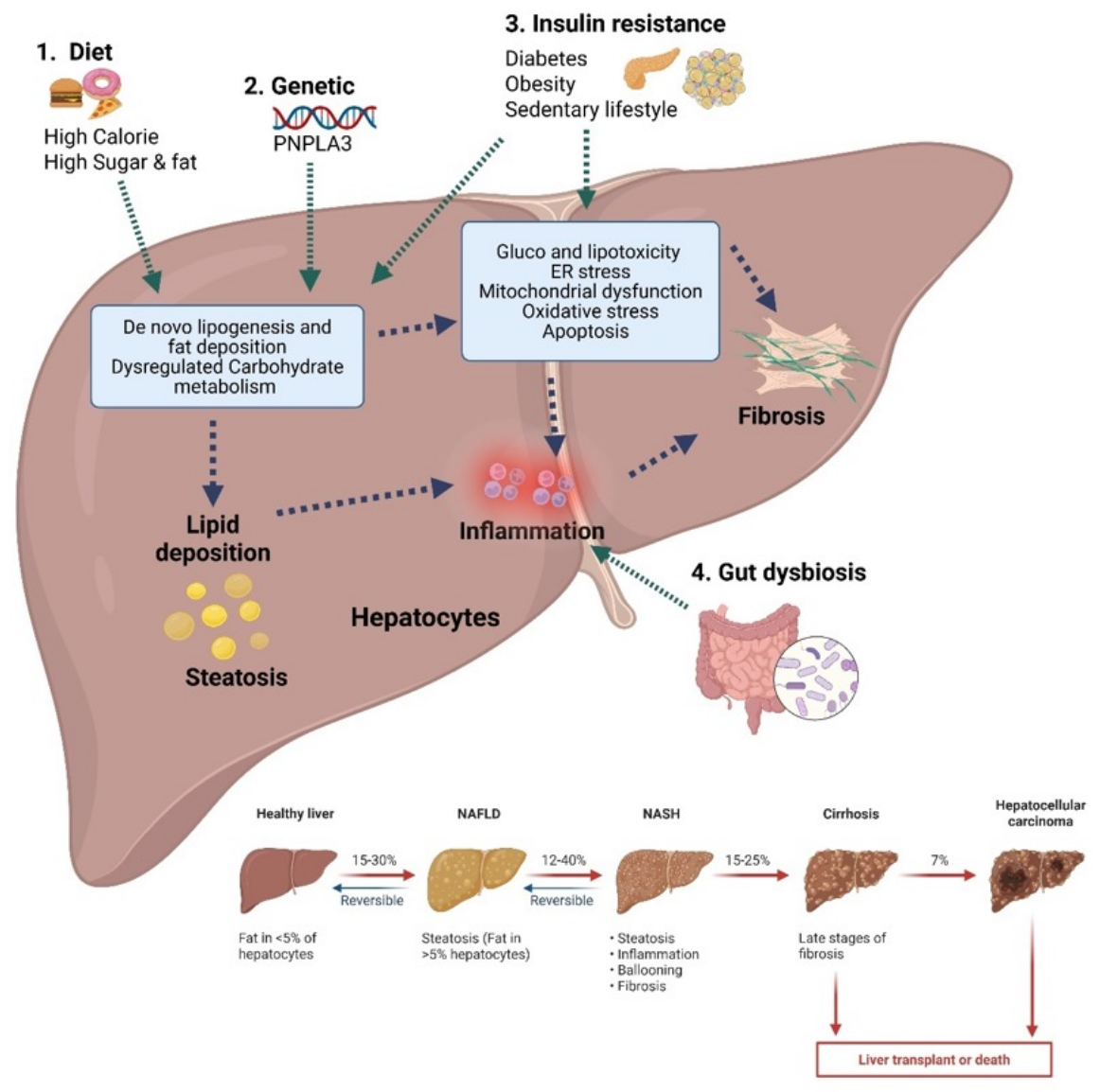
Multiple metabolic, genetic, and environmental risk factors contribute to MAFLD development and disease progression, as illustrated in Figure 2.
Figure 2. Schematic presentation of MAFLD pathogenesis and disease progression.
Earlier concepts of fatty liver disease pathogenesis were based on the “two-hit” hypothesis, in which the first hit involved hepatic lipid accumulation, followed by a second hit characterized by inflammatory cytokine release, adipokine imbalance, mitochondrial dysfunction, and oxidative stress [22]. These sequential insults were thought to drive progression from NAFLD to NASH and ultimately to advanced fibrosis [22].
However, the NAFLD–NASH–hepatocellular carcinoma (HCC) continuum exhibits substantial biological heterogeneity, making it difficult to attribute disease progression to a linear sequence of events. Consequently, the “multiple-hit” hypothesis has emerged, incorporating the combined effects of insulin resistance, lipotoxicity, chronic inflammation, cytokine dysregulation, activation of innate immune pathways, and alterations in the gut microbiota.
High-carbohydrate diets enriched in fructose or sucrose are strongly associated with MAFLD development [23,24]. Sustained hyperglycemia induces endoplasmic reticulum (ER) and mitochondrial stress—collectively termed glucotoxicity. In rodent models, hepatic exposure to elevated levels of glucose, fructose, or sucrose induces insulin resistance [25,26]. This impairment is mediated by reduced insulin receptor expression and/or increased serine phosphorylation of insulin receptor substrate-1 (IRS-1), resulting in defective insulin signaling [27,28]. Chronic persistence of glucotoxicity, as observed in type 2 diabetes mellitus (T2DM), promotes oxidative stress, inflammation, and additional ER stress, thereby exacerbating hepatic injury.
Consistent with these findings, circulating cells from patients with poorly controlled T2DM exhibit evidence of ER stress and reduced nuclear factor erythroid 2-related factor 2 (Nrf2) activity, supporting a role for ER stress in diabetic complications [29]. This chronic low-grade inflammation induced by glucotoxicity contributes to insulin resistance and represents a shared pathogenic mechanism linking T2DM and MAFLD.
Hepatic lipid accumulation in MAFLD arises from increased fatty acid uptake, enhanced de novo lipogenesis, impaired mitochondrial β-oxidation, and reduced lipid export. Fatty acid-binding protein 1 (FABP1), which is predominantly expressed in the liver, facilitates hepatic lipid uptake in concert with fatty acid transport proteins (FATPs) and CD36 [30]. Elevated FATP expression has been positively correlated with hepatic steatosis in patients with MAFLD, although its precise contribution to disease progression remains incompletely defined [31].
De novo lipogenesis in the liver produces three major fatty acids—palmitate, oleate, and palmitoleate—through enzymatic pathways involving acetyl-CoA carboxylase (ACC), fatty acid synthase (FAS), and stearoyl-CoA desaturase-1 (SCD1) [32]. These fatty acids are either stored as triglycerides or exported as very-low-density lipoproteins (VLDLs) [33]. Excessive accumulation of these lipid species leads to hepatic steatosis and hypertriglyceridemia.
The liver plays a central role in systemic metabolic regulation through transcription factors such as sterol regulatory element-binding protein 1c (SREBP-1c), which governs genes involved in glucose and lipid metabolism and contributes to visceral obesity and insulin resistance [34]. Carbohydrate-responsive element-binding protein (ChREBP) is another key regulator, highly expressed in the liver, intestine, and adipose tissue. ChREBP controls lipid metabolism by regulating lipogenic enzymes and hepatokines and has been implicated in T2DM, dyslipidemia, MAFLD, and hepatocarcinogenesis [35].
Impaired mitochondrial fatty acid β-oxidation in MAFLD further contributes to disease progression by generating excess reactive oxygen species (ROS), leading to mitochondrial dysfunction, reduced insulin sensitivity, and chronic inflammation [36].
Hepatic lipid export represents another critical regulatory pathway. Apolipoprotein B100 (apoB100) and microsomal triglyceride transfer protein (MTTP) are essential for VLDL assembly and secretion. Increased dietary fat intake elevates the hepatic fatty acid levels, inducing ER stress that suppresses apoB100 secretion and exacerbates steatosis [37]. Because apoB100 synthesis is a rate-limiting step in lipid export, its insufficiency promotes lipid retention, lipotoxicity, and progression toward NASH [38].
Disruption of the gut microbiota—commonly associated with fatty liver disease, inflammatory bowel disease, obesity, T2DM, and certain malignancies—also contributes to MAFLD pathogenesis. Gut dysbiosis alters lipid and glucose metabolism and promotes hepatic inflammation. Microbial products such as lipopolysaccharides (LPSs) can activate hepatic inflammatory pathways and disrupt bile acid homeostasis, leading to increased hepatic lipid accumulation [39,40]. These observations highlight the importance of the gut–liver axis in modulating immune and metabolic responses in MAFLD.
Finally, genetic susceptibility plays a significant role in MAFLD development and progression. Polymorphisms in genes such as PNPLA3 (promoting hepatic lipid accumulation), TM6SF2 (impairing VLDL secretion), and MBOAT7 (enhancing steatosis and fibrosis) are strongly associated with increased MAFLD risk [41]. Together, genetic and epigenetic factors modulate disease severity and interindividual variability.
Importantly, the diverse pathogenic pathways implicated in MAFLD are differentially represented across experimental animal models. Diet-induced models incorporating high fat, fructose, and cholesterol best reproduce systemic insulin resistance, lipotoxicity, and gut–liver axis dysregulation. In contrast, nutrient-deficient models [e.g., methionine- and choline-deficient (MCD) and choline-deficient, L-amino acid-defined (CDAA)] preferentially recapitulate oxidative stress, inflammation, and fibrosis but fail to reflect metabolic syndrome. Genetic models enable the targeted interrogation of lipid-handling and insulin-signaling pathways, whereas chemical and combination models accelerate fibrogenesis and hepatocarcinogenesis. These mechanistic distinctions provide a conceptual framework for the rational selection of experimental models based on specific disease processes and research objectives.

4. Animal Models of Liver Disease

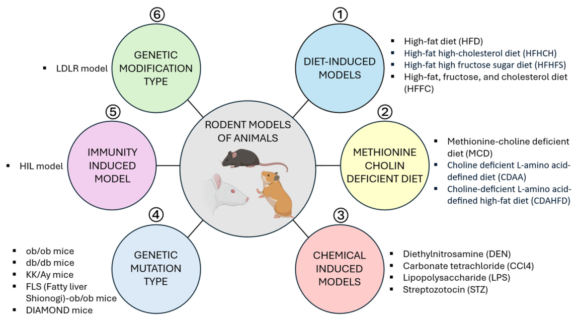
Several experimental models have been developed to understand the etiological, biological, pathological, and metabolic processes that drive MAFLD and its progression to MASH, fibrosis, and hepatocellular carcinoma (HCC). These models are also extremely valuable for mechanistic studies and the preclinical evaluation of therapeutic candidates. Commonly used rodent models and induction strategies are presented in Figure 3.
Figure 3. Overview of approaches used to generate rodent models for MAFLD/MASH studies.
Among the rodent models, mice are particularly widely used because of their genetic tractability and the availability of well-characterized dietary, chemical, and transgenic approaches. Throughout this section, we use MAFLD/MASH terminology for conceptual consistency while retaining legacy NAFLD/NASH terminology when it appears in established model names or cited studies.

4.1. Diet-Induced Models

Diet-induced models are widely used to study MAFLD and MASH, utilizing dietary regimens that differ in fat type and percentage, cholesterol content, and carbohydrate source (e.g., fructose/sucrose). In general, obesogenic diets best reproduce metabolic syndrome and insulin resistance, whereas nutrient-deficiency diets accelerate steatohepatitis and fibrosis but often lack systemic metabolic features. Accordingly, diet choice should reflect the targeted disease stage and research objective (e.g., metabolic dysfunction, fibrosis progression, or drug efficacy testing).

4.1.1. High-Fat Diet (HFD) or Diet-Induced Obesity (DIO) Models

The most commonly employed DIO models involve feeding rodents a diet in which fat constitutes ≥45–60% of the total caloric intake, typically for ≥16 weeks. These diets reliably induce obesity, insulin resistance, and hepatic steatosis; however, inflammation and fibrosis are often mild and develop slowly, limiting their utility for modeling advanced MASH. With prolonged feeding (up to ~60 weeks), some models may develop progressive fibrosis, and in certain settings, HCC, but with substantial time and variability.
To enhance disease severity and reproducibility, researchers have modified dietary compositions to include cholesterol and refined sugars (fructose/sucrose/glucose), and historically, trans fats. Because diet formulations vary considerably across studies, careful reporting of macronutrient composition and units (preferably % kcal) is essential for comparability.
Several modified “Western-style” diets have been developed to better replicate human disease phenotypes:
  • Western diet (WD): typically, ~40% kcal from fat, with variable carbohydrate and cholesterol content.
  • American lifestyle-induced obesity syndrome (ALIOS) diet: 45% kcal fat with 2% trans fats, often combined with fructose in drinking water.
  • Amylin liver NASH (AMLN) diet: ~40% kcal fat (historically including 18% trans fats), 20% fructose, and 2% cholesterol.
  • Gubra-Amylin NASH (GAN) diet: a trans fat–free version of AMLN (often using palm oil), developed following restrictions on trans fats [42].
The GAN DIO-NASH model has shown strong translational relevance, reproducing histopathological, transcriptional, and metabolic signatures observed in human MASH. Consequently, GAN and AMLN-type diets are widely used in translational research and interventional studies, particularly for therapeutic target identification and preclinical drug testing [43]. Diets combining high fat and fructose are considered particularly relevant to human MASH pathophysiology [44]. Importantly, fructose co-administered with glucose may be more hepatotoxic than fructose alone, and sucrose can be a stronger inducer of hepatic inflammation, underscoring the importance of carbohydrate composition in model design.
The C57BL/6 mouse strain is frequently used due to its susceptibility to metabolic disease. In practice, diets containing ≥45% kcal fat and added cholesterol (commonly ≥0.5%) are more likely to induce steatohepatitis and fibrosis than fat alone [45]. Fructose also contributes to hepatic lipogenesis through gut dysbiosis and endotoxemia (via LPS), exacerbating liver inflammation and advancing MAFLD/MASH [46]. Long-term HFD feeding induces obesity, hyperinsulinemia, hypercholesterolemia, and steatohepatitis in mice, often following the onset of metabolic abnormalities [47]. Diets rich in saturated fatty acids (≥12%), cholesterol (2%), and fructose reproduce multiple aspects of human metabolic syndrome and liver fibrosis, providing a relevant model for progressive disease [48].
HFD-based models are essential tools for studying metabolic dysfunction and early-stage disease, as mice fed HFDs consistently develop characteristics of metabolic syndrome, including visceral adiposity, hyperlipidemia, and insulin resistance, along with variable degrees of hepatic inflammation and fibrosis [49,50]. In rat models, male Lewis, Wistar, and Sprague-Dawley (SD) rats fed an HFD for 3 weeks developed grade 3 hepatic steatosis (>66% of hepatocytes affected). Notably, strain differences can influence phenotype: Lewis rats exhibited microvesicular steatosis, hepatocellular injury, and fibrosis, whereas male SD rats showed pronounced steatosis, supporting their use for late-stage steatosis-focused studies [49]. In another study, male MS-NASH mice maintained on an HFD for 16 weeks developed increased body and liver weight, elevated serum aminotransferases, and histological evidence of macrovesicular steatosis, ballooning, lobular inflammation, and collagen deposition, closely mimicking features of human MASH [51].

4.1.2. High-Fat and High-Cholesterol Diet (HFHCh)

Dietary cholesterol plays a critical role in progression from steatosis to steatohepatitis, often acting synergistically with saturated fats to exacerbate hepatic injury. Diets enriched in both fat and cholesterol contribute to lipid dysregulation and hepatic triglyceride accumulation, thereby triggering hepatic inflammation and fibrogenesis and driving progression to MASH [45,52].
Several preclinical models have employed HFHCh diets to investigate disease pathogenesis and therapeutic interventions. One model utilized male Golden Syrian hamsters fed an HFHCh diet supplemented with 10% fructose in drinking water for 10–20 weeks. This model developed hallmark features of MASH and cardiac dysfunction, characterized through biochemical profiling, histology, and echocardiography. Administration of Elafibranor (15 mg/kg/day orally for five weeks) led to the resolution of steatohepatitis and improvement in diastolic function, demonstrating the model’s utility for evaluating cardiometabolic therapies [53].
Another widely used model involves Sprague-Dawley rats fed an HFHCh diet for up to 20 weeks, with serial sacrifice to assess disease progression. This model exhibited steatosis and inflammation beginning around weeks 4–20, with fibrosis emerging between weeks 12–20. Notably, elevated lipopolysaccharide (LPS) levels were detected after 12 weeks, implicating gut–liver axis involvement [54]. Numerous studies have shown that adding cholesterol (0.5–10%) to high-fat diets accelerates disease progression by enhancing steatosis, lobular inflammation, and hepatic fibrosis. For instance, Sprague-Dawley rats fed 2.5% cholesterol developed histological features of MASH and progressed to cirrhosis [55].
In a long-term study, an HFD composed of 88 g standard chow supplemented with 10 g lard oil and 2 g cholesterol was administered to male Sprague-Dawley rats for 48 weeks. This regimen resulted in perisinusoidal fibrosis by 24 weeks, hepatic fibrosis by 36 weeks, and severe fibrosis by 48 weeks. This stepwise trajectory closely parallels key features of human MASH progression, supporting its use for investigating mechanisms and anti-fibrotic therapies [56].

4.1.3. High-Fat, High-Fructose Diet (HFFD)

The HFFD model is widely used to study dietary drivers of MAFLD/MASH, typically incorporating fructose (commonly 10% in drinking water) alongside a high-fat diet to mimic Western dietary patterns. Excessive dietary fat and fructose promote hepatic steatosis, insulin resistance, and ER stress via multiple mechanisms, including FFA-induced lipotoxicity and the induction of proinflammatory mediators such as TNF-α, IL-6, and MCP-1 [57,58,59]. Fructose is strongly linked to oxidative stress and fibrogenesis and is considered a key dietary driver of progression beyond simple steatosis.
Experimental studies demonstrate dose- and time-dependent relationships between sugar exposure and disease severity. In Wistar rats, a 30% sucrose solution induced grade 2 steatosis within 1–6 weeks and progressed to grade 3 by 20 weeks. Higher sucrose concentrations (40–50%) over 20 weeks produced more severe NASH-like phenotypes with fibrosis [59]. These findings underscore that both dose and duration are major determinants of phenotype in sugar-driven models.
Golden Syrian hamsters fed a high-fat, high-fructose, high-cholesterol diet (HFFCD) for six weeks developed metabolic disturbances, including increased body weight, visceral adiposity, and extensive hepatic lipid accumulation. This model is well-suited to studies of early metabolic dysfunction and lipid dysregulation, as well as therapeutic testing targeting metabolic pathways [44,60].

4.1.4. High-Fat, High-Fructose, and High-Cholesterol (HFFC) Diet

The high-fat, high-fructose, and high-cholesterol (HFFC) diet is commonly used to establish animal models of metabolic syndrome [61]. This diet typically contains 40% fat, 20% fructose, and 2% cholesterol. Mice fed the HFFC diet for 34–36 weeks exhibited increased hepatic triglyceride accumulation, collagen deposition, and elevated expression of fibrosis-related genes (e.g., Col1a1, Acta2/α-SMA, and Loxl2) [61,62]. Therefore, HFFC-type diets are particularly useful for modeling progressive disease with fibrosis in the context of metabolic dysfunction.

4.1.5. Methionine- and Choline-Deficient (MCD) Diet

The MCD diet is a well-established model for inducing steatohepatitis in rodents and produces severe steatosis, inflammation, and fibrosis within approximately 4–8 weeks. The pathology arises due to impaired β-oxidation and lipoprotein synthesis, pathways in which methionine and choline are essential. Because the methionine cycle is critical for methylation and redox homeostasis, disruption of this pathway contributes to oxidative stress and liver injury [63].
Rodents fed the MCD diet exhibit reduced body weight and decreased fasting glucose and insulin levels, alongside steatosis, steatohepatitis, and fibrosis [64]. In Sprague-Dawley rats, the diet induces steatosis, lobular inflammation, and mild perisinusoidal fibrosis. A key limitation is poor metabolic fidelity, as the model does not reproduce obesity or systemic insulin resistance, and animals frequently lose weight due to reduced caloric intake [64]. Attempts to mitigate weight loss by combining MCD with HFD have generally been unsuccessful.
In mice, steatosis and inflammation can emerge within two weeks, followed by periportal fibrosis by ~10 weeks and bridging fibrosis by ~16 weeks [65]. Species, strain, and sex significantly influence the outcomes. Among the Wistar, Long-Evans, and Sprague-Dawley rats and C57BL/6 mice fed MCD for four weeks, male Wistar rats showed pronounced steatosis, whereas male C57BL/6 mice developed greater inflammation, necrosis, lipid peroxidation, and ultrastructural injury [66]. Despite its limitations, the MCD diet remains useful for mechanistic studies focusing on oxidative stress, inflammation, and fibrogenesis, but its metabolic divergence from human disease must be considered.
Additionally, although the MCD model has been used to explore gut microbiota and immune responses, the intestinal microbiota alterations induced by MCD feeding do not closely match human MAFLD-associated dysbiosis, limiting translational interpretation [67].

4.1.6. Choline-Deficient, L-Amino Acid-Defined (CDAA) Diet

The CDAA diet is widely used in pharmacological and genetic research. In this semisynthetic diet, protein is replaced with an L-amino acid mixture, and the methionine content is typically normal or only slightly reduced [68]. In female Fischer 344 rats fed CD versus CDAA diets for up to 12 weeks, CDAA induced more diffuse steatosis, increased hepatocyte death and proliferation, and greater disease severity, with potential progression toward HCC [68]. Male Wistar rats fed CDAA for 24 weeks developed cirrhosis, progressing to HCC in 54.6% by 48 weeks [69].
Feeding mice and rats a CDAA diet (e.g., 0.17% methionine) for 12 weeks produces severe steatosis with modest steatohepatitis and variable fibrosis; continued feeding can lead to progressive fibrosis and liver cancer, supporting its utility for studying transition from steatohepatitis to HCC [61,64,70].

4.1.7. Choline-Deficient, L-Amino Acid-Defined, High-Fat (CDAHFD) Diet

The CDAHFD model recapitulates key histopathological features of human MASH by combining high fat with reduced methionine. Male C57BL/6J mice fed CDAHFD (60% kcal fat, 0.1% methionine) gain weight rapidly and develop hepatic steatosis early, with hepatomegaly and significant fibrosis by approximately six weeks [71]. This model offers a practical balance between induction speed and fibrotic severity, making it useful for fibrosis-focused studies and therapeutic testing. Short-term feeding (e.g., one week) can induce steatohepatitis with mitochondrial dysfunction and severe oxidative stress without established fibrosis [72], highlighting the importance of induction duration for matching the desired disease stage.

4.2. Chemically Induced Models

Several chemicals, including diethylnitrosamine (DEN), carbon tetrachloride (CCl4), sodium nitrite, LPS, and streptozotocin (STZ), have been employed to develop experimental models of advanced MASH, fibrosis, and HCC, often by accelerating inflammatory and fibrotic pathways.

4.2.1. Diethylnitrosamine (DEN)

DEN is a potent hepatocarcinogen capable of inducing oxidative stress and DNA damage and has long been used to establish HCC models, often in conjunction with fibrogenic injury [73,74]. Dietary factors strongly influence DEN-induced tumorigenesis; for example, an HFD combined with DEN exposure impairs liver function, with DEN acting as the major driver of tumor formation. This approach provides mechanistic insight into how metabolic dysfunction can exacerbate hepatocarcinogenesis [74].
In a DEN + high-energy diet (HED) model, lipid metabolite transport to the liver activates macrophages and promotes inflammatory responses that aggravate HCC progression. C3H mice fed a diet supplemented with 5% shortening, 5% lard, and 1% cholesterol and given DEN in drinking water (30 mg/mL) for 22 weeks developed tumors with 100% incidence (survival rate 46.15%) [75].

4.2.2. Carbon Tetrachloride (CCl4)

CCl4 is a highly toxic agent that induces hepatic injury and fibrosis and is among the most widely used chemicals for experimental liver fibrosis [76,77]. When combined with obesogenic diets, CCl4 markedly accelerates progression to advanced fibrosis and HCC, improving feasibility for studying later disease stages.
In one combined dietary–chemical model, nine-week-old C57BL/6 mice were maintained on normal chow or WD with weekly intraperitoneal CCl4 for 12–24 weeks. The WD contained 21.1% fat, 41% sucrose, and 1.25% cholesterol (by weight), with a high-sugar solution (fructose and glucose). CCl4 was administered at 0.2 µL/g body weight once weekly. The combination of WD + CCl4 significantly worsened steatohepatitis, fibrosis, and tumor development [78].
Similarly, WD combined with weekly CCl4 produced F3 fibrosis within 8–12 weeks and HCC by ~24 weeks, shortening latency and strengthening utility for preclinical evaluation. In MS-NASH mice, a Western diet with fructose (WDF) combined with CCl4 (low or high dosing regimens) promoted fibrosis progression [79].

4.2.3. Sodium Nitrite (NaNO2)

Sodium nitrite has been employed to accelerate the progression of MAFLD to MASH with fibrosis. In Wistar rats, a choline-deficient high-fat diet (CDHFD) combined with intraperitoneal NaNO2 (25 mg/kg, three times weekly from week 4) induced marked steatosis and fibrosis by week 10. This model has been used for antifibrotic testing, including cilofexor and propranolol, both of which reduced fibrosis [80].

4.2.4. Lipopolysaccharides (LPS)

LPS, a Gram-negative bacterial endotoxin, has been used as a “second hit” to amplify hepatic inflammation. In C57BL/6 mice fed HFD for 12 weeks, intraperitoneal LPS (10 mg/kg) exacerbated steatosis, inflammation, and lobular necrosis, highlighting the contribution of intestinal permeability and metabolic endotoxemia to MASH pathogenesis [81].

4.2.5. Streptozotocin (STZ) Models (STAM)

STZ selectively damages pancreatic β-cells. When combined with high-fat feeding, low-dose STZ produces models that mimic diabetes-associated MASH and HCC, but the underlying mechanism can differ from human MAFLD because STZ induces insulin deficiency rather than systemic insulin resistance.
The STAM model is generated by administering STZ (200 µg, subcutaneous) to 2-day-old male C57BL/6 mice, followed by high-fat feeding from four weeks. Animals develop steatosis, MASH by ~5 weeks, fibrosis by ~10 weeks, and spontaneous HCC thereafter [82]. This model is particularly useful for studying MASH-to-HCC progression, while acknowledging mechanistic differences relative to human disease.

4.3. Genetically Modified Models

4.3.1. Leptin-Related Models (ob/ob and db/db)

Genetic alterations in leptin signaling provide classic models of obesity and diabetes.
  • ob/ob mice carry a mutation preventing leptin synthesis and develop severe obesity and insulin resistance but typically show limited spontaneous hepatic inflammation and fibrosis.
  • db/db mice lack functional leptin receptors and develop obesity, hyperglycemia, and liver injury, with phenotype severity influenced by genetic background (e.g., C57BL/6 vs. C57BLKS/J) [83].
These models are especially valuable for studying metabolic drivers of steatosis and insulin resistance; however, additional dietary or chemical insults are often required to induce robust fibrosis. A spontaneous mutation in the leptin receptor (Lepr) produces severe obesity, hyperphagia, and polydipsia, with the C57BLKS/J background exhibiting more severe phenotypes [83].

4.3.2. KK-Ay Mice

KK-Ay mice, generated by crossing diabetic KK mice with heterozygous agouti (Ay) mice, are widely used to model obesity and type 2 diabetes. When fed a NAFLD diet (40% kcal fat, 20% kcal fructose, 2% cholesterol, 0.5% cholic acid), KK-Ay mice rapidly develop obesity, insulin resistance, dyslipidemia, and steatohepatitis within 4 weeks, progressing to fibrosis by 12 weeks [84]. This model is useful for metabolic syndrome-driven MASH with relatively rapid fibrosis development.

4.3.3. Fatty Liver Shionogi (FLS) Mice

FLS mice develop genetic fatty liver and progressive steatosis without obesity or diabetes. FLS-ob/ob mice combine leptin deficiency with hepatic lipid accumulation, resulting in more severe steatohepatitis. FLS-ob/ob mice show steatosis at ~12 weeks and oxidative stress with advanced fibrosis by 24–36 weeks, progressing to cirrhosis by ~48 weeks [85]. Thus, FLS-derived strains can be used to study progressive steatohepatitis and fibrosis with defined genetic susceptibility.

4.3.4. Diamond Mice

The Diamond mouse model (C57BL/6J × 129S1/SvImJ) is used to study progression to MASH and HCC. Mice fed a high-fat diet with access to glucose and fructose develop steatosis within 4–8 weeks, steatohepatitis by 16–24 weeks, fibrosis thereafter, and spontaneous HCC by ~48 weeks [86]. This model is highly valuable for translational research because it reproduces metabolic, histological, and transcriptomic features relevant to human disease.

4.4. Immunity-Induced Models

Humanized immune system models have been developed by engrafting human immune cells into immunodeficient mice (HIL mice). When fed a high-fat, high-calorie diet for ~20 weeks, these mice exhibit steatosis, inflammation, and fibrosis, enabling the investigation of immune contributions to disease and the preclinical testing of immunomodulatory therapies [87].

4.5. Genetically Modified or Gene-Edited Models

The LDL receptor knockout (LDLR/) mouse is commonly used to study dyslipidemia and atherosclerosis but can develop MASH-like features under dietary stress. When fed a modified CDAA diet containing 1% cholesterol and 41% fat, LDLR/ mice develop steatosis within one week and progress to inflammation and fibrosis by ~8 weeks. By 24–39 weeks, animals may develop hepatic hyperplasia and hepatocellular adenomas/carcinomas [88]. Accordingly, this model is useful for studying rapid progression to advanced disease in a dyslipidemic context.
Integrative model comparison
While individual rodent models reproduce selected aspects of MAFLD/MASH, no single model fully captures the entire human disease spectrum. Obesogenic diet-induced models (e.g., GAN, AMLN-type diets) provide the greatest translational relevance for metabolic dysfunction and therapeutic evaluation. Nutrient-deficient diets induce steatohepatitis and fibrosis rapidly but lack systemic metabolic fidelity. Genetic models are best suited for the mechanistic dissection of defined pathways, whereas combination models (diet + toxin/chemical) are most effective for studying advanced fibrosis and hepatocellular carcinoma within feasible experimental timeframes. Accordingly, model selection should be guided by disease stage, dominant pathogenic pathways, and experimental feasibility rather than nomenclature alone.
We summarize commonly used rodent models in Table 1 and provide a decision-aiding guide for model selection in Table 2.
Table 1. Summary of commonly used rodent models in MAFLD/MASH studies.
Table 2. Decision-aiding guide for selecting models by research objective.

5. Concluding Remarks

Although multiple rodent models of MAFLD have enhanced the understanding of disease mechanisms, no single model fully captures the heterogeneity and complexity of human MAFLD/MASH. Ideally, models should recapitulate (i) systemic metabolic dysfunction (e.g., obesity and insulin resistance), (ii) liver histopathology (steatosis, inflammation, ballooning, fibrosis), and (iii) clinically relevant progression to cirrhosis and hepatocellular carcinoma (HCC). Accordingly, model selection should be guided by the specific disease stage and research objective rather than by terminology alone.
Key considerations for model selection include the following:
  • Diet-induced models: High-fat, high-sugar, and cholesterol-enriched diets best mirror human lifestyle-related risk factors and are particularly useful for studying metabolic syndrome-associated MAFLD/MASH and for therapeutic evaluation [44,45].
  • Nutrient-deficiency diets (MCD and CDAA/CDAHFD): These diets induce rapid steatohepatitis and fibrosis but often fail to reproduce systemic metabolic abnormalities observed in human MAFLD/MASH (e.g., obesity and insulin resistance) [89].
  • Chemical “second-hit” models (e.g., CCl4, DEN, STZ, LPS): These approaches are valuable when combined with dietary stressors to accelerate fibrosis and/or HCC development, improving feasibility for late-stage studies and drug testing [78,90].
  • Humanized and immune-focused models: Humanized mice provide improved translational relevance for studying immune mechanisms, biomarker discovery, and immunomodulatory therapeutics [87,91,92].
  • Species and strain differences: Alternative species such as hamsters, which express CETP and exhibit a lipoprotein metabolism more similar to humans, may offer improved metabolic fidelity compared with standard mouse and rat strains [93,94,95,96]. Within mice, substrain differences (e.g., C57BL/6J vs. C57BL/6N) should be explicitly considered when comparing outcomes and reproducibility.
Present Status of Rodent Models
Current experimental rodent models of MAFLD/MASH rely predominantly on diet-based induction (high fat with added fructose and/or cholesterol), genetic susceptibility, chemical acceleration, or combinations. These models have been instrumental for studying early-stage steatosis, obesity, and metabolic syndrome; however, many fail to fully reproduce progressive fibrosis and hepatocellular carcinoma within practical experimental timeframes. Therefore, selecting an appropriate model based on the study aims remains critical for generating interpretable and translatable data.
At present, key focus areas in MAFLD/MASH modeling include:
(a) Choice of species and strain.
Mice and rats remain the preferred laboratory species due to low cost, short lifespan, and the availability of genetic tools. Susceptibility varies across strains; for example, C57BL/6 and A/J strains are often more susceptible than BALB/c to diet-induced metabolic dysfunction, and C57BL/6 mice are widely used due to reproducibility under high-fat/high-fructose diets with added cholesterol [64,97]. Importantly, substrain differences (C57BL/6J vs. C57BL/6N) can influence metabolic and inflammatory phenotypes and should be reported explicitly.
Wistar and Sprague-Dawley rats are commonly used, whereas Fischer 344 (F344) rats often require specific dietary challenges (e.g., CDAA diets) to develop MAFLD-like pathology. Zucker fatty rats model obesity and insulin resistance but typically require additional dietary or chemical “hits” to induce advanced steatohepatitis and fibrosis. Zucker diabetic fatty (ZDF) rats are useful for studying MAFLD in the context of type 2 diabetes [98,99].
Golden Syrian hamsters are increasingly used because their lipoprotein metabolism is closer to humans than that of mice or rats, including CETP activity, which supports translational studies of dyslipidemia and cardiometabolic disease [93,94,95,96].
(b) Dietary considerations and clinical relevance.
Diet-based approaches are increasingly emphasized because they reflect human metabolic dysregulation. High-fat, high-sugar, and cholesterol-rich diets mimic key risk factors and are generally preferred for translational studies [44,45]. For example, Western-style diets enriched in saturated fat, cholesterol, and sugars are often favored over MCD diets because MCD diets are misaligned with human dietary patterns and induce weight loss rather than obesity. Although MCD diets rapidly induce steatohepatitis and fibrosis, they do not induce metabolic syndrome and therefore have limited clinical relevance. High-fat, high-fructose (HFHF) diets reliably promote obesity and insulin resistance but may yield less severe hepatic pathology unless combined with cholesterol or longer feeding durations [64,65].
(c) Rapid progression models for advanced fibrosis and HCC.
To model advanced disease stages such as bridging fibrosis, cirrhosis, portal hypertension, and HCC within feasible timelines, investigators are increasingly using combination models. Western diet plus low-dose CCl4 accelerates fibrosis and can promote HCC development, while STZ combined with high-fat feeding can model diabetes-associated MASH progression and HCC [78,79,90]. These models are particularly useful for the preclinical testing of antifibrotic and anticancer therapeutics while recognizing that chemical injury introduces non-physiological components.
(d) Genetically predisposed and genetically modified models.
Genetic susceptibility influences disease severity, and certain strains (e.g., C57BL/6) demonstrate greater injury under cholesterol-enriched Western diets, supporting their use in progressive disease modeling [100]. Gene-edited or transgenic models enable the mechanistic interrogation of defined pathways. For example, A-ZIP/F-1 mice develop severe lipoatrophic diabetes and exhibit marked hepatic lipid accumulation [101]. Classic leptin pathway models (ob/ob and db/db) reproduce obesity and insulin resistance but frequently require additional dietary or inflammatory “hits” to develop robust steatohepatitis and fibrosis, highlighting an important distinction from human MAFLD/MASH [61].
Overall, a central challenge remains the lack of standardization across models, including variability in diet formulations, induction methods, animal strains, and outcome measures. Some models reproduce histology well, while others capture metabolic dysfunction more faithfully, and only a subset reflects both domains, contributing to limited reproducibility and incomplete clinical translation [61,97,102]. Consequently, the prevailing trend is toward clinically relevant diet-induced and combination models that incorporate key elements of human disease (e.g., fat, fructose, cholesterol, and metabolic dysfunction) while enabling the progression to fibrosis and HCC within practical timeframes.
Future Perspectives
Future development of MAFLD/MASH rodent models should be guided by defined experimental use cases rather than phenotypic breadth alone. Diet-induced models remain optimal for studying disease initiation, metabolic dysregulation, and early-stage steatosis, whereas combination and genetic models are often best suited for modeling progressive fibrosis, cirrhosis, and hepatocellular carcinoma. Humanized and gene-edited models are expected to play an increasing role in translational studies, immune mechanisms, and biomarker discovery. Multi-omics approaches and advanced in vitro systems (e.g., organoids and microphysiological platforms) will complement in vivo models by enabling mechanistic precision and scalable drug screening.
A key practical direction is the continued shift away from models that induce steatohepatitis without metabolic syndrome (e.g., MCD-only diets) toward more clinically relevant diet-based and combination strategies that better capture the metabolic and pathological complexity of human disease. Ultimately, aligning model choice with the study goal—mechanistic discovery, disease-stage modeling, or therapeutic evaluation—will be essential for improving reproducibility and translational success.

Author Contributions

Conceptualization, K.K.B.; methodology, K.K.B.; software, M.P.S.; validation, K.K.B. and M.P.S.; investigation, K.K.B.; resources, M.P.S.; writing—original draft preparation, K.K.B.; writing—review and editing, K.K.B. and M.P.S.; visualization, K.K.B. All authors have read and agreed to the published version of the manuscript.

Funding

This research received no external funding.

Institutional Review Board Statement

Not applicable.

Data Availability Statement

No new data were created or analyzed in this study.

Acknowledgments

The authors are grateful to Ichiaki Ito, Anderson Cancer Center, for his time and thoughtful review of the manuscript. The figures were created using Biorender.com.

Conflicts of Interest

The authors declare no conflicts of interest.

References

  1. Eslam, M.; Sanyal, A.J.; George, J.; Sanyal, A.; Neuschwander-Tetri, B.; Tiribelli, C.; Kleiner, D.E.; Brunt, E.; Bugianesi, E.; Yki-Järvinen, H.; et al. MAFLD: A Consensus-Driven Proposed Nomenclature for Metabolic Associated Fatty Liver Disease. Gastroenterology 2020, 158, 1999–2014.e1. [Google Scholar] [CrossRef]
  2. Eslam, M.; Newsome, P.N.; Sarin, S.K.; Anstee, Q.M.; Targher, G.; Romero-Gomez, M.; Zelber-Sagi, S.; Wong, V.W.-S.; Dufour, J.-F.; Schattenberg, J.M.; et al. A new definition for metabolic dysfunction-associated fatty liver disease: An international expert consensus statement. J. Hepatol. 2020, 73, 202–209. [Google Scholar] [CrossRef]
  3. Gofton, C.; Upendran, Y.; Zheng, M.-H.; George, J. MAFLD: How is it different from NAFLD? Clin. Mol. Hepatol. 2023, 29, S17–S31. [Google Scholar] [CrossRef]
  4. Finucane, M.M.; Stevens, G.A.; Cowan, M.J.; Danaei, G.; Lin, J.K.; Paciorek, C.J.; Singh, G.M.; Gutierrez, H.R.; Lu, Y.; Bahalim, A.N.; et al. National, regional, and global trends in body-mass index since 1980: Systematic analysis of health examination surveys and epidemiological studies with 960 country-years and 9·1 million participants. Lancet 2011, 377, 557–567. [Google Scholar] [CrossRef]
  5. Bhopale, K.K.; Srinivasan, M.P. Therapeutics for Metabolic Dysfunction-Associated Fatty Liver Disease (MAFLD). Livers 2023, 3, 597–617. [Google Scholar] [CrossRef]
  6. Spengler, E.K.; Loomba, R. Recommendations for Diagnosis, Referral for Liver Biopsy, and Treatment of Nonalcoholic Fatty Liver Disease and Nonalcoholic Steatohepatitis. Mayo Clin. Proc. 2015, 90, 1233–1246. [Google Scholar] [CrossRef] [PubMed]
  7. Rinella, M.E.; Lazarus, J.V.; Ratziu, V.; Francque, S.M.; Sanyal, A.J.; Kanwal, F.; Romero, D.; Abdelmalek, M.F.; Anstee, Q.M.; Arab, J.P.; et al. A multisociety Delphi consensus statement on new fatty liver disease nomenclature. Hepatology 2023, 78, 1966–1986. [Google Scholar] [CrossRef]
  8. Younossi, Z.M.; Blissett, D.; Blissett, R.; Henry, L.; Stepanova, M.; Younossi, Y.; Racila, A.; Hunt, S.; Beckerman, R. The economic and clinical burden of nonalcoholic fatty liver disease in the United States and Europe. Hepatology 2016, 64, 1577–1586. [Google Scholar] [CrossRef] [PubMed]
  9. Allen, A.M.; Lazarus, J.V.; Younossi, Z.M. Healthcare and socioeconomic costs of NAFLD: A global framework to navigate the uncertainties. J. Hepatol. 2023, 79, 209–217. [Google Scholar] [CrossRef]
  10. Okunogbe, A.; Nugent, R.; Spencer, G.; Powis, J.; Ralston, J.; Wilding, J. Economic impacts of overweight and obesity: Current and future estimates for 161 countries. BMJ Glob. Health 2022, 7, e009773. [Google Scholar] [CrossRef]
  11. Mantovani, A.; Petracca, G.; Beatrice, G.; Csermely, A.; Lonardo, A.; Schattenberg, J.M.; Tilg, H.; Byrne, C.D.; Targher, G. Non-alcoholic fatty liver disease and risk of incident chronic kidney disease: An updated meta-analysis. Gut 2022, 71, 156–162. [Google Scholar] [CrossRef]
  12. Niederseer, D.; Wernly, B.; Aigner, E.; Stickel, F.; Datz, C. NAFLD and Cardiovascular Diseases: Epidemiological, Mechanistic and Therapeutic Considerations. J. Clin. Med. 2021, 10, 467. [Google Scholar] [CrossRef]
  13. Piazzolla, V.A.; Mangia, A. Noninvasive Diagnosis of NAFLD and NASH. Cells 2020, 9, 1005. [Google Scholar] [CrossRef]
  14. Heinemann, F.; Gross, P.; Zeveleva, S.; Qian, H.S.; Hill, J.; Hofer, A.; Jonigk, D.; Diehl, A.M.; Abdelmalek, M.; Lenter, M.C.; et al. Deep learning-based quantification of NAFLD/NASH progression in human liver biopsies. Sci. Rep. 2022, 12, 19236. [Google Scholar] [CrossRef] [PubMed]
  15. Singh, S.; Allen, A.M.; Wang, Z.; Prokop, L.J.; Murad, M.H.; Loomba, R. Fibrosis progression in nonalcoholic fatty liver vs nonalcoholic steatohepatitis: A systematic review and meta-analysis of paired-biopsy studies. Clin. Gastroenterol. Hepatol. 2015, 13, 643–654.e9. [Google Scholar] [CrossRef] [PubMed]
  16. Caussy, C.; Soni, M.; Cui, J.; Bettencourt, R.; Schork, N.; Chen, C.-H.; Ikhwan, M.A.; Bassirian, S.; Cepin, S.; Gonzalez, M.P.; et al. Nonalcoholic fatty liver disease with cirrhosis increases familial risk for advanced fibrosis. J. Clin. Investig. 2017, 127, 2697–2704. [Google Scholar] [CrossRef] [PubMed]
  17. Watanabe, A.; Sohail, M.A.; Gomes, D.A.; Hashmi, A.; Nagata, J.; Sutterwala, F.S.; Mahmood, S.; Jhandier, M.N.; Shi, Y.; Flavell, R.A.; et al. Inflammasome-mediated regulation of hepatic stellate cells. Am. J. Physiol. Gastrointest. Liver Physiol. 2009, 296, G1248–G1257. [Google Scholar] [CrossRef] [PubMed]
  18. Powell, E.E.; Wong, V.W.; Rinella, M. Non-alcoholic fatty liver disease. Lancet 2021, 397, 2212–2224. [Google Scholar] [CrossRef]
  19. Friedman, S.L.; Neuschwander-Tetri, B.A.; Rinella, M.; Sanyal, A.J. Mechanisms of NAFLD development and therapeutic strategies. Nat. Med. 2018, 24, 908–922. [Google Scholar] [CrossRef]
  20. Zhong, F.; Zhou, X.; Xu, J.; Gao, L. Rodent Models of Nonalcoholic Fatty Liver Disease. Digestion 2020, 101, 522–535. [Google Scholar] [CrossRef]
  21. Thomas, M.C.; Brownlee, M.; Susztak, K.; Sharma, K.; Jandeleit-Dahm, K.A.; Zoungas, S.; Rossing, P.; Groop, P.H.; Cooper, M.E. Diabetic kidney disease. Nat. Rev. Dis. Primers 2015, 1, 15018. [Google Scholar] [CrossRef] [PubMed]
  22. Day, C.P.; James, O.F.W. Steatohepatitis: A tale of two “hits”? Gastroenterology 1998, 114, 842–845. [Google Scholar] [CrossRef]
  23. Basaranoglu, M.; Basaranoglu, G.; Bugianesi, E. Carbohydrate intake and nonalcoholic fatty liver disease: Fructose as a weapon of mass destruction. Hepatobiliary Surg. Nutr. 2015, 4, 109–116. [Google Scholar]
  24. Jensen, T.; Abdelmalek, M.F.; Sullivan, S.; Nadeau, K.J.; Green, M.; Roncal, C.; Nakagawa, T.; Kuwabara, M.; Sato, Y.; Kang, D.H.; et al. Fructose and sugar: A major mediator of non-alcoholic fatty liver disease. J. Hepatol. 2018, 68, 1063–1075. [Google Scholar] [CrossRef]
  25. Pagliassotti, M.J.; Prach, P.A.; Koppenhafer, T.A.; Pan, D.A. Changes in insulin action, triglycerides, and lipid composition during sucrose feeding in rats. Am. J. Physiol. Regul. Integr. Comp. Physiol. 1996, 271, R1319–R1326. [Google Scholar] [CrossRef]
  26. Thresher, J.S.; Podolin, D.A.; Wei, Y.; Mazzeo, R.S.; Pagliassotti, M.J. Comparison of the effects of sucrose and fructose on insulin action and glucose tolerance. Am. J. Physiol. Regul. Integr. Comp. Physiol. 2000, 279, R1334–R1340. [Google Scholar] [CrossRef] [PubMed]
  27. Bezerra, R.M.N.; Ueno, M.; Silva, M.S.; Tavares, D.Q.; Carvalho, C.R.O.; Saad, M.J.A. A High Fructose Diet Affects the Early Steps of Insulin Action in Muscle and Liver of Rats. J. Nutr. 2000, 130, 1531–1535. [Google Scholar] [CrossRef]
  28. Rebollo, A.; Roglans, N.; Baena, M.; Padrosa, A.; Sanchez, R.M.; Merlos, M.; Alegret, M.; Laguna, J.C. Liquid fructose down-regulates liver insulin receptor substrate 2 and gluconeogenic enzymes by modifying nutrient sensing factors in rats. J. Nutr. Biochem. 2014, 25, 250–258. [Google Scholar] [CrossRef] [PubMed]
  29. Mozzini, C.; Garbin, U.; Stranieri, C.; Pasini, A.; Solani, E.; Tinelli, I.A.; Cominacini, L.; Pasini, A.M.F. Endoplasmic reticulum stress and Nrf2 repression in circulating cells of type 2 diabetic patients without the recommended glycemic goals. Free Radic. Res. 2015, 49, 244–252. [Google Scholar] [CrossRef] [PubMed]
  30. Ipsen, D.H.; Lykkesfeldt, J.; Tveden-Nyborg, P. Molecular mechanisms of hepatic lipid accumulation in non-alcoholic fatty liver disease. Cell. Mol. Life Sci. 2018, 75, 3313–3327. [Google Scholar] [CrossRef]
  31. Auinger, A.; Valenti, L.; Pfeuffer, M.; Helwig, U.; Herrmann, J.; Fracanzani, A.L.; Dongiovanni, P.; Fargion, S.; Schrezenmeir, J.; Rubin, D. A Promoter Polymorphism in the Liver-specific Fatty Acid Transport Protein 5 is Associated with Features of the Metabolic Syndrome and Steatosis. Horm. Metab. Res. 2010, 42, 854–859. [Google Scholar] [CrossRef]
  32. Hong, S.; Gordon, D.; Stec, D.E.; Hinds, T.D. Bilirubin: A Ligand of the PPARα Nuclear Receptor; Badr, M.Z., Ed.; Nuclear Receptors; Springer International Publishing: Cham, Switzerland, 2021; pp. 463–482. [Google Scholar]
  33. Sanders, F.W.; Griffin, J.L. De novo lipogenesis in the liver in health and disease: More than just a shunting yard for glucose. Biol. Rev. Camb. Philos. Soc. 2016, 91, 452–468. [Google Scholar] [CrossRef]
  34. Knebel, B.; Haas, J.; Hartwig, S.; Jacob, S.; Köllmer, C.; Nitzgen, U.; Muller, D.; Kotzka, J. Liver-Specific Expression of Transcriptionally Active SREBP-1c Is Associated with Fatty Liver and Increased Visceral Fat Mass. PLoS ONE 2012, 7, e31812. [Google Scholar] [CrossRef] [PubMed]
  35. Iizuka, K.; Takao, K.; Yabe, D. ChREBP-Mediated Regulation of Lipid Metabolism: Involvement of the Gut Microbiota, Liver, and Adipose Tissue. Front. Endocrinol. 2020, 11, 587189. [Google Scholar] [CrossRef]
  36. Clare, K.; Dillon, J.F.; Brennan, P.N. Reactive Oxygen Species and Oxidative Stress in the Pathogenesis of MAFLD. J. Clin. Transl. Hepatol. 2022, 10, 939–946. [Google Scholar] [CrossRef] [PubMed]
  37. Ota, T.; Gayet, C.; Ginsberg, H.N. Inhibition of apolipoprotein B100 secretion by lipid-induced hepatic endoplasmic reticulum stress in rodents. J. Clin. Investig. 2008, 118, 316–332. [Google Scholar] [CrossRef] [PubMed]
  38. Charlton, M.; Sreekumar, R.; Rasmussen, D.; Lindor, K.; Nair, S.K. Apolipoprotein synthesis in nonalcoholic steatohepatitis. Hepatology 2002, 35, 898–904. [Google Scholar] [CrossRef]
  39. Krishnan, S.; Ding, Y.; Saedi, N.; Choi, M.; Sridharan, G.V.; Sherr, D.H.; Yarmush, M.L.; Alaniz, R.C.; Jayaraman, A.; Lee, K. Gut Microbiota-Derived Tryptophan Metabolites Modulate Inflammatory Response in Hepatocytes and Macrophages. Cell Rep. 2018, 23, 1099–1111. [Google Scholar] [CrossRef]
  40. de Vos, W.M.; Tilg, H.; Van Hul, M.; Cani, P.D. Gut microbiome and health: Mechanistic insights. Gut 2022, 71, 1020–1032. [Google Scholar] [CrossRef]
  41. Zhu, X.; Xia, M.; Gao, X. Update on genetics and epigenetics in metabolic associated fatty liver disease. Ther. Adv. Endocrinol. Metab. 2022, 13, 20420188221132138. [Google Scholar] [CrossRef]
  42. Boland, M.L.; Oro, D.; Tolbol, K.S.; Thrane, S.T.; Nielsen, J.C.; Cohen, T.S.; Tabor, D.E.; Fernandes, F.; Tovchigrechko, A.; Veidal, S.S.; et al. Towards a standard diet-induced and biopsy-confirmed mouse model of non-alcoholic steatohepatitis: Impact of dietary fat source. World J. Gastroenterol. 2019, 25, 4904–4920. [Google Scholar] [CrossRef]
  43. Hansen, H.H.; Ægidius, H.M.; Oró, D.; Evers, S.S.; Heebøll, S.; Eriksen, P.L.; Thomsen, K.L.; Bengtsson, A.; Veidal, S.S.; Feigh, M.; et al. Human translatability of the GAN diet-induced obese mouse model of non-alcoholic steatohepatitis. BMC Gastroenterol. 2020, 20, 210. [Google Scholar] [CrossRef]
  44. Im, Y.R.; Hunter, H.; de Gracia Hahn, D.; Duret, A.; Cheah, Q.; Dong, J.; Fairey, M.; Hjalmarsson, C.; Li, A.; Lim, H.K.; et al. A Systematic Review of Animal Models of NAFLD Finds High-Fat, High-Fructose Diets Most Closely Resemble Human NAFLD. Hepatology 2021, 74, 1884–1901. [Google Scholar] [CrossRef]
  45. Eng, J.M.; Estall, J.L. Diet-Induced Models of Non-Alcoholic Fatty Liver Disease: Food for Thought on Sugar, Fat, and Cholesterol. Cells 2021, 10, 1805. [Google Scholar] [CrossRef]
  46. Vasques-Monteiro, I.M.L.; Silva-Veiga, F.M.; Miranda, C.S.; De Andrade Gonçalves, É.C.B.; Daleprane, J.B.; Souza-Mello, V. A rise in Proteobacteria is an indicator of gut-liver axis-mediated nonalcoholic fatty liver disease in high-fructose-fed adult mice. Nutr. Res. 2021, 91, 26–35. [Google Scholar] [CrossRef] [PubMed]
  47. Ito, M.; Suzuki, J.; Tsujioka, S.; Sasaki, M.; Gomori, A.; Shirakura, T.; Hirose, H.; Ito, M.; Ishihara, A.; Iwaasa, H.; et al. Longitudinal analysis of murine steatohepatitis model induced by chronic exposure to high-fat diet. Hepatol. Res. 2007, 37, 50–57. [Google Scholar] [CrossRef]
  48. Charlton, M.; Krishnan, A.; Viker, K.; Sanderson, S.; Cazanave, S.; McConico, A.; Masuoko, H.; Gores, G. Fast food diet mouse: Novel small animal model of NASH with ballooning, progressive fibrosis, and high physiological fidelity to the human condition, American journal of physiology. Gastrointest. Liver Physiol. 2011, 301, G825–G834. [Google Scholar] [CrossRef]
  49. Stöppeler, S.; Palmes, D.; Fehr, M.; Hölzen, J.P.; Zibert, A.; Siaj, R.; Schmidt, H.H.-J.; Spiegel, H.-U.; Bahde, R. Gender and strain-specific differences in the development of steatosis in rats. Lab. Anim. 2013, 47, 43–52. [Google Scholar] [CrossRef] [PubMed]
  50. Rosenstengel, S.; Stoeppeler, S.; Bahde, R.; Spiegel, H.-U.; Palmes, D. Type of Steatosis Influences Microcirculation and Fibrogenesis in Different Rat Strains. J. Investig. Surg. 2011, 24, 273–282. [Google Scholar] [CrossRef] [PubMed]
  51. Varani, J.; McClintock, S.D.; Knibbs, R.N.; Harber, I.; Zeidan, D.; Jawad-Makki, M.A.H.; Aslam, M.N. Liver Protein Expression in NASH Mice on a High-Fat Diet: Response to Multi-Mineral Intervention. Front. Nutr. 2022, 9, 859292. [Google Scholar] [CrossRef]
  52. Mari, M.; Caballero, F.; Colell, A.; Morales, A.; Caballeria, J.; Fernandez, A.; Enrich, C.; Fernandez-Checa, J.C.; Garcia-Ruiz, C. Mitochondrial free cholesterol loading sensitizes to TNF- and Fas-mediated steatohepatitis. Cell Metab. 2006, 4, 185–198. [Google Scholar] [CrossRef] [PubMed]
  53. Briand, F.; Maupoint, J.; Brousseau, E.; Breyner, N.; Bouchet, M.; Costard, C.; Leste-Lasserre, T.; Petitjean, M.; Chen, L.; Chabrat, A.; et al. Elafibranor improves diet-induced nonalcoholic steatohepatitis associated with heart failure with preserved ejection fraction in Golden Syrian hamsters. Metabolism 2021, 117, 154707. [Google Scholar] [CrossRef]
  54. Maciejewska, D.; Lukomska, A.; Dec, K.; Skonieczna-Zydecka, K.; Gutowska, I.; Skorka-Majewicz, M.; Styburski, D.; Misiakiewicz-Has, K.; Pilutin, A.; Palma, J.; et al. Diet-Induced Rat Model of Gradual Development of Non-Alcoholic Fatty Liver Disease (NAFLD) with Lipopolysaccharides (LPS) Secretion. Diagnostics 2019, 9, 205. [Google Scholar] [CrossRef]
  55. Ichimura, M.; Kawase, M.; Masuzumi, M.; Sakaki, M.; Nagata, Y.; Tanaka, K.; Suruga, K.; Tamaru, S.; Kato, S.; Tsuneyama, K.; et al. High-fat and high-cholesterol diet rapidly induces non-alcoholic steatohepatitis with advanced fibrosis in Sprague-Dawley rats. Hepatol. Res. 2015, 45, 458–469. [Google Scholar] [CrossRef] [PubMed]
  56. Xu, Z.-J.; Fan, J.-G.; Ding, X.-D.; Qiao, L.; Wang, G.-L.; High-Fat, C.O. Diet-Induced, Non-alcoholic Steatohepatitis with Fibrosis in Rats. Dig. Dis. Sci. 2010, 55, 931–940. [Google Scholar] [CrossRef] [PubMed]
  57. Chen, Q.; Wang, T.; Li, J.; Wang, S.; Qiu, F.; Yu, H.; Zhang, Y.; Wang, T. Effects of Natural Products on Fructose-Induced Nonalcoholic Fatty Liver Disease (NAFLD). Nutrients 2017, 9, 96. [Google Scholar] [CrossRef]
  58. Abdelmalek, M.F.; Suzuki, A.; Guy, C.; Unalp-Arida, A.; Colvin, R.; Johnson, R.J.; Diehl, A.M. Increased Fructose Consumption Is Associated With Fibrosis Severity in Patients With Nonalcoholic Fatty Liver Disease. Hepatology 2010, 51, 1961–1971. [Google Scholar] [CrossRef]
  59. Acosta-Cota, S.J.; Aguilar-Medina, E.M.; Ramos-Payan, R.; Ruiz-Quinonez, A.K.; Romero-Quintana, J.G.; Montes-Avila, J.; Rendon-Maldonado, J.G.; Sánchez-López, A.; Centurión, D.; Osuna-Martínez, U.; et al. Histopathological and biochemical changes in the development of nonalcoholic fatty liver disease induced by high-sucrose diet at different times. Can. J. Physiol. Pharmacol. 2019, 97, 23–36. [Google Scholar] [CrossRef]
  60. Zhao, C.Z.; Jiang, L.; Li, W.Y.; Wu, G.; Chen, J.; Dong, L.H.; Li, M.; Jiang, W.; Zhu, J.X.; Gao, Y.P.; et al. Establishment and metabonomics analysis of nonalcoholic fatty liver disease model in golden hamster. Z. Naturforsch C J. Biosci. 2022, 77, 197–206. [Google Scholar] [CrossRef] [PubMed]
  61. Fang, T.; Wang, H.; Pan, X.; Little, P.J.; Xu, S.; Weng, J. Mouse models of nonalcoholic fatty liver disease (NAFLD): Pathomechanisms and pharmacotherapies. Int. J. Biol. Sci. 2022, 18, 5681–5697. [Google Scholar] [CrossRef]
  62. Perakakis, N.; Joshi, A.; Peradze, N.; Stefanakis, K.; Li, G.; Feigh, M.; Veidal, S.S.; Rosen, G.; Fleming, M.; Mantzoros, C.S. The Selective Peroxisome Proliferator-Activated Receptor Gamma Modulator CHS-131 Improves Liver Histopathology and Metabolism in a Mouse Model of Obesity and Nonalcoholic Steatohepatitis. Hepatol. Commun. 2020, 4, 1302–1315. [Google Scholar] [CrossRef]
  63. Wang, H.; Wu, Y.; Tang, W. Methionine cycle in nonalcoholic fatty liver disease and its potential applications. Biochem. Pharmacol. 2022, 200, 115033. [Google Scholar] [CrossRef]
  64. Radhakrishnan, S.; Ke, J.Y.; Pellizzon, M.A. Targeted Nutrient Modifications in Purified Diets Differentially Affect Nonalcoholic Fatty Liver Disease and Metabolic Disease Development in Rodent Models. Curr. Dev. Nutr. 2020, 4, nzaa078. [Google Scholar] [CrossRef]
  65. Itagaki, H.; Shimizu, K.; Morikawa, S.; Ogawa, K.; Ezaki, T. Morphological and functional characterization of non-alcoholic fatty liver disease induced by a methionine-choline-deficient diet in C57BL/6 mice. Int. J. Clin. Exp. Pathol. 2013, 6, 2683–2696. [Google Scholar]
  66. Kirsch, R.; Clarkson, V.; Shephard, E.G.; Marais, D.A.; Jaffer, M.A.; Woodburne, V.E.; Kirsch, R.E.; Pde, L.H. Rodent nutritional model of non-alcoholic steatohepatitis: Species, strain and sex difference studies. J. Gastroenterol. Hepatol. 2003, 18, 1272–1282. [Google Scholar] [CrossRef]
  67. Schneider, K.M.; Mohs, A.; Kilic, K.; Candels, L.S.; Elfers, C.; Bennek, E.; Schneider, L.B.; Heymann, F.; Gassler, N.; Penders, J.; et al. Intestinal Microbiota Protects against MCD Diet-Induced Steatohepatitis. Int. J. Mol. Sci. 2019, 20, 308. [Google Scholar] [CrossRef] [PubMed]
  68. Nakae, D.; Mizumoto, Y.; Andoh, N.; Tamura, K.; Horiguchi, K.; Endoh, T.; Kobayashi, E.; Tsujiuchi, T.; Denda, A.; Lombardi, B.; et al. Comparative Changes in the Liver of Female Fischer-344 Rats after Short-Term Feeding of a Semipurified or a Semisynthetic L-Amino Acid-Defined Choline-Deficient Diet. Toxicol. Pathol. 1995, 23, 583–590. [Google Scholar] [CrossRef]
  69. Tamaki, Y.; Nakade, Y.; Yamauchi, T.; Makino, Y.; Yokohama, S.; Okada, M.; Aso, K.; Kanamori, H.; Ohashi, T.; Sato, K.; et al. Angiotensin II type 1 receptor antagonist prevents hepatic carcinoma in rats with nonalcoholic steatohepatitis. J. Gastroenterol. 2013, 48, 491–503. [Google Scholar] [CrossRef]
  70. Farrell, G.; Schattenberg, J.M.; Leclercq, I.; Yeh, M.M.; Goldin, R.; Teoh, N.; Schuppan, D. Mouse Models of Nonalcoholic Steatohepatitis: Toward Optimization of Their Relevance to Human Nonalcoholic Steatohepatitis. Hepatology 2019, 69, 2241–2257. [Google Scholar] [CrossRef] [PubMed]
  71. Matsumoto, M.; Hada, N.; Sakamaki, Y.; Uno, A.; Shiga, T.; Tanaka, C.; Ito, T.; Katsume, A.; Sudoh, M. An improved mouse model that rapidly develops fibrosis in non-alcoholic steatohepatitis. Int. J. Exp. Pathol. 2013, 94, 93–103. [Google Scholar] [CrossRef] [PubMed]
  72. Sugasawa, T.; Ono, S.; Yonamine, M.; Fujita, S.I.; Matsumoto, Y.; Aoki, K.; Nakano, T.; Tamai, S.; Yoshida, Y.; Kawakami, Y.; et al. One Week of CDAHFD Induces Steatohepatitis and Mitochondrial Dysfunction with Oxidative Stress in Liver. Int. J. Mol. Sci. 2021, 22, 5851. [Google Scholar] [CrossRef]
  73. Wu, J. Utilization of animal models to investigate nonalcoholic steatohepatitis-associated hepatocellular carcinoma. Oncotarget 2016, 7, 42762–42776. [Google Scholar] [CrossRef]
  74. Arboatti, A.S.; Lambertucci, F.; Sedlmeier, M.G.; Pisani, G.; Monti, J.; Álvarez, M.D.L.; Francés, D.E.A.; Ronco, M.T.; Carnovale, C.E. Diethylnitrosamine enhances hepatic tumorigenic pathways in mice fed with high fat diet (Hfd). Chem. Biol. Interact. 2019, 303, 70–78. [Google Scholar] [CrossRef]
  75. Fu, H.; Tang, B.; Lang, J.; Du, Y.; Cao, B.; Jin, L.; Fang, M.; Hu, Z.; Cheng, C.; Liu, X.; et al. High-Fat Diet Promotes Macrophage-Mediated Hepatic Inflammation and Aggravates Diethylnitrosamine-Induced Hepatocarcinogenesis in Mice. Front. Nutr. 2020, 7, 585306. [Google Scholar] [CrossRef]
  76. Scholten, D.; Trebicka, J.; Liedtke, C.; Weiskirchen, R. The carbon tetrachloride model in mice. Lab. Anim. 2015, 49, 4–11. [Google Scholar] [CrossRef] [PubMed]
  77. Oligschlaeger, Y.; Shiri-Sverdlov, R. NAFLD Preclinical Models: More than a Handful, Less of a Concern? Biomedicines 2020, 8, 28. [Google Scholar] [CrossRef] [PubMed]
  78. Tsuchida, T.; Lee, Y.A.; Fujiwara, N.; Ybanez, M.; Allen, B.; Martins, S.; Fiel, M.I.; Goossens, N.; Chou, H.-I.; Hoshida, Y.; et al. A simple diet- and chemical-induced murine NASH model with rapid progression of steatohepatitis, fibrosis and liver cancer. J. Hepatol. 2018, 69, 385–395. [Google Scholar] [CrossRef]
  79. Zhang, G.; Wang, X.; Chung, T.Y.; Ye, W.; Hodge, L.; Zhang, L.; Chng, K.; Xiao, Y.F.; Wang, Y.J. Carbon tetrachloride (CCl) accelerated development of non-alcoholic fatty liver disease (NAFLD)/steatohepatitis (NASH) in MS-NASH mice fed western diet supplemented with fructose (WDF). BMC Gastroenterol. 2020, 20, 339. [Google Scholar] [CrossRef]
  80. Schwabl, P.; Hambruch, E.; Budas, G.R.; Supper, P.; Burnet, M.; Liles, J.T.; Birkel, M.; Brusilovskaya, K.; Konigshofer, P.; Peck-Radosavljevic, M.; et al. The Non-Steroidal FXR Agonist Cilofexor Improves Portal Hypertension and Reduces Hepatic Fibrosis in a Rat NASH Model. Biomedicines 2021, 9, 60. [Google Scholar] [CrossRef]
  81. Wang, Y.G.; Fang, W.L.; Wei, J.; Wang, T.; Wang, N.; Ma, J.L.; Shi, M. The involvement of NLRX1 and NLRP3 in the development of nonalcoholic steatohepatitis in mice. J. Chin. Med. Assoc. JCMA 2013, 76, 686–692. [Google Scholar] [CrossRef] [PubMed]
  82. Saito, T.; Muramatsu, M.; Ishii, Y.; Saigo, Y.; Konuma, T.; Toriniwa, Y.; Miyajima, K.; Ohta, T. Pathophysiological Analysis of the Progression of Hepatic Lesions in STAM Mice. Physiol. Res. 2017, 66, 791–799. [Google Scholar] [CrossRef]
  83. Suriano, F.; Vieira-Silva, S.; Falony, G.; Roumain, M.; Paquot, A.; Pelicaen, R.; Regnier, M.; Delzenne, N.M.; Raes, J.; Muccioli, G.G.; et al. Novel insights into the genetically obese (ob/ob) and diabetic (db/db) mice: Two sides of the same coin. Microbiome 2021, 9, 147. [Google Scholar] [CrossRef] [PubMed]
  84. Sakuma, T.; Nakamura, M.; Chiba, T.; Iwanaga, T.; Kan, M.; Kojima, R.; Ao, J.; Ma, Y.; Unozawa, H.; Fujita, N.; et al. A diet-induced murine model for non-alcoholic fatty liver disease with obesity and insulin resistance that rapidly develops steatohepatitis and fibrosis. Lab. Investig. 2022, 102, 1150–1157. [Google Scholar] [CrossRef] [PubMed]
  85. Sugihara, T.; Koda, M.; Kishina, M.; Kato, J.; Tokunaga, S.; Matono, T.; Ueki, M.; Murawaki, Y. Fatty liver Shionogi-ob/ob mouse: A new candidate for a non-alcoholic steatohepatitis model. Hepatol. Res. 2013, 43, 547–556. [Google Scholar] [CrossRef]
  86. Asgharpour, A.; Cazanave, S.C.; Pacana, T.; Seneshaw, M.; Vincent, R.; Banini, B.A.; Kumar, D.P.; Daita, K.; Min, H.K.; Mirshahi, F.; et al. A diet-induced animal model of non-alcoholic fatty liver disease and hepatocellular cancer. J. Hepatol. 2016, 65, 579–588. [Google Scholar] [CrossRef]
  87. Her, Z.; Tan, J.H.L.; Lim, Y.S.; Tan, S.Y.; Chan, X.Y.; Tan, W.W.S.; Liu, M.; Yong, K.S.M.; Lai, F.; Ceccarello, E.; et al. CD4(+) T Cells Mediate the Development of Liver Fibrosis in High Fat Diet-Induced NAFLD in Humanized Mice. Front. Immunol. 2020, 11, 580968. [Google Scholar] [CrossRef] [PubMed]
  88. Erni, B. Glucose transport in Escherichia coli. FEMS Microbiol. Rev. 1989, 5, 13–23. [Google Scholar] [CrossRef]
  89. Anstee, Q.M.; Goldin, R.D. Mouse models in non-alcoholic fatty liver disease and steatohepatitis research. Int. J. Exp. Pathol. 2006, 87, 1–16. [Google Scholar] [CrossRef]
  90. Zheng, Q.; Zhu, M.; Zeng, X.; Liu, W.; Fu, F.; Li, X.; Liao, G.; Lu, Y.; Chen, Y. Comparison of Animal Models for the Study of Nonalcoholic Fatty Liver Disease. Lab. Investig. 2023, 103, 100129. [Google Scholar] [CrossRef]
  91. Patouraux, S.; Rousseau, D.; Bonnafous, S.; Lebeaupin, C.; Luci, C.; Canivet, C.M.; Schneck, A.S.; Bertola, A.; Saint-Paul, M.C.; Iannelli, A.; et al. CD44 is a key player in non-alcoholic steatohepatitis. J. Hepatol. 2017, 67, 328–338. [Google Scholar] [CrossRef]
  92. Iyer, S.; Upadhyay, P.K.; Majumdar, S.S.; Nagarajan, P. Animal Models Correlating Immune Cells for the Development of NAFLD/NASH. J. Clin. Exp. Hepatol. 2015, 5, 239–245. [Google Scholar] [CrossRef]
  93. Guo, X.; Gao, M.; Wang, Y.; Lin, X.; Yang, L.; Cong, N.; An, X.; Wang, F.; Qu, K.; Yu, L.; et al. LDL Receptor Gene-ablated Hamsters: A Rodent Model of Familial Hypercholesterolemia With Dominant Inheritance and Diet-induced Coronary Atherosclerosis. EBioMedicine 2018, 27, 214–224. [Google Scholar] [CrossRef]
  94. Liu, G.; Lai, P.; Guo, J.; Wang, Y.; Xian, X. Genetically-engineered hamster models: Applications and perspective in dyslipidemia and atherosclerosis-related cardiovascular disease. Med. Rev. 2021, 2021, 92–110. [Google Scholar] [CrossRef]
  95. Zhao, Y.; Qu, H.; Wang, Y.; Xiao, W.; Zhang, Y.; Shi, D. Small rodent models of atherosclerosis. Biomed. Pharmacother. 2020, 129, 110426. [Google Scholar] [CrossRef]
  96. Dong, Z.; Shi, H.; Zhao, M.; Zhang, X.; Huang, W.; Wang, Y.; Zheng, L.; Xian, X.; Liu, G. Loss of LCAT activity in the golden Syrian hamster elicits pro-atherogenic dyslipidemia and enhanced atherosclerosis. Metabolism 2018, 83, 245–255. [Google Scholar] [CrossRef] [PubMed]
  97. Zhang, Q.; Jin, Y.; Xin, X.; An, Z.; Hu, Y.Y.; Li, Y.; Feng, Q. A high trans-fat, high-carbohydrate, high-cholesterol, high-cholate diet induced NASH mouse model. Nutr. Metab. 2023, 20, 28. [Google Scholar] [CrossRef] [PubMed]
  98. Kucera, O.; Cervinkova, Z. Experimental models of non-alcoholic fatty liver disease in rats. World J. Gastroenterol. 2014, 20, 8364–8376. [Google Scholar] [CrossRef] [PubMed] [PubMed Central]
  99. Saito, T.; Toriniwa, Y.; Ishii, Y.; Uemura, A.; Miyajima, K.; Uno, K.; Shirai, Y.; Nakae, D.; Ohta, T. Hepatic lesions induced by feeding Western diets to Zucker fatty rats, an insulin-resistant model. J. Toxicol. Pathol. 2018, 31, 283–291. [Google Scholar] [CrossRef] [PubMed] [PubMed Central]
  100. Maddie, N.; Chacko, N.; Matatov, D.; Carrillo-Sepulveda, M.A. Western diet promotes the progression of metabolic dysfunction-associated steatotic liver disease in association with ferroptosis in male mice. Physiol. Rep. 2024, 12, e70139. [Google Scholar] [CrossRef] [PubMed] [PubMed Central]
  101. Kim, J.K.; Gavrilova, O.; Chen, Y.; Reitman, M.L.; Shulman, G.I. Mechanism of insulin resistance in A-ZIP/F-1 fatless mice. J. Biol. Chem. 2000, 275, 8456–8460. [Google Scholar] [CrossRef] [PubMed]
  102. Martin-Grau, M.; Marrachelli, V.G.; Monleon, D. Rodent models and metabolomics in non-alcoholic fatty liver disease: What can we learn? World J. Hepatol. 2022, 14, 304–318. [Google Scholar] [CrossRef] [PubMed] [PubMed Central]
Disclaimer/Publisher’s Note: The statements, opinions and data contained in all publications are solely those of the individual author(s) and contributor(s) and not of MDPI and/or the editor(s). MDPI and/or the editor(s) disclaim responsibility for any injury to people or property resulting from any ideas, methods, instructions or products referred to in the content.

Article Metrics

Citations

Article Access Statistics

Multiple requests from the same IP address are counted as one view.