Review

Intestinal Microbiota and Hepatitis C: Dysbiosis During the Natural History of the Disease and Treatment


by
Fabiola Justina Fumero León
and
Flor Helene Pujol
*
Laboratorio de Virología Molecular, Centro de Microbiología y Biología Celular (CMBC), Instituto Venezolano de Investigaciones Científicas (IVIC), Caracas 1020A, Venezuela
*
Author to whom correspondence should be addressed.
Livers 2026, 6(1), 11; https://doi.org/10.3390/livers6010011
Submission received: 21 July 2025 / Revised: 5 December 2025 / Accepted: 4 February 2026 / Published: 11 February 2026

Abstract

It is known that the composition of the intestinal microbiota (IM) is associated with the pathogenesis of viral hepatitis. Hepatitis C virus (HCV) is an RNA virus that affects about 50 million people worldwide. HCV infection is considered a major risk factor for developing liver cirrhosis and hepatocellular carcinoma. The liver is closely related to bacterial components derived from the bacteria of the IM through the gut–liver axis, influencing host susceptibility to certain diseases, such as the development of hepatopathy associated with HCV infection. This review specifically evaluates the association of HCV infection with the bacterial IM, focusing on key aspects, such as the evolving intestinal dysbiosis during the natural history of the infection and the effect of treatment (antibiotics, direct-acting antivirals, pre/probiotics, and diet) in the management of these patients, in the different stages of the disease, up to HCC.

1. Introduction

The intestine harbors a diverse population of microorganisms (intestinal microbiota, IM), predominantly non-pathogenic (80–85%), which play critical roles in the proper functioning of the human body: protection against enteropathogens, metabolic functions, the use of micronutrients and energy, and an optimal immune function [1]. Of particular interest is the role of the IM in lipid metabolism, through its positive effect in increasing lipolysis and its protective effect against obesity [2]. The IM plays a fundamental role in shaping the biochemical profile of the diet and has a profound impact on host health [3]. Prolonged changes in the microbiota (dysbiosis) are generally associated with a reduced diversity of the microbiota and an increased presence of facultative anaerobes (for example, Enterobacteriaceae) [4]. Dysbiosis is known to be associated with many diseases: this association is particularly evident and severe in the gut [5] during chronic infections, such as chronic hepatitis.
Hepatitis is an inflammation of the liver: according to its etiology, it can be classified into viral hepatitis (VH), which is mainly caused by infection with some of the hepatitis viruses (HAV, HBV, HCV, HDV, and HEV), and non-viral hepatitis, which includes alcoholic hepatitis, drug-induced hepatitis and autoimmune hepatitis [6]. Hepatitis C virus (HCV) affects about 50 million people worldwide. This infection is transmitted by the parenteral route. HCV infection is considered a major risk factor for developing liver cirrhosis and hepatocellular carcinoma (HCC). Around 60–80% of infected patients progress to chronic infection, often characterized by a laboratory liver profile with normal or mildly elevated parameters, amid a severe liver damage. However, 20–40% of cases with HCV infection achieve a spontaneous resolution of the infection [7,8]. In the absence of a vaccine against this virus, the availability of highly effective treatments based on direct-acting antivirals (DAAs) has allowed the WHO to propose a plan for HCV eradication by 2030 [9].
It is known that the composition of the gut microbiota is associated with the pathogenesis of viral hepatitis, although the alterations in this microbiota are distinct depending on the pathology [10,11]. This narrative review aims to specifically assess how HCV infection affects the bacterial IM and, in turn, how this impacts disease progression. The IM is not only affected during HCV infection, but also the alterations are modulated during the natural history of the disease. This review highlights another reason for starting DAA treatment as soon as HCV infection is detected. The role of probiotics and other emerging therapies related to the IM balance restoration is also discussed.

2. Gut Microbiota

The IM is considered by some authors to be a hidden organ, due to the fact that it can comprise up to 100 trillion cells [12] of bacteria of various species. The bacterial flora in the mouth and upper respiratory tract is most frequently composed of species of Streptococcus, Moraxella, Neisseria, and Hemophilus. Very few bacteria are present in the stomach and small intestine, while in the colon, there is a relevant diversity of commensal microorganisms such as bacteria, viruses, fungi, and archaea [1,13]. Firmicutes, Bacteroidetes, Actinobacteria, and Proteobacteria are the four dominant bacterial phyla present in the colon. The most important genera are Bacteroides, Clostridium, Faecalibacterium, Eubacterium, Ruminococcus, Peptococcus, Peptostreptococcus, and Bifidobacterium. Some of the fungal species that also coexist are Candida, Saccharomyces, Aspergillus, Penicillium, Rhodotorula, Trametes, Pleospora, Sclerotina, Bullera, and Galactomyces, among others [14].
The first known studies on microbiota began in 1676 with the study of the oral microbiota: Antoni van Leeuwenhoek used a single-lens microscope to report the existence of “animalcules” in samples like plaque from his teeth [14]. In 1970, Carl Woese proposed the use of ribosomal RNA genes as microbial classification markers: 16S rRNA16. Then, in 1977, Sanger DNA sequencing emerged, which later, with the emergence of PCR in 1980, became one of the main methods for the study of the microbiota [15]. Among the sequencing methods currently eligible for study is amplicon sequencing, which is used to obtain an overview of a microbial community. Next-generation sequencing provides a greater taxonomic resolution and potential functionality and is at present the most widely used method [15,16].
In addition to its collective metabolic cooperative activity, the IM exerts influence on host susceptibility to certain diseases, including the development of liver diseases associated with HCV infection. Due to its anatomical location, the liver is closely related to the bacterial components derived from the bacteria that make up the IM through the intestine–liver axis. Previous studies indicate that the IM alterations such as intestinal dysbiosis (ID) affect the course and development of both alcoholic and non-alcoholic liver disease; the latter includes hepatitis associated with viral infections such as hepatitis C and metabolic syndrome, among others. Recently, alterations in the IM have been correlated with the unbalanced proliferation of bacteria whose metabolic waste products or certain structural components, far from being beneficial to the organism, could exert a negative influence on the development of severe cases of liver disease and its complications [17].

3. Hepatitis C Virus

HCV is a small, positive-sense RNA virus (30–38 nm) characterized by an icosahedral nucleocapsid and an envelope, classified under the genus Hepacivirus of the family Flaviviridae. It has a single-stranded positive-sense genome of 9.6 Kb. The structural protein core and glycoproteins E1 and E2 are the major components of the HCV particle [18]. The non-structural proteins, NS3, NS4A, NS4B, NS5A, and NS5B, form the replication complex, whereas Vp7 (a viroporin) and NS2 (a multifunctional protein with protease activity) are involved in viral assembly and release (Figure 1). NS3 is a serine protease and a helicase, with NS4A as a cofactor. NS4B is a hydrophobic protein that induces important changes in the membranes associated with the replication complex, to form the membranous network (Figure 1), and participates in the assembly process. NS5A is involved in both replication and virion assembly. NS5B is an RNA-dependent RNA polymerase and the main component of the replication complex, in which host-specific factors participate [19,20].
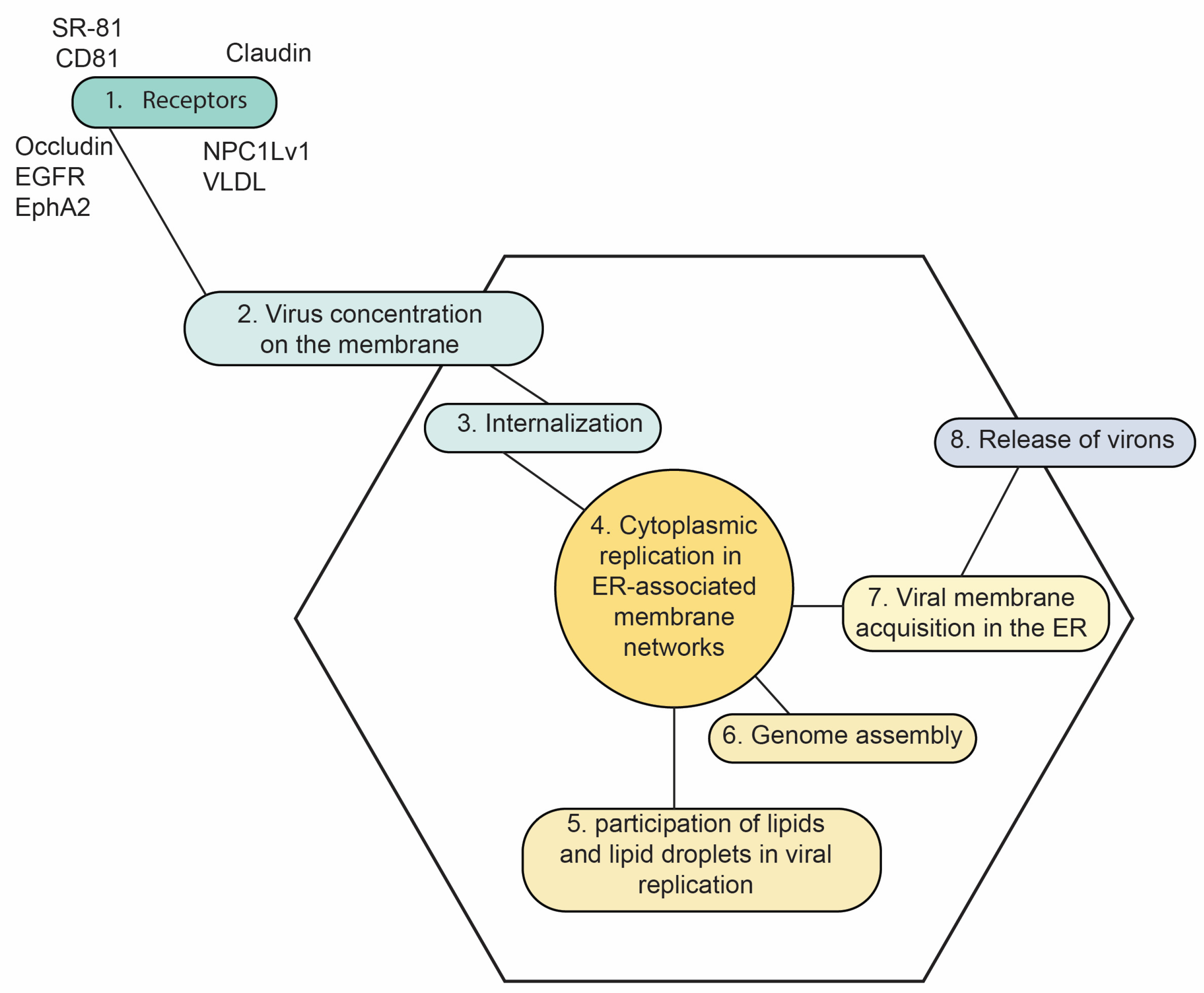
The HCV life cycle begins with recognition and binding to the host cell; this process is mediated by numerous cellular factors such as proteins, lipids, and glycans that promote the entry of viral particles into hepatocytes (Figure 1). The initial interaction of the virion with the cell membrane is mediated by glycosaminoglycans (GAGs) such as heparan sulfate, which help to concentrate the virus on the cell surface. As a consequence of the association between HCV and lipoproteins, it has also been proposed that the LDL receptor may be involved in the HCV entry into hepatocytes. HCV then interacts specifically and sequentially with SR-B1 (scavenger receptor B1) tetraspanin CD81 and the tight junction proteins claudin-1 and occludin. Recently, two tyrosine kinase receptors, EGFR (epidermal growth factor receptor) and EphA2 (ephrin type-A receptor 2), have been identified as the cofactors for HCV entry, and the NPC1L1 (Niemann–Pick C1-like1) receptor, involved in intestinal cholesterol absorption, was also shown to play a role in HCV entry, probably in the fusion of viral and cell membranes. HCV enters hepatocytes by clathrin-dependent endocytosis. Fusion of the virus envelope with the endosome membrane takes place in the early endosomes and is a process mediated by the acidic pH of the endosomes [21].
Replication of this virus is preferably carried out in the cytoplasm of hepatocytes in membranous networks adherent to the endoplasmic reticulum (ER) [21]. The presence of HCV has also been described in other cell types, such as peripheral blood mononuclear cells, dendritic cells, or the central nervous system [22]. The HCV replication complex generates the RNA progeny, which is used for translation, replication, or encapsidation into new virions. Finally, HCV uses the very-low-density lipoprotein (VLDL) lipid biosynthesis pathway for the assembly of new viral particles, which are ready to exit the cell, without a direct cytopathic effect on the cell [23]. The new viruses may also egress as lipoviral particles, which are hybrid molecules composed of viruses and lipoproteins [24].
Of paramount importance is the association of the HCV replication with cellular lipids (Figure 1). The interaction of HCV with lipid droplets and lipid metabolic pathways leads to hepatic steatosis (HS) [23,24]. The core protein inhibits microsomal triglyceride-transfer protein, which is required for VLDL formation, and the inhibition of this enzyme might contribute to HS [23].
HS is associated with severe histological damage and higher fibrosis scores, and it is correlated with the degree of fibrosis in CHC. HCV genotype 3 is associated with a higher frequency of HS. Other HCV genotypes are also associated with HS, although in a more dependent association with the presence of underlying metabolic conditions [24].
Figure 1. Key highlights of the HCV life cycle. 1. The virion interacts with several type of proteins at the cell surface in a sequential manner. 2. The interaction with GAGs leads to the concentration of viral particles at the surface of the cell. The virus then interacts specifically and sequentially with SR-B1, CD81 and the tight junction proteins claudin-1 and occludin. 3. Endocytosis is mediated by clathrin. 4. The entire replication occurs in the cytoplasm. 5. However, this process is tightly related to membranous networks and lipid droplets adhered to the ER. 6. HCV RNA progeny is encapsidated into new virions. 7. The virions acquire the envelope proteins inserted in the membrane of the ER. 8. Non-cytosolic egress of the cell [20,21,24,25].
Livers 06 00011 g001
As an RNA virus, HCV displays significant genetic diversity. A manifestation of this diversity is the emergence of genotypes over the evolutionary time [26,27]. Eight genotypes, and a great number of subtypes for each genotype, have been described for HCV [26]. HCV genotype 1 is the most common and exhibits a worldwide distribution (particularly subtypes 1a and 1b) [25]. Genotype 1, along with genotypes 2, 4, 5 and 7, originated probably in Africa [27], while genotypes 3, 6, and 8 probably originated in Asia. Some HCV subtypes are called epidemics (1a, 1b, 2a, 2b, 2c and 3a), since they display a worldwide distribution [27]. In the era of interferon-based therapies, both HCV genotypes 1 and 4 were frequently non-responders, requiring a longer duration of treatment, with limited success [28]. This limitation seems to be overcome by the availability of pan-genotypic direct-acting antivirals (DAAs, see below) [28]. Another consequence of the high genetic diversity of this virus is the intra-host diversity, also called quasispecies [26]. This diversity impacts virus transmission, pathogenesis, immune evasion and drug resistance [26].

4. Dysbiosis in HCV Infection

4.1. Pathogenic Pathways

Changes in the composition of IM may impact the progression and development of chronic liver disease associated with HCV infection [29]. Some studies have proposed that in vitro and in vivo models of the gut–liver axis in chronic hepatitis C have shown basically two pathogenic pathways: The first pathway induces liver damage directly through reduced bile salt production and protein synthesis. Bile is produced in the liver, being composed of cholesterol, phospholipids, proteins, bicarbonate, and bile acids that the liver has conjugated with taurine or glycine. Bile is directed to the proximal small intestine through the bile ducts, where it aids in the digestion and absorption of lipids. In the colon, intestinal bacteria convert primary bile acids into secondary bile acids by dihydroxylation, and finally, both primary and secondary bile acids are reabsorbed back into the liver via the portal circulation. The portal circulation also transports absorbed nutrients, lipids, microbial products such as lipopolysaccharides (LPSs), and microbial metabolites such as short-chain fatty acids (SCFAs) back to the liver. Toxic metabolites such as acetaldehyde and inflammatory cytokines produced by the liver enter into the systemic circulation, also carrying pathogen-associated molecular patterns, trimethylamine N-oxide, and VLDL; in the presence of intestinal ID, these alterations result in decreased bile salt conjugation that causes disturbances in lipid metabolism contributing to liver damage known as HS, favoring the progression of chronic HCV infection [30,31].
Although inflammation is considered a normal defense mechanism against the viral infection of hepatocytes, ongoing and unregulated inflammation is the primary cause of chronic liver damage induced by HCV infection, including fibrosis, cirrhosis, and HCC. Chronic HCV infection is characterized by the release of different inflammatory mediators expressed by liver cells, Kupffer cells, and immune cells, leading to the induction of inflammatory responses. A known inflammatory mediator that plays a crucial role in the induction of fibrosis is TGF-β1, which induces fibrogenesis-related gene expression in hepatic stellate cells (HSCs). Another inflammatory mediator is the pyrin domain (domain present in inflammasome-associated proteins) of the NOD 3 (NLRP3) family of receptors, which is involved in the activation of HSCs and the induction of fibrosis [32].
The prolonged induction of inflammatory signals in hepatocytes leads to the stimulation of HSCs to secrete the extracellular matrix (ECM) in the liver, resulting in the induction of liver fibrosis and cirrhosis. Chronic liver damage and the accumulation of ECM in the liver lead to an imbalance between fibrogenesis and fibrolysis [33].
The second proposed pathogenic pathway suggests that, following the establishment of a chronic HCV infection, the activation of B lymphocytes modulates the intestinal production of IgA by plasma cells, subsequently leading to an increase in intestinal permeability. This increased permeability then induces ID, facilitating the passage of molecules like lipopolysaccharides (LPSs) and other pathogen-associated molecular patterns (PAMPs) into the portal circulation. These antigens stimulate a further proinflammatory immune response mediated by interleukins such as IL-6 and TNF-α, thereby exacerbating the degree of hepatic fibrosis. The sustained activation of these cells, compounded by direct HCV damage, continues to stimulate this proinflammatory response and drive liver fibrosis [34]. Moreover, throughout this review, we will see that ID itself can alter the pathogenic pathways induced by HCV infection, creating a vicious cycle in the complex IM–HCV relationship.

4.2. Dysbiosis and Stage of Disease

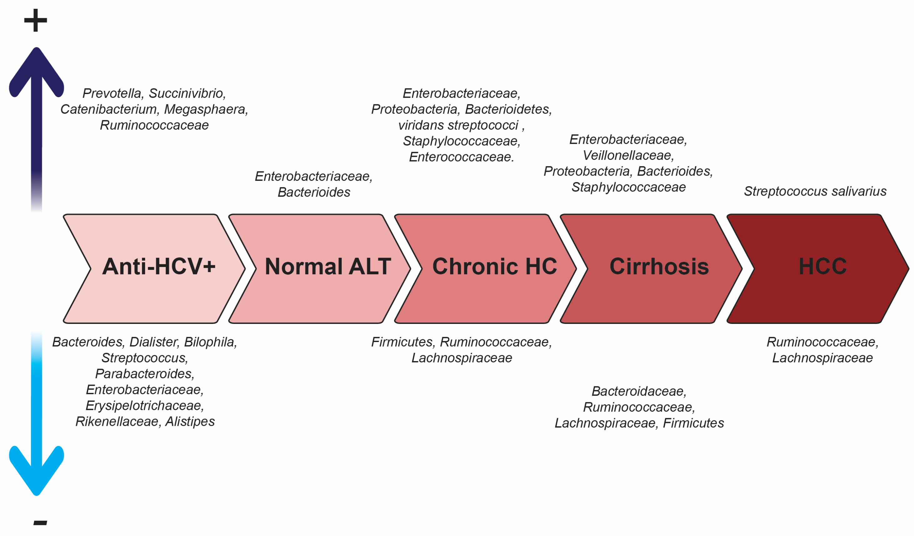
A review conducted by El-Mowafy et al. in 2021 showed that ID varies according to the clinical stage of hepatitis C, recording alterations in the composition (diversity) and quantity of microorganisms present [32]. Figure 2 schematically details the families, genera and species of bacteria mainly involved in this process. Hepatitis C infection is generally associated with a lower bacterial diversity. The order Clostridiales is significantly depleted during HCV infection, while both Lactobacillus and Streptococcus genera are significantly increased. An increase in the phylum of Bacterioidetes, the family of Enterobacteriaceae, and Viridans streptococci, is observed during infection, while the phylum of Firmicutes is decreased. A significant reduction in potential beneficial microorganisms, such as Ruminococcaceae and Lachnospiraceae families, has been reported in chronic hepatitis, liver cirrhosis, and HCC patients, with a significant increase in Lactobacilli and viridans streptococci. Streptococcus salivarius increases in HCC patients with liver cirrhosis, suggesting that this bacterium may play a role in the progression of disease. This bacterium has been shown to affect innate immunity, and this may increase the progression to HCC [35]. The evolving changes in IM during chronic HCV infection might promote disease progression and the development of liver complications and intestinal inflammation. The sense of the cause–effect between HCV infection and ID is still unknown [32].
As stated before, HS is a main characteristic associated with the pathogenesis of HCV infection. ID contributes greatly to the development of HS: an imbalance in the microbial community leads to conditions such as metabolic-associated fatty liver disease or metabolic-associated steatotic liver disease [45,46]. The progression to steatohepatitis, fibrosis, cirrhosis and, eventually, hepatocellular carcinoma is closely associated with ID [46]. Proteobacteria and Fusobacteria are commonly found elevated in the IM composition at the different stages of steatotic disease [45,46]. Proteobacteria have also been found to be elevated in the chronic stages of hepatitis C (Figure 2) [32,43].

4.3. Hepatitis C, Extrahepatic Manifestations and IM

Many HCV-infected patients can develop extrahepatic manifestations, including autoimmune and rheumatic disorders, and many of them are immune-related [46,47,48]. Mixed cryoglobulinemic vasculitis is one of the most clinically relevant complications [46,47]. Up to 30% of patients with chronic hepatitis C may also carry autoantibodies to different organs or even non-organ-specific autoantibodies [48]. In the era of IFN-based treatments, these autoimmune disorders were an additional challenge in the control of this disease [48]. There is promising evidence that the treatment with direct-acting antivirals (DAAs) may also reduce these sequelae, although an early treatment is recommended [46,47].
There is growing evidence on the relationship between host immunity and IM. ID has been implied in the progression of various chronic conditions, including hepatic disorders [49]. The IM influences not only the local mucosal immunity, but also systemic innate and adaptative immune responses [49,50]. The IM is recognized as an important player in the development and progression of autoimmune diseases [49]. The IM modulates systemic immunity in particular by affecting the differentiation of adaptive immune cells, particularly T lymphocytes. The IM influences the balance among T-helper subsets, including Th1, Th2, and Th17 cells, and the induction of regulatory T cells. One of the mechanisms responsible for these effects are the IM metabolites. SCFAs, particularly butyrate, favors the differentiation of peripherally induced T regulatory cells, suppressing the systemic inflammatory responses. Another microbial metabolite, pentanoate, promotes regulatory B cell differentiation while inhibiting Th17 cell expansion. These aspects are particularly relevant in the development of autoimmune and inflammatory disorders [49,50].

5. Gut Microbiota and HCV-Induced Hepatocellular Carcinoma

Hepatocellular carcinoma (HCC) is an aggressive type of liver cancer that typically develops in individuals with chronic liver disease, particularly those with hepatitis B, C, and D virus infections. The incidence of HCC associated with viral infections has been decreasing due to HBV vaccination [51,52], in contrast to the increase in incidence and prevalence due to HCV infection, predominantly in Africa, Asia and Latin America [52]. Multiple proofs have demonstrated the strong correlation between chronic HCV infection, its progression to liver cirrhosis, and the development of liver fibrosis with the risk and progression to HCC. It has been reported that ALT values greater than 70 IU/L may be associated with liver cirrhosis. Another factor related to HCC in patients with chronic HCV is the presence of obesity and type 2 diabetes, as it has been observed in prospective studies that patients with these comorbidities have a higher progression to cirrhosis and HCC [52].
Alcohol consumption in patients with chronic hepatitis C has been considered an aggravating factor for the development of HCC. On the other hand, age and gender have been described as variables of interest when studying HCV associated with HCV infection since patients who become infected with HCV earlier in life have a lower risk of liver fibrosis and cirrhosis throughout a chronic infection, while adult patients older than 45 years have an increased risk of HCC. The higher prevalence of HCC associated with chronic hepatitis C has been observed in male patients worldwide, although it is not considered a risk factor for its development. As for the virus, HCV genotype 3 has been associated with the increased progression to HS and this, in turn, with the accelerated progression to cirrhosis as well as HCC [53].
The mechanism of hepatocellular carcinogenesis associated with HCV is complex and multifactorial. It develops mainly through a combination of chronic liver damage, persistent inflammation, and genetic alterations, which contribute to the development of HCC. HCV does not have a direct cytopathic effect, but chronic infection generates a persistent immune response. This continuous response causes chronic inflammation in the liver, leading to the death of hepatocytes and their subsequent regeneration. This cycle of cell damage and regeneration creates a favorable environment for the appearance of mutations (indirect oncogenic action) [52].
At least 30 genes (and their mutations) involved in the development of HCC in chronic HCV infection have been studied. These regulate the activation of molecular mechanisms and cell signaling pathways related to the development of cirrhosis and/or a high degree of liver fibrosis. Somatic point mutations in the P53-RB pathway related to apoptosis, alterations and mutations in the β-catenin pathway, MAPK, chromatin, and transcription modulators, among other pathways, can be found altered in patients with chronic HCV and HCC [54].
More directly, some viral proteins seem to play a leading role in the development of HCC: such is the case of the capsid protein that can interact with some cellular factors necessary for cell proliferation and survival and can also inhibit the action of the tumor suppressor protein p53, essential for cell cycle control. HCV NS5A and NS3 proteins are also involved in inhibiting apoptosis and promoting cell replication, which increases the risk of malignant transformation [55].
Several studies have revealed the crucial role of IM as a determining factor in the development of HCC related to chronic HCV infection. The progression of these patients into cirrhosis is associated with communication pathways involving bile salts and the portal vein system. The altered IM and impaired intestinal permeability contribute significantly to the pathogenesis of all stages of chronic liver disease. Additionally, these alterations in IM may cause some important effects on the progression of liver disease through the gut–liver axis [56].
Regarding the clinical profile, some investigators have recently studied the IM of patients with HCC associated with chronic HCV infection and have observed that bacterially produced SCFAs are decreased in patients with chronic HCV infection who develop HCC and/or cirrhosis, in agreement with the observations of Kakiyama et al. [6] on the modulation of the bile acid in the stool of patients with cirrhosis. LPSs and other metabolites may be increased in these patients due to the significant ID that leads to the translocation of some opportunistic pathogens such as Streptococcus spp. and Enterobacteriace, among others [57].
The IM alterations are observed not only in HCC and HCV patients but also in those with liver pathologies of other etiologies. However, the nature of these alterations varies depending on the underlying causes, and the composition of IM in each case usually differs in terms of the microorganisms present. For this reason, it is suggested that, with the analysis of IM, it is possible to approach a specific cause or trigger of HCC to guide a presumptive etiologic diagnosis and that it can also be performed precociously. For example, certain metabolites of some IM microorganisms, mainly bacteria, can be detected in peripheral blood and/or feces and serve as the early biomarkers of chronic liver disease, cirrhosis, and HCC [58].
Thananya et al. investigated differences in the fecal IM diversity and composition between viral and non-viral, non-B, non-C HCC subgroups (NBNC-HCC). They showed that the α-diversity (the degree of diversity) of IM was significantly lower in NBNC-HCC compared to viral HCC and healthy controls. Sixteen bacterial genera were identified that differed significantly between viral HCC and NBNC-HCC, 11 genera were enriched in viral HCC (e.g., including Faecalibacterium, Agathobacter, and Coprococcus), and 5 genera were increased in NBNC-CHC (e.g., Bacteroides, Streptococcus, Ruminococcus gnavus group, Parabacteroides, and Erysipelatoclostridium). The fecal BCoAT gene levels and various fecal SCFA-producing bacteria were shown to decrease significantly in NBNC-HCC compared to viral HCC and controls. The plasma levels of lipopolysaccharide-binding protein were higher in NBNC-CHC when compared with viral HCC and controls. Predictive functional analysis showed differences in metabolic pathways between the subgroups, with dioxin degradation being the most distinctive pathway in NBNC-CHC. In this study, they highlighted 16 different genera that allow distinguishing between viral HCC and NBNC-HCC [59].

6. Treatment

6.1. Antibiotics

A recent cross-sectional study explored the effect of rifaximin (an antibiotic that does not cross the intestinal mucosa) on the ID observed in cirrhotic patients infected with HCV. Rifaximin caused a significant decrease in the secondary/primary bile acid ratio after treatment and a microbiota modulated towards a reduction in the abundance of Veillonellaceae, with no significant changes in other bacteria [31]. Based on this, it could be concluded that the ID of cirrhotic patients correlates with changes in the bile acid profile. Rifaximin treatment modifies the function of IM by reducing the conversion of primary to secondary bile acids, and these changes in the bile acid profile and IM could have important implications for the pathogenesis and progression of cirrhosis. This study suggests that the correction of ID may have a beneficial effect on HCV disease progression [31].
Immune checkpoints inhibitors (ICIs), alone or in combination with other therapies, have emerged as a promising therapy for different cancers [54]. It has been reported that treatment with antibiotics before ICI treatment leads to a worse pronostic in oncologic patients, compared to the ones who did not receive antibiotics before [60]. In the case of HCC, a study involving 4100 patients with HCC from different etiologies (including HCV) showed that treatment with antibiotics 30 days before or during the anti-oncologic therapy led to worse outcomes in patients treated with ICIs or tyrosine kinase inhibitors or even those in the placebo group [61]. Further studies are warranted on the role of ID in modulating the outcome in patients with HCC undergoing systemic therapy [61].

6.2. Direct-Acting Antivirals

DAAs have represented a revolution in the field of hepatitis C treatment. Previously, interferon- and ribavirin-based treatments were the first line of therapy used until the second decade of the 2000s. With the emergence of new therapeutic regimens based on DAAs, HCV eradication, known as sustained viral response (SVR), is achieved in 95% of patients diagnosed with the infection, regardless of genotype. DAAs have become the current first-line treatment of choice for hepatitis C, offering relatively short treatment regimens with few adverse effects [62]. Table 1 describes the viral targets affected by DAAs.
One of the first studies examining the short-term effects of DAAs on the IM in non-cirrhotic HCV patients showed that neither the use of DAAs nor 3 months of SVR were able to counteract completely the changes in IM caused by HCV infection, although partial restoration in inflammation, α-diversity, and some bacterial genera was observed in patients with very low degrees of fibrosis. Inflammatory markers associated with HCV infection were also evaluated in this study; treatment with DAAs failed to counteract the inflammatory state, especially the TNFα levels, namely, in patients with low degrees of fibrosis. Another marker of the inflammatory profile studied was IL-6, the levels of which seem to be more associated with the presence of advanced liver fibrosis [63]. The same observation was reported shortly after treatment in patients infected with different genotypes of HCV [64] (Table 2).
Thanks to the contributions of this study on changes in the IM composition, HCV infection was associated with changes in the phylum (Firmicutes, Actinobacteria), genus (Lachnospira, Blautia, Coprococcus, Dorea, and Veillonella), and other bacterial species (Akkermansia muciniphila). DAAs and SVR did not produce significant modifications in the composition of IM. However, the sample size of each study group should be expanded in future research, and if possible, other factors that could affect the composition of the IM, such as the exact diet, alcohol consumption, and medications, should be considered [63]. In contrast, a longitudinal study on the long-term effects of DAAs on IM conducted by Chuaypen et al. in 2023 in a cohort of 50 patients with HCV infection but without liver cirrhosis who received treatment with DAAs and achieved SVR observed that the ID of these patients appeared to recover to normal composition and function within 72 weeks after the completion of treatment [65]. Other studies support the observation that, although SVR after DAA treatment can induce a reduction in the ID, this amelioration is less apparent in patients with cirrhosis [66,67]. This dysbiosis seem to persist even after several years after antiviral treatment in cirrhotic patients [68].
These results underscore the need to initiate treatment with DAAs in HCV-infected patients as early as possible; if liver damage has already occurred, it is equally important to initiate treatment early. It is also suggested to monitor patients even after HCV eradication. In this context, additional interventions, such as the use of specific probiotics designed to normalize the ID caused by hepatitis C, may be necessary to fully normalize or restore the IM both during and at the end of treatment with DAAs [63].

6.3. Probiotics and Other Emerging Adjuvants

It is clear that ID appears at any stage of HCV infection. The use of probiotics may be then advisable as cofactors to reduce the pathogenic effect of this disease. A preliminary study on HCV-positive asymptomatic subjects was conducted with the probiotic FK-23 (heat-treated Enterococcus faecalis strain FK-23). A decrease in the levels of transaminases was observed, with no significant change in the viral load. More studies are needed to confirm these preliminary results [69].
Probiotics may also mitigate the risk of HCC by modulating host IM to promote the growth of beneficial microbes and inhibit HCC-associated dysbiosis, thereby preventing pathogen-associated molecular pattern-mediated liver inflammation. Probiotics have indirect antiviral activities against HBV and HCV infections by ameliorating obesity and non-alcoholic liver disease (NAFLD) risk; their antioxidant, antiproliferative, antiangiogenic, and antimetastatic effects could prevent HCC pathogenesis. Probiotics also positively regulate the expression of tumor suppressor genes and decrease the expression of oncogenes. In addition, metabolites generated by probiotics through the degradation of dietary phytochemicals may mitigate the risk of HCC development. These multiple anticancer mechanisms illustrate the potential of probiotics as an adjuvant strategy for HCV-associated HCC risk management [70]. Although probiotics are not currently recommended as a treatment for any of the stages of viral hepatitis, their use as adjuvants in the treatment with DAAs has been shown to improve ID in these patients [70]. Several clinical trials are ongoing to test the utility of probiotics as adjuvants in the treatment of several cancers: none of them however are used in HCC [71].
In addition to antibiotics and probiotics, other future prospects include diets, fecal microbiota transplantation (no study yet) [11,72], and nano-delivery systems [73]. Two studies comparing patients with cirrhosis from Brazil or Turkey with those from the USA (many of them with HCV infection) showed that the diet of the non-USA patients was richer in cereals, rice, and yogurt, compared to that of the patients from the USA. A higher α-diversity was observed in the IM of the Brazilian or Turkish patients: in addition, these patients experienced less hospitalization episodes than the patients from the USA [74,75].
Prebiotics are indigestible dietary components that enhance the colonization of gut with beneficial microorganisms. Prebiotics and symbiotics (a combination of prebiotics and probiotics) have also been proposed for the IM restoration in patients with cirrhosis, with preliminary promising results [76].
Finally, given the emerging therapeutic potential of bacteriophages, their role in modulating or restoring the IM is warranted for future therapeutic approaches [46].

7. Concluding Remarks

HCV infection causes ID. This dysbiosis is not uniform across the disease spectrum: it is more pronounced in patients with advanced liver damage, including cirrhosis and HCC. Dysbiosis is, in turn, a poor prognostic factor for the progression of hepatitis C. With DAA treatment, the IM is not restored in patients with advanced disease (both viral infection and liver damage). This observation further highlights the importance of early hepatitis C treatment. Further studies on the IM and HCV infection with a larger sample size are required to elucidate some clinical and virological aspects, as well as the implications of IM per se on disease progression and evolution. As a limitation, we find that most of the studies available and/or cited address the study of IM: it would be interesting to compare these results with the study of the hepatic microbiota in order to establish a correlation of the gut–liver axis connection and to have a closer look at the changes that occur at the intrahepatic level [46]. Promising emerging treatment or diet may be crucial for restoring the IM after cure of an HCV infection.

Author Contributions

Conceptualization, F.H.P.; literature review, F.J.F.L.; writing—original draft preparation, F.J.F.L.; final editing, F.H.P. All authors have read and agreed to the published version of the manuscript.

Funding

This research received no external funding.

Institutional Review Board Statement

Not applicable.

Informed Consent Statement

Not applicable.

Data Availability Statement

No new data were created or analyzed in this study. Data sharing is not applicable to this article.

Acknowledgments

The authors expressed their gratitude to the postgraduate program of Microbiology, IVIC, Venezuela, for motivating the publication of this manuscript.

Conflicts of Interest

The authors declare no conflicts of interest.

Abbreviations

The following abbreviations are used in this manuscript:
ALTalanine aminotransferase
Chronic HCchronic hepatitis C
DAAsdirect-acting antivirals
ECMextracellular matrix
EGFRepidermal growth factor receptor
EphA2 ephrin type-A receptor 2
ERendoplasmic reticulum
GAGsglycosaminoglycans
HCChepatocellular carcinoma
HCVhepatitis C virus
HShepatic steatosis
HSCshepatic stellate cells
ICIsimmune checkpoint inhibitors
IL-6 interleukin-6
IDintestinal dysbiosis
IMintestinal microbiota
LPSslipopolysaccharides
NBNC-HCCnon-B non-C HCC
SCFAsshort-chain fatty acids
SR-B1scavenger receptor B1
SVRsustained viral response
TNF-αtumor necrosis factor α
VHviral hepatitis
VLDLvery-low-density lipoprotein

References

  1. Lozupone, C.A.; Stombaugh, J.I.; Gordon, J.I.; Jansson, J.K.; Knight, R. Diversity, stability and resilience of the human gut microbiota. Nature 2012, 489, 220–230. [Google Scholar] [CrossRef]
  2. Marascio, N.; Scarlata, G.G.M.; Romeo, F.; Cicino, C.; Trecarichi, E.M.; Quirino, A.; Torti, C.; Matera, G.; Russo, A. The Role of Gut Microbiota in the Clinical Outcome of Septic Patients: State of the Art and Future Perspectives. Int. J. Mol. Sci. 2023, 24, 9307. [Google Scholar] [CrossRef] [PubMed]
  3. Rowland, I.; Gibson, G.; Heinken, A.; Scott, K.; Swann, J.; Thiele, I.; Tuohy, K. Gut microbiota functions: Metabolism of nutrients and other food components. Eur. J. Nutr. 2018, 57, 1–24. [Google Scholar] [CrossRef] [PubMed]
  4. Carías Domínguez, A.M.; de Jesús Rosa Salazar, D.; Stefanolo, J.P.; Cruz Serrano, M.C.; Casas, I.C.; Zuluaga Peña, J.R. Intestinal Dysbiosis: Exploring Definition, Associated Symptoms, and Perspectives for a Comprehensive Understanding—A Scoping Review. Probiotics Antimicrob. Proteins 2025, 17, 440–449. [Google Scholar] [CrossRef] [PubMed]
  5. Origüela, V.; Lopez-Zaplana, A. Gut Microbiota: An Immersion in Dysbiosis, Associated Pathologies, and Probiotics. Microorganisms 2025, 13, 1084. [Google Scholar] [CrossRef]
  6. Pisano, M.B.; Giadans, C.G.; Flichman, D.M.; Re, V.E.; Preciado, M.V.; Valva, P. Viral hepatitis update: Progress and perspectives. World J. Gastroenterol. 2021, 27, 4018–4044. [Google Scholar] [CrossRef]
  7. Pujol, F.H.; Trepo, C.; Chemin, I. Épidémiologie et histoire naturelle de l’hépatite C. EMC—Hépatologie 2024, 39, 1–6. [Google Scholar] [CrossRef]
  8. Stroffolini, T.; Stroffolini, G. Prevalence and Modes of Transmission of Hepatitis C Virus Infection: A Historical Worldwide Review. Viruses 2024, 16, 1115. [Google Scholar] [CrossRef]
  9. World Health Organization: Hepatitis. Available online: https://www.who.int/health-topics/hepatitis/elimination-of-hepatitis-by-2030#tab=tab_1 (accessed on 17 June 2025).
  10. Sehgal, R.; Bedi, O.; Trehanpati, N. Role of Microbiota in Pathogenesis and Management of Viral Hepatitis. Front. Cell. Infect. Microbiol. 2020, 10, 341. [Google Scholar] [CrossRef]
  11. Yu, J.X.; Wu, J.; Chen, X.; Zang, S.G.; Li, X.B.; Wu, L.P.; Xuan, S.H. Gut microbiota in liver diseases: Initiation, development and therapy. Front. Med. 2025, 12, 1615839. [Google Scholar] [CrossRef]
  12. Clemente, J.C.; Ursell, L.K.; Parfrey, L.W.; Knight, R. The impact of the gut microbiota on human health: An integrative view. Cell 2012, 148, 1258–1270. [Google Scholar] [CrossRef] [PubMed]
  13. Khanna, S.; Tosh, P.K. A clinician’s primer on the role of the microbiome in human health and disease. Mayo Clin. Proc. 2014, 89, 107–114. [Google Scholar] [CrossRef] [PubMed]
  14. Raimondi, S.; Amaretti, A.; Gozzoli, C.; Simone, M.; Righini, L.; Candeliere, F.; Brun, P.; Ardizzoni, A.; Colombari, B.; Paulone, S.; et al. Longitudinal Survey of Fungi in the Human Gut: ITS Profiling, Phenotyping, and Colonization. Front. Microbiol. 2019, 10, 1575. [Google Scholar] [CrossRef] [PubMed]
  15. Escobar-Zepeda, A.; Vera-Ponce de Leon, A.; Sanchez-Flores, A. The Road to Metagenomics: From Microbiology to DNA Sequencing Technologies and Bioinformatics. Front. Genet. 2015, 6, 348. [Google Scholar] [CrossRef]
  16. Qian, X.B.; Chen, T.; Xu, Y.P.; Chen, L.; Sun, F.X.; Lu, M.P.; Liu, Y.X. A guide to human microbiome research: Study design, sample collection, and bioinformatics analysis. Chin. Med. J. 2020, 133, 1844–1855. [Google Scholar] [CrossRef]
  17. Schnabl, B.; Brenner, D.A. Interactions Between the Intestinal Microbiome and Liver Diseases. Gastroenterology 2014, 146, 1513–1524. [Google Scholar] [CrossRef]
  18. Koutsoudakis, G.; Forns, X.; Pérez-Del-Pulgar, S. The molecular biology of hepatitis C virus. Gastroenterol. Y Hepatol. 2013, 36, 280–293. [Google Scholar] [CrossRef]
  19. Popescu, C.I.; Rouillé, Y.; Dubuisson, J. Hepatitis C virus assembly imaging. Viruses 2011, 3, 2238–2254. [Google Scholar] [CrossRef]
  20. Bartenschlager, R.; Penin, F.; Lohmann, V.; André, P. Assembly of infectious hepatitis C virus particles. Trends Microbiol. 2011, 19, 95–103. [Google Scholar] [CrossRef]
  21. Chigbu, D.I.; Loonawat, R.; Sehgal, M.; Patel, D.; Jain, P. Hepatitis C Virus Infection: Host–Virus Interaction and Mechanisms of Viral Persistence. Cells 2019, 8, 376. [Google Scholar] [CrossRef]
  22. Dahari, H.; Feliu, A.; Garcia-Retortillo, M.; Forns, X.; Neumann, A.U. Second hepatitis C replication compartment indicated by viral dynamics during liver transplantation. J. Hepatol. 2005, 42, 491–498. [Google Scholar] [CrossRef] [PubMed]
  23. Dempsey, J.L.; Ioannou, G.N.; Carr, R.M. Mechanisms of Lipid Droplet Accumulation in Steatotic Liver Diseases. Semin. Liver Dis. 2023, 43, 367–382. [Google Scholar] [CrossRef] [PubMed]
  24. Elgretli, W.; Chen, T.; Kronfli, N.; Sebastiani, G. Hepatitis C Virus-Lipid Interplay: Pathogenesis and Clinical Impact. Biomedicines 2023, 11, 271. [Google Scholar] [CrossRef] [PubMed]
  25. Moradpour, D.; Penin, F.; Rice, C.M. Replication of hepatitis C virus. Nat. Rev. Microbiol. 2007, 5, 453–463. [Google Scholar] [CrossRef]
  26. Martinez, M.A.; Franco, S. Therapy Implications of Hepatitis C Virus Genetic Diversity. Viruses 2020, 13, 41. [Google Scholar] [CrossRef]
  27. Vo-Quang, E.; Pawlotsky, J.M. ‘Unusual’ HCV genotype subtypes: Origin, distribution, sensitivity to direct-acting antiviral drugs and behaviour on antiviral treatment and retreatment. Gut 2024, 73, 1570–1582. [Google Scholar] [CrossRef]
  28. Di Marco, L.; Cannova, S.; Ferrigno, E.; Landro, G.; Nonni, R.; Mantia, C.; Cartabellotta, F.; Calvaruso, V.; Di Marco, V. A Comprehensive Review of Antiviral Therapy for Hepatitis C: The Long Journey from Interferon to Pan-Genotypic Direct-Acting Antivirals (DAAs). Viruses 2025, 17, 163. [Google Scholar] [CrossRef]
  29. Preveden, T.; Scarpellini, E.; Milić, N.; Luzza, F.; Abenavoli, L. Gut microbiota changes and chronic hepatitis C virus infection. Expert Rev. Gastroenterol. Hepatol. 2017, 11, 813–819. [Google Scholar] [CrossRef]
  30. Hsu, C.L.; Schnabl, B. The gut–liver axis and gut microbiota in health and liver disease. Nat. Rev. Microbiol. 2023, 21, 719–733. [Google Scholar] [CrossRef]
  31. Kakiyama, G.; Pandak, W.M.; Gillevet, P.M.; Hylemon, P.B.; Heuman, D.M.; Daita, K.; Takei, H.; Muto, A.; Nittono, H.; Ridlon, J.M.; et al. Modulation of the fecal bile acid profile by gut microbiota in cirrhosis. J. Hepatol. 2013, 58, 949–955. [Google Scholar] [CrossRef]
  32. El-Mowafy, M.; Elgaml, A.; El-Mesery, M.; Sultan, S.; Ahmed, T.A.E.; Gomaa, A.I.; Aly, M.; Mottawea, W. Changes of Gut-Microbiota-Liver Axis in Hepatitis C Virus Infection. Biology 2021, 10, 55. [Google Scholar] [CrossRef] [PubMed]
  33. Khatun, M.; Ray, R.B. Mechanisms Underlying Hepatitis C Virus-Associated Hepatic Fibrosis. Cells 2019, 8, 1249. [Google Scholar] [CrossRef] [PubMed]
  34. Fang, J.; Yu, C.H.; Li, X.J.; Yao, J.M.; Fang, Z.Y.; Yoon, S.H.; Yu, W.Y. Gut dysbiosis in nonalcoholic fatty liver disease: Pathogenesis, diagnosis, and therapeutic implications. Front. Cell. Infect. Microbiol. 2022, 12, 997018. [Google Scholar] [CrossRef] [PubMed]
  35. Cosseau, C.; Devine, D.A.; Dullaghan, E.; Gardy, J.L.; Chikatamarla, A.; Gellatly, S.; Yu, L.L.; Pistolic, J.; Falsafi, R.; Tagg, J.; et al. The commensal Streptococcus salivarius K12 downregulates the innate immune responses of human epithelial cells and promotes host-microbe homeostasis. Infect. Immun. 2008, 76, 4163–4175. [Google Scholar] [CrossRef]
  36. Sultan, S.; El-Mowafy, M.; Elgaml, A.; El-Mesery, M.; El Shabrawi, A.; Elegezy, M.; Hammami, R.; Mottawea, W. Alterations of the Treatment-Naive Gut Microbiome in Newly Diagnosed Hepatitis C Virus Infection. ACS Infect. Dis. 2021, 7, 1059–1068. [Google Scholar] [CrossRef]
  37. Ponziani, F.R.; Putignani, L.; Paroni Sterbini, F.; Petito, V.; Picca, A.; Del Chierico, F.; Reddel, S.; Calvani, R.; Marzetti, E.; Sanguinetti, M.; et al. Influence of hepatitis C virus eradication with direct-acting antivirals on the gut microbiota in patients with cirrhosis. Aliment. Pharmacol. Ther. 2018, 48, 1301–1311. [Google Scholar] [CrossRef]
  38. Iwata, R.; Stieger, B.; Mertens, J.C.; Muller, T.; Baur, K.; Frei, P.; Braun, J.; Vergopoulos, A.; Martin, I.V.; Schmitt, J.; et al. The role of bile acid retention and a common polymorphism in the ABCB11 gene as host factors affecting antiviral treatment response in chronic hepatitis C. J. Viral Hepat. 2011, 18, 768–778. [Google Scholar] [CrossRef]
  39. Nakamoto, N.; Sasaki, N.; Aoki, R.; Miyamoto, K.; Suda, W.; Teratani, T.; Suzuki, T.; Koda, Y.; Chu, P.S.; Taniki, N.; et al. Gut pathobionts underlie intestinal barrier dysfunction and liver T helper 17 cell immune response in primary sclerosing cholangitis. Nat. Microbiol. 2019, 4, 492–503. [Google Scholar] [CrossRef]
  40. Sanduzzi Zamparelli, M.; Rocco, A.; Compare, D.; Nardone, G. The gut microbiota: A new potential driving force in liver cirrhosis and hepatocellular carcinoma. United Eur. Gastroenterol. J. 2017, 5, 944–953. [Google Scholar] [CrossRef]
  41. Chen, Y.; Yang, F.; Lu, H.; Wang, B.; Chen, Y.; Lei, D.; Wang, Y.; Zhu, B.; Li, L. Characterization of fecal microbial communities in patients with liver cirrhosis. Hepatology 2011, 54, 562–572. [Google Scholar] [CrossRef]
  42. Tuomisto, S.; Pessi, T.; Collin, P.; Vuento, R.; Aittoniemi, J.; Karhunen, P.J. Changes in gut bacterial populations and their translocation into liver and ascites in alcoholic liver cirrhotics. BMC Gastroenterol. 2014, 14, 40. [Google Scholar] [CrossRef] [PubMed]
  43. Inoue, T.; Nakayama, J.; Moriya, K.; Kawaratani, H.; Momoda, R.; Ito, K.; Iio, E.; Nojiri, S.; Fujiwara, K.; Yoneda, M.; et al. Gut Dysbiosis Associated With Hepatitis C Virus Infection. Clin. Infect. Dis. 2018, 67, 869–877. [Google Scholar] [CrossRef] [PubMed]
  44. Qin, N.; Yang, F.; Li, A.; Prifti, E.; Chen, Y.; Shao, L.; Guo, J.; Le Chatelier, E.; Yao, J.; Wu, L.; et al. Alterations of the human gut microbiome in liver cirrhosis. Nature 2014, 513, 59–64. [Google Scholar] [CrossRef] [PubMed]
  45. Pasta, A.; Formisano, E.; Calabrese, F.; Marabotto, E.; Furnari, M.; Bodini, G.; Torres, M.C.P.; Pisciotta, L.; Giannini, E.G.; Zentilin, P. From Dysbiosis to Hepatic Inflammation: A Narrative Review on the Diet-Microbiota-Liver Axis in Steatotic Liver Disease. Microorganisms 2025, 13, 241. [Google Scholar] [CrossRef]
  46. Lau, H.C.; Zhang, X.; Yu, J. Gut microbiome in metabolic dysfunction-associated steatotic liver disease and associated hepatocellular carcinoma. Nat. Rev. Gastroenterol. Hepatol. 2025, 22, 619–638. [Google Scholar] [CrossRef]
  47. Mazzaro, C.; Quartuccio, L.; Adinolfi, L.E.; Roccatello, D.; Pozzato, G.; Nevola, R.; Tonizzo, M.; Gitto, S.; Andreone, P.; Gattei, V. A Review on Extrahepatic Manifestations of Chronic Hepatitis C Virus Infection and the Impact of Direct-Acting Antiviral Therapy. Viruses 2021, 13, 2249. [Google Scholar] [CrossRef]
  48. Balta, A.A.S.; Ignat, M.D.; Barbu, R.E.; Dumitru, C.; Radaschin, D.S.; Bulza, V.; Mateescu Costin, S.A.; Pleșea-Condratovici, C.; Baroiu, L. Impact of Direct-Acting Antivirals on Extrahepatic Manifestations in Chronic Hepatitis C: A Narrative Review with a Hermeneutic Approach. Healthcare 2025, 13, 1953. [Google Scholar] [CrossRef]
  49. Bianchi, F.B.; Muratori, P.; Granito, A.; Pappas, G.; Ferri, S.; Muratori, L. Hepatitis C and autoreactivity. Dig. Liver Dis. 2007, 39, S22–S24. [Google Scholar] [CrossRef]
  50. Frumento, D.; Țălu, S. Interaction Between Human Microbiota, Immune System, and Hepatitis C Virus Infection: A Narrative Review. Appl. Sci. 2025, 15, 3157. [Google Scholar] [CrossRef]
  51. Pujol, F.H.; Toyé, R.M.; Loureiro, C.L.; Jaspe, R.C.; Chemin, I. Hepatitis B eradication: Vaccine as a key player. Am. J. Transl. Res. 2023, 15, 4971–4983. [Google Scholar]
  52. El-Serag, H.B.; Rudolph, K.L. Hepatocellular carcinoma: Epidemiology and molecular carcinogenesis. Gastroenterology 2007, 132, 2557–2576. [Google Scholar] [CrossRef] [PubMed]
  53. Ampuero, J.; Romero-Gómez, M.; Reddy, K.R. Review article: HCV genotype 3—The new treatment challenge. Aliment. Pharmacol. Ther. 2014, 39, 686–698. [Google Scholar] [CrossRef] [PubMed]
  54. Fujimoto, A.T.; Totoki, Y.; Abe, T.; Boroevich, K.A.; Hosoda, F.; Nguyen, H.H.; Aoki, M.; Hosono, N.; Kubo, M.; Miya, F.; et al. Whole-genome sequencing of liver cancers identifies etiological influences on mutation patterns and recurrent mutations in chromatin regulators. Nat. Genet. 2012, 44, 760–764. [Google Scholar] [CrossRef] [PubMed]
  55. D’Souza, S.; Lau, K.C.; Coffin, C.S.; Patel, T.R. Molecular mechanisms of viral hepatitis induced hepatocellular carcinoma. World J. Gastroenterol. 2020, 26, 5759–5783. [Google Scholar] [CrossRef]
  56. Piñero, F.; Vazquez, M.; Baré, P.; Rohr, C.; Mendizabal, M.; Sciara, M.; Alonso, C.; Fay, F.; Silva, M. A different gut microbiome linked to inflammation found in cirrhotic patients with and without hepatocellular carcinoma. Ann. Hepatol. 2019, 18, 480–487. [Google Scholar] [CrossRef]
  57. Ponziani, F.R.; Bhoori, S.; Castelli, C.; Putignani, L.; Rivoltini, L.; Del Chierico, F.; Sanguinetti, M.; Morelli, D.; Paroni Sterbini, F.; Petito, V.; et al. Hepatocellular carcinoma is associated with gut microbiota profle and infammation in nonalcoholic fatty liver disease. Hepatology 2019, 69, 107–120. [Google Scholar] [CrossRef]
  58. Ren, Z.; Li, A.; Jiang, J.; Zhou, L.; Yu, Z.; Lu, H.; Xie, H.; Chen, X.; Shao, L.; Zhang, R.; et al. Gut microbiome analysis as a tool towards targeted non-invasive biomarkers for early hepatocellular carcinoma. Gut 2019, 68, 1014–1023. [Google Scholar] [CrossRef]
  59. Jinato, T.; Anuntakarun, S.; Satthawiwat, N.; Chuaypen, N.; Tangkijvanich, P. Distinct alterations of gut microbiota between viral- and non-viral-related hepatocellular. Appl. Microb. Cell Physiol. 2024, 108, 34. [Google Scholar] [CrossRef]
  60. Elkrief, A.; Derosa, L.; Kroemer, G.; Zitvogel, L.; Routy, B. The negative impact of antibiotics on outcomes in cancer patients treated with immunotherapy: A new independent prognostic factor? Ann. Oncol. 2019, 30, 1572–1579. [Google Scholar] [CrossRef]
  61. Pinato, D.J.; Li, X.; Mishra-Kalyani, P.; D’Alessio, A.; Fulgenzi, C.A.M.; Scheiner, B.; Pinter, M.; Wei, G.; Schneider, J.; Rivera, D.R.; et al. Association between antibiotics and adverse oncological outcomes in patients receiving targeted or immune-based therapy for hepatocellular carcinoma. JHEP Rep. 2023, 5, 100747. [Google Scholar] [CrossRef]
  62. European Association for the Study of the Liver. EASL recommendations on treatment of hepatitis C: Final update of the series. J. Hepatol. 2020, 73, 1170–1218. [Google Scholar] [CrossRef] [PubMed]
  63. Perez-Matute, P.; Iniguez, M.; Villanueva-Millan, M.J.; Recio-Fernandez, E.; Vazquez, A.M.; Sanchez, S.C.; Morano, L.E.; Oteo, J.A. Short-term effects of direct-acting antiviral agents on inflammation and gut microbiota in hepatitis C-infected patients. Eur. J. Intern. Med. 2019, 67, 47–58. [Google Scholar] [CrossRef] [PubMed]
  64. Yilmaz, B.; Ruckstuhl, L.; Müllhaupt, B.; Magenta, L.; Kuster, M.H.; Clerc, O.; Torgler, R.; Semmo, N. Pilot Sub-Study of the Effect of Hepatitis C Cure by Glecaprevir/Pibrentasvir on the Gut Microbiome of Patients with Chronic Hepatitis C Genotypes 1 to 6 in the Mythen Study. Pharmaceuticals 2021, 14, 931. [Google Scholar] [CrossRef] [PubMed]
  65. Chuaypen, N.; Jinato, T.; Avihingsanon, A.; Nookaew, I.; Tanaka, Y.; Tangkijvanich, P. Long-term benefit of DAAs on gut dysbiosis and microbial translocation in HCV-infected patients with and without HIV coinfection. Sci. Rep. 2023, 13, 14413. [Google Scholar] [CrossRef]
  66. Pinchera, B.; Scotto, R.; Zappulo, E.; Buonomo, A.R.; Maraolo, A.E.; Schiano Moriello, N.; Viceconte, G.; Cattaneo, L.; Villari, R.; Gison, F.; et al. Impact of oral antiviral therapy against HCV on gut microbiota. A prospective study. New Microbiol. 2023, 46, 196–201. [Google Scholar]
  67. Wellhöner, F.; Döscher, N.; Woelfl, F.; Vital, M.; Plumeier, I.; Kahl, S.; Potthoff, A.; Manns, M.P.; Pieper, D.H.; Cornberg, M.; et al. Eradication of Chronic HCV Infection: Improvement of Dysbiosis Only in Patients Without Liver Cirrhosis. Hepatology 2021, 74, 72–82. [Google Scholar] [CrossRef]
  68. Midori, Y.; Nosaka, T.; Hiramatsu, K.; Akazawa, Y.; Tanaka, T.; Takahashi, K.; Naito, T.; Matsuda, H.; Ohtani, M.; Nakamoto, Y. Isolation of mucosa-associated microbiota dysbiosis in the ascending colon in hepatitis C virus post-sustained virologic response cirrhotic patients. Front. Cell. Infect. Microbiol. 2024, 14, 1371429. [Google Scholar] [CrossRef]
  69. Oo, K.M.; Lwin, A.A.; Kyaw, Y.Y.; Tun, W.M.; Fukada, K.; Goshima, A.; Shimada, T.; Okada, S. Safety and long-term effect of the probiotic FK-23 in patients with hepatitis C virus infection. Biosci. Microbiota Food Health 2016, 35, 123–128. [Google Scholar] [CrossRef]
  70. Thilakarathna, W.P.D.W.; Rupasinghe, H.P.V.; Ridgway, N.D. Mechanisms by Which Probiotic Bacteria Attenuate the Risk of Hepatocellular Carcinoma. Int. J. Mol. Sci. 2021, 22, 2606. [Google Scholar] [CrossRef]
  71. Chen, P.; Yang, C.; Ren, K.; Xu, M.; Pan, C.; Ye, X.; Li, L. Modulation of gut microbiota by probiotics to improve the efficacy of immunotherapy in hepatocellular carcinoma. Front. Immunol. 2024, 15, 1504948. [Google Scholar] [CrossRef]
  72. Shah, Y.R.; Ali, H.; Tiwari, A.; Guevara-Lazo, D.; Nombera-Aznaran, N.; Pinnam, B.S.M.; Gangwani, M.K.; Gopakumar, H.; Sohail, A.H.; Kanumilli, S.; et al. Role of fecal microbiota transplant in management of hepatic encephalopathy: Current trends and future directions. World J. Hepatol. 2024, 16, 17–32. [Google Scholar] [CrossRef]
  73. Yu, J.; Chen, X.; Yang, X.; Zhang, B. Understanding gut dysbiosis for hepatocellular carcinoma diagnosis and treatment. Trends Endocrinol. Metab. 2024, 35, 1006–1020. [Google Scholar] [CrossRef]
  74. Bajaj, J.S.; Idilman, R.; Mabudian, L.; Hood, M.; Fagan, A.; Turan, D.; White, M.B.; Karakaya, F.; Wang, J.; Atalay, R.; et al. Diet affects gut microbiota and modulates hospitalization risk differentially in an international cirrhosis cohort. Hepatology 2018, 68, 234–247. [Google Scholar] [CrossRef]
  75. Álvares-da-Silva, M.R.; Oliveira, C.P.; Fagan, A.; Longo, L.; Thoen, R.U.; Yoshimura Zitelli, P.M.; Tanaka Ferreira, R.M.; Mcgeorge, S.; Shamsaddini, A.; Farias, A.Q.; et al. Interaction of Microbiome, Diet, and Hospitalizations Between Brazilian and American Patients With Cirrhosis. Clin. Gastroenterol. Hepatol. 2022, 20, 930–940. [Google Scholar] [CrossRef]
  76. Laivacuma, S.; Oblate, O.; Derovs, A. Gut Microbiota and the Gut-Liver Axis in Liver Disease: From Chronic Viral Hepatitis to Cirrhosis, Hepatocellular Carcinoma, and Microbiome-Based Therapies. Microorganisms 2025, 13, 1053. [Google Scholar] [CrossRef]
Figure 2. Alterations in IM family, genus, and species composition in HCV infection associated with the clinical stage of disease. Blue arrows mean increase (+) or decrease (-) in the bacteria mentioned. A decrease in the Ruminococcaceae and Lachnospiraceae families is highlighted in the chronic stage of the infection. At the same time, an increase in Enterobacteriaceae and Bacterioides occurs and remains present throughout the clinical course of the disease, from patients with normal alanine aminotransferase (ALT) values to advanced cirrhosis stages, to finally find a decrease in this pattern in HCC, where an increase in Streptococcus salivarius is observed (adapted from [32]). Chronic hepatitis C (chronic HC); positive anti-HCV antibodies (anti-HCV+). References: Anti-HCV + [36]; normal ALT [35,37]; chronic HC [37,38,39]; cirrhosis [31,37,39,40,41,42,43]; HCC [35,43,44].
Livers 06 00011 g002
Table 1. DAAs according to viral target protein and mechanism of action [62].
NS5A InhibitorNS3/4A Protease InhibitorNS5B Polymerase Inhibitor (NA 1)NS5B Polymerase Inhibitor (NNA 2)
DaclatasvirGlecaprevirSofosbuvirDesabuvir
ElbasvirVoxilaprevirDeleobuvir
LedipasvirGrazoprevir
OmbitasvirParitaprevir
PribrentasvirSimeprevir
Velpatasvir
1 NA: nucleoside analog. 2 NNA: non-nucleoside analog.
Table 2. Effects of DAAs on ID during HCV infection.
Stage of DiseaseEffect of DAAs
Chronic HCV infectionShort-term effect: no amelioration of ID [63,64]
Long-term effect: restoration of normal IM [65]
CirrhosisLow or no amelioration even after long-term SVR [66,67,68]
Disclaimer/Publisher’s Note: The statements, opinions and data contained in all publications are solely those of the individual author(s) and contributor(s) and not of MDPI and/or the editor(s). MDPI and/or the editor(s) disclaim responsibility for any injury to people or property resulting from any ideas, methods, instructions or products referred to in the content.