Review

Cutaneous Manifestations of Liver Cirrhosis: Clinical Significance and Diagnostic Implications

1
Boonshoft School of Medicine, Wright State University, Dayton, OH 45324, USA
2
Department of Internal Medicine, Rutgers New Jersey Medical School, Newark, NJ 07103, USA
3
Department of Dermatology, Boonshoft School of Medicine, Wright State University, Dayton, OH 45324, USA
*
Author to whom correspondence should be addressed.
Livers 2025, 5(3), 37; https://doi.org/10.3390/livers5030037
Submission received: 3 June 2025 / Revised: 1 August 2025 / Accepted: 5 August 2025 / Published: 15 August 2025
(This article belongs to the Special Issue Liver Fibrosis: Mechanisms, Targets, Assessment and Treatment)

Abstract

Liver cirrhosis, a progressive and often irreversible condition, exerts widespread systemic effects, with the skin frequently serving as a visible window into the extent of hepatic dysfunction. Cutaneous manifestations, such as spider angiomas, palmar erythema, jaundice, and pruritus, not only reflect underlying pathophysiologic changes but also serve as important, non-invasive diagnostic and prognostic markers of disease severity. Early detection of such cutaneous findings may allow for early treatment, optimize patient management, and improve outcomes. This review addresses the various cutaneous manifestations of liver cirrhosis, their pathogenesis, and their prognostic and diagnostic importance, emphasizing the need for heightened clinical awareness of the improvement in patient care.

1. Introduction

Liver cirrhosis represents the end stage of chronic liver injury from a variety of etiologies, including alcohol use, viral hepatitis, and metabolic-dysfunction-associated steatotic liver disease (MASLD), and other etiologies [1,2]. As the liver progressively fails, systemic manifestations emerge, often offering the clinician early and critical clues to the underlying disease [3]. Among these, dermatologic signs are especially valuable, providing a non-invasive and accessible means for detecting hepatic dysfunction [4].
There are several etiologies that can lead to liver cirrhosis. These include but are not limited to metabolic-dysfunction-associated steatotic liver disease (MASLD), alcohol-associated liver disease, hepatitis C virus (HCV), hepatitis B virus (HBV), autoimmune hepatitis, primary biliary cholangitis (PBC), primary sclerosing cholangitis (PSC), drug-induced liver injury, and genetic syndromes, such as hemochromatosis, Wilson’s disease, and alpha-1 antitrypsin deficiency. Among these etiologies, MASLD is currently the fastest-growing indication of liver transplantation in Western countries, with high prevalence in the United States.
Cutaneous manifestations, such as spider angiomas, palmar erythema, jaundice, and pruritus, are common in cirrhotic patients and often precede more severe clinical deterioration. These signs reflect a complex interplay between vascular, hormonal, and metabolic disturbances resulting from impaired liver function [5]. Furthermore, the presence and severity of certain skin findings have been correlated with disease progression and prognosis, making them essential components of the clinical assessment [6].
Despite their diagnostic significance, the recognition of these cutaneous clues is frequently overlooked or underestimated, delaying appropriate intervention [7]. In order to address this gap in the literature, we provide this review article after conducting a comprehensive PubMed search to identify original and review articles discussing skin manifestations of cirrhosis, with a focus on their pathophysiology and prognostic significance. Relevant studies were screened to ensure inclusion of the most current and clinically meaningful evidence. This review aims to explore the spectrum of skin manifestations in liver cirrhosis, delve into their underlying pathophysiological mechanisms, and highlight their clinical implications in diagnosis, disease staging, and management.
The current review article summarizes up-to-date data on various skin manifestations of liver cirrhosis. It offers a one-stop shop for hepatologists and dermatologists to review current evidence on the pathophysiology of different cutaneous manifestations in patients with liver cirrhosis. Furthermore, it summarizes the diagnostic and prognostic value of various skin findings in liver cirrhosis.

2. Overview of Liver Cirrhosis and Systemic Manifestations

Cirrhosis is due to chronic liver inflammation that leads to diffuse fibrosis [8]. Three main cell types contribute to the progression of fibrosis and alteration of the hepatic architecture, as follows: hepatic stellate cells (HSCs), myofibroblasts, and liver sinusoidal endothelial cells (LSECs). HSCs and myofibroblasts secrete extracellular matrix, while LSECs lose their permeability and undergo new capillary formation and vasoconstriction. Another major component of the development of fibrosis is the presence of inflammatory cells, such as macrophages and neutrophils, that release pro-inflammatory cytokines and produce reactive oxygen species [9]. The combination of inflammation and vasoconstriction leads to high resistance and pressure in the portal vasculature. As a result, there is increased production of nitric oxide and vasodilation in the splanchnic circulation as an attempt to increase portal blood flow. Increased resistance and blood flow lead to the development of portal hypertension, a sign of cirrhosis that is associated with a high risk of complications, such as ascites, gastrointestinal bleeding, hepatic encephalopathy, and renal dysfunction [8]. A compensatory mechanism in response to portal hypertension is the creation of alternate pathways for blood to travel between the portal and systemic circulation. The most clinically relevant examples are esophageal or gastric varices, which are prone to bleeding if the pressure exceeds the capacity of the vessel wall [10].
Portal hypertension and hepatic insufficiency lead to the distinctive clinical manifestations of cirrhosis. One of the most common symptoms of cirrhosis is ascites, the accumulation of fluid in the peritoneal cavity. Ascites fluid formation is due to a downstream effect of hypoperfusion of the renal system, leading to activation of the renin–angiotensin–aldosterone system and retention of fluid. The rise in blood volume causes more filtration out of mesenteric vessels into the peritoneum [10]. Another common complication of cirrhosis is hepatic encephalopathy (HE), which is reported in 30% of patients with cirrhosis. While its pathophysiology is not entirely understood, HE is a spectrum of neuropsychiatric abnormalities that is thought to be due to decreased metabolism of ammonia by the liver. Ammonia is capable of crossing the blood–brain barrier and precipitates encephalopathy by causing swelling of astrocytes, altering pH, and disrupting cellular metabolism [11]. Acute kidney injuries (AKIs) are also prevalent in patients with cirrhosis and manifest as a rise in serum creatinine greater than 50% from baseline and a decrease in glomerular filtration rate. Other manifestations include hepatorenal syndrome, which is a specific type of renal failure in patients with cirrhosis [8]. Other non-specific systemic manifestations of cirrhosis include fatigue, weakness, and weight loss. Cirrhosis can also present asymptomatically, and laboratory findings can aid in establishing a diagnosis, such as elevated liver enzymes, thrombocytopenia, anemia, and coagulopathy [9].

3. Cutaneous Manifestations: Clinical Features

  • Spider Angiomas/Telangiectasias
Telangiectasias refer to dilated small blood vessels visible on the surface of skin or mucous membranes. Spider angiomas, also referred to as spider telangiectasias, are the most common telangiectatic lesion seen in patients with cirrhosis. They appear as a central erythematous to violaceous brown macule surrounded by clusters of web-like vessels (Figure 1A,B). They are vascular dermatologic lesions which arise when the sphincteric muscle surrounding a cutaneous arteriole fails, resulting in dilation of the arteriole and its associated numerous thin-walled capillary branches, resulting in their spider-leg-like appearance. They are blanchable and most commonly observed on the face, neck, upper chest, and arms in adults, possibly due to the proximity to the superior vena cava. It is estimated that one-third of patients with liver cirrhosis present with spider angiomas, and they are more common in women than men [12].
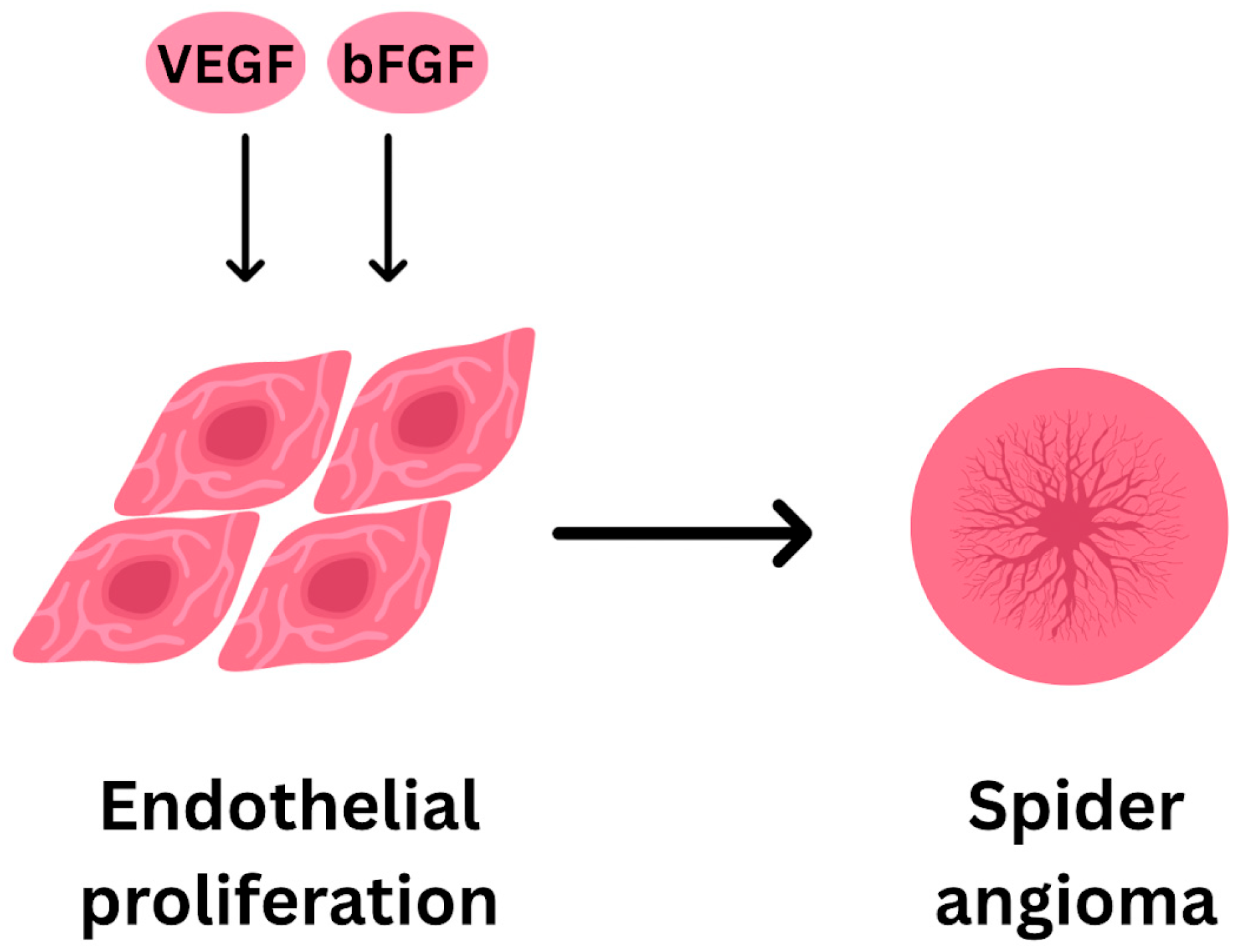
While the exact pathogenesis is still unknown, neovascularization is a likely contributor of spider angioma formation through vascular endothelial growth factor (VEGF) and basic fibroblast growth factor (bFGF), which stimulate endothelial cell proliferation and angiogenesis (Figure 2). Elevated levels of both VEGF and bFGF were found in the plasma of cirrhotic patients compared to healthy controls, and the elevation of VEGF was correlated with the size of patients’ spider angiomas [13]. Elevated levels of substance P have also been detected in patients with MASLD cirrhosis [14]. However, spider angiomas can also be observed in non-cirrhotic conditions, including pregnancy and patients taking oral contraceptives, which are thought to occur due to increased estrogen levels, resulting in increased dilation, permeability, and proliferation of vessels, which often resolve spontaneously following childbirth or cessation of birth control [12]. The impaired ability of the liver to metabolize estrogen in a cirrhotic state thus also likely contributes to the formation of spider angiomas in cirrhosis.
The presence of spider angiomas has been shown to correlate with and can act as skin markers for hepatopulmonary syndrome and esophageal varices, two serious complications of cirrhosis [12,15]. While a prior study suggested that the total number of spiders angiomas and their locations could be associated with the severity of chronic liver disease, more research is needed to determine the overall prognostic importance [16].
While spider angiomata is the most common telangiectatic lesion seen in patients with cirrhosis, diffuse cutaneous telangiectasias, including acquired bilateral telangiectatic macules (ABTMs), and telangiectasia macularis eruptiva perstans (TMEP) have also been described in the setting of chronic liver disease [17].
ABTM refers to a pattern of red brown macules with telangiectasia along the upper arms, notably, the telangiectatic nature of the macules is typically only visible via dermoscopy. In a study characterizing the etiology of these lesions, Kim et al. found that 54% of patients had hepatic disease, including alcoholic cirrhosis. Treatment for ABTM involves addressing the underlying liver disease [17]. Differential diagnoses for ABTM include erythematotelangiectatic rosacea, generalized essential telangiectasia, spider angiomata, hereditary hemorrhagic telangiectasia, and pigmented purpuric dermatoses (PPD).
TMEP is a type of cutaneous mastocytosis that presents with pruritic erythematous to brownish macules seen with telangiectasia, typically distributed on the trunk and upper extremities [18]. Unlike other mast-cell-mediated disorders, TMEP has a negative Darier’s sign, meaning physical disruption of lesions do not result in swelling and urticaria. Histopathology traditionally shows perivascular mast cell infiltrate. A case report by Huang et al. describes a patient with cirrhosis and TMEP and highlights the importance of consideration of telangiectatic disorders in cirrhosis outside of the predominant spider angiomata. TMEP is managed with antihistamines and avoidance of hepatotoxic substances. Differential diagnoses for TMEP include urticaria pigmentosa (presents without telangiectasia), spider angiomata (presents with spiraling vessels extending from central arteriole and without increased mast cells on biopsy), and systemic mastocytosis [18]
  • Palmar Erythema
Another cutaneous manifestation of cirrhosis is palmar erythema (Figure 3). Palmar erythema presents as bilateral and symmetric, non-pruritic, and non-painful erythema along the palmar surface, most commonly along both the thenar and hypothenar eminences. It has been estimated that about 23% of cirrhotic patients present with palmar erythema [19]. It is thought to occur most frequently in the palms due to the higher density of arteriovenous shunts and microscopic evaluation in patients with palmar erythema has shown an increase in the dilation of capillaries and increased palmar superficial arterial and venous plexi [20]. Again, increased estrogen levels are thought to be the mechanism associated with the vasodilation leading to palmar erythema, as estradiol stimulates the production of nitric oxide via nitric oxide synthase which induces vasodilation [15,20].
As palmar erythema is associated with increased free estrogen levels, it can be a physiologic change observed in pregnancy. Palmar erythema is also associated with autoimmune diseases, including rheumatoid arthritis, Kawasaki disease, diabetes, and sarcoidosis. In addition, palmar erythema can be drug-induced with normal liver function, with offending medications including albuterol and topiramate or other medications that induce hepatic damage, such as amiodarone, statins, fibric acid derivatives, and biologics [19,21].
  • Jaundice
Jaundice refers to yellow to green discoloration of skin caused by the deposition of bilirubin and its precursors. Jaundice is typically seen when serum bilirubin exceeds between 2 and 3 mg/dL. The face, sclera, and mucous membranes (tissues with high elastin content) are most often affected, though discoloration can be seen throughout the body [5].
The liver is the primary organ involved in bilirubin metabolism as part of the hemoglobin breakdown process. The conjugation state of the bilirubin allows for differentiation of jaundice etiologies, as follows: prehepatic, with unconjugated hyperbilirubinemia, intrahepatic, with conjugated and unconjugated hyperbilirubinemia, and post hepatic, with conjugated hyperbilirubinemia. Jaundice in cirrhosis is classified as intrahepatic, and the degree of jaundice generally correlates with advancement of liver disease [5]. Jaundice is an extremely clinically significant sign. It is a poor prognostic marker and indicates an increased risk of complications. New-onset jaundice in a patient with cirrhosis warrants evaluation for acute-on-chronic causes of hepatic decompensation, including drug-induced hepatitis, infection, or biliary stricture. Treatment of jaundice involves treatment of the underlying pathology. In the case of intrahepatic causes, treatment should prioritize appropriate volume status and prevention of infection or gastrointestinal bleeding. In cases where biliary obstruction is noted, endospcoical or surgical drainage may be necessary [22,23].
  • Pruritus
Pruritus is a common symptom and often an early symptom of chronic liver disease. Pruritus typically affects the palms and soles, though it can become generalized throughout the back, abdomen, and legs [24]. It is often chronic, though periodic, as it has been matched to circadian rhythm and shown to be worse in the evenings. Warm weather can trigger itching and advanced age, concomitant diabetes, and worsening liver disease are risk factors for development of pruritus [25]. Associated excoriation, lichenification, and secondary infection can mimic prurigo nodularis, lichen simplex chronicus, and contact dermatitis.
Pruritus is most prevalent in patients with cholestatic pathologies, affecting 70% of those with primary biliary cirrhosis (PBC). Further, patients with hepatitis B and C, intrahepatic cholestasis of pregnancy (ICP), sclerosing cholangitis, bile duct carcinoma, and biliary obstructions have been shown to be affected at greater levels [24]. The mechanism of cholestatic pruritus is multifaceted and likely involves bile acid accumulation, various cytokine release, endogenous opioids, and activation of specific sensory receptors. Prior research has established that bile salts can cause mast cell degranulation, which leads to itching [26].
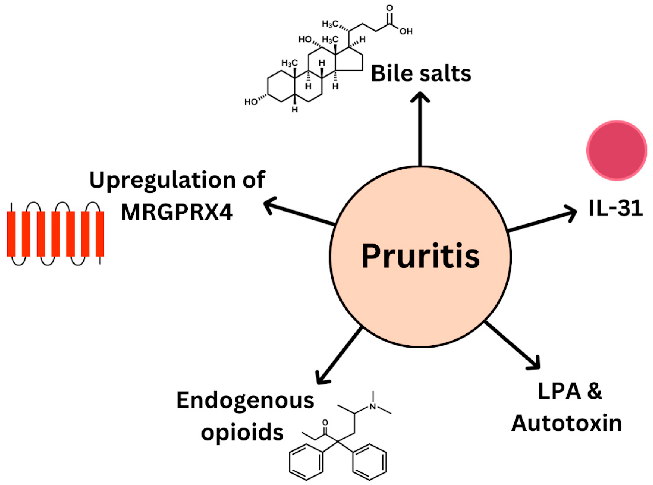
T-cell involvement is likely, as when bile acids activated farnesoid X receptor, a nuclear regulator of bile acid metabolism, increased release of IL-31, a cytokine known to induce itching, was seen. Further, when patients with cholestatic liver pathologies were given Cilefexor, a nonsteroidal FXR agonist, they had increased levels of IL-31 and pruritus [27]. Notably, not all patients with pruritus and chronic liver disease have elevated bile acids; thus, newer theories implicate the release of other pruritogens lysophosphatidic acid (LPA) and autotaxin due to hepatocyte injury in the pathogenesis. Endogenous opioids are thought to be involved in cholestatic pruritus, as impaired clearance leads to subsequent systemic elevation of compounds such as enkephalins and b-endorphin. These compounds act on the known pruritis inducing mu-opioid receptor (MOR) and may downregulate the kappa opioid receptor (KOR), an inhibitor of itch. Studies have investigated MOR inhibitors naloxone and naltrexone for the treatment of cholestatic pruritus and showed a decrease in scratching activity [28,29]. Recent research has shown that bile acids activate sensory neuron receptor Mas-related G protein-coupled receptor X4 (MRGPRX4), and murine studies have demonstrated that upregulation of MRGPRX4 results in increased pruritus (Figure 4) [30].
Treatment of pruritis in hepatic disease focuses on removal of bile acids from systemic circulation. Cholestyramine, an anion exchange resin, which binds bile acids in the gut lumen, is the first-line treatment [26]. Alternate therapies focus on disrupting metabolism of potential pruritogens or blockage of the itch response and include rifampicin, naltrexone, and sertraline [26].
  • Stasis ulcers
Stasis ulcers, also referred to as venous ulcers, occur in cirrhotic patients secondarily to portal hypertension, resulting in edema and swelling from venous incompetency.
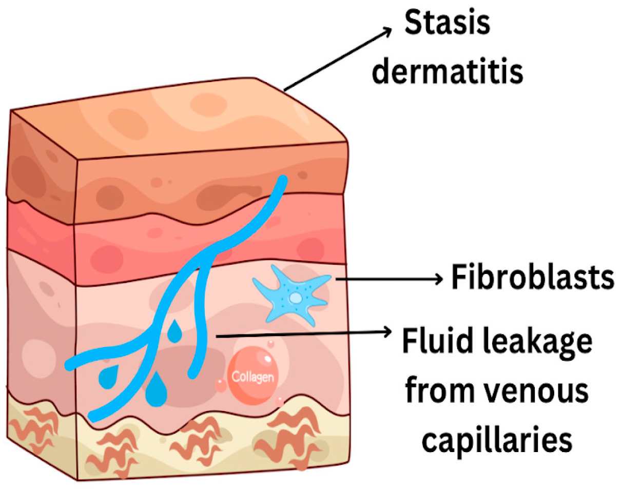
Most commonly, the skin will initially experience statis dermatitis due to fluid leakage from the venous capillaries, causing inflammation and thickening of the skin. The edematous fluid has impaired immune cell trafficking, resulting in chronic inflammation with mononuclear cells, fibroblast proliferation and collagen deposition, and progressive lymphatic duct obliteration. As inflammation and interstitial edema progresses, open ulcers may arise (Figure 5). Most commonly, the ulcers are shallow and flat, with irregular borders that may be surrounded by discoloration [31].
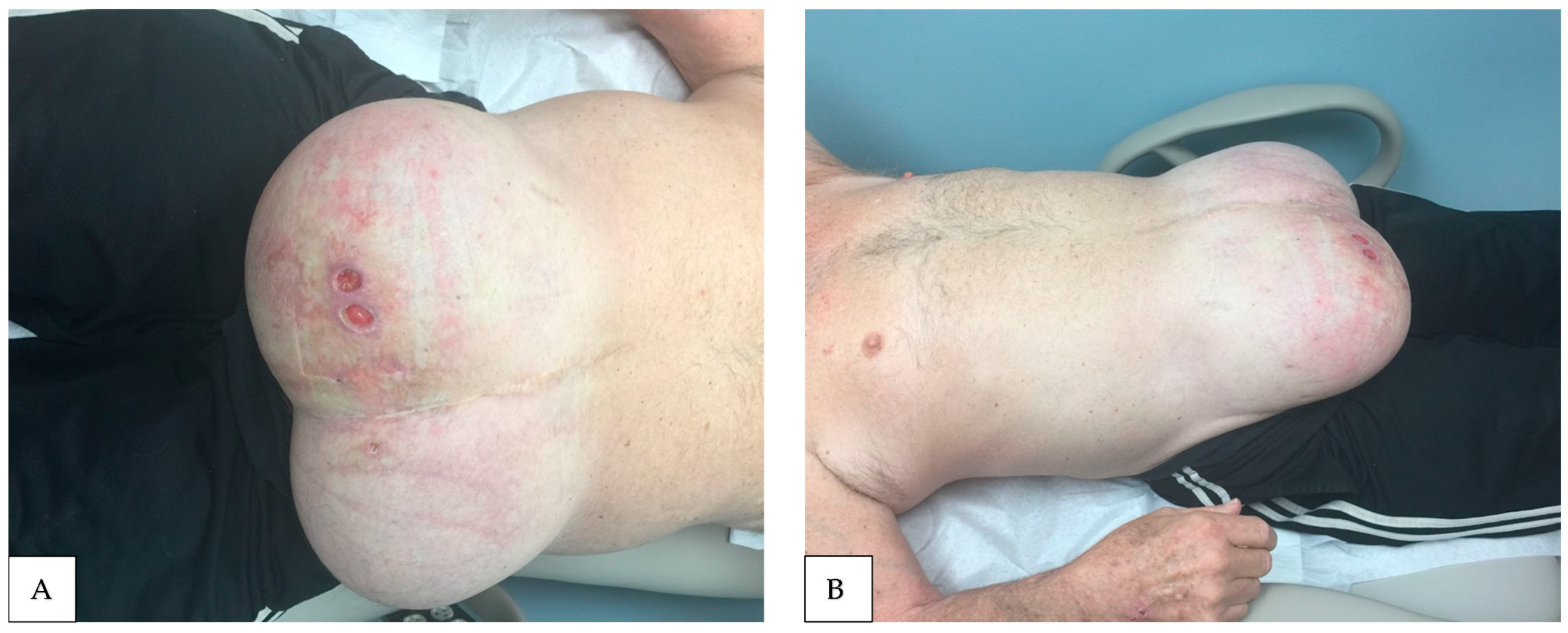
Stasis ulcers are most common on the lower legs due to the effects of gravity on the microcirculation [31]. Classically, these lower leg ulcers are often refractory and difficult to treat, but a case series revealed that decompression of the portal circulation via the placement of a transjugular intrahepatic portosystemic shunt (TIPS) leads to complete and sustained ulcer healing [32]. While cirrhosis is a known cause of stasis ulcers, venous hypertension resulting in stasis ulcers can also arise secondary to heart failure, renal failure, use of vasodilator drugs such as amlodipine, and with diseases of the lymph vessels [31]. More research is needed to better elucidate the relationship and prevalence between cirrhosis and statis ulcer development (Figure 6).
  • Other Skin and Nails Findings:
○ Terry’s nails
Terry’s nails, coined in 1954, refers to proximal whitish discoloration of the nail bed with a narrow (0.5–3 mm) brown to red band at the onychodermal region, seen in patients with alcoholic cirrhosis [33].
Prevalence of Terry’s nails in cirrhosis is approximately 25% and is hypothesized to be caused by telangiectatic changes in the nail and hypoalbuminemia [34,35]. Though initially discovered in cirrhosis, Terry’s nails have been described in patients with congestive heart failure, renal disease, diabetes, tuberculosis, reactive arthritis, and as an age-related phenomenon [34]. (Notably, when identified in younger patients, Terry’s nails are more likely to be caused by hepatic disease, thus a high degree of clinical suspicion must be maintained in these individuals [34].) Key differential diagnoses of Terry’s nails include Lindsay’s nails, proximal white discoloration of approximately 50% of the nails with blanchable distal red-brown discoloration (half and half nails) strongly associated with chronic kidney disease and thought to be caused by azotemia. Both Terry’s and Lindsay’s nails are a form of proximal apparent leukonychia (whitish discoloration due to disruption of the nail plate), and one study found that 43.8% of patients at a liver disease treatment center had leukonychia, predominantly Terry’s nails type though Lindsay’s nail type was noted [36]. Degree of distal discoloration and whether it is blanchable (Terry’s nails are typically non-blanching) are useful differentiating features. Treatment for Terry’s nails involves treatment of underlying hepatic disease, though visible nail changes may take months and lag behind resolution of other symptoms.
○ Clubbing
Clubbing refers to rounded, bulging growth of the distal nail with a loss or increase in the nail bed angle associated with chronic systemic disease.
In the case of cirrhosis, long standing hypoxia with subsequent increased blood flow and growth factor release result in the spongy, bulbous appearance of fingernails (Figure 7). One study found clubbing in 35% of patients with liver cell failure [37]. Clubbing has been noted in cirrhosis in cases of advanced disease and thought to be connected to release of hepatocyte growth factor [38]. Differential diagnoses for etiology of clubbing include pulmonary, cardiac, and gastrointestinal pathologies. Other nail changes seen in cirrhosis include brittle nails, onychorrhexis, and increased longitudinal striations [37]. The myriad nail changes described in association with cirrhosis warrant comprehensive nail examination for patients with suspected hepatic dysfunction.
○ Xanthomas/xanthelasma
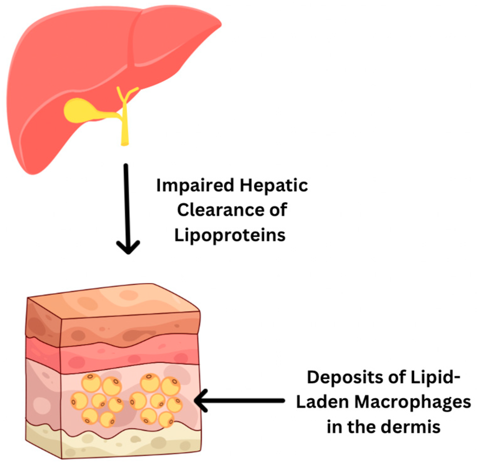
Xanthomas refer to yellow to orange papules, plaques, or nodules, which are cutaneous cholesterol deposits. Pathogenesis involves impaired hepatic clearance of lipoproteins, leading to deposits of lipid-laden macrophages in the dermis (Figure 8). Lesions are typically soft, fluctuant in growth, and asymptomatic, though they can be pruritic in the setting of cholestatic disease [5]. Xanthomas can occur throughout the body and in any pathology with hypercholesterolemia, as well as secondary dyslipidemia in the setting of hepatic disease. In patients with cirrhosis, lesions are noted to appear as eruptive xanthomas, clusters of papules typically distributed on extensor surfaces, which appear and resolve in the course of weeks, xanthelasma, and tendinous or planar xanthomas [39]. Xanthelasma refers to the distribution of xanthomas along the medial upper eyelid and is associated with PBC [40]. Tendinous xanthomas (the Achilles tendon is often affected) and xanthoma striatum palmare, in which the creases of the palms and digits appear with soft yellow deposits, are also associated with PBC [41].
Treatment of xanthomas involves addressing hyperlipidemia; therapeutic options that have shown success in cirrhosis-associated lesions include cholestyramine and ursodeoxycholic acid (UDCA), as well as plasmapheresis in severe cases [42]. Xanthelasma may not be refractory to pharmacological therapy, and surgical excision and laser treatment may be necessary [43]. Differential diagnoses for xanthomas include but are not limited to syringoma, sebaceous gland hyperplasia, milia, cutaneous sarcoidosis, and necrobiotic xanthogranuloma [44].
○ Caput medusae
Caput medusae refers to engorgement of superficial periumbilical veins and formation of collateral vessels in response to severe portal hypertension and rerouting through portocaval anastomoses.
The name is derived from the tortuous and snake-like appearance of the vessels and is a cutaneous sign of severe portal hypertension; studies have shown the presence of caput medusa in patients with hepatic dysfunction to have a positive likelihood ratio of 9.5 for cirrhosis [45]. It is associated with increased risk of complications including gastrointestinal bleeding and hepatic encephalopathy. When evaluating caput medusae, blood flow toward the head and limbs is indicative of portal hypertension. General enlargement of abdominal wall venous structures is not seen as often as caput medusa but is also a highly specific cutaneous sign of cirrhosis [46]. Treatment for caput medusae typically includes non-selective beta blockers carvedilol or propranolol to address portal hypertension. Intractable cases may necessitate transjugular intrahepatic portosystemic shunt (TIPS), and liver transplantation is definitive therapy in cirrhosis with severe portal hypertension [47]. Differential diagnoses for caput medusae encompass any pathology that elevates portal venous pressure, including portal vein thrombosis, right-sided heart failure, abdominal mass or malignancy, inferior vena cava obstruction, schistosomiasis, and Budd-Chiari syndrome [48].
○ Pellagra
Pellagra refers to the syndrome caused by deficiency of niacin (vitamin B3), an important co-factor in metabolism. The triad of pellagra involves skin manifestations and neurological and gastrointestinal symptoms. Cutaneous manifestations are often marked photosensitivity and symmetrical erythematous-to-hyperpigmented plaques in sun-exposed areas. Classically, there is involvement of the C3–C4 dermatome with a “collar-like” distribution. Neurologic and gastrointestinal symptoms associated with pellagra include irritability, insomnia, general cognitive decline, encephalopathy, diarrhea, and glossitis. Pellagra is relevant in the context of alcoholic cirrhosis, as alcohol interferes with hepatic metabolism of B3 and with the absorption of the precursor to niacin—tryptophan. Further, alcoholic cirrhosis is often associated with malnutrition, and, thus, insufficient intake of vitamin B3 further predisposes individuals to deficiencies. Pellagra may present as altered mental status, and the triad of symptoms may be subtle, and, thus, clinicians must have a high degree of suspicion to identify this syndrome.
○ Acanthosis Nigricans
Acanthosis nigricans (AN) presents as hyperpigmented, velvety plaques in intertriginous regions, including the axilla, groin, and neck. The pathophysiology of AN, which is strongly associated with metabolic syndrome and T2DM, is likely due to hyperinsulinemia and subsequent activation of various fibroblast growth factors. MASLD involves insulin resistance, and, thus, AN is a potential cutaneous manifestation of this disease. A recent cross-sectional study demonstrated that AN correlated to a greater degree of steatosis in individuals with MASLD cirrhosis.
○ Erythema Nodosum and Pyoderma Gangrenosum
Inflammatory bowel disease (IBD), with or without primary sclerosing cholangitis (PSC), is associated with erythema nodosum or pyoderma gangrenosum. Erythema nodosum presents as red, tender nodules on the lower extremities. Pyoderma gangrenosum manifests first as erythematous pustules or nodules and then spreads to adjacent skin and develops into burrowing ulcers. Both of these skin manifestations are extrahepatic complications of PSC, and when seen clinically, should raise suspicion for underlying cirrhotic disorders.

4. Pathophysiological Mechanisms Linking Liver Dysfunction to Skin Changes

The liver plays a vital role in numerous physiological processes, including the synthesis of hormone-transporting proteins, conjugation of bilirubin, detoxification of drugs and toxins like alcohol, and the metabolism of lipids, carbohydrates, and other nutrients [43]. Hepatic injury can impair any of these functions, potentially leading to systemic effects that can involve the skin [43].
Hormonal imbalances resulting from liver cirrhosis have widespread effects, particularly on sex hormone regulation. In the bloodstream, both testosterone and estradiol are bound to albumin and sex-hormone-binding globulin (SHBG), with testosterone having a higher affinity for SHBG [49]. Damage from cirrhosis results in impaired hepatic clearance of estrogen secondary to portal hypertension, decreased testosterone production, elevated SHBG levels, and dysfunction of the hypothalamic–pituitary axis [50]. Increased SHBG levels are thought to result from upregulated hepatic synthesis, driven by excess estrogen and diminished testosterone-mediated suppression [50]. Because SHBG preferentially binds testosterone, free testosterone levels are further disproportionately reduced [51,52]. Excessive estrogen contributes to several dermatological manifestations commonly seen in cirrhosis, such as spider angiomas and palmar erythema [52].
Dysfunction of the bilirubin cycle due to liver cirrhosis can cause elevation of both unconjugated and conjugated bilirubin [53]. Fibrosis of hepatic parenchyma from liver cirrhosis disrupts liver structure and function. Damage to hepatocytes results in decreased hepatic intake of unconjugated bilirubin and decreased conjugation [54]. Hepatocellular dysfunction and scar tissue compression of bile ducts also result in impairment of excretion of conjugated bilirubin into bile [54]. The elevation of bilirubin levels beyond 2.5–3.0 mg/dL leads to jaundice [5].
Fat malabsorption due to hyperbilirubinemia from liver cirrhosis can lead to nutritional deficiencies in fat-soluble vitamins A, D, E, and K [55]. Damage to hepatic parenchyma also contributes to decreased storage and impairment of the conversion of vitamins to their metabolically active forms [55]. In the skin, vitamin A is integral to maintaining the specialized epithelial surfaces of the body and deficiency can cause inappropriate keratinization of stratified squamous epithelium [56]. Low vitamin D has been associated with alopecia areata [15]. Collagen cross-linking was accelerated in rats with vitamin E deficiency [57]. Vitamin K is necessary for the formation of clotting factors in the coagulation cascade; deficiency can present as purpura, ecchymoses, and gingival bleeding [58].
Autoimmune liver conditions, including primary biliary cholangitis (PBC), primary sclerosing cholangitis (PSC), and autoimmune hepatitis (AIH), have also been associated with dermatologic conditions. For example, PBC causes destruction of intrahepatic bile ducts leading to cholestasis and resultant dyslipidemia, which can lead to the formation of cutaneous xanthomas. Xanthomas often appear as soft yellow-brown papules/plaques and are commonly found on the eyelids (referred to as xanthelasma) or skin folds, such as on the palmar creases and are most commonly described in patients with familial hypercholesterolemia. Interestingly, xanthomas in PBC have been associated with an abnormal lipoprotein called lipoprotein X (Harris). Additionally, AIH has been associated with a variety of additional autoimmune-mediated cutaneous disorders, including vitiligo; psoriasis; alopecia; autoimmune bullous diseases such as pemphigus vulgaris/dermatitis herpetiform; neutrophilic dermatoses such as pyoderma gangrenosum; and lichen planus. As patients with AIH often receive immunosuppressive treatment, they are also at an increased risk of non-melanoma skin cancer.
Additional cutaneous manifestations of liver cirrhosis include cyanosis and digital clubbing, typically only seen in patients with pulmonary complications, such as hepatopulmonary syndrome (HPS) and portopulmonary hypertension (POPH). Though the exact pathophysiology remains unclear, the occurrence of intrapulmonary vascular dilatations in advanced hepatic disease disrupts pulmonary gas exchange, which can lead to ventilation–perfusion mismatch. In patients presenting with vague pulmonary symptoms, the presence of cyanosis and digital clubbing, in conjunction with other cutaneous findings of advanced liver disease, can aid in identification of HPS or POPH in advanced stages of cirrhosis. Identification of these complications is critical, particularly in evaluation for liver transplantation.

5. Diagnostic and Prognostic Value of Skin Signs

Cutaneous skin findings are one of the earliest indicators of hepatic dysfunction that leads clinicians to suspect a diagnosis of cirrhosis. The presence of multiple cutaneous skin manifestations, such as jaundice, pruritus, palmar erythema, and spider angiomas, further suggests cirrhosis [5]. Understanding these dermatologic manifestations and using them as critical diagnosis and prognostic indicators allows clinicians to help optimize patient care and improve clinical outcomes in cirrhotic patients. Jaundice stems from hepatocyte failure or biliary obstruction that causes the elevation of bilirubin accumulation reflecting advanced-stage cirrhosis [8]. Cholestasis and bile acid deposition on the skin leads to pruritus, which is also associated with advanced cirrhosis and worse quality of life measures [59]. Palmer erythema with symmetric redness on the hands due to elevated nitric oxide and estrogen levels reflects peripheral vasodilation [60]. Portal hypertension and impaired hepatic metabolism of estrogen in advanced stages of cirrhosis lead to spider angiomas on the face, neck, upper chest, and arms [16]. A large area, a significant number, or atypical locations of spider angiomas are correlated with higher severity of chronic liver disease [61].
Distinguishing skin findings that occur in patients with liver cirrhosis from those that can also occur in patients without liver cirrhosis can be challenging. It requires a holistic approach, assessing the patient’s clinical picture while integrating the skin manifestations with other physical exam findings and diagnostic modalities. While certain findings, such as Terry nails, spider angiomata, palmar erythema, jaundice, and caput medusae, can be more specific in cirrhosis, they can also occur in other diseases. However, their presence should prompt a comprehensive liver evaluation. In contrast, there are many other less specific findings, such as pruritus and xerosis, that may be seen in various systemic conditions. An astute clinician should utilize physical exam findings along with other data to evaluate for different etiologies that may be causing those skin manifestations to improve early diagnosis.
Although pruritus and other skin manifestations of cirrhosis may have limited diagnostic value, they play a crucial role in assessing patients’ quality of life. These symptoms hold significant clinical importance, particularly for symptom management and improving patient outcomes. Having said that, there have been data showing some association of certain skin manifestations with advanced disease. Prior studies have showed that patients who have more spider angiomas tend to have higher serum bilirubin and longer prothrombin time, both of which are markers of more advanced liver disease. Furthermore, there have been reports suggesting that the severity of cutaneous manifestations often parallels that of hepatic dysfunction which is measured by Model for End-Stage Liver Disease (MELD) scores and Child–Pugh classifications. Serum bilirubin quantifies jaundice and directly contributes to both MELD and Child–Pugh calculations. Pruritus intensity shows a positive association with bilirubin levels and MELD scores in cholestatic cirrhosis [59]. When spider angiomas appear on the chest or abdomen, it is associated with a higher MELD score (10.77 ± 6.76 versus 7.68 ± 5.42, p = 0.003), indicating a higher mortality [16]. Child–Pugh classification can be integrated with cutaneous manifestations to assess disease severity. These data, while scarce, suggest a possible role of cutaneous manifestations in evaluating the degree of cirrhosis. Thus, clinicians need to recognize and identify the presence of these characteristic skin changes and their associations with the stage of cirrhosis. Further research is warranted to better elucidate the diagnostic and prognostic value of different skin manifestations in liver cirrhosis.
The following table summarizes the key dermatologic manifestations, their prevalence in liver cirrhosis, and their clinical relevance based on available literature (Table 1).
While the data evaluating the prognostic and diagnostic value of cutaneous entities in cirrhosis are limited, there is a growing body of literature showing the impact of skin symptoms on patients’ quality of life. For instance, pruritus is associated with poor sleep and psychological impairment, which lead to worse quality of life. Skin manifestations in cirrhosis impose a significant burden, negatively impacting physical comfort, sleep, mental well-being, and social life. Regular evaluation and focused treatment of these symptoms are vital to enhancing quality of life and patient-centered outcomes.

6. Management Implications

The management of cutaneous manifestations of cirrhosis relies on addressing the underlying hepatic dysfunction. Antiviral therapies that treat hepatitis B and C reduce liver inflammation and, thus, reduce spider angiomas and pruritis. Abstinence from alcohol and nutritional support can resolve palmer erythema in alcoholic cirrhosis [62]. Liver transplantation remains a definitive treatment for end-stage cirrhosis, which can lead to the resolution of these cutaneous symptoms [61]. Management of pruritis relies on the traditional first-line treatment of cholestyramine, which is a bile acid sequestrant that can decrease the amount of bile acid in the serum [63]. However, other studies have suggested a transition of first-line treatment to fibrates, as it provides a safer profile and anticholestatic properties [64]. Antihistamines and topical corticosteroids remain as other options for the symptomatic management of pruritis. Jaundice treatment primarily follows treating the underlying liver dysfunction. Monitoring cutaneous signs provides a simple, non-invasive method to assess the disease progression and treatment response. At baseline, these cutaneous signs should be identified and continually documented throughout to establish any systemic improvement or worsening of hepatic function.

7. Overall Importance of Recognizing and Addressing Cutaneous Manifestations of Liver Cirrhosis

Cutaneous signs of advanced liver disease may provide some insight into the severity of hepatic dysfunction and overall prognosis. For example, one study determined that the presence of at least one of four characteristic skin findings (spider naevi, palmar erythema, nail changes, or bleeding) was associated with the presence of severe fibrosis or cirrhosis. Conversely, only a low percentage of patients were found to have advanced liver disease in the absence of these findings [6]. Patients with spider nevi in particular were more than four times more likely to have severe liver disease [65]. Furthermore, the presence of subcutaneous collateral vessels upon physical exam predicted worse outcomes in patients with cirrhosis [66].
Skin findings may serve as early non-invasive indicators of advanced liver disease. Given their potential prognostic significance, they could provide a more accessible and cost-effective tool in the assessment of advanced liver disease. Therefore, dermatologists may play an important role in the multidisciplinary care team for these patients. For one, early identification of characteristic skin findings by dermatologists can prompt further testing, facilitate more rapid diagnosis, and improve long-term outcomes [60,67]. Additionally, in patients with pre-existing liver disease, dermatologists can play a role in monitoring for findings suggestive of illness progression.
Despite the potential clinical utility of cutaneous findings in characterization of severe liver disease, there are still significant limitations to its application. Variation among individual patients may impact the prognostic value of skin findings. Moreover, many of these findings lack sensitivity for advanced liver disease, thus limiting their usefulness for screening otherwise asymptomatic individuals without a high suspicion for advanced liver disease. For example, spider angiomas have a high association with pregnancy due to the hormonal changes that take place during gestation [5]. Lastly, the identification of any cutaneous lesion can vary widely based on skin tone, further necessitating the incorporation of diversity in research and medical education materials.
One of the essential components to provide a holistic approach to patient care is the collaboration between dermatologists and hepatologists for patients with skin manifestations of liver cirrhosis. Early recognition of the skin presentations of liver cirrhosis by dermatologists can lead to timely referral and early diagnosis, potentially preventing disease progression and improving patient outcomes. On the other hand, hepatologists should also consider prompt dermatology referrals that can help in further addressing patients’ symptoms and improving quality of life. A multidisciplinary collaborative approach can help in improving patient education and understanding of their disease and even adherence to treatment and follow-up.

8. Future Directions

The integration of dermatologic screening into the routine clinical care of patients suspected to have cirrhosis can facilitate earlier diagnosis. Cirrhosis management reduces the risk of serious complications and liver decompensation if initiated early, while significantly improving a patient’s quality of life [62]. Further research is needed to establish how specific cutaneous manifestations correlate to the severity and progression of cirrhosis. For example, spider angiomas can be seen in one-third of patients with cirrhosis and correlates with the frequency of esophageal varices, a complication of liver cirrhosis that can lead to hemorrhage [15,62]. Additionally, examining the psychosocial impact of cutaneous manifestations could help determine if dermatological interventions should be incorporated as part of the standard of care for cirrhosis patients.
Integrating the skin manifestations of liver cirrhosis and their underlying pathophysiology, diagnostic significance, and prognostic value into medical curricula and training programs offers a valuable opportunity to enhance early recognition among healthcare trainees. Educating medical students and residents on these cutaneous signs and their clinical implications can build clinical skills needed for the detection of liver disease at an earlier stage, prompting timely referrals, diagnosis, and management. Understanding the pathophysiology behind these disorders can also help in fostering further research studies to explore these biochemical pathways. This can also foster a collaborative multidisciplinary approach between hepatology and dermatology, providing a more holistic approach to patient care.

9. Conclusions

Cutaneous manifestations of liver cirrhosis reflect the underlying systemic dysfunction and can serve as early indicators of hepatic pathology. Non-specific signs such as spider angiomas, palmar erythema, jaundice, and pruritus may precede more overt clinical symptoms and offer valuable diagnostic and prognostic insights. Given their accessibility during physical examination, these dermatologic features should be routinely assessed in patients at risk for chronic liver disease. Regular skin evaluations can facilitate earlier detection of liver cirrhosis, allowing for timely diagnostic workup, initiation of management strategies, and improved patient outcomes. A thorough dermatological examination can aid in the assessment of liver cirrhosis by identifying skin manifestations that are more specific to liver disease, such as spider angioma, palmar erythema, and Terry’s nails. These findings, when interpreted in the context of other clinical and laboratory data, are often associated with advanced liver disease and clinically significant portal hypertension, and their presence should prompt a comprehensive evaluation for signs of decompensation. Increasing clinician awareness and education regarding the dermatologic signs of liver cirrhosis is essential. By recognizing these cutaneous clues, healthcare providers can intervene earlier, mitigating complications and enhancing the quality of care for individuals with or at risk for liver cirrhosis.

Author Contributions

Conceptualization, R.K., Y.A., and J.B.T.; methodology, R.K., Y.A., and J.B.T.; software, R.K.; validation, C.R., and J.B.T.; formal analysis, R.K.; investigation, R.K., and J.B.T.; resources, R.K.; data curation, R.K.; writing—original draft preparation, R.K., R.R., R.A., T.C., C.D., A.P., and A.L.; writing—review and editing, C.R., and J.B.T.; visualization, C.R., and J.B.T.; supervision, C.R., and J.B.T.; project administration, R.K. All authors have read and agreed to the published version of the manuscript.

Funding

This research received no external funding.

Institutional Review Board Statement

Our review article involved only two patients and was limited to descriptive reporting without hypothesis testing or systematic investigation. As such, it does not meet the definition of human subjects research per institutional guidelines and was exempt from IRB/ethics committee review. Informed consent was obtained from both patients for the publication of their clinical information and images. No identifiable personal information was included in the manuscript. All other data included in the manuscript were based on available medical literature.

Informed Consent Statement

Informed consent was obtained from the patients to share deidentifed data.

Data Availability Statement

No new data were created or analyzed in this study. Data sharing is not applicable to this article.

Conflicts of Interest

The authors declare no conflicts of interest.

References

  1. Tsochatzis, E.A.; Bosch, J.; Burroughs, A.K. Liver cirrhosis. Lancet 2014, 383, 1749–1761. [Google Scholar] [CrossRef] [PubMed]
  2. Schuppan, D.; Afdhal, N.H. Liver cirrhosis. Lancet 2008, 371, 838–851. [Google Scholar] [CrossRef] [PubMed]
  3. Horvatits, T.; Drolz, A.; Trauner, M.; Fuhrmann, V. Liver Injury and Failure in Critical Illness. Hepatology 2019, 70, 2204–2215. [Google Scholar] [CrossRef] [PubMed]
  4. Martínez Jiménez, S. Cutaneous Manifestations of Liver Disease: A Narrative Review. Cureus 2024, 16, e70357. [Google Scholar] [CrossRef] [PubMed]
  5. Liu, Y.; Zhao, Y.; Gao, X.; Liu, J.; Ji, F.; Hsu, Y.-C.; Li, Z.; Nguyen, M.H. Recognizing skin conditions in patients with cirrhosis: A narrative review. Ann. Med. 2022, 54, 3016–3028. [Google Scholar] [CrossRef] [PubMed]
  6. Niederau, C.; Lange, S.; Frühauf, M.; Thiel, A. Cutaneous signs of liver disease: Value for prognosis of severe fibrosis and cirrhosis. Liver Int. 2008, 28, 659–666. [Google Scholar] [CrossRef]
  7. Runyon, B.A. A Primer on Detecting Cirrhosis and Caring for These Patients without Causing Harm. Int. J. Hepatol. 2011, 2011, 801983. [Google Scholar] [CrossRef]
  8. Ginès, P.; Krag, A.; Abraldes, J.G.; Solà, E.; Fabrellas, N.; Kamath, P.S. Liver cirrhosis. Lancet 2021, 398, 1359–1376. [Google Scholar] [CrossRef]
  9. Somnay, K.; Wadgaonkar, P.; Sridhar, N.; Roshni, P.; Rao, N.; Wadgaonkar, R. Liver Fibrosis Leading to Cirrhosis: Basic Mechanisms and Clinical Perspectives. Biomedicines 2024, 12, 2229. [Google Scholar] [CrossRef]
  10. Jagdish, R.K.; Roy, A.; Kumar, K.; Premkumar, M.; Sharma, M.; Rao, P.N.; Reddy, D.N.; Kulkarni, A.V. Pathophysiology and management of liver cirrhosis: From portal hypertension to acute-on-chronic liver failure. Front. Med. 2023, 10, 1060073. [Google Scholar] [CrossRef]
  11. Fallahzadeh, M.A.; Rahimi, R.S. Hepatic Encephalopathy: Current and Emerging Treatment Modalities. Clin. Gastroenterol. Hepatol. 2022, 20, S9–S19. [Google Scholar] [CrossRef]
  12. Samant, H.; Kothadia, J.P. Spider Angioma. In StatPearls [Internet]; StatPearls Publishing: Treasure Island, FL, USA, 2023. [Google Scholar] [PubMed]
  13. Li, C.P. Spider angiomas in patients with liver cirrhosis: Role of vascular endothelial growth factor and basic fibroblast growth factor. World J. Gastroenterol. 2003, 9, 2832. [Google Scholar] [CrossRef]
  14. Li, C.P.; Lee, F.Y.; Hwang, S.J.; Chang, F.-Y.; Lin, H.-C.; Lu, R.-H.; Hou, M.-C.; Chu, C.-J.; Chan, C.-C.; Luo, J.-C.; et al. Role of Substance P in The Pathogenesis of Spider Angiomas in Patients with Nonalcoholic Liver Cirrhosis. Am. J. Gastroenterol. 1999, 94, 502–507. [Google Scholar] [CrossRef]
  15. Bhandari, A.; Mahajan, R. Skin Changes in Cirrhosis. J. Clin. Exp. Hepatol. 2022, 12, 1215–1224. [Google Scholar] [CrossRef] [PubMed]
  16. Terp, K.; Izquierdo-Pretel, G. Spider Angioma Number and Location as Potential Prognostic Indicators in Chronic Liver Disease: A Case Report. Cureus 2023, 15, e34193. [Google Scholar] [CrossRef] [PubMed]
  17. Kim, G.-W.; Shin, K.; Kim, T.-W.; Kim, T.-W.; You, H.-S.; Jin, H.-J.; Shim, W.-H.; Kim, H.-S.; Ko, H.-C.; Kim, B.-S.; et al. The importance of dermoscopy for the diagnosis of acquired bilateral telangiectatic macules: The angioid streak pattern reveals underlying chronic liver disease. J. Eur. Acad. Dermatol. Venereol. 2018, 32, 1597–1601. [Google Scholar] [CrossRef] [PubMed]
  18. Huang, A.; Desai, A.; Brinster, N.; Marmon, S. Telangiectasia macularis eruptiva perstans in the presence of liver cirrhosis. JAAD Case Rep. 2020, 6, 438–440. [Google Scholar] [CrossRef] [PubMed]
  19. Serrao, R.; Zirwas, M.; English, J.C. Palmar Erythema. Am. J. Clin. Dermatol. 2007, 8, 347–356. [Google Scholar] [CrossRef]
  20. Nautiyal, A.; Chopra, K.B. Liver Palms (Palmar Erythema). Am. J. Med. 2010, 123, 596–597. [Google Scholar] [CrossRef]
  21. Milam, P.; Berger, M.; Ramaswamy, B.; Reinbolt, R.; Wesolowski, R.; Kaffenberger, B.H. Spider Telangiectases and Palmar Erythema as Harbingers of Structural Liver Changes in Three Breast Cancer Patients on Ado-trastuzumab Emtansine. J. Clin. Aesthet Dermatol. 2019, 12, 23. [Google Scholar]
  22. Gustot, T.; Stadlbauer, V.; Laleman, W.; Alessandria, C.; Thursz, M. Transition to decompensation and acute-on-chronic liver failure: Role of predisposing factors and precipitating events. J. Hepatol. 2021, 75, S36–S48. [Google Scholar] [CrossRef]
  23. Harrison, P.M. Management of patients with decompensated cirrhosis. Clin. Med. 2015, 15, 201–203. [Google Scholar] [CrossRef] [PubMed]
  24. Selim, R.; Ahn, J. Pruritus in Chronic Liver Disease. Clin. Liver Dis. 2023, 27, 47–55. [Google Scholar] [CrossRef]
  25. Bassari, R. Jaundice associated pruritis: A review of pathophysiology and treatment. World J. Gastroenterol. 2015, 21, 1404. [Google Scholar] [CrossRef]
  26. Düll, M.M.; Kremer, A.E. Newer Approaches to the Management of Pruritus in Cholestatic Liver Disease. Curr. Hepatol. Rep. 2020, 19, 86–95. [Google Scholar] [CrossRef]
  27. Xu, J.; Wang, Y.; Khoshdeli, M.; Peach, M.; Chuang, J.; Lin, J.; Tsai, W.; Mahadevan, S.; Minto, W.; Diehl, L.; et al. IL-31 levels correlate with pruritus in patients with cholestatic and metabolic liver diseases and is farnesoid X receptor responsive in NASH. Hepatology 2023, 77, 20–32. [Google Scholar] [CrossRef]
  28. Terg, R.; Coronel, E.; Sordá, J.; Muñoz, A.E.; Findor, J. Efficacy and safety of oral naltrexone treatment for pruritus of cholestasis, a crossover, double blind, placebo-controlled study. J. Hepatol. 2002, 37, 717–722. [Google Scholar] [CrossRef]
  29. Bergasa, N.V.; Talbot, T.L.; Alling, D.W.; Schmitt, J.M.; Walker, E.C.; Baker, B.L.; Korenman, J.C.; Park, Y.; Hoofnagle, J.H.; Jones, E. A controlled trial of naloxone infusions for the pruritus of chronic cholestasis. Gastroenterology 1992, 102, 544–549. [Google Scholar] [CrossRef] [PubMed]
  30. Meixiong, J.; Vasavda, C.; Snyder, S.H.; Dong, X. MRGPRX4 is a G protein-coupled receptor activated by bile acids that may contribute to cholestatic pruritus. Proc. Natl. Acad. Sci. USA 2019, 116, 10525–10530. [Google Scholar] [CrossRef] [PubMed]
  31. Naschitz, J.E. Stasis Ulcers—The Unifying Concept. J. Clin. Res. Rep. 2021, 9, 1–5. [Google Scholar] [CrossRef]
  32. El Younis, C.M.; Bergasa, N.V. Healing of leg ulcers associated with transjugular intrahepatic portosystemic shunt in decompensated cirrhosis: Case series of a possible hepatodermal syndrome. Gastroenterol Hepatol. 2008, 4, 211. [Google Scholar]
  33. Terry, R. White nails in hepatic cirrhosis. Lancet 1954, 263, 757–759. [Google Scholar] [CrossRef] [PubMed]
  34. Nia, A.M.; Ederer, S.; Dahlem, K.M.; Gassanov, N.; Er, F. Terry’s Nails: A Window to Systemic Diseases. Am. J. Med. 2011, 124, 602–604. [Google Scholar] [CrossRef] [PubMed]
  35. Sack, J.S.; Liu, A.F.; Gray, M.; Roat, J.; Zucker, S.D. Association of Terry Nails with Liver Cirrhosis. Am. J. Gastroenterol. 2021, 116, 2455–2458. [Google Scholar] [CrossRef]
  36. Fernandez-Somoza, J.-M.; Ginarte, M.; Otero, E.; Tomé, S.; Soutullo, C.; Martínez-Ulloa, A.; Gonzalez-Quintela, A.; Sepehrimanesh, M. Clinical and capillaroscopic findings in patients with liver disease and proximal apparent leukonychia (Terry nails and its variants). Medicine 2021, 100, e26207. [Google Scholar] [CrossRef]
  37. Salem, A.; Al Mokadem, S.; Attwa, E.; Abd El Raoof, S.; Ebrahim, H.; Faheem, K. Nail changes in chronic renal failure patients under haemodialysis. J. Eur. Acad. Dermatol. Venereol. 2008, 22, 1326–1331. [Google Scholar] [CrossRef] [PubMed]
  38. Hojo, S.; Fujita, J.; Yamadori, I.; Ezaki, T.; Watanabe, S.; Yamanouchi, H.; Miyawaki, H.; Yamaji, Y.; Nishioka, M.; Takahara, J. Hepatocyte Growth Factor and Digital Clubbing. Intern. Med. 1997, 36, 44–46. [Google Scholar] [CrossRef]
  39. Baila-Rueda, L.; Mateo-Gallego, R.; Lamiquiz-Moneo, I.; Cenarro, A.; Civeira, F. Severe hypercholesterolemia and phytosterolemia with extensive xanthomas in primary biliary cirrhosis: Role of biliary excretion on sterol homeostasis. J. Clin. Lipidol. 2014, 8, 520–524. [Google Scholar] [CrossRef]
  40. Al Aboud, A.M.; Shah, S.S.; Blair, K.; Al Aboud, D.M. Xanthelasma Palpebrarum. In StatPearls [Internet]; StatPearls Publishing: Treasure Island, FL, USA, 2025. Available online: https://www.ncbi.nlm.nih.gov/books/NBK531501/ (accessed on 1 July 2025).
  41. Sexton, F.; Shanahan, W.; Ryan, J.D. Xanthoma striatum palmare: Getting a handle on hyperlipidaemia in primary biliary cholangitis. Clin. Res. Hepatol. Gastroenterol. 2024, 48, 102255. [Google Scholar] [CrossRef]
  42. Suzuki, L.; Hirayama, S.; Fukui, M.; Sasaki, M.; Hiroi, S.; Ayaori, M.; Terai, S.; Tozuka, M.; Watada, H.; Miida, T. Lipoprotein-X in cholestatic patients causes xanthomas and promotes foam cell formation in human macrophages. J. Clin. Lipidol. 2017, 11, 110–118. [Google Scholar] [CrossRef]
  43. Hazin, R.; Tamimi, T.I.A.R.; Abuzetun, J.Y.; Zein, N.N. Recognizing and treating cutaneous signs of liver disease. Cleve Clin. J. Med. 2009, 76, 599–606. [Google Scholar] [CrossRef] [PubMed]
  44. Van Alfen, B.; Hatch, J.; Conley, M.; Seeliyur Duraiswamy, S. A Case Report of Xanthelasma and the Associated Differential Diagnosis. Cureus 2025, 17, e81971. [Google Scholar] [CrossRef]
  45. Saunders Elsevier. Evidence-Based Physical Diagnosis; Saunders Elsevier: Philadelphia, PA, USA, 2012. [Google Scholar]
  46. Wilson, R.; Williams, D.M. Cirrhosis. Med. Clin. N. Am. 2022, 106, 437–446. [Google Scholar] [CrossRef]
  47. Abraldes, J.G.; Caraceni, P.; Ghabril, M.; Garcia-Tsao, G. Update in the Treatment of the Complications of Cirrhosis. Clin. Gastroenterol. Hepatol. 2023, 21, 2100–2109. [Google Scholar] [CrossRef]
  48. Simonetto, D.A.; Liu, M.; Kamath, P.S. Portal Hypertension and Related Complications: Diagnosis and Management. Mayo Clin. Proc. 2019, 94, 714–726. [Google Scholar] [CrossRef] [PubMed]
  49. Nelson, L.R.; Bulun, S.E. Estrogen production and action. J. Am. Acad. Dermatol. 2001, 45, S116–S124. [Google Scholar] [CrossRef] [PubMed]
  50. Neong, S.F.; Billington, E.O.; Congly, S.E. Sexual Dysfunction and Sex Hormone Abnormalities in Patients with Cirrhosis: Review of Pathogenesis and Management. Hepatology 2019, 69, 2683–2695. [Google Scholar] [CrossRef]
  51. Sinclair, M.; Grossmann, M.; Gow, P.J.; Angus, P.W. Testosterone in men with advanced liver disease: Abnormalities and implications. J. Gastroenterol. Hepatol. 2015, 30, 244–251. [Google Scholar] [CrossRef]
  52. Patel, A.D.; Katz, K.; Gordon, K.B. Cutaneous Manifestations of Chronic Liver Disease. Clin. Liver Dis. 2020, 24, 351–360. [Google Scholar] [CrossRef]
  53. Roche, S.P.; Kobos, R. Jaundice in the adult patient. Am. Fam. Phys. 2004, 69, 299–304. [Google Scholar]
  54. Berumen, J.; Baglieri, J.; Kisseleva, T.; Mekeel, K. Liver fibrosis: Pathophysiology and clinical implications. WIREs Mech Dis. 2021, 13, e1499. [Google Scholar] [CrossRef] [PubMed]
  55. Dogra, S.; Jindal, R. Cutaneous Manifestations of Common Liver Diseases. J. Clin. Exp. Hepatol. 2011, 1, 177–184. [Google Scholar] [CrossRef]
  56. Roche, F.C.; Harris-Tryon, T.A. Illuminating the Role of Vitamin A in Skin Innate Immunity and the Skin Microbiome: A Narrative Review. Nutrients. 2021, 13, 302. [Google Scholar] [CrossRef]
  57. Igarashi, A.; Uzuka, M.; Nakajima, K. The effects of vitamin E deficiency on rat skin. Br. J. Dermatol. 1989, 121, 43–49. [Google Scholar] [CrossRef]
  58. Ballal, S.; Prathibha, J.P.; Pinto, A.V.; Srinivasa, S.; Augustine, M. Unusual Manifestation of Vitamin K Deficiency, Nodular Purpura: A Case Series. Indian Dermatol. Online J. 2025, 16, 132–136. [Google Scholar] [CrossRef]
  59. Mayo, M.J.; Carey, E.; Smith, H.T.; Mospan, A.R.; McLaughlin, M.; Thompson, A.; Morris, H.L.; Sandefur, R.; Kim, W.R.; Bowlus, C.; et al. Impact of Pruritus on Quality of Life and Current Treatment Patterns in Patients with Primary Biliary Cholangitis. Dig. Dis. Sci. 2023, 68, 995–1005. [Google Scholar] [CrossRef] [PubMed]
  60. Schwarz, M.; Schwarz, C.; Burghart, L.; Pfisterer, N.; Bauer, D.; Hübl, W.; Mandorfer, M.; Gschwantler, M.; Reiberger, T.; Elbahrawy, A. Late-stage presentation with decompensated cirrhosis is alarmingly common but successful etiologic therapy allows for favorable clinical outcomes. PLoS ONE 2023, 18, e0290352. [Google Scholar] [CrossRef] [PubMed]
  61. Jindal, A.; Gupta, A.; Bhadoria, A.S. Resolution of Multiple Large Spider Angiomas After Liver Transplantation in Severe Alcoholic Hepatitis. Indian J. Dermatol. 2022, 67, 837. [Google Scholar] [CrossRef]
  62. Fadlallah, H.; El Masri, D.; Bahmad, H.F.; Abou-Kheir, W.; El Masri, J. Update on the Complications and Management of Liver Cirrhosis. Med. Sci. 2025, 13, 13. [Google Scholar] [CrossRef]
  63. Ebhohon, E.; Chung, R.T. Systematic review: Efficacy of therapies for cholestatic pruritus. Therap. Adv. Gastroenterol. 2023, 16. [Google Scholar] [CrossRef]
  64. Bolier, R.; de Vries, E.S.; Parés, A.; Helder, J.; Kemper, E.M.; Zwinderman, K.; Elferink, R.P.O.; Beuers, U. Fibrates for the treatment of cholestatic itch (FITCH): Study protocol for a randomized controlled trial. Trials 2017, 18, 230. [Google Scholar] [CrossRef]
  65. Udell, J.A.; Wang, C.S.; Tinmouth, J.; FitzGerald, J.M.; Ayas, N.T.; Simel, D.L.; Schulzer, M.; Mak, E.; Yoshida, E.M. Does This Patient with Liver Disease Have Cirrhosis? JAMA 2012, 307, 832. [Google Scholar] [CrossRef] [PubMed]
  66. Li, H.; Wang, R.; Méndez-Sánchez, N.; Peng, Y.; Guo, X.; Qi, X. Impact of spider nevus and subcutaneous collateral vessel of chest/abdominal wall on outcomes of liver cirrhosis. Arch. Med. Sci. 2019, 15, 434–448. [Google Scholar] [CrossRef] [PubMed]
  67. Ginès, P.; Castera, L.; Lammert, F.; Graupera, I.; Serra-Burriel, M.; Allen, A.M.; Wong, V.W.; Hartmann, P.; Thiele, M.; Caballeria, L.; et al. Population screening for liver fibrosis: Toward early diagnosis and intervention for chronic liver diseases. Hepatology 2022, 75, 219–228. [Google Scholar] [CrossRef] [PubMed]
Figure 1. Telangiectasises overlying erythematous patches on the right lower eyelid (A) and upper chest (B).
Livers 05 00037 g001
Figure 2. Simplified figure illustrating the pathophysiology of spider angioma in liver cirrhosis. VEGF: vascular endothelial growth factor; bFGF: basic fibroblast growth factor.
Livers 05 00037 g002
Figure 3. Simplified figure illustrating the pathophysiology of palmar erythema in liver cirrhosis.
Livers 05 00037 g003
Figure 4. Simplified figure illustrating the pathophysiology of pruritus in liver cirrhosis.
Livers 05 00037 g004
Figure 5. Simplified figure illustrating the pathophysiology of stasis dermatitis in liver cirrhosis.
Livers 05 00037 g005
Figure 6. A well-demarcated venous stasis ulcer on the lower abdominal wall (A) and chronic ascites in the lower abdomen (B).
Livers 05 00037 g006
Figure 7. Simplified figure illustrating the pathophysiology of clubbing in liver cirrhosis.
Livers 05 00037 g007
Figure 8. Simplified figure illustrating the pathophysiology of xanthomas in liver cirrhosis.
Livers 05 00037 g008
Table 1. Key skin findings in liver cirrhosis, their prevalence, and clinical relevance.
Skin ManifestationPrevalence in CirrhosisClinical Relevance
Spider Angiomas33–40%More specific for cirrhosis; associated with advanced disease and portal hypertension
Palmar Erythema23–30%More specific for cirrhosis; reflects hyperestrogenism and advanced disease
Jaundice28–47%Indicates impaired bilirubin metabolism; maker of decompensation
Pruritus39%Nonspecific; common in cholestatic liver disease; impacts quality of life
Stasis Ulcers<5%Nonspecific; may indicate severe hypoalbuminemia or venous insufficiency
Terry’s Nails25.6%Highly specific for cirrhosis; associated with advanced disease
Clubbing5–15%Nonspecific
Xanthomas<5%Nonspecific
Caput Medusae1–5%More specific for portal hypertension and advanced cirrhosis
Disclaimer/Publisher’s Note: The statements, opinions and data contained in all publications are solely those of the individual author(s) and contributor(s) and not of MDPI and/or the editor(s). MDPI and/or the editor(s) disclaim responsibility for any injury to people or property resulting from any ideas, methods, instructions or products referred to in the content.