Article

Serum Level of Glypican-3 in Patients with Hepatocellular Carcinoma and Advanced Chronic Liver Disease: A Pilot Study

by
Irina Ivanova
1,*,
Sonya Banova-Chakyrova
1,
Pavlina Boykova-Vylcheva
1 and
Yana Bocheva
2
1
Clinic of Gastroenterology, University Hospital “St. Marina”, Medical University “Prof. D-r Paraskev Stoyanov”, 9002 Varna, Bulgaria
2
Clinical Laboratory, University Hospital “St. Marina”, Medical University “Prof. D-r Paraskev Stoyanov”, 9002 Varna, Bulgaria
*
Author to whom correspondence should be addressed.
Livers 2025, 5(3), 36; https://doi.org/10.3390/livers5030036
Submission received: 30 June 2025 / Revised: 26 July 2025 / Accepted: 31 July 2025 / Published: 8 August 2025

Abstract

Background: Early diagnosis of hepatocellular carcinoma (HCC) and monitoring of therapeutic results remain clinical challenges. Methods: In a prospective study, we evaluated the diagnostic capabilities of the serum level of glypican-3 in 70 patients with chronic advanced compensated liver disease: 40 cases with confirmed HCC and 30 cases with chronic viral hepatitis with bridging fibrosis or cirrhosis as a control group. The glypican-3 concentration was analyzed in the context of the disease characteristics. Results: The mean level of glypican-3 in HCC patients was 50.84 ± 75.98 ng/mL, significantly higher compared to the control group of 5.69 ± 10.43 ng/mL. A progressive increase in alpha-fetoprotein in accordance with the stage of neoplastic disease was observed, but this tendency was not assessed for glypican-3. Two cut-off levels can be suggested for glypican-3: 2.5 ng/mL to exclude HCC with an optimal sensitivity of 85%, and 33.7 ng/mL for confirmation of HCC, with a specificity of 96.7%. The diagnostic accuracy of serum glypican-3 was 80.0% for HCC, 82.1% for alpha-fetoprotein, and 87.4% for both tumor markers. Conclusions: This pilot study suggests a complementary role of glypican-3 with alpha-fetoprotein and better diagnostic performance when combining tumor biomarkers.

1. Introduction

Hepatocellular carcinoma (HCC) is a rising cause of neoplastic-related mortality. The current increasing trend in the burden related to metabolic dysfunction-associated steatotic liver disease (MASLD) highlights a need to develop reliable and simple biomarkers for malignant transformation [1,2]. The degree of fibrosis is a well-established important determinant of the risk of HCC; therefore, correct staging of chronic liver disease before the initiation of therapies, such as antivirals, and abstinence from alcohol are recommended [3]. HCC development also depends on host and environmental factors, such as age, presence of diabetes, platelet count, and therapy response [4]. Further, gene and serum-based signatures have been increasingly recognized as valuable parameters for precise HCC screening. The global HCC BRIDGE study clearly showed that, in countries that have adopted a comprehensive approach to patients at risk of HCC, namely Japan and Taiwan, approximately 70% of HCCs were detected at early to very early stages, corresponding to a 5-year survival rate of 50–70% [5]. Guideline-concordant surveillance for HCC in Asia-Pacific countries primarily relies on imaging methods, mainly biannual abdominal ultrasound (US), with contrast-enhanced US (CEUS), optional contrast-enhanced computed tomography (CT), and magnetic resonance (MR) performed every 6–12 months [6,7]. Imaging modalities are combined with serum alpha-fetoprotein (AFP) measurement. A broader panel of tumor markers, including additional testing for des-gamma carboxyprothrombin, DCP, and lectin-bound AFP (AFP-L3), may enhance the diagnostic performance of AFP, but this would increase costs [6]. However, the Japan Society of Hepatology expert panel determined that the combination of these three serum markers is useful for the detection and diagnosis of HCC and improves understanding of its dynamics [8]. The recommended cutoff values of AFP, AFP-L3, and DCP for HCC surveillance and diagnosis are 10 ng/mL, 10%, and 40 mAU/mL, respectively [8].
EASL Clinical Practice Guidelines for HCC recommend surveillance for cirrhotic patients with Child–Pugh stage A and B, and some Child–Pugh stage C patients awaiting liver transplantation. Due to the low incidence rate of HCC in patients with advanced fibrosis (as in patients with non-cirrhotic MASLD and HCV after achieving a sustained virological response), monitoring for HCC in this population is not currently recommended due to insufficient evidence of the benefits [3]. The recommended modality for HCC surveillance is abdominal ultrasound (US) at 6-month intervals. US has an optimal sensitivity of 84% for detecting HCC at any stage, but its sensitivity for early-stage HCC is only 47%. A meta-analysis has shown that combining AFP measurement with US for early HCC detection in patients with cirrhosis yields a slight improvement, increasing the sensitivity to 63% [9]. Despite the combination of AFP measurement and US being mentioned as a reasonable option in European recommendations, the use of other serum biomarkers is not supported for HCC surveillance due to limited evidence [3]. HCC may potentially develop at earlier stages in MASLD, and a large proportion of metabolic dysfunction-related liver cancers might present as low-AFP tumors [10].
Glypican-3 (GPC3), part of a family of proteoglycans, is attached to the cell surface through a glycosylphosphatidylinositol anchor. GPC3 mRNA is significantly upregulated in HCC tumor tissues, whereas it is absent or has a weak expression in the surrounding liver cirrhotic and non-cirrhotic parenchyma. The expression of GPC3 in the serum of HCC patients (at both mRNA and protein levels) was higher than in the serum of healthy adults or patients with non-malignant liver disease [11]. Most authors have stated that GPC3 levels do not correlate with the serum concentration of AFP [12,13,14].
In the presented study, we focused on the clinical role of GPC3 for HCC detection and tested the hypothesis that a combination of AFP and GPC3 could enhance the diagnostic performance of the tumor markers.

2. Materials and Methods

In a prospective manner, we analyzed 40 consecutive patients diagnosed with HCC from a single referral center (University Hospital “St. Marina”) over a 2-year period (2015–2016). The control group included 30 patients with advanced chronic liver disease of viral etiology who were part of a 6-month program and underwent ultrasound for HCC screening but showed no imaging evidence of liver lesions. The diagnostic criteria for HCC were based on imaging hallmarks (intense arterial hyperenhancement followed by hypoenhancement in a contrast-enhanced examination (CEUS, CT, or MR)). In 25 of 40 patients, HCC was also confirmed by US or CT-guided biopsy of the focal lesions. CEUS was the prevalent imaging modality for the diagnosis of HCC, performed and recorded after a 2.4 mL SonoVue contrast injection using an Aloka α7 ProSound machine. The CEUS criteria for HCC characterization included homogeneous, non-rim arterial phase hyperenhancement and late onset mild washout (occurring more than 60 s after contrast injection) [15]. The CEUS investigation was subsequently followed by abdominal CT and/or MR for further evaluation and staging. The Barcelona Clinic Liver Cancer (BCLC) staging and allocation system was applied accordingly [16]. For the purposes of this study, very early and stage A cases were combined under the category “early HCC”, while stages C and D were classified as “advanced HCC”. Additionally, patients were staged according to the Cancer of the Liver Italian Program (CLIP) scoring system [17]. Morphological assessment was available in 25 cases (62.5%), and carcinomas were graded using a simplified three-scale system: well-differentiated, moderately differentiated, and poorly differentiated HCC.
GPC3 levels were measured by quantitative enzyme immunoassay (EIA) in serum samples stored at −20 °C. The commercial kit used for human GPC3 quantification (Cusabio®) had a detection range from 0.625 ng/mL to 40 ng/mL. AFP was measured simultaneously with GPC3, with normal values defined by the manufacturer as 0.6 to 6.0 ng/mL.
Continuous variables are presented as mean values with standard deviation, minimum to maximum range, and 95% confidence interval (CI). Differences between variables were analyzed using the Mann–Whitney test or the Kruskal–Wallis test (for the comparison involving more than two independent variables). Associations between GPC3 and disease characteristics were assessed using Spearman correlation coefficients. Receiver operating characteristic (ROC) curve analysis was performed to evaluate overall test performance. Statistical analyses were conducted using the trial version of GraphPad Prism 5.0.

3. Results

3.1. Analysis of Disease Characteristics in Patients with HCC and Advanced Stages of Fibrosis and Cirrhosis

Over a two-year period (2015–2016), we enrolled 40 consecutive patients diagnosed with HCC—30 males and 10 females—with a mean age of 61.5 ± 11.15 years (ranging from 36 to 82 years). Cirrhosis was present as an underlying condition in 37 of the 40 patients (92%), with most individuals staged as Child–Pugh class A or B (score 5 to 9). The primary etiologies of liver disease were chronic viral infections (HBV—62.5%; HCV—27.5%) and alcohol-related causes (10%). In 14 out of 40 patients (35%), HCC was identified as the first complication of chronic advanced liver disease. Clinical and laboratory characteristics of HCC patients are summarized in Table 1.
According to the characteristics of HCC, the number of nodules ranged from 1 to 6 (mean 2.65 ± 1.88), and their size ranged from 1 to 10 cm (mean 3.90 ± 2.12 cm). In total, 30% of tumors were classified as BCLC stage A, 32.5% as intermediate BCLC stage B, and the remaining 37.5% of patients as BCLC stages C or D. Among tumors with available biopsy analysis (11 out of 25; 44%), most were moderately differentiated HCCs.
The control group consisted of 17 males and 13 females, with a mean age of 55.4 ± 11.6 years. Of these, 60% had cirrhosis classified as Child–Pugh A or B, and 40% had chronic hepatitis B with advanced fibrosis.

3.2. Analysis of Tumor Markers

The serum levels of GPC3 in patients with HCC were highly variable, with a mean value of 50.84 ± 75.98 ng/mL (range: 0.6-240 ng/mL;95% CI: 26.54–75.14 ng/mL). This was significantly higher than the mean GPC3 level observed in the control group, which was 5.69 ± 10.43 ng/mL (range: 0.6 to 37.13 ng/mL; 95% CI: 1.79–9.58 ng/mL), p < 0.0001 (Figure 1).
Further analysis of GPC3 serum concentrations according to patient characteristics and tumor features did not reveal any statistically significant difference (Table 2).
In contrast to the progressive increase in AFP in accordance with the neoplastic disease stage, this tendency was not observed for the concentration of GPC3. There was a trend of higher values of GPC3 in the early stage of HCC, although it was not confirmed to be significant (Figure 2).
Interestingly, the GPC3 concentration correlated inversely with GGT (r = −0.454, p = 0.004) and Carbohydrate antigen CA 19–9 (r = −0.664, p = 0.01), but was not associated with age, Child–Pugh score, the size and number of the tumor nodules, or the AFP level. The level of AFP in HCC patients correlated with Child–Pugh score (r = 0.439, p = 0.005), liver stiffness (r = 0.786, p = 0.048), and tumor nodule number (r = 0.56, p < 0.0001).
The mean AFP serum concentration in patients with advanced chronic viral liver disease was 31.8 ng/mL, ranging from 2.2 to 107 ng/mL. AFP levels > 6 ng/mL were found in30% of the control group and 65% of the HCC group. Importantly, in eight of nine patients with proven HCC but normal AFP, we observed increased GPC3 ≥ 2.5 ng/mL. The trend for discordant concentrations of GPC3 and AFP was also found in the control group. Thus, all nine patients without malignancy but with estimated AFP > 6 ng/mL were GPC3 “negative” (GPC3 < 2.5 ng/mL).
The ROC curve analysis established a cut-off level of 2.5 ng/mL of GPC3 with an optimal sensitivity of 85% (CI 70.1–94.3%) to exclude HCC and a threshold of 33.7 ng/mL of GPC3 for confirmation of HCC, with a specificity of 96.7% (82.8–99.9%) (Table 3).
The diagnostic accuracy of GPC3 for HCC was 80.0% (area under the ROC curve 0.80, 95% CI 0.69–0.91, p < 0.0001), of AFP—82.1% (0.821, 95% CI 0.724–0.917, p < 0.0001), and of both tumor markers—87.4% (0.874, 95% CI 0.796–0.952, p < 0.0001). Table 4 outlines the associations between the two biomarkers—GPC3 and AFP in patients with HCC.
Therefore, we suggest a complementary role of GPC3 and better diagnostic performance of GPC3 and AFP in combination, considering the significant performance of GPC3 in HCCs without AFP expression and higher values of GPC3 in early-stage tumors.

4. Discussion

The diagnosis and monitoring of therapeutic outcomes in hepatocellular carcinoma (HCC) remain clinically challenging. Current imaging methods with contrast enhancement establish criteria for HCC diagnosis based on liver cirrhosis, but their limitations are well described [18,19,20]. The HCC appears on nodularity of parenchyma and vascular distortion. Also, coagulation abnormalities often increase the risk of complications following targeted biopsy. The importance of precise risk stratification and appropriate surveillance for early diagnosis and favorable prognosis of HCC was clearly established [3,5,6].
For over twenty years, abdominal US has been the cornerstone for monitoring HCC development in high- and very-high-risk patients with chronic liver disease. However, the sensitivity of conventional US for detecting early HCCs ranges from 47% to 63% [9,19,20]. AFP is commonly used in clinical practice to evaluate patients with focal liver lesions. A consistent increase in AFP level above 300 ng/mL is indicative of HCC, but its sensitivity for tumors smaller than 3 cm drops to 40%. Only 10–20% of early HCCs present with abnormal AFP levels. A randomized controlled trial in China demonstrated that a biannual US + AFP screening strategy reduced mortality from primary liver cancer by 37% [21]. According to a comparison of surveillance strategies using a Markov model, the simultaneous approach involving US and AFP showed a favorable outcome with a higher cost-effectiveness ratio per incremental quality-adjusted life-year [22]. Thus, the multinational guidelines recommend follow-up for HCC development in patients with advanced chronic liver disease using US + AFP [3,6,20]. However, experts recognize the need for novel biomarkers to define early-stage HCC and assess prognosis and therapeutic response.
A prospective, longitudinal study involving 689 patients with cirrhosis and/or hepatitis B investigated the HCC follow-up strategy by monitoring AFP, AFP-L3, and DCP levels. Of the 42 newly detected cases of HCC, 73.8% were at a very early stage. The study found that the combination of AFP and AFP-L3 had the highest diagnostic accuracy. Moreover, a threshold of 5 ng/mL for AFP and 4% for AFP-L3 provided optimal sensitivity and specificity for HCC detection at 79% and 87%, respectively [23]. In our observational trial, the cut-off value of AFP with optimal sensitivity for HCC was 5.2 ng/mL. Overall, systematic reviews indicate that a higher threshold of 20 ng/mL for AFP ensures the best sensitivity related to the specificity of this marker in HCC surveillance programs [24]. In our study, 35% of cases received antiviral treatment, which, along with the small sample size, could explain the lower AFP levels. Another approach to HCC screening is the combination of biomarkers with clinical indicators in risk prediction models, such as GALAD (including gender, age, AFP, AFP-L3, and DCP). In this system, AFP contributes as a continuous variable rather than a binary threshold for the diagnosis of HCC. A prospective trial demonstrated that single-time-point and longitudinal GALAD in 397 cirrhotic patients had high sensitivity for assessment of early HCC [25]. The results of new extensive prospective studies (including phase 3 trials) are underway to validate the benefit of the combined use of AFP-L3 and DCP in HCC detection [26].
Glypican-3 (GPC3) is an oncofetal proteoglycan, similar to AFP. The glycosyl-phosphatidyl-inositol-anchored heparan sulfate proteoglycans, such as GPC3, play an important role in cellular growth, differentiation, and migration. It is hypothesized that GPC3 is silenced in adult tissues, including the liver, due to promoter region hypermethylation after embryogenesis. GPC3 is significantly upregulated in HCC tumor tissues compared to its absence or weak expression in the surrounding cirrhotic and non-cirrhotic liver parenchyma. GPC3 immunostaining, which is usually cytoplasmic but may also be membranous or canalicular, is identified in HCC, but not in liver resection specimens for intrahepatic cholangiocarcinoma [27]. A panel of three histochemical markers—GPC3, Heat shock protein 70 (HSP70), and Glutamine synthetase (GS)—can assist in the pathology diagnosis of very early HCC (less than 2 cm in size) and is recommended by the International Consensus Group for Hepatocellular Neoplasia. The positivity of two markers in this panel has a sensitivity of 72% and a specificity of 100% for detecting malignancy [28]. GPC3 mRNA levels are more frequently elevated in HCC tissue compared to AFP (88% versus 55%), with an even greater difference in HCCs smaller than 3 cm (77% versus 43%) [11,29,30].
Importantly, serum GPC3 concentration has been found to correlate with immunohistochemistry detection in HCC tissues [31,32]. Further studies put forth the hypothesis that serum GPC3 may be superior to the AFP marker for early detection of HCC, and GPC3 measurement increases the sensitivity of AFP for the diagnosis of HCC, although the number of included patients was very small [31,33,34]. A cut-off value of 132 ng/mL of plasma GPC3 has a suboptimal sensitivity of 40.0% but very high specificity of 92.3% for early (stage I) HCC. The summary analysis of diagnostic accuracy of GPC3 confirms a pooled specificity of 97.0% (95.2–98.2%) for early-stage tumors [35]. Four meta-analyses on biomarkers in HCC established moderate performance of GPC3 as a single diagnostic tool, with area under summary receiver operating characteristic curves of 0.82, 0.70, and 0.78 [12,31,35,36]. However, diagnosis of HCC in clinical practice is a complex process of interpretation of contrast-enhanced imaging studies, morphology analysis, and tumor markers like AFP, mainly in settings of liver cirrhosis. Thus, we decided to evaluate the role of GPC3 as a potential and complementary tool in the clinical management of patients with advanced chronic liver disease. Our HCC cohort’s established GPC3 serum concentration of 50.84 ng/mL was lower than in studies of Ofuji et al. [34] (346.2 ng/mL) and Sun et al. [13] (272.5 ng/mL). The GPC3 serum level did not correlate with HCC stage, features, or AFP concentration. Interestingly, the highest GPC3 values were found in early-stage tumors, but this was not statistically significant, perhaps due to the small sample size. In contrast, only 2 patients (22%) with early HCC (BCLC stage 0 and A) had elevated AFP. Our trial showed that GPC3 had moderate diagnostic accuracy (AUC 0.80), similar to AFP (AUC 0.82). Moreover, the combination of two biomarkers (AFP plus GPC3) was associated with increased diagnostic value (AUC of 0.874). Notably, 89% of proven HCCs with normal AFP were GPC3-positive at a threshold of 2.5 ng/mL. Other studies also highlight the importance of GPC3 serum level or GPC3 tissue expression in AFP-negative HCCs (AFP < 20 ng/mL) [37,38]. A published paper on HCC patients in comparison to a control group of cases of hepatitis and cirrhosis, evaluating nine biomarkers, established an optimal specificity of GPC3 for HCC (96%) at a similar threshold of 2 ng/mL. Furthermore, no significant GPC3-AFP relationship was found in the study that also assessed GPC3’s value in AFP-negative tumors [39]. One of the key findings from our pilot study is that 60% of the tumors were GPC3-positive (defined by serum level above 33 ng/mL). Tissue GPC3 expression has been proven to be a strong indicator of malignancy [28]. GPC3 may be involved in the stem cell biology of HCC. Two recent papers have reported a possible clinical significance and a relation to worse survival in HCC if co-expression of GPC3/Cytokeratin-19 or GPC3/Spalt-like transcription factor (SALL-4) is confirmed [40,41]. This concept is encouraging for future research on GPC3 as a marker of origin of HCC from hepatic progenitor cells, thus as a prognostic factor, and perhaps as a therapeutic target [42,43,44]. A meta-analysis of 17 studies involving 2618 HCC patients found a significant association between higher tumor tissue expression of GPC3 and the presence of vascular invasion, advanced stage of the neoplasm, and shorter overall and disease-free survival, indicating a poor prognosis of HCC [45]. Perhaps the most remarkable role of GPC3 and its affiliated proteins is as a target for immunotherapy, an area under extensive investigation.
According to our results, gamma-glutamyl transferase (GGT) levels moderately and inversely correlated with GPC3 concentration in patients with HCC (r = −0.454, p = 0.004). GGT is a glycoprotein and membrane-bound enzyme that exhibits tissue-specific expression, which is modified under various physiological and pathologic conditions, such as development and carcinogenesis. GGT is a well-established marker for the severity of liver disease and baseline insulin resistance. Several malignancies are associated with increased GGT serum activity, but overall, GGT comprises various isoforms. Hepatoma-specific GGT (HS-GGT) is a component of total GGT activity that is exclusively found in the sera of HCC patients and has been confirmed as a useful specific molecular marker for HCC at a cut-off level > 5.5 IU/mL. Overexpression of HS-GGT in HCC has been linked to alteration in GGT gene methylation status, with a trend towards hypomethylation [46]. We hope that further trials on the combination of serum level of GGT, HS-GGT, and GPC3 as potential biomarkers for primary hepatic malignancy will elucidate the intriguing issue of early diagnosis of HCC.
An indisputable limitation of our study is the small sample size of patients with HCC. Additionally, the control group has a risk profile for the development of HCC due to the baseline advanced chronic liver disease. Our pilot trial design does not include a group of healthy controls or monitoring of GPC3 dynamics. Furthermore, a protocol that includes a DCP test would be of interest. We believe that serum markers warrant attention in the surveillance of patients for HCC development, given the suboptimal role of US alone in some cirrhotic patients.

5. Conclusions

Glypican-3 can be supplemental to AFP in the diagnosis of HCC. Further studies are needed to evaluate the role of GPC3 levels in the early diagnosis of HCC and to explore its prognostic and therapeutic potential.

Author Contributions

Concept and methodology, all authors; formal analysis, I.I. and S.B.-C.; investigations: all authors; writing—original draft preparation, I.I.; writing—review and editing—all authors. All authors have read and agreed to the published version of the manuscript.

Funding

This research is part of the project entitled “Hepatocellular carcinoma—a current diagnostic and therapeutic approach” funded by the MEDICAL UNIVERSITY OF VARNA, grant number 1402.

Institutional Review Board Statement

The study was conducted in accordance with the Declaration of Helsinki and approved by the Ethics Committee of the MEDICAL UNIVERSITY OF VARNA (protocol 11/Nov 2015).

Informed Consent Statement

Informed consent was obtained from all subjects involved in the study.

Data Availability Statement

The database generated for the purpose of that study was archived in digital format and is available only for the primary investigator and for the purpose of the trial due to privacy and ethical restrictions.

Acknowledgments

The authors wish to thank Mariya Tzaneva from the Clinic of General and Clinical Pathology of University Hospital “St. Marina” for collaboration and reviewing the histology samples of available biopsies.

Conflicts of Interest

The authors declare no conflicts of interest. The funders had no role in the design of the study; in the collection, analyses, or interpretation of data; in the writing of the manuscript; or in the decision to publish the results.

Abbreviations

The following abbreviations are used in this manuscript:
HCCHepatocellular carcinoma
MASLDMetabolic dysfunction-associated steatotic liver disease
AFPAlpha-fetoprotein
DCPDes-gamma carboxyprothrombin
USUltrasound
CEUSContrast-enhanced ultrasound
CTComputed tomography
MRMagnetic resonance
EASLEuropean Association of Study of the Liver
HCVHepatitis C virus
HBVHepatitis B virus
GPC3Glypican-3
RNARibonucleic acid
BCLCBarcelona Clinic Liver Cancer
CLIPCancer of the Liver Italian Program
ROCReceiver operating characteristic curve
CTPChild-Turcotte-Pugh classification system
CIConfidence interval
SEMStandard error of mean
SDStandard deviation
SALL-4Spalt-like transcription factor
AUCArea under the curve
GGTGamma-glutamyl transferase
HS-GGTHepatoma-specific gamma-glutamyl transferase
HSP70Heat shock protein 70
GSGlutamine synthetase

References

  1. Goldberg, D.; Ditah, I.C.; Saeian, K.; Lalehzari, M.; Aronsohn, A.; Gorospe, E.C.; Charlton, M. Changes in the Prevalence of Hepatitis C Virus Infection, Nonalcoholic Steatohepatitis, and Alcoholic Liver Disease Among Patients with Cirrhosis or Liver Failure on the Waitlist for Liver Transplantation. Gastroenterology 2017, 152, 1090–1099.e1. [Google Scholar] [CrossRef]
  2. Makarova-Rusher, O.V.; Altekruse, S.F.; McNeel, T.S.; Ulahannan, S.; Duffy, A.G.; Graubard, B.I.; Greten, T.F.; McGlynn, K.A. Population attributable fractions of risk factors for hepatocellular carcinoma in the United States. Cancer 2016, 122, 1757–1765. [Google Scholar] [CrossRef]
  3. Sangro, B.; Argemi, J.; Ronot, M.; Paradis, V.; Meyer, T.; Mazzaferro, V.; Jepsen, P.; Golfieri, R.; Galle, P.; Dawson, L.; et al. EASL Clinical Practice Guidelines on the management of hepatocellular carcinoma. J. Hepatol. 2025, 82, 315–374. [Google Scholar] [CrossRef]
  4. van der Meer, A.J.; Feld, J.J.; Hofer, H.; Almasio, P.L.; Calvaruso, V.; Fernández-Rodríguez, C.M.; Aleman, S.; Ganne-Carrié, N.; D’aMbrosio, R.; Pol, S.; et al. Risk of cirrhosis-related complications in patients with advanced fibrosis following hepatitis C virus eradication. J. Hepatol. 2017, 66, 485–493. [Google Scholar] [CrossRef]
  5. Park, J.; Chen, M.; Colombo, M.; Roberts, L.R.; Schwartz, M.; Chen, P.; Kudo, M.; Johnson, P.; Wagner, S.; Orsini, L.S.; et al. Global patterns of hepatocellular carcinoma management from diagnosis to death: The BRIDGE Study. Liver Int. 2015, 35, 2155–2166. [Google Scholar] [CrossRef]
  6. Omata, M.; Cheng, A.L.; Kokudo, N.; Kudo, M.; Lee, J.M.; Jia, J.; Tateishi, R.; Han, K.H.; Chawla, Y.K.; Shiina, S.; et al. Asia-Pacific clinical practice guidelines on the management of hepatocellular carcinoma: A 2017 update. Hepatol. Int. 2017, 11, 317–370. [Google Scholar] [CrossRef]
  7. Cho, Y.; Kim, B.H.; Park, J.-W. Overview of Asian clinical practice guidelines for the management of hepatocellular carcinoma: An Asian perspective comparison. Clin. Mol. Hepatol. 2023, 29, 252–262. [Google Scholar] [CrossRef]
  8. Kudo, M.; Kawamura, Y.; Hasegawa, K.; Tateishi, R.; Kariyama, K.; Shiina, S.; Toyoda, H.; Imai, Y.; Hiraoka, A.; Ikeda, M.; et al. Management of Hepatocellular Carcinoma in Japan: JSH Consensus Statements and Recommendations 2021 Update. Liver Cancer 2021, 10, 181–223. [Google Scholar] [CrossRef]
  9. Tzartzeva, K.; Obi, J.; Rich, N.E.; Parikh, N.D.; Marrero, J.A.; Yopp, A.; Waljee, A.K.; Singal, A.G. Surveillance Imaging and Alpha Fetoprotein for Early Detection of Hepatocellular Carcinoma in Patients With Cirrhosis: A Meta-analysis. Gastroenterology 2018, 154, 1706–1718.e1. [Google Scholar] [CrossRef]
  10. Calderaro, J.; Couchy, G.; Imbeaud, S.; Amaddeo, G.; Letouze, E.; Blanc, J.-F.; Laurent, C.; Hajji, Y.; Azoulay, D.; Bioulac-Sage, P.; et al. Histological subtypes of hepatocellular carcinoma are related to gene mutations and molecular tumour classification. J. Hepatol. 2017, 67, 727–738. [Google Scholar] [CrossRef]
  11. Capurro, M.; Wanless, I.R.; Sherman, M.; Deboer, G.; Shi, W.; Miyoshi, E.; Filmus, J. Glypican-3: A novel serum and histochemical marker for hepatocellular carcinoma. Gastroenterology 2003, 125, 89–97. [Google Scholar] [CrossRef]
  12. Liu, J.-W.; Zuo, X.-L.; Wang, S. Diagnosis accuracy of serum Glypican-3 level in patients with hepatocellular carcinoma and liver cirrhosis: A meta-analysis. Eur. Rev. Med. Pharmacol. Sci. 2015, 19, 3655–3673. [Google Scholar]
  13. Sun, B.; Huang, Z.; Wang, B.; Yu, Y.; Lin, S.; Luo, L.; Wang, Y.; Huang, Z. Significance of Glypican-3 (GPC3) Expression in Hepatocellular Cancer Diagnosis. Med. Sci. Monit. 2017, 23, 850–855. [Google Scholar] [CrossRef]
  14. Wang, Y.-Y.; Zhou, C.-J.; Li, J.; Zhou, L.; Li, M.-S.; Xiao, B. Value of detection of serum glypican-3 level in diagnosis and therapeutic effect evaluation of primary hepatocellular carcinoma. Nan Fang Yi Ke Da Xue Xue Bao 2017, 37, 1060–1065. [Google Scholar] [CrossRef]
  15. Dietrich, C.F.; Nolsøe, C.P.; Barr, R.G.; Berzigotti, A.; Burns, P.N.; Cantisani, V.; Chammas, M.C.; Chaubal, N.; Choi, B.I.; Clevert, D.-A.; et al. Guidelines and Good Clinical Practice Recommendations for Contrast-Enhanced Ultrasound (CEUS) in the Liver–Update 2020 WFUMB in Cooperation with EFSUMB, AFSUMB, AIUM, and FLAUS. Ultrasound Med. Biol. 2020, 46, 2579–2604. [Google Scholar] [CrossRef]
  16. Reig, M.; Forner, A.; Rimola, J.; Ferrer-Fàbrega, J.; Burrel, M.; Garcia-Criado, Á.; Kelley, R.K.; Galle, P.R.; Mazzaferro, V.; Salem, R.; et al. BCLC strategy for prognosis prediction and treatment recommendation: The 2022 update. J. Hepatol. 2022, 76, 681–693. [Google Scholar] [CrossRef]
  17. The Cancer of the Liver Italian Program (CLIP) Investigators. Prospective validation of the CLIP score: A new prognostic system for patients with cirrhosis and hepatocellular carcinoma. Hepatology 2000, 31, 840–845. [Google Scholar] [CrossRef]
  18. Bruix, J.; Reig, M.; Sherman, M. Evidence-Based Diagnosis, Staging, and Treatment of Patients with Hepatocellular Carcinoma. Gastroenterology 2016, 150, 835–853. [Google Scholar] [CrossRef]
  19. Forner, A.; Vilana, R.; Ayuso, C.; Bianchi, L.; Sole, M.; Ayuso, J.R.; Boix, L.; Sala, M.; Varela, M.; Llovet, J.M.; et al. Diagnosis of hepatic nodules 20 mm or smaller in cirrhosis: Prospective validation of the noninvasive diagnostic criteria for hepatocellular carcinoma. Hepatology 2008, 47, 97–104. [Google Scholar] [CrossRef]
  20. Singal, A.G.; Llovet, J.M.; Yarchoan, M.; Mehta, N.; Heimbach, J.K.; Dawson, L.A.; Jou, J.H.; Kulik, L.M.; Agopian, V.G.; Marrero, J.A.; et al. AASLD Practice Guidance on prevention, diagnosis, and treatment of hepatocellular carcinoma. Hepatology 2023, 78, 1922–1965. [Google Scholar] [CrossRef]
  21. Zhang, B.-H.; Yang, B.-H.; Tang, Z.-Y. Randomized controlled trial of screening for hepatocellular carcinoma. J. Cancer Res. Clin. Oncol. 2004, 130, 417–422. [Google Scholar] [CrossRef]
  22. Parikh, N.D.; Singal, A.G.; Hutton, D.W.; Tapper, E.B. Cost-Effectiveness of Hepatocellular Carcinoma Surveillance: An Assessment of Benefits and Harms. Am. J. Gastroenterol. 2020, 115, 1642–1649. [Google Scholar] [CrossRef]
  23. Choi, J.; Kim, G.; Han, S.; Lee, W.; Chun, S.; Lim, Y. Longitudinal Assessment of Three Serum Biomarkers to Detect Very Early-Stage Hepatocellular Carcinoma. Hepatology 2019, 69, 1983–1994. [Google Scholar] [CrossRef]
  24. Tateishi, R.; Yoshida, H.; Matsuyama, Y.; Mine, N.; Kondo, Y.; Omata, M. Diagnostic accuracy of tumor markers for hepatocellular carcinoma: A systematic review. Hepatol. Int. 2008, 2, 17–30. [Google Scholar] [CrossRef]
  25. Singal, A.G.; Tayob, N.; Mehta, A.; Marrero, J.A.; El-Serag, H.; Jin, Q.; de Viteri, C.S.; Fobar, A.; Parikh, N.D. GALAD demonstrates high sensitivity for HCC surveillance in a cohort of patients with cirrhosis. Hepatology 2022, 75, 541–549. [Google Scholar] [CrossRef]
  26. Norman, J.S.; Mehta, N. The Role of AFP-L3 and DCP Biomarkers in the Diagnosis and Management of Hepatocellular Carcinoma. Curr. Hepatol. Rep. 2025, 24, 16. [Google Scholar] [CrossRef]
  27. Shirakawa, H.; Kuronuma, T.; Nishimura, Y.; Hasebe, T.; Nakano, M.; Gotohda, N.; Takahashi, S.; Nakagohri, T.; Konishi, M.; Kobayashi, N.; et al. Glypican-3 is a useful diagnostic marker for a component of hepatocellular carcinoma in human liver cancer. Int. J. Oncol. 2009, 34, 649–656. [Google Scholar] [CrossRef] [PubMed]
  28. Kojiro, M.; Wanless, I.R.; Alves, V.; Badve, S.; Balabaud, C.; Bedosa, P.; Bhathal, P.; Bioulac-Sage, P.; Brunt, E.M.; Burt, A.D.; et al. Pathologic diagnosis of early hepatocellular carcinoma: A report of the International Consensus Group for Hepatocellular Neoplasia. Hepatology 2009, 49, 658–664. [Google Scholar] [CrossRef] [PubMed]
  29. Singal, A.; Volk, M.L.; Waljee, A.; Salgia, R.; Higgins, P.; Rogers, M.A.M.; Marrero, J.A. Meta-analysis: Surveillance with ultrasound for early-stage hepatocellular carcinoma in patients with cirrhosis. Aliment. Pharmacol. Ther. 2009, 30, 37–47. [Google Scholar] [CrossRef]
  30. Hsu, H.C.; Cheng, W.; Lai, P.L. Cloning and expression of a developmentally regulated transcript MXR7 in hepatocellular carcinoma: Biological significance and temporospatial distribution. Cancer Res. 1997, 57, 5179–5184. [Google Scholar]
  31. Liu, X.-F.; Hu, Z.-D.; Liu, X.-C.; Cao, Y.; Ding, C.-M.; Hu, C.-J. Diagnostic accuracy of serum glypican-3 for hepatocellular carcinoma: A systematic review and meta-analysis. Clin. Biochem. 2014, 47, 196–200. [Google Scholar] [CrossRef]
  32. Xiao, W.-K.; Qi, C.-Y.; Chen, D.; Li, S.-Q.; Fu, S.-J.; Peng, B.-G.; Liang, L.-J. Prognostic significance of glypican-3 in hepatocellular carcinoma: A meta-analysis. BMC Cancer 2014, 14, 104. [Google Scholar] [CrossRef]
  33. Liu, H.; Li, P.; Zhai, Y.; Qu, C.-F.; Zhang, L.-J.; Tan, Y.-F.; Li, N.; Ding, H.-G. Diagnostic value of glypican-3 in serum and liver for primary hepatocellular carcinoma. World J. Gastroenterol. 2010, 16, 4410–4415. [Google Scholar] [CrossRef] [PubMed]
  34. Ofuji, K.; Saito, K.; Suzuki, S.; Shimomura, M.; Shirakawa, H.; Nobuoka, D.; Sawada, Y.; Yoshimura, M.; Tsuchiya, N.; Takahashi, M.; et al. Perioperative plasma glypican-3 level may enable prediction of the risk of recurrence after surgery in patients with stage I hepatocellular carcinoma. Oncotarget 2017, 8, 37835–37844. [Google Scholar] [CrossRef] [PubMed]
  35. Jia, X.; Liu, J.; Gao, Y.; Huang, Y.; Du, Z. Diagnosis accuracy of serum glypican-3 in patients with hepatocellular carcinoma: A systematic review with meta-analysis. Arch. Med. Res. 2014, 45, 580–588. [Google Scholar] [CrossRef] [PubMed]
  36. Xu, D.; Su, C.; Sun, L.; Gao, Y.; Li, Y. Performance of Serum Glypican 3 in Diagnosis of Hepatocellular Carcinoma: A meta-analysis. Ann. Hepatol. 2018, 18, 58–67. [Google Scholar] [CrossRef] [PubMed]
  37. Batbaatar, B.; Gurbadam, U.; Tuvshinsaikhan, O.; Narmandakh, N.-E.; Khatanbaatar, G.; Radnaabazar, M.; Erdene-Ochir, D.; Boldbaatar, M.; Byambaragchaa, M.; Amankyeldi, Y.; et al. Evaluation of glypican-3 in patients with hepatocellular carcinoma. Mol. Clin. Oncol. 2025, 22, 1–8. [Google Scholar] [CrossRef]
  38. Liu, S.; Wang, M.; Zheng, C.; Zhong, Q.; Shi, Y.; Han, X. Diagnostic value of serum glypican-3 alone and in combination with AFP as an aid in the diagnosis of liver cancer. Clin. Biochem. 2020, 79, 54–60. [Google Scholar] [CrossRef]
  39. Malov, S.; Malov, I.; Kuvshinov, A.; Marche, P.; Decaens, T.; Macek-Jilkova, Z.; Yushchuk, N. Search for effective serum tumor markers for early diagnosis of hepatocellular carcinoma associated with hepatitis C. Sovrem. Tehnol. V Med. 2021, 13, 27–33. [Google Scholar] [CrossRef]
  40. Feng, J.; Chen, J.; Zhu, R.; Yu, L.; Zhang, Y.; Feng, D.; Kong, H.; Song, C.; Xia, H.; Wu, J.; et al. Prediction of early recurrence of hepatocellular carcinoma within the Milan criteria after radical resection. Oncotarget 2017, 8, 63299–63310. [Google Scholar] [CrossRef]
  41. Sideras, K.; Bots, S.J.; Biermann, K.; Sprengers, D.; Polak, W.G.; Ijzermans, J.N.M.; A de Man, R.; Pan, Q.; Sleijfer, S.; Bruno, M.J.; et al. Tumour antigen expression in hepatocellular carcinoma in a low-endemic western area. Br. J. Cancer 2015, 112, 1911–1920. [Google Scholar] [CrossRef] [PubMed]
  42. Haruyama, Y.; Kataoka, H. Glypican-3 is a prognostic factor and an immunotherapeutic target in hepatocellular carcinoma. World J. Gastroenterol. 2016, 22, 275–283. [Google Scholar] [CrossRef] [PubMed]
  43. Guo, M.; Zhang, H.; Zheng, J.; Liu, Y. Glypican-3: A New Target for Diagnosis and Treatment of Hepatocellular Carcinoma. J. Cancer 2020, 11, 2008–2021. [Google Scholar] [CrossRef] [PubMed]
  44. Zheng, X.; Liu, X.; Lei, Y.; Wang, G.; Liu, M. Glypican-3: A Novel and Promising Target for the Treatment of Hepatocellular Carcinoma. Front. Oncol. 2022, 12, 824208. [Google Scholar] [CrossRef]
  45. Moudi, B.; Heidari, Z.; Mahmoudzadeh-Sagheb, H. Meta-analysis and systematic review of prognostic significance of Glypican-3 in patients with hepatitis B-related hepatocellular carcinoma. VirusDisease 2019, 30, 193–200. [Google Scholar] [CrossRef]
  46. Yao, D.; Jiang, D.; Huang, Z.; Lu, J.; Tao, Q.; Yu, Z.; Meng, X. Abnormal expression of hepatoma specific γ-glutamyl transferase and alteration of γ-glutamyl transferase gene methylation status in patients with hepatocellular carcinoma. Cancer 2000, 88, 761–769. [Google Scholar] [CrossRef]
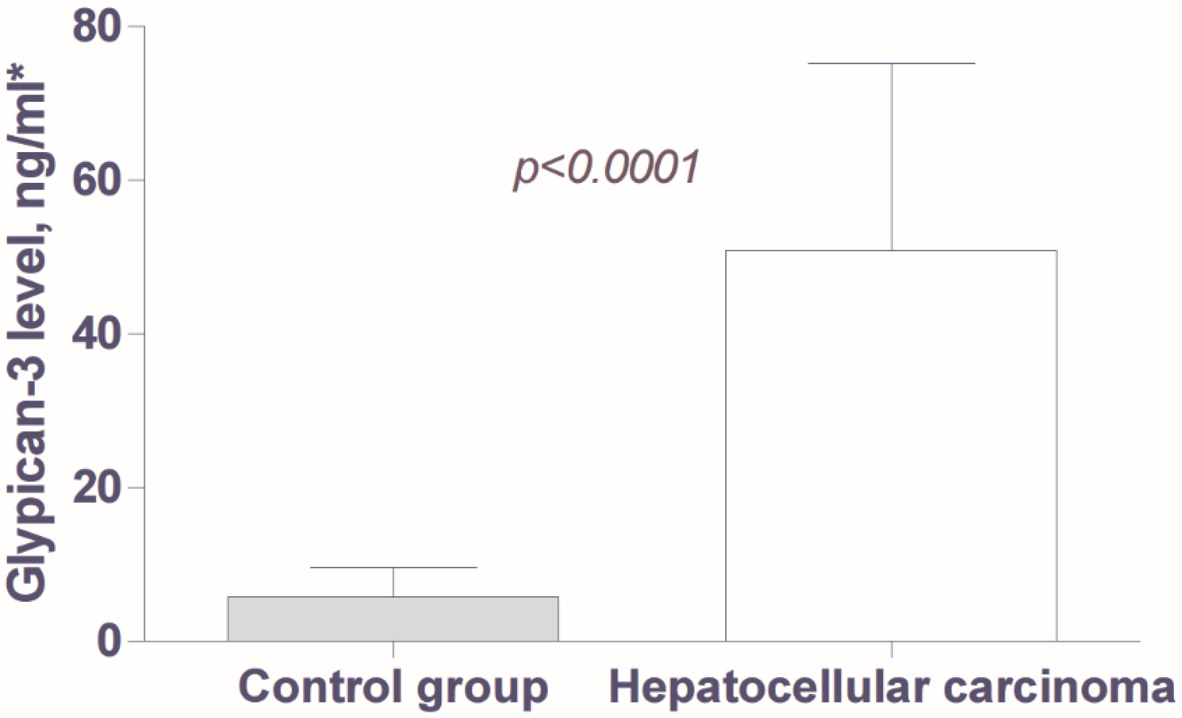
Figure 1. Glypican-3 serum levels in patients with hepatocellular carcinoma (n = 40) and with advanced chronic liver disease (n = 30). * Data in the graph are presented as mean values with 95% confidence intervals.
Livers 05 00036 g001
Figure 2. Levels of Glypican-3 and Alpha-Fetoprotein, according to the staging of hepatocellular carcinoma (HCC).
Livers 05 00036 g002
Table 1. Clinical and laboratory characteristics of patients with hepatocellular carcinoma (n = 40).
Parameters of Clinical and Laboratory Characteristics
Sex: male/female (n)30/10
Age (mean +/− SD, years)61.5 ± 11.15
Etiology of liver disease: HBV/HCV/alcohol (n)25/11/4
Diabetes (n, %)9 (22.5)
Child–Turcotte–Pugh class: A/B (n)16/24
Ascites: no/mild/moderate (n)18/15/7
Esophageal varices: no/small/large (n)16/10/12
Liver stiffness, measured by FibroScan, kPa *24.77 ± 15.30 (4.40–53.20)
Hemoglobin, g/L *130.5 ± 21.12 (82–165)
Platelets, ×109/L *160.5 ± 96.66 (35–502)
Prothrombin index, % *69.1 ± 17.4 (35–112)
Aspartate aminotransferase, U/L *90.63 ± 76.29 (23–289)
Alanine aminotransferase, U/L *56.10 ± 45.97 (17–253)
Gamma-glutamyl transferase, U/L *159.6 ± 180.7 (15–808)
Alkaline phosphatase, U/L *205.2 ± 262.4 (41–1685)
Cholinesterase, U/L *5270 ± 5044 (1023–31,000)
Bilirubin, mcg/L *38.95 ± 44.74 (6–267)
Albumin, g/L *36.10 ± 6.55 (25–49)
Alpha-fetoprotein (mean +/− SD, ng/mL)155.9 ± 165.1
Carbohydrate antigen CA 19–9 (mean +/− SD, U/mL)122.4 ± 144
* Data are presented as mean values with standard deviation (SD), minimum to maximum range.
Table 2. Glypican-3 levels, according to patient demographics and tumor features in forty cases with hepatocellular carcinoma.
ParameterCategoryNGlypican-3
(Mean ± SEM), ng/mL
p
Age<60 years
≥60 years
16
24
44.83 ± 17.31
54.85 ± 16.61
0.67
SexMales
Females
30
10
46.50 ± 13.69
63.84 ± 25.81
0.68
HBsAg statusHBV infection
Other etiology
25
15
48.93 ± 14.19
54.01 ± 22.26
0.39
Child–Turcotte–Pugh classA (compensated)
B (decompensated)
16
24
52.59 ± 19.85
49.67 ± 15.37
0.79
Barcelona Clinic Liver Cancer stageEarly HCC
Intermediate HCC
Advanced HCC
12
13
15
60.37 ± 27.63
35.54 ± 11.02
56.47 ± 21.91
0.64
Size of the tumor≤5 cm
>5 cm
26
14
61.04 ± 15.91
34.07 ± 18.25
0.27
Number of nodulesSolitary
2–3 nodules
Multiple, >3
20
8
12
61.72 ± 19.88
40.41 ± 15.89
39.65 ± 20.62
0.80
Macrovascular invasionNo
Yes
32
8
56.83 ± 14.48
26.86 ± 14.22
0.72
Extrahepatic metastasisNo
Yes
30
10
56.57 ± 15.22
33.63 ± 14 74
0.29
CLIP score0–1
2
3–4
5–6
12
11
10
6
84.70 ± 20.82
30.01 ± 16.55
38.60 ± 22.64
30.63 ± 19.02
0.39
Wash-out in CEUS portal venous phaseNo
Yes
15
25
55.00 ± 19.83
55.61 ± 18.93
0.49
HCC differentiation gradeWell-differentiated
Moderately differentiated
Poorly differentiated
9
11
5
37.96 ± 20.52
105.5 ± 44.93
28.96 ± 15.40
0.32
Table 3. Diagnostic accuracy of Glypican-3 and Alpha-Fetoprotein levels for hepatocellular carcinoma.
BiomarkerCut-Off Level (ng/mL)Sensitivity, % (95% CI)Specificity, % (95% CI)
Glypican-32.585.0 (70.1–94.3) 73.3 (54.1–87.7)
33.730.0 (16.5–46.5) 96.7 (82.8–99.9)
Alpha-fetoprotein5.278.9 (62.9–90.4) 76.7 (57.7–90.0)
86.544.7 (28.6–61.7) 96.7 (82.8–99.9)
Table 4. Relationship between Glypican-3 and Alpha-Fetoprotein levels in patients with hepatocellular carcinoma (n = 40).
Alpha-Fetoprotein (ng/mL)NGlypican-3 (>33.7 ng/mL)
PositiveNegative
<6 ng/mL955.5%44.5%
6–200 ng/mL1625%75%
>200 ng/mL1520%80%
Disclaimer/Publisher’s Note: The statements, opinions and data contained in all publications are solely those of the individual author(s) and contributor(s) and not of MDPI and/or the editor(s). MDPI and/or the editor(s) disclaim responsibility for any injury to people or property resulting from any ideas, methods, instructions or products referred to in the content.