Review

Sarcopenia and Metabolic Dysfunction-Associated Steatotic Liver Disease: A Narrative Review

by
Ludovico Abenavoli
1,*,
Michael Statsenko
2,
Giuseppe Guido Maria Scarlata
1,
Domenico Morano
1,
Roman Myazin
2 and
Dmitriy Emelyanov
2
1
Department of Health Sciences, University “Magna Graecia”, 88100 Catanzaro, Italy
2
Federal State Budgetary Educational Institution, Higher Education “Volgograd State Medical University”, the Ministry of Health of the Russian Federation, 400066 Volgograd, Russia
*
Author to whom correspondence should be addressed.
Livers 2024, 4(4), 495-506; https://doi.org/10.3390/livers4040035
Submission received: 6 August 2024 / Revised: 26 September 2024 / Accepted: 29 September 2024 / Published: 30 September 2024

Abstract

The primary objective of modern medicine is to extend human life expectancy. Currently, the majority of hospital patients across various clinical settings are elderly or advanced-age individuals, often with multiple comorbidities and age-related alterations in peripheral tissues. One such alteration is sarcopenia, a progressive decline in muscle mass, strength, and function, which significantly increases the risk of disability and mortality in older adults. Sarcopenia is associated with numerous adverse outcomes, and its underlying mechanisms are the subject of ongoing research. This narrative review discusses the epidemiology, pathophysiology, and diagnostic criteria for sarcopenia. It also examines the connections between sarcopenia and metabolic dysfunction-associated steatotic liver disease (MASLD), highlighting potential treatment approaches for the coexistence of these two pathologies.

1. Sarcopenia: What Is the Problem?

A key objective of modern medicine is to extend human life expectancy. Currently, most hospitalized patients are elderly or senile individuals who often have multiple comorbidities along with age-related alterations in peripheral tissues. One of these changes is sarcopenia—a syndrome characterized by the progressive and widespread loss of skeletal muscle mass, strength, and function [1,2]. By 2050, sarcopenia will pose a significant global challenge due to the increase in elderly population, making healthcare costs surge, and raising rates of morbidity and disability [2]. In a 2023 United Nations report titled “Don’t Leave Anyone Behind in an Aging Population World,” it was emphasized that in 2021, worldwide, there were 761 million people aged 65 and older. This figure is 5.9 times higher than it was in 1950 and 2.3 times higher than in 1991. In this regard, the number of elderly people is expected to more than double over the coming years, reaching 1.664 billion by 2051 [3]. It is well understood that all tissues in the human body are formed from nutrients absorbed through the digestive tract and then delivered to the bloodstream. The ratio of essential nutrients varies throughout different life stages. For example, protein requirements are significantly higher in children than in adults, as proteins, particularly from animal sources, provide the essential amino acids necessary for growth [4]. For adults, maintaining or increasing muscle mass is also dependent on an adequate protein intake. Skeletal muscle primarily consists of proteins, facilitates metabolic reactions for energy production using glucose and lipids, and has the ability to repair itself after physical activity [5]. Muscle tissue serves as the body’s main protein reserve during periods of insufficient nutritional support or stress. In prolonged catabolic states such as starvation, muscle and fat mass diminish, leading to cachexia. During these periods, muscle protein is broken down and used as a source of amino acids for maintaining body welfare and energy production via gluconeogenesis, resulting in muscle wasting, weakness, and decreased physical function [4,5]. The term “sarcopenia” was coined in 1989 by American professor I. Rosenberg to describe the loss of skeletal muscle mass in elderly individuals [6,7]. Recent studies reveal that sarcopenia is driven by epigenetic dysregulation of several molecular pathways, including protein turnover, insulin resistance (IR), mitochondrial dysfunction, and chronic low-grade systemic inflammation [8]. As sarcopenia progresses, muscle mass, strength, and function decline, with muscle protein degradation surpassing its synthesis. This is often accompanied by an increase or maintenance of adipose tissue, leading to sarcopenic obesity. Some researchers define sarcopenic obesity as a decrease in muscle mass and strength that co-occurs with an increase in body fat—over 25% in men and 38% in women—according to dual-energy X-ray absorptiometry (DXA) or bioelectrical impedance analysis [9,10]. Sarcopenia reduces physical activity, which further increases fat mass. Obesity exacerbates sarcopenia through the production of pro-inflammatory cytokines, dysregulation of leptin and adiponectin secretion, and decreased muscle sensitivity to insulin, creating a cycle that worsens the condition, as illustrated in Figure 1.

2. Sarcopenia and Systemic Risks

It has been well established that individuals with sarcopenic obesity are at increased risk of developing cardiovascular diseases and heart failure, with risks increased by as much as 23% and 42%, respectively [11]. Additionally, there is a significant increase in the risk of metabolic syndrome, arterial hypertension, and dyslipidemia in these patients [12]. For example, a study involving 4252 British men aged 60–79 demonstrated that sarcopenic obesity was strongly associated with at least a twofold increase in the overall risk of death, including death from cardiovascular causes [13]. Sarcopenic obesity is closely linked to inflammation and an increased risk of fractures, and it is commonly seen in patients with visceral obesity. Interestingly, patients with excessive subcutaneous fat tend to have a lower mortality rate, a phenomenon often referred to as the “obesity paradox”. However, it is important to note that the literature contains over 500 articles with conflicting findings regarding the effects of obesity on sarcopenia. Large-scale studies aimed at identifying diagnostic criteria for different sarcopenic obesity phenotypes, such as visceral and subcutaneous, could lead to more personalized approaches to the treatment and prevention of this condition in the future [14]. Until recently, the inevitable age-related loss of muscle mass in elderly and senile individuals was primarily considered a geriatric issue. However, due to the aging of the global population, consensus documents on sarcopenia in older adults have been developed in recent years [1,2,15,16]. The European Working Group on Sarcopenia in Older People (EWGSOP) defines sarcopenia as “a syndrome characterized by progressive and generalized loss of muscle mass and strength, with an increased risk of adverse outcomes such as disability, reduced quality of life, and death” [1]. The prevalence of sarcopenia varies, affecting between 5% and 13% of people aged 60–70 years and reaching up to 50% in individuals over 80 years old [1,2]. Muscle health is a significant factor in predicting life expectancy in older adults. In a two-year survival analysis, hand grip strength and the ability to perform self-care were the strongest predictors of survival [17]. Participants with low muscle strength and balance issues faced a much higher risk of future falls. Furthermore, as people age, each additional decade increases the mortality risk due to decrease in body weight. The lowest mortality rates among older women were seen in those with a body mass index (BMI) of 31.7 kg/m2, while for men of the same age group, the optimal BMI was 28.8 kg/m2 [18]. Age-related sarcopenia and physical weakness are driven by inflammatory processes, immune sensitization, anabolic resistance, and oxidative stress. These factors often lead to a sedentary lifestyle and protein-energy malnutrition, further exacerbated by age-related loss of appetite [19]. Additionally, several chronic diseases, such as chronic heart failure, chronic obstructive pulmonary disease, and chronic kidney disease, are also known to contribute to sarcopenia [20].

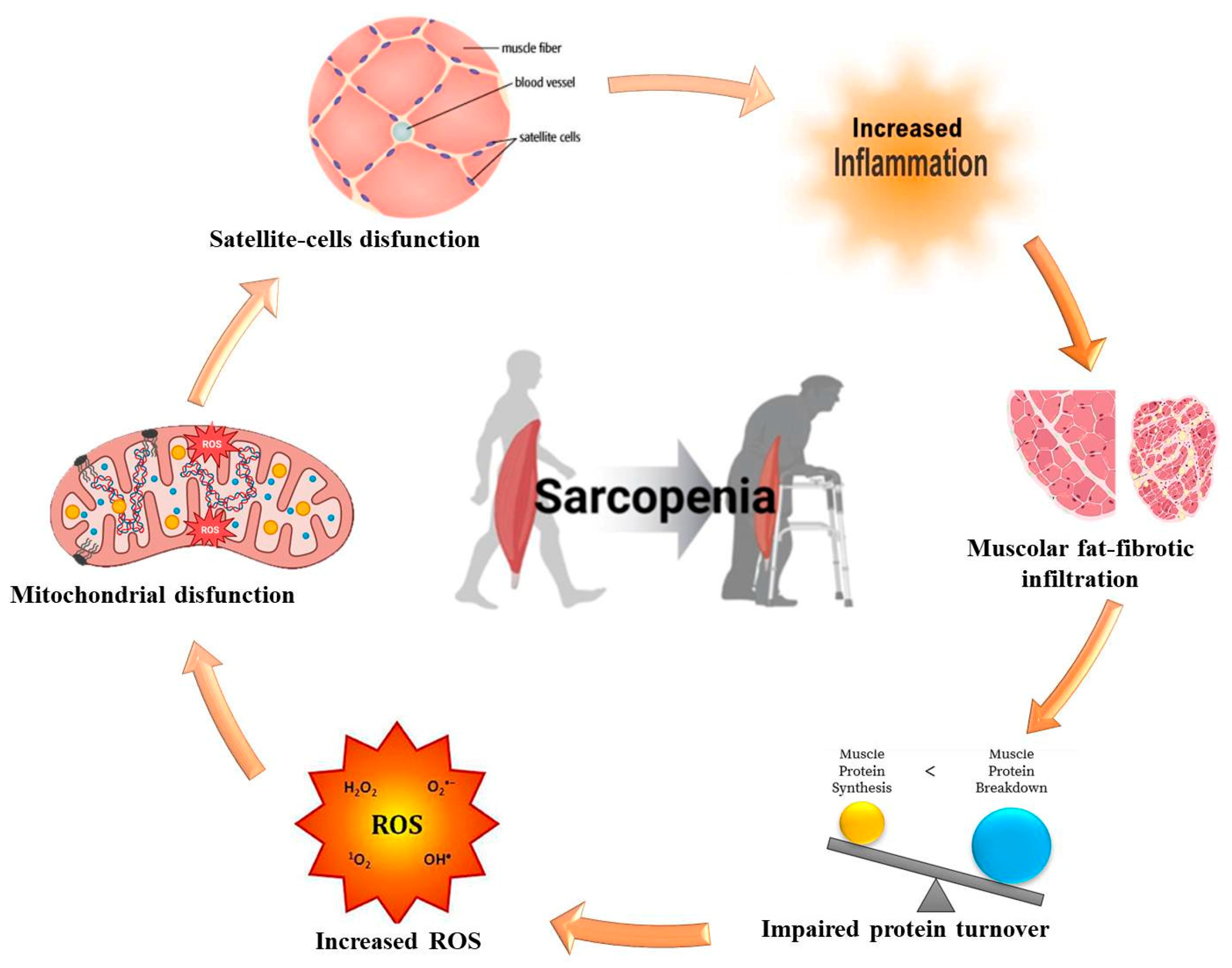
3. Molecular and Pathophysiological Mechanisms of Sarcopenia

Currently, sarcopenia is not only a geriatric issue. Indeed, today, sarcopenia is diagnosed in middle-aged and even young individuals, including children with severe illnesses [20]. In such cases, muscle wasting is seen as an inflammatory condition. Sarcopenia may also be associated with osteoporosis and obesity, where muscle tissue is replaced by fat. This leads to myosteatosis, the accumulation of fat within muscle, a result of increased systemic anabolic resistance [5]. Myosteatosis contributes to functional muscle failure, which, in turn, raises the risk of mortality [16,19]. Sarcopenia can be classified into two types: primary and secondary. Primary sarcopenia is age-related, while secondary sarcopenia arises from several conditions, such as inadequate protein/energy intake, the use of glucocorticoids or muscle relaxants, prolonged immobility, or chronic diseases [2,21,22]. Sarcopenia may either result from or contribute to various diseases. Its pathogenesis is linked to hyperammonemia and low levels of branched-chain amino acids, which negatively affect the patient’s prognosis [23,24]. Ammonia is involved in sarcopenia development through mechanisms that increase the expression of myostatin and autophagy markers, leading to disrupted muscle metabolism. Skeletal muscle becomes the primary site for ammonia detoxification instead of the liver, due to changes in the expression of genes responsible for reducing ammonia levels. This creates a vicious cycle in which hyperammonemia damages the muscle, leading to sarcopenia, which in turn impairs the muscle’s ability to clear ammonia, perpetuating the cycle [25,26]. Although muscle is not typically regarded as an endocrine organ, it is metabolically active [27]. In recent years, researchers have identified over 650 myokines secreted by muscles, though only 5% have been thoroughly studied [28,29,30,31]. During muscle aging, changes in the expression of myokines, especially interleukin-15 and myostatin, contribute to the development of sarcopenia [15]. Muscle atrophy is characterized by increased protein degradation, with growth factor signaling playing a key role. Peroxisome proliferator-activated receptors (PPARs), which are activated by fatty acids, regulate genes involved in development, metabolism, and inflammation. PPARs are expressed in muscle and have diverse effects. There are three PPAR isotypes: PPAR-α, -β/δ, and -γ. PPAR-α is highly expressed in tissues that metabolize fatty acids, including skeletal muscle, while PPAR-β/δ is found throughout the body and predominantly influences energy metabolism and muscle fiber type switching. PPAR-γ is expressed in adipocytes and plays a role in lipid deposition in muscles and other organs. Collectively, these PPAR isotypes are crucial for muscle homeostasis. The shared pathogenesis of sarcopenia and metabolic dysfunction-associated steatotic liver disease (MASLD) increasingly points to the gut microbiota as a critical mediator, influencing both muscle and liver health through complex interactions. In sarcopenia, microbial dysbiosis contributes to systemic inflammation, nutrient malabsorption, and altered metabolic signaling, which accelerates muscle wasting. Similarly, MASLD is characterized by metabolic dysfunction, with emerging evidence highlighting the role of the gut microbiota in its pathogenesis. Specific bacterial species, such as Bacteroides, Firmicutes, and Lactobacillus, have been implicated in modulating lipid metabolism and inflammation, directly affecting liver fat deposition and muscle degradation [5]. Key microbial metabolites, including short-chain fatty acids like butyrate and propionate, exert protective effects on muscle and liver function by modulating insulin sensitivity and reducing inflammation. In contrast, increased levels of trimethylamine N-oxide, a product of gut microbial metabolism of dietary choline, carnitine, and phosphatidylcholine, have been linked to IR and liver fat accumulation, exacerbating both sarcopenia and MASLD. Furthermore, gut dysbiosis can alter bile acid signaling pathways via FXR and G-protein coupled bile acid receptor 1, disrupting lipid metabolism and contributing to both conditions [14,19,20,32,33,34,35]. However, there are other common pathogenetic mechanisms that we discuss in the following sections.

4. Sarcopenia Classification and Diagnostic Models

In its 2010 consensus, the European Working Group on Sarcopenia in Older People (EWGSOP) recommended using three criteria to diagnose sarcopenia: (1) low muscle mass, assessed through computed tomography (CT), magnetic resonance imaging (MRI), dual-energy x-ray absorptiometry (DXA), ultrasound, bioimpedance analysis, or caliperometry; (2) low muscle strength, determined by dynamometry; and (3) reduced muscle function, measured by chair-rise or walking-speed tests [1]. According to the EWGSOP, sarcopenia is classified into three stages: stage I (pre-sarcopenia) with reduced muscle mass without loss of strength or function; stage II (sarcopenia) with reduced muscle mass and either decreased strength or function; and stage III (severe sarcopenia) with reduced muscle mass, strength, and function [2,36,37]. Sarcopenia staging is essential for determining treatment and rehabilitation strategies. Diagnostic guidelines were updated in the 2019 EWGSOP-2 consensus, which suggests that diagnosis should start with an assessed score of 4 or more on the SARC-F questionnaire [38]. The main indicator of probable sarcopenia is reduced muscle strength, measured by dynamometry [2,26]. The diagnosis is confirmed by reduced muscle mass. If muscle mass, strength, and function are all decreased, the sarcopenia is considered severe [22]. CT, MRI, the assessment of musculoskeletal index, and ultrasound are considered the gold standards for diagnosing muscle mass loss [39,40,41]. The EWGSOP-2 consensus emphasizes that ultrasound is a reliable method for detecting sarcopenia in older patients with comorbidities [16,36,42,43]. Ultrasound techniques such as measuring the thickness of the rectus abdominis muscle are more accurate than anthropometric methods and more accessible than CT, DXA, or bioimpedance analysis, making them suitable for use in virtually any patient [44,45,46,47,48,49,50].

5. MASLD and Sarcopenia: Shared Pathogenetic Mechanisms

Recent studies have explored the role of sarcopenia, or age-related muscle loss, in metabolic disorders, specifically its link to MASLD. This condition, formerly non-alcoholic fatty liver disease (NAFLD), involves liver fat accumulation and cardiometabolic risk factors without significant alcohol consumption. It ranges from simple steatosis to more severe conditions such as metabolic dysfunction-associated steatohepatitis (MASH), fibrosis, cirrhosis, and hepatocellular carcinoma (HCC) [51,52,53,54]. Approximately 30% of MASLD patients with compensated cirrhosis progress to decompensation within 8–10 years, with fibrosis being a key predictor of adverse outcomes [55,56,57]. Risk factors include age over 50, metabolic syndrome, IR, type 2 diabetes mellitus (T2DM), and genetic factors like PNPLA3 polymorphisms [58,59]. A 2010 study first identified the link between sarcopenia and MASLD, noting higher MASLD prevalence in individuals with sarcopenia, especially those with elevated BMI [60]. Sarcopenia has been associated with increased liver fibrosis in MASLD patients, regardless of BMI [61]. While obesity is a significant MASLD risk factor, 40% of MASLD patients are non-obese, with 10% having a normal BMI and 30% being overweight. Despite their lower BMI, non-obese MASLD patients remain at risk for MASH, cardiovascular disease, and HCC. Reduced muscle mass may impair BMI’s diagnostic accuracy in detecting obesity [61]. Sarcopenia worsens liver fibrosis and cirrhosis outcomes, even in non-obese individuals. The muscle–liver axis involves mediators like myostatin, testosterone, and adiponectin, influencing both muscle and liver health [62,63,64]. Hyperammonemia in MASLD further exacerbates sarcopenia and liver inflammation, contributing to a cycle of worsening muscle and liver dysfunction [65]. IR plays a central role in the pathogenesis of several metabolic disorders, including sarcopenia, sarcopenic obesity, and MASLD. These conditions share common mechanisms driven by IR, which disrupts skeletal muscle function, glucose metabolism, and lipid regulation, further contributing to metabolic dysfunction. Skeletal muscle is vital for whole-body glucose homeostasis, as it is the primary site for insulin-mediated glucose uptake. Under normal conditions, insulin promotes glucose transporter type 4 translocation to the muscle cell membrane, facilitating glycogen synthesis and enhancing protein metabolism. However, in individuals with IR, this mechanism is impaired, leading to metabolic inflexibility—the inability to switch between glucose and fatty acids as energy sources. This metabolic inflexibility promotes hyperinsulinemia and worsens IR, contributing to MASLD development. IR is also associated with myosteatosis, characterized by fat infiltration into muscle, reduced protein synthesis, and increased muscle breakdown, exacerbating sarcopenia [66]. Anabolic resistance, or the diminished capacity of skeletal muscle to synthesize proteins in response to stimuli like insulin and exercise, is a key factor in sarcopenia. This resistance worsens with aging, obesity, and IR, leading to further decline in muscle mass. In MASLD and MASH, IR impairs muscle protein synthesis and increases proteolysis, perpetuating a cycle of muscle loss and fat accumulation in both muscle and liver tissues [67,68,69]. Lipid accumulation in muscle, particularly bioactive intermediates like diacylglycerol (DAG), contributes to lipid-induced IR by inhibiting insulin signaling. Excess lipids generate reactive oxygen species and endoplasmic reticulum stress, leading to mitochondrial dysfunction and sustained IR. Additionally, intermuscular fat secretes pro-inflammatory cytokines such as tumor necrosis factor-α (TNF-α) and interleukin (IL)-6, which exacerbate IR and lipotoxicity [55]. Hepatic fat accumulation in MASLD is closely linked to both liver and skeletal muscle IR. Elevated hepatic DAG levels activate the protein kinase C pathway, worsening insulin sensitivity. Systemic inflammation driven by adipose tissue and macrophage-derived cytokines further impairs liver and muscle insulin sensitivity, promoting the progression of metabolic disorders like T2DM. MASLD and T2DM are also associated with hyperleptinemia and decreased adiponectin levels, both of which contribute to increased IR and liver inflammation [70]. Chronic low-grade inflammation is a hallmark of both MASLD and obesity. Macrophage infiltration into adipose tissue and into the liver leads to elevated levels of TNF-α and IL-6, which inhibit anabolic signaling in muscle, promote muscle atrophy, and worsen IR. This inflammatory state also promotes liver fat accumulation, further exacerbating MASLD progression. Hepatokines, including fetuin-A, fibroblast growth factor 21, and retinol-binding protein 4, mediate liver–muscle crosstalk and contribute to IR and lipid dysregulation in metabolic diseases [66]. Shared pathogenetic mechanisms are summarized in Figure 2.

6. Potential Treatments

Regenerating skeletal muscle is a slow process, and temporarily reducing ammonia levels in the blood does not necessarily lower ammonia levels in the muscles or prevent metabolic and molecular damage. Therefore, long-term strategies for lowering ammonia levels must be put in place to effectively reverse muscle loss and improve contractile function. Promising methods include using cell-permeable esters of alpha-ketoglutarate, which react with ammonia to form glutamine, aiding in its removal. Isoleucine and valine have also been suggested as substrates, since they can eliminate one mole of ammonia per mole of amino acid, but these interventions have not yet been evaluated in clinical trials [71]. Sarcopenia is common in nearly half of cirrhotic patients, with ammonia playing a significant role in its development. Increased expression of myostatin and autophagy markers, triggered by hyperammonemia, disrupts muscle metabolism, contributing to sarcopenia. Skeletal muscle becomes the primary site for ammonia detoxification in cirrhotic patients, leading to a vicious cycle where muscle damage from hyperammonemia further reduces the muscle’s ability to clear ammonia from the blood [72,73]. This exacerbates muscle loss, reduces patients’ quality of life, increases mortality, and raises the risk of hepatic encephalopathy [26,74,75]. Ammonia has toxic effects on both the brain and skeletal muscles. In animal models, hyperammonemia due to liver failure and portal hypertension impairs muscle protein homeostasis, leading to ATP depletion and activation of muscle-degrading pathways like nuclear factor kappa-B and myostatin. Reducing ammonia levels in preclinical cirrhosis models has been shown to mitigate sarcopenia [76]. Therefore, monitoring and managing ammonia levels in cirrhotic patients is of paramount importance, as high ammonia concentrations are associated with severe complications and increased mortality [24,77]. Hyperammonemia disrupts muscle protein synthesis through autophagy and myostatin activation, which worsens sarcopenia [78]. Physical exercise has been shown to reduce the likelihood of MASLD (46% vs. 55%, p < 0.001) in obese individuals with maintained muscle mass. Mitochondrial dysfunction in skeletal muscle contributes to muscle atrophy and may play a role in MASLD development. Correcting this dysfunction through increased physical activity could be an effective approach to treating MASLD and other metabolic disorders [51]. Recently, a study on the effects of L-ornithine and L-arginine supplementation in male volunteers undergoing intense physical training was performed. Those receiving the supplements showed significant gains in muscle mass, endurance, and lower urinary hydroxyproline levels compared with a control group [51]. Other studies have also demonstrated increased serum hormone levels after exercise with L-ornithine supplementation [79]. L-ornithine-L-aspartate (LOLA) has shown promise in treating patients with NAFLD and muscle loss. By lowering ammonia levels, LOLA has been found to increase muscle mass, improve cognitive function, and shorten hospital stays, leading to an improved quality of life [80]. In rodent models of MASH, LOLA significantly increased lean body mass and muscle strength. This suggests that hyperammonemia plays a role in the pathogenesis of sarcopenia in MASLD patients, and LOLA may be an effective treatment for both steatohepatitis and sarcopenic obesity [81]. LOLA therapy has also been effective in reducing ammonia levels and improving outcomes in cirrhotic patients with sarcopenia. In a clinical trial involving 463 patients, including those with NAFLD, oral LOLA significantly lowered liver enzyme levels over 60 days. Another study showed a dose-dependent reduction in transaminase levels and triglycerides after 12 weeks of LOLA therapy, with improved liver microcirculation in patients with MASH [82]. The mechanisms behind LOLA’s effectiveness include stimulating ammonia conversion to urea in the liver and promoting glutamine synthesis in muscles. This dual action helps reduce muscle ammonia levels and improve muscle function. Clinical trials have confirmed that LOLA reduces hyperammonemia and sarcopenia in cirrhotic patients by improving muscle protein synthesis and cognitive function while also reducing mortality rates [26,54,83,84,85,86,87,88]. Along with exercise, other treatments for cirrhosis-related sarcopenia include antibiotics, lactulose, branched-chain amino acids, testosterone, vitamin D, zinc, and dietary interventions [89,90]. Further research is necessary to explore the relationship between sarcopenia and MASLD and to develop new treatment strategies for these comorbid conditions [91,92]. Table 1 summarize the different clinical features and potential treatments related to sarcopenia.

7. Conclusions

Managing sarcopenia, particularly in patients with MASLD, poses significant challenges for clinicians. Sarcopenia’s complex pathophysiology, driven by factors like hyperammonemia, systemic inflammation, and anabolic resistance, complicates both its diagnosis and its treatment. Despite the availability of diagnostic tools like DXA and MRI, inconsistent criteria and limited access to these technologies often delay or miss diagnoses. Treatment remains difficult, with exercise and nutrition offering variable results, especially in severe cases. Ammonia-lowering therapies like LOLA show promise, but larger clinical trials are needed to confirm their efficacy in reducing muscle loss and improving outcomes in MASLD patients. Moreover, a better understanding of the molecular mechanisms underlying sarcopenia, including the roles of myostatin, autophagy, and the gut microbiota, could make it possible to devise new management strategies. Further research into the interplay between sarcopenia and MASLD may lead to more personalized treatments that combine pharmacological interventions with lifestyle modifications. Addressing these challenges through early diagnosis, targeted therapies, and multidisciplinary approaches is key to improving patient outcomes.

Author Contributions

Conceptualization, R.M. and D.E.; methodology L.A. and M.S., investigation, G.G.M.S. and D.M.; resources, M.S., R.M. and D.E.; writing—original draft preparation, L.A., M.S., R.M. and D.E.; writing—review and editing, L.A., G.G.M.S. and D.M.; supervision, L.A. All authors have read and agreed to the published version of the manuscript.

Funding

This research received no external funding.

Acknowledgments

We would like to thank Simone Scarlata for his critical review of the English language in this article.

Conflicts of Interest

The authors declare no conflicts of interest.

References

  1. Cruz-Jeentoft, A.J.; Baeyens, J.P.; Bauer, J.M.; Boirie, Y.; Cederholm, T.; Landi, F.; Martin, F.C.; Michel, J.P.; Rolland, Y.; Schneider, S.M.; et al. Sarcopenia: European consensus on definition and diagnosis. Report of the European Working Group on Sarcopenia in Older People. Age Ageing 2010, 39, 412–423. [Google Scholar] [CrossRef] [PubMed]
  2. Cruz-Jentoft, A.J.; Bahat, G.; Bauer, J.; Boirie, Y.; Bruyère, O.; Cederholm, T.; Cooper, C.; Landi, F.; Rolland, Y.; Sayer, A.A.; et al. Sarcopenia: Revised European consensus on definition and diagnosis. Age Ageing 2019, 48, 16–31. [Google Scholar] [CrossRef] [PubMed]
  3. United Nations. Department of Economic and Social Affairs 2023. World Social Report 2023: Leaving No One behind in an Ageing World. Available online: https://repository.gheli.harvard.edu/repository/12779/ (accessed on 30 June 2024).
  4. Zavyalova, A.N.; Novikova, V.P.; Ignatova, P.D. Axis «microbiota-muscle». Exp. Clin. Gastroenterol. 2022, 11, 60–69. [Google Scholar] [CrossRef]
  5. Manickam, R.; Duszka, K.; Wahli, W. PPARs and Microbiota in Skeletal Muscle Health and Wasting. Int. J. Mol. Sci. 2020, 21, 8056. [Google Scholar] [CrossRef] [PubMed]
  6. Lee, J.; Kim, H.J. Normal Aging Induces Changes in the Brain and Neurodegeneration Progress: Review of the Structural, Biochemical, Metabolic, Cellular, and Molecular Changes. Front. Aging Neurosci. 2022, 14, 931536. [Google Scholar] [CrossRef] [PubMed]
  7. Chen, Q.; Shen, J.; Gao, Y.; Wang, X.X.; Jia, Q.Y.; Li, M. Nutritional status of the older adults in nursing homes: A cross-sectional study. J. Clin. Nurs. 2024, 33, 2640–2648. [Google Scholar] [CrossRef]
  8. Livshits, G.; Kalinkovich, A. Restoration of epigenetic impairment in the skeletal muscle and chronic inflammation resolution as a therapeutic approach in sarcopenia. Ageing Res. Rev. 2024, 96, 102267. [Google Scholar] [CrossRef] [PubMed]
  9. Mokrysheva, N.G.; Krupinova, J.A.; Volodicheva, V.L.; Mirnaya, S.S.; Melnichenko, G.A. A view at sarcopenia by endocrinologist. Obes. Metab. 2018, 15, 21–27. [Google Scholar] [CrossRef]
  10. Perna, S.; Spadaccini, D.; Rondanelli, M. Sarcopenic Obesity: Time to target the phenotypes. J. Cachexia Sarcopenia Muscle 2019, 10, 710–711. [Google Scholar] [CrossRef]
  11. Velarde, M.C. Mitochondrial and sex steroid hormone crosstalk during aging. Longev. Healthspan 2014, 3, 2. [Google Scholar] [CrossRef] [PubMed]
  12. Choi, K.M. Sarcopenia and sarcopenic obesity. Korean J. Intern. Med. 2016, 31, 1054–1060. [Google Scholar] [CrossRef] [PubMed]
  13. Atkins, J.L.; Whincup, P.H.; Morris, R.W.; Lennon, L.T.; Papacosta, O.; Wannamethee, S.G. Sarcopenic obesity and risk of cardiovascular disease and mortality: A populationbased cohort study of older men. J. Am. Geriatr. Soc. 2014, 62, 253–260. [Google Scholar] [CrossRef] [PubMed]
  14. Axelrod, C.L.; Dantas, W.S.; Kirwan, J.P. Sarcopenic obesity: Emerging mechanisms and therapeutic potential. Metabolism 2023, 146, 155639. [Google Scholar] [CrossRef]
  15. Dao, T.; Green, A.E.; Kim, Y.A.; Bae, S.J.; Ha, K.T.; Gariani, K.; Lee, M.R.; Menzies, K.J.; Ryu, D. Sarcopenia and Muscle Aging: A Brief Overview. Endocrinol. Metab. 2020, 35, 716–732. [Google Scholar] [CrossRef] [PubMed]
  16. Picca, A.; Calvani, R.; Cesari, M.; Landi, F.; Bernabei, R.; Coelho- Júnior, H.J.; Marzetti, E. Biomarkers of Physical Frailty and Sarcopenia: Coming up to the Place? Int. J. Mol. Sci. 2020, 21, 5635. [Google Scholar] [CrossRef] [PubMed]
  17. Morley, J.E. Sarcopenia: Diagnosis and treatment. J. Nutr. Health Aging 2008, 12, 452–456. [Google Scholar] [CrossRef] [PubMed]
  18. Paddon-Jones, D.; Short, K.R.; Campbell, W.W.; Volpi, E.; Wolfe, R.R. Role of dietary protein in the sarcopenia of aging. Am. J. Clin. Nutr. 2008, 87, 1562S–1566S. [Google Scholar] [CrossRef]
  19. Ticinesi, A.; Nouvenne, A.; Cerundolo, N.; Catania, P.; Prati, B.; Tana, C.; Meschi, T. Gut Microbiota, Muscle Mass and Function in Aging: A Focus on Physical Frailty and Sarcopenia. Nutrients 2019, 11, 1633. [Google Scholar] [CrossRef]
  20. De Sire, R.; Rizzatti, G.; Ingravalle, F.; Pizzoferrato, M.; Petito, V.; Lopetuso, L.; Graziani, C.; de Sire, A.; Mentella, M.C.; Mele, M.C.; et al. Skeletal muscle-gut axis: Emerging mechanisms of sarcopenia for intestinal and extra intestinal diseases. Minerva Gastroenterol. Dietol. 2018, 64, 351–362. [Google Scholar] [CrossRef]
  21. Narici, M.V.; Maffulli, N. Sarcopenia: Characteristics, mechanisms and functional significance. Br. Med. Bull. 2010, 95, 139–159. [Google Scholar] [CrossRef]
  22. Franzke, B.; Neubauer, O.; Cameron-Smith, D.; Wagner, K.H. Dietary protein, muscle and physical function in the very old. Nutrients 2018, 10, 935. [Google Scholar] [CrossRef]
  23. Bhatia, V.; Singh, R.; Acharya, S.K. Predictive value of arterial ammonia for complications and outcome in acute liver failure. Gut 2006, 55, 98–104. [Google Scholar] [CrossRef] [PubMed]
  24. Shalimar Sheikh, M.F.; Mookerjee, R.P.; Agarwal, B.; Acharya, S.K.; Jalan, R. Prognostic Role of Ammonia in Patients with Cirrhosis. Hepatology 2019, 70, 982–994. [Google Scholar] [CrossRef] [PubMed]
  25. Goodpaster, B.H.; Park, S.W.; Harris, T.B.; Kritchevsky, S.B.; Nevitt, M.; Schwartz, A.V.; Simonsick, E.M.; Tylavsky, F.A.; Visser, M.; Newman, A.B. The loss of skeletal muscle strength, mass, and quality in older adults: The health, aging and body composition study. J. Gerontol. A Biol. Sci. Med. Sci. 2006, 61, 1059–1064. [Google Scholar] [CrossRef] [PubMed]
  26. Yang, Y.J.; Kim, D.J. An Overview of the Molecular Mechanisms Contributing to Musculoskeletal Disorders in Chronic Liver Disease: Osteoporosis, Sarcopenia, and Osteoporotic Sarcopenia. Int. J. Mol. Sci. 2021, 22, 2604. [Google Scholar] [CrossRef] [PubMed]
  27. Giudice, J.; Taylor, J.M. Muscle as a paracrine and endocrine organ. Curr. Opin. Pharmacol. 2017, 34, 49–55. [Google Scholar] [CrossRef]
  28. Paltsyn, A.A. Myokines. Patol. Fiziol. 2020, 64, 135–141. [Google Scholar]
  29. Severinsen, M.C.K.; Pedersen, B.K. Muscle-Organ Crosstalk: The Emerging Roles of Myokines. Endocr. Rev. 2020, 41, 594–609. [Google Scholar] [CrossRef]
  30. Mancinelli, R.; Checcaglini, F.; Coscia, F.; Gigliotti, P.; Fulle, S.; Fanò- Illic, G. Biological Aspects of Selected Myokines in Skeletal Muscle: Focus on Aging. Int. J. Mol. Sci. 2021, 22, 8520. [Google Scholar] [CrossRef] [PubMed]
  31. Suriano, F.; Van Hul, M.; Cani, P.D. Gut microbiota and regulation of myokine- adipokine function. Curr. Opin. Pharmacol. 2020, 52, 9–17. [Google Scholar] [CrossRef] [PubMed]
  32. Lahiri, S.; Kim, H.; Garcia-Perez, I.; Reza, M.M.; Martin, K.A.; Kundu, P.; Cox, L.M.; Selkrig, J.; Posma, J.M.; Zhang, H.; et al. The gut microbiota influences skeletal muscle mass and function in mice. Sci. Transl. Med. 2019, 11, eaan5662. [Google Scholar] [CrossRef] [PubMed]
  33. Grosicki, G.J.; Fielding, R.A.; Lustgarten, M.S. Gut Microbiota Contribute to Age- Related Changes in Skeletal Muscle Size, Composition, and Function: Biological Basis for a Gut- Muscle Axis. Calcif. Tissue Int. 2018, 102, 433–442. [Google Scholar] [CrossRef]
  34. Spychala, M.S.; Venna, V.R.; Jandzinski, M.; Doran, S.J.; Durgan, D.J.; Ganesh, B.P.; Ajami, N.J.; Putluri, N.; Graf, J.; Bryan, R.M.; et al. Age-related changes in the gut microbiota influence systemic inflammation and stroke outcome. Ann. Neurol. 2018, 84, 23–36. [Google Scholar] [CrossRef]
  35. Gizard, F.; Fernandez, A.; De Vadder, F. Interactions between gut microbiota and skeletal muscle. Nutr. Metab. Insights 2020, 13, 1178638820980490. [Google Scholar] [CrossRef]
  36. Erokhina, A.S.; Golovanova, E.D. The relationship between comorbidity and sarcopenia: Impact on mortality and survival. Doctor 2021, 6, 60–64. [Google Scholar] [CrossRef]
  37. Lera, L.; Angel, B.; Marquez, C.; Saguez, R.; Albala, C. Besides Sarcopenia, Pre-Sarcopenia Also Predicts All-Cause Mortality in Older Chileans. Clin. Interv. Aging 2021, 16, 611–619. [Google Scholar] [CrossRef] [PubMed]
  38. Malmstrom, T.K.; Morley, J.E. SARC-F: A Simple Questionnaire to Rapidly Diagnose Sarcopenia. JAMDA 2013, 14, 531–532. [Google Scholar] [CrossRef] [PubMed]
  39. Issa, D.; Alkhouri, N.; Tsien, C.; Shah, S.; Lopez, R.; McCullough, A.; Dasarathy, S. Presence of sarcopenia (muscle wasting) in patients with nonalcoholic steatohepatitis. Hepatology 2014, 60, 428–429. [Google Scholar] [CrossRef] [PubMed]
  40. Bredella, M.A.; Ghomi, R.H.; Thomas, B.J.; Torriani, M.; Brick, D.J.; Gerweck, A.V.; Misra, M.; Klibanski, A.; Milleret, K.K. Comparison of DXA and CT in the assessment of body composition in premenopausal women with obesity and anorexia nervosa. Obesity 2010, 18, 2227–2233. [Google Scholar] [CrossRef] [PubMed]
  41. Grinevich, V.B.; Sas, E.I. The role of sarcopenia in the development of non-alcoholic fatty liver disease. Clin. Exp. Gastroenterol. 2020, 183, 70–73. [Google Scholar] [CrossRef]
  42. Landi, F.; Cruz-Jentoft, A.J.; Liperoti, R.; Russo, A.; Giovannini, S.; Tosato, M.; Capoluongo, E.; Bernabei, R.; Onder, G. Sarcopenia and mortality risk in frail older persons aged 80 years and older: Results from ilSIRENTE study. Age Ageing 2013, 42, 203–209. [Google Scholar] [CrossRef] [PubMed]
  43. Grigorieva, I.I.; Raskina, T.A.; Letaeva, M.V.; Malyshenko, O.S.; Averkieva, Y.V.; Masenko, V.L.; Kokov, A.N. Sarcopenia: Pathogenesis and diagnosis. Fundam. Clin. Med. 2019, 4, 105–116. [Google Scholar] [CrossRef]
  44. Erokhina, A.S.; Golovanova, E.D.; Miloserdov, M.A. Ultrasound assessment of muscle mass in the diagnosis of sarcopenia in cardiovascular patients. Cardiovasc. Ther. Prev. 2021, 20, 2699. [Google Scholar] [CrossRef]
  45. Ido, A.; Nakayama, Y.; Ishii, K.; Iemitsu, M.; Sato, K.; Fujimoto, M.; Kurihara, T.; Hamaoka, T.; Satoh-Asahara, N.; Sanadaet, K. Ultrasound-derived abdominal muscle thickness better detects metabolic syndrome risk in obese patients than skeletal muscle index measured by dual energy X-ray absorptiometry. PLoS ONE 2015, 10, 0143858. [Google Scholar] [CrossRef] [PubMed]
  46. Sanada, K.; Kuchiki, T.; Miyachi, M.; McGrath, K.; Higuchi, M.; Ebashi, H. Effects of age on ventilator threshold and peak oxygen uptake normalized for regional skeletal muscle mass in Japanese men and women aged 20–80 years. Eur. J. Appl. Physiol. 2007, 99, 475–483. [Google Scholar] [CrossRef] [PubMed]
  47. Nijholt, W.; Scafoglieri, A.; Jager-Wittenaar, H.; Van der Schans, C.P. The reliability and validity of ultrasound to quantify muscles in older adults: A systematic review. J. Cachexia Sarkopenia Muscle 2017, 8, 702–712. [Google Scholar] [CrossRef]
  48. Stenholm, S.; Harris, T.B.; Rantanen, T.; Visser, M.; Kritchevsky, S.B.; Ferrucci, L. Sarcopenic obesity: Definition, cause and consequences. Curr. Opin. Clin. Nutr. Metab. Care 2008, 11, 693–700. [Google Scholar] [CrossRef] [PubMed]
  49. González-Correa, C.H.; Pineda-Zuluaga, M.C.; Marulanda-Mejía, F. Skeletal Muscle Mass by Bioelectrical Impedance Analysis and Calf Circumference for Sarcopenia Diagnosis. J. Electr. Bioimpedance 2020, 11, 57–61. [Google Scholar] [CrossRef]
  50. Van Kan, G.A.; Rolland, Y.; Andrieu, S.; Bauer, J.; Beauchet, O.; Bonnefoy, M.; Cesari, M.; Donini, L.M.; Gillette-Guyonnet, S.; Inzitari, M.; et al. Gait speed at usual pace as a predictor of adverse outcomes in community-dwelling older people an International Academy on Nutrition and Aging (IANA) Task Force. J. Nutr. Health Aging 2009, 13, 881–889. [Google Scholar] [CrossRef] [PubMed]
  51. Sas, E.I.; Barnakova, V.A. Skeletal muscle hormone activity and non-alcoholic fatty liver disease. Med. Alph. 2020, 17, 31–34. [Google Scholar] [CrossRef]
  52. European Association for the Study of the Liver (EASL); European Association for the Study of Diabetes (EASD); European Association for the Study of Obesity (EASO). EASL–EASD–EASO Clinical Practice Guidelines on the management of metabolic dysfunction-associated steatotic liver disease (MASLD). J. Hepatol. 2024, 81, 492–542. [Google Scholar] [CrossRef] [PubMed]
  53. European Association for the Study of the Liver (EASL); European Association for the Study of Diabetes (EASD); European Association for the Study of Obesity (EASO). Clinical Practice Guidelines for the management of non-alcoholic fatty liver disease. J. Hepatol. 2016, 64, 1388–1402. [Google Scholar] [CrossRef]
  54. Rojas, A.; García-Lozano, M.R.; Gil-Gómez, A.; Romero-Gómez, M.; Ampuero, J. Glutaminolysis-ammonia-urea Cycle Axis, Non-alcoholic Fatty Liver Disease Progression and Development of Novel Therapies. J. Clin. Trans. Hepatol. 2022, 10, 356–362. [Google Scholar] [CrossRef] [PubMed]
  55. Mishina, E.E.; Mayorov, A.Y.; Bogomolov, P.O.; Matsievich, M.V.; Kokina, K.Y.; Bogolyubova, A.V. Nonalcoholic fatty liver disease: Cause or consequence of insulin resistance? Diabetes Mellit. 2017, 20, 335–343. [Google Scholar] [CrossRef]
  56. Angulo, P.; Kleiner, D.E.; Dam-Larsen, S.; Adams, L.A.; Bjornsson, E.S.; Charatcharoenwitthaya, P.; Mills, P.R.; Keach, J.C.; Lafferty, H.D.; Stahler, A.; et al. Liver fibrosis, but no other histologic features, is associated with long-term outcomes of patients with nonalcoholic fatty liver disease. Gastroenterology 2015, 149, 389–397. [Google Scholar] [CrossRef]
  57. Chalasani, N.; Younossi, Z.; Lavine, J.E.; Charlton, M.; Cusi, K.; Rinella, M.; Harrison, S.A.; Brunt, E.M.; Arun, S.J. The diagnosis and management of nonalcoholic fatty liver disease: Practice guidance from the American Association for the Study of Liver Diseases. Hepatology 2018, 67, 328–357. [Google Scholar] [CrossRef] [PubMed]
  58. Chitturi, S.; Wong, V.W.S.; Chan, W.K.; Wong, G.L.H.; Wong, S.K.H.; Sollano, J.; Ni, Y.H.; Liu, C.J.; Lin, Y.C.; Lesmana, L.A.; et al. The Asia–Pacific Working Party on Non-alcoholic Fatty Liver Disease guidelines 2017—Part 2: Management and special groups. J. Gastroenterol. Hepatol. 2018, 33, 86–98. [Google Scholar] [CrossRef] [PubMed]
  59. Chelakkot, C.; Ghim, J.; Ryu, S.H. Mechanisms regulating intestinal barrier integrity and its pathological implications. Exp. Mol. Med. 2018, 50, 1–9. [Google Scholar] [CrossRef]
  60. Lim, S.; Kim, J.H.; Yoon, J.W.; Kang, S.M.; Choi, S.H.; Park, Y.J.; Kim, K.W.; Lim, J.Y.; Park, K.S.; Jang, H.C. Sarcopenic obesity: Prevalence and association with metabolic syndrome in the Korean Longitudinal Study on Health and Aging (KLoSHA). Diabetes Care 2010, 33, 1652–1654. [Google Scholar] [CrossRef] [PubMed]
  61. Kim, S.W.; Jung, H.W. Which one is associated with nonalcoholic fatty liver disease? Small muscle mass or large fat mass. Hepatology 2015, 61, 1764. [Google Scholar] [CrossRef] [PubMed]
  62. Zhang, C.; McFarlane, C.; Lokireddy, S.; Bonala, X.G.; Masuda, S.; Gluckman, P.D.; Sharma, M.; Kambadur, R. Myostatin-deficient mice exhibit reduced insulin resistance through activating the AMP-activated protein kinase signalling pathway. Diabetologia 2011, 54, 1491–1501. [Google Scholar] [CrossRef]
  63. Dasarathy, S.; Muc, S.; Runkana, A.; Mullen, K.D. Alteration in body composition in the portacaval anastamosis rat is mediated by increased expression of myostatin. Am. J. Physiol. Gastrointest. Liver Physiol. 2011, 301, G731–G738. [Google Scholar] [CrossRef] [PubMed]
  64. Seldin, M.M.; Peterson, J.M.; Byerly, M.S.; Wei, Z.; Wong, G.W. Myonectin (CTRP15), a novel myokine that links skeletal muscle to systemic lipid homeostasis. J. Biol. Chem. 2012, 287, 11968–11980. [Google Scholar] [CrossRef] [PubMed]
  65. Qiu, J.; Tsien, C.; Thapalaya, S.; Narayanan, A.; Weihl, C.C.; Ching, J.K.; Eghtesad, B.; Singh, K.; Fu, X.; Dubyak, G.; et al. Hyperammonemia-mediated autophagy in skeletal muscle contributes to sarcopenia of cirrhosis. Am. J. Physiol. Endocrinol. Metab. 2012, 303, E983–E993. [Google Scholar] [CrossRef] [PubMed]
  66. Lee, Y.H.; Jung, K.S.; Kim, S.U.; Yoon, H.J.; Yun, Y.J.; Lee, B.W.; Kang, E.S.; Han, K.H.; Lee, H.C.; Cha, B.S. Sarcopaenia is associated with NAFLD independently of obesity and insulin resistance: Nationwide surveys (KNHANES 2008–2011). J. Hepatol. 2015, 63, 486–493. [Google Scholar] [CrossRef]
  67. Whillier, S. Exercise and Insulin Resistance. Adv. Exp. Med. Biol. 2020, 1228, 137–150. [Google Scholar] [PubMed]
  68. Zykina, E.J.; Simonova, Z.G. Hyperammonemia in patients with stable angina pectoris and nonalcoholic fatty liver disease at the steatosis stage. Exp. Clin. Gastroenterol. 2023, 8, 57–65. [Google Scholar] [CrossRef]
  69. Lazebnik, L.B.; Golovanova, E.V.; Alekseenko, S.A.; Bueverov, A.O.; Plotnikova, E.Y.; Dolgushina, A.I.; Ilchenko, L.Y.; Ermolova, T.V.; Tarasova, L.V.; Lee, E.D.; et al. Russian Consensus “Hyperammonemia in Adults” (Version 2021). Exp. Clin. Gastroenterol. 2021, 97–118. [Google Scholar] [CrossRef]
  70. Kim, T.N.; Park, M.S.; Yang, S.J.; Yoo, H.J.; Kang, H.J.; Song, W.; Seo, J.A.; Kim, S.G.; Kim, N.H.; Baik, S.H.; et al. Prevalence and determinant factors of sarcopenia in patients with type 2 diabetes: The Korean Sarcopenic Obesity Study (KSOS). Diabetes Care 2010, 33, 1497–1499. [Google Scholar] [CrossRef] [PubMed]
  71. Dasarathy, S.; Merli, M. Sarcopenia from mechanism to diagnosis and treatment in liver disease. J. Hepatol. 2016, 65, 1232–1244. [Google Scholar] [CrossRef]
  72. Cruz-Jentoft, A.J.; Sayer, A.A. Sarcopenia. Lancet 2019, 393, 2636–2646, Erratum in Lancet 2019, 393, 2590. [Google Scholar] [CrossRef] [PubMed]
  73. Reijnierse, E.M.; Verlaan, S.; Pham, V.K.; Lim, W.K.; Meskers, C.G.M.; Maier, A.B. Lower Skeletal Muscle Mass at Admission Independently Predicts Falls and Mortality 3 Months Post-discharge in Hospitalized Older Patients. J. Gerontol. A Biol. Sci. Med. Sci. 2019, 74, 1650–1656. [Google Scholar] [CrossRef] [PubMed]
  74. Goates, S.; Du, K.; Arensberg, M.B.; Gaillard, T.; Guralnik, J.; Pereira, S.L. Economic Impact of Hospitalizations in US Adults with Sarcopenia. J. Frailty Aging 2019, 8, 93–99. [Google Scholar] [CrossRef] [PubMed]
  75. Gale, C.R.; Martyn, C.N.; Cooper, C.; Sayer, A.A. Grip strength, body composition, and mortality. Int. J. Epidemiol. 2007, 36, 228–235. [Google Scholar] [CrossRef]
  76. Pichon, C.; Nachit, M.; Gillard, J.; Velde, G.V.; Lanthier, N.; Leclercq, I.A. Impact of L-ornithine L-aspartate on non-alcoholic steatohepatitis-associated hyperammonemia and muscle alterations. Front. Nutr. 2022, 9, 1051157. [Google Scholar] [CrossRef] [PubMed]
  77. Dasarathy, S.; Muc, S.; Hisamuddin, K.; Edmison, J.M.; Dodig, M.; McCullough, A.J.; Kalhan, S.C. Altered expression of genes regulating skeletal muscle mass in the portacaval anastomosis rat. Am. J. Physiol. Gastrointest. Liver Physiol. 2007, 292, G1105–G1113. [Google Scholar] [CrossRef] [PubMed]
  78. Cohen, S.M.; Bevan, C.; Gollapudi, B.; Klaunig, J.E. Evaluation of the carcinogenicity of carbon tetrachloride. J. Toxicol. Environ. Health B Crit. Rev. 2023, 26, 342–370. [Google Scholar] [CrossRef] [PubMed]
  79. Demura, S.; Yamada, T.; Yamaji, S.; Komatsu, M.; Morishita, K. The effect of L-ornithine hydrochloride ingestion on performance during incremental exhaustive ergometer bicycle exercise and ammonia metabolism during and after exercise. Eur. J. Clin. Nutr. 2010, 64, 1166–1171. [Google Scholar] [CrossRef] [PubMed]
  80. Butterworth, R.F.; Canbay, A. Hepatoprotection by L-Ornithine L-Aspartate in Non-Alcoholic Fatty Liver Disease. Dig. Dis. 2019, 37, 63–68. [Google Scholar] [CrossRef]
  81. Wang, Z.X.; Wang, M.Y.; Yang, R.X.; Zhao, Z.H.; Xin, F.Z.; Li, Y.; Ren, T.Y.; Fan, J.G. Ammonia Scavenger Restores Liver and Muscle Injury in a Mouse Model of Non-alcoholic Steatohepatitis With Sarcopenic Obesity. Front. Nutr. 2022, 9, 808497. [Google Scholar] [CrossRef]
  82. Butterworth, R.F. L-Ornithine L-Aspartate for the Treatment of Sarcopenia in Chronic Liver Disease: The Taming of a Vicious Cycle. Can. J. Gastroenterol. Hepatol. 2019, 2019, 8182195. [Google Scholar] [CrossRef] [PubMed]
  83. Kumar, A.; Davuluri, G.; Silva, R.N.E.; Engelen, M.P.K.J.; Ten, H.; Gabrie, A.M.; Prayson, R.; Deutz, N.E.P.; Dasarathy, S. Ammonia lowering reverses sarcopenia of cirrhosis by restoring skeletal muscle proteostasis. Hepatology 2017, 65, 2045–2058. [Google Scholar] [CrossRef] [PubMed]
  84. Butterworth, R.F.; Kircheis, G.; Hilger, N.; McPhail, M.J.W. Efficacy of l-ornithine l-aspartate for the treatment of hepatic encephalopathy and hyperammonemia in cirrhosis: Systematic review and meta-analysis of randomized controlled trials. J. Clin. Exp. Hepatol. 2018, 8, 301–313. [Google Scholar] [CrossRef]
  85. Canbay, A.; Sowa, J.P. L-ornithine L-aspartate (LOLA) as a novel approach for therapy of Non-alcoholic Fatty Liver Disease. Drugs 2019, 79, S39–S44. [Google Scholar] [CrossRef]
  86. Gairing, S.J.; Müller, L.; Kloeckner, R.; Galle, P.R.; Labenz, C. Review article: Post-TIPSS hepatic encephalopathy-current knowledge and future perspectives. Aliment. Pharmacol. Ther. 2022, 55, 1265–1276. [Google Scholar] [CrossRef] [PubMed]
  87. Jain, A.; Sharma, B.C.; Mahajan, B.; Srivastava, S.; Kumar, A.; Sachdeva, S.; Sonika, U.; Dalal, A. L-ornithine L-aspartate in acute treatment of severe hepatic encephalopathy: A double-blind randomized controlled trial. Hepatology 2022, 75, 1194–1203. [Google Scholar] [CrossRef] [PubMed]
  88. Butterworth, R.F. L-Ornithine L-Aspartate: Multimodal Therapeutic Agent for Hyperammonemia and Hepatic Encephalopathy in Cirrhosis. J. Pharmacol. Pharm. Res. 2019, 2, 1–9. [Google Scholar]
  89. Kamimura, H.; Sato, T.; Natsui, K.; Kobayashi, T.; Yoshida, T.; Kamimura, K.; Tsuchiya, A.; Murayama, T.; Yokoyama, J.; Kawai, H.; et al. Molecular Mechanisms and Treatment of Sarcopenia in Liver Disease: A Review of Current Knowledge. Int. J. Mol. Sci. 2021, 22, 1425. [Google Scholar] [CrossRef] [PubMed]
  90. Abenavoli, L.; Gambardella, M.L.; Scarlata, G.G.M.; Lenci, I.; Baiocchi, L.; Luzza, F. The Many Faces of Metabolic Dysfunction-Associated Fatty Liver Disease Treatment: From the Mediterranean Diet to Fecal Microbiota Transplantation. Medicina 2024, 60, 563. [Google Scholar] [CrossRef] [PubMed]
  91. Colaci, C.; Gambardella, M.L.; Scarlata, G.G.M.; Boccuto, L.; Colica, C.; Luzza, F.; Scarpellini, E.; Mendez-Sanchez, N.; Abenavoli, L. Dysmetabolic comorbidities and non-alcoholic fatty liver disease: A stairway to metabolic dysfunction-associated steatotic liver disease. Hepatoma Res. 2024, 10, 16. [Google Scholar] [CrossRef]
  92. Abenavoli, L.; Spagnuolo, R.; Scarlata, G.G.M.; Gambardella, M.L.; Boccuto, L.; Méndez-Sánchez, N.; Luzza, F. Metabolic Dysfunction-Associated Steatotic Liver Disease in Patients with Inflammatory Bowel Diseases: A Pilot Study. Life 2024, 14, 1226. [Google Scholar] [CrossRef]
Figure 1. Pathological processes involved in the development of sarcopenia.
Livers 04 00035 g001
Figure 2. Pathogenic mechanisms related to sarcopenia development in the context of MASLD. Abbreviations: TNF-α, tumor necrosis factor alpha; IL-1: interleukin-1; FFAs, free fatty acids; ROS, reactive oxygen species; up arrow: high levels; down arrow: low levels.
Livers 04 00035 g002
Table 1. Summary of different clinical features and potential treatments related to sarcopenia.
TopicDetailsReferences
Ammonia-lowering strategiesLong-term reduction is needed, as temporary reductions are ineffective.[71]
Role of ammonia in sarcopeniaAmmonia is a key factor in muscle damage and sarcopenia, especially in cirrhotic patients. It triggers myostatin and autophagy, worsening muscle metabolism.[72,73]
Cycle of muscle damageHyperammonemia causes muscle damage, reducing the muscle’s ability to detoxify ammonia, worsening sarcopenia, quality of life, and risk of mortality.[26,74,75]
Pre-clinical findingsReducing ammonia mitigates sarcopenia in liver disease models. Hyperammonemia disrupts muscle protein synthesis.[76,77,78]
Physical exerciseReduces MASLD likelihood (46% vs. 55%); improves mitochondrial function, addressing metabolic disorders.[51]
LOLA therapyImproves muscle mass and cognitive function, and reduces hospital stays in NAFLD and cirrhotic patients. Increases lean body mass in rodent MASH models.[80,81,82]
Clinical evidence of LOLAReduces ammonia, sarcopenia, and mortality in cirrhotic patients, improving protein synthesis and cognitive function.[26,82,86,87,88]
Abbreviations: MASLD, metabolic dysfunction-associated steatotic liver disease; LOLA, L-ornithine L-aspartate; NAFLD: non-alcoholic fatty liver disease; MASH: metabolic dysfunction-associated steatohepatitis.
Disclaimer/Publisher’s Note: The statements, opinions and data contained in all publications are solely those of the individual author(s) and contributor(s) and not of MDPI and/or the editor(s). MDPI and/or the editor(s) disclaim responsibility for any injury to people or property resulting from any ideas, methods, instructions or products referred to in the content.

