Review

Nutrition Therapy in Critically Ill Patients with Liver Disease: A Narrative Review

by
Miguel Ángel Hidalgo-Blanco
1,
Juan Carlos Lopez-Delgado
1,2,* and
José Antonio Sarria-Guerrero
1
1
Escola d’ Infermeria (Facultat de Medicina i Ciències de la Salut), Department d’ Infermeria Fonametal i Medicoquirúrgica, University of Barcelona, C/Feixa Llarga, s/n, 08907 L’Hospitalet de Llobregat, Barcelona, Spain
2
Hospital Clínic de Barcelona, Área de Vigilancia Intensiva (ICMiD), C/Villarroel, 170, 08036 Barcelona, Barcelona, Spain
*
Author to whom correspondence should be addressed.
Livers 2023, 3(3), 529-544; https://doi.org/10.3390/livers3030036
Submission received: 31 July 2023 / Revised: 10 September 2023 / Accepted: 13 September 2023 / Published: 21 September 2023

Abstract

:
Nutrition therapy in critically ill patients with liver disease represents a challenge for Intensive Care Units (ICUs). Nutritional status is correlated with the degree of hepatic dysfunction and the presence of malnutrition worsens outcomes in these patients. The nutritional risk that critically ill patients represent, together with the pathophysiological alterations of liver disease, especially in terms of nutrition intake and protein depletion, leads to malnutrition and sarcopenia. Nutrition therapy improves the survival of these patients; however, this is challenging since they more frequently experience difficulties with nutrition delivery. In consequence, both evaluation of nutritional status and an individualized approach seem mandatory for achieving nutrition objectives. The present narrative review discusses the importance of nutrition therapy, the recommendations of contemporary clinical practice guidelines, and a practical approach to provide the best possible nutrition therapy in patients with liver disease admitted to ICUs.

1. Introduction

The liver is a major organ involved in maintaining an appropriate nutritional status with different roles that include the metabolism of proteins, carbohydrates, and fat; malnutrition is the most common comorbidity associated with chronic liver disease [1]. Indeed, all patients with cirrhosis are malnourished to some degree due to changes in nutrient ingestion, absorption, and utilization [2]. The severity of malnutrition correlates with the degree of hepatic dysfunction and is associated with the presence of complications, survival, and outcomes [2,3,4,5].
Patients with liver disease are admitted to an Intensive Care Unit (ICU) based on the presence or absence of preexisting liver-related complications. However, the main reasons for the ICU admission of these patients are liver related to a greater or lesser extent: acute liver failure (ALF), acute-on-chronic liver failure (ACLF) (i.e., acute hepatic decompensation, such as upper bleeding, in patients with preexisting chronic liver disease), and liver transplantation [6]. The syndromes these patients develop are common to all ICU patients (e.g., septic shock, bleeding, respiratory insufficiency, renal failure, etc.); however, the etiology behind the specific reason for ICU admission is liver related from a pathophysiological basis (e.g., immune dysfunction, portal hypertension, hepatorenal syndrome) [5,6].
The prevalence of malnutrition in these patients is variable, with rates ranging from 24% to 66% of hospitalized patients with liver disease, depending on the method used to estimate the nutritional status [2,3]. The prevalence also increases with the severity of the liver disease, but it may occur even in early stages [4]. Chronic liver disease involves a process of continuous inflammation and regeneration that eventually results in permanent fibrosis and cirrhosis, where hepatitis C virus infection is the most common cause of this condition [4]. Other common causes include alcoholic liver disease, non-alcoholic fatty liver disease (NAFLD), and hepatitis B virus infection [7]. Chronic and acute inflammation produced by liver disease affects gastrointestinal function, leading to malnutrition, and malnutrition is more intense in the presence of alcohol as the etiological cause of liver disease due to the poor oral intake and vitamin deficits of alcoholic patients [7].
Malnutrition in all forms of liver disease is associated with higher rates of mortality and morbidity, yet it is often under-recognized and under-treated, even though appropriate nutrition therapy can improve outcomes [8]. An example of this is the impact of malnutrition on liver transplantation candidates: the degree of malnutrition itself has a significant independent impact on outcomes after transplantation (e.g., postoperative complications, length of hospital stay, and mortality) [5]. Patients suffering from chronic or acute liver disease need to receive adequate metabolic and nutritional therapy to enhance their recovery via control or reversal of the metabolic alterations (e.g., hypoalbuminemia) [9]. Indeed, patients with ALF who exhibit a high nutritional risk (≈80%) and high rates of inadequate nutritional therapy may improve their liver function (e.g., higher albumin levels, lower prothrombin time, and lower bilirubin levels) and outcomes (e.g., 28-day mortality, length of stay, nosocomial infections, and liver-related complications) with appropriate nutrition therapy [10].
The application of nutritional assessment exhibits significant differences in the definition and prevalence of malnutrition depending on the method used, which makes it difficult to evaluate nutritional status in patients with liver disease admitted to an ICU. In addition, nutrition therapy seems crucial, as ensuring adequate nutritional repletion, as well as correcting vitamin and micronutrient deficiencies, is central to maintaining the remaining hepatic function, thus improving the patient’s metabolic reserves and the survival of these patients [9].
This narrative review discusses the impact of nutritional status and the importance of nutrition medical therapy in liver disease based on the pathophysiological alterations seen in these patients when admitted to an ICU. We also propose a practical approach for adequate nutrition therapy in patients with liver disease.

2. Materials and Methods

The review of the indexed articles related to patients with liver disease in an ICU was performed using the Ovid MEDLINE® interface until January 2023. The present review aims to select manuscripts addressing malnutrition or the nutritional status of patients with liver disease admitted to an ICU. The selection of articles focusing on the pathophysiological characteristics of malnutrition or nutritional status in these patients was performed based on scientific importance, the latest publication, and the citation of the manuscripts based on the opinion of the present reviewers. The terms liver disease, ALF, ACLF, and liver transplantation were selected to include those patients with a variable spectrum of deteriorated liver function who were admitted to an ICU, and it also included liver cirrhosis since it represents a stage of irreversible liver damage. Secondary liver injury during ICU admission without previous liver disease, which represents a common cause of cholestasis and liver damage (i.e., transaminitis), was not considered in the present review. All manuscripts selected for the development of the present review have been included and cited within the text.

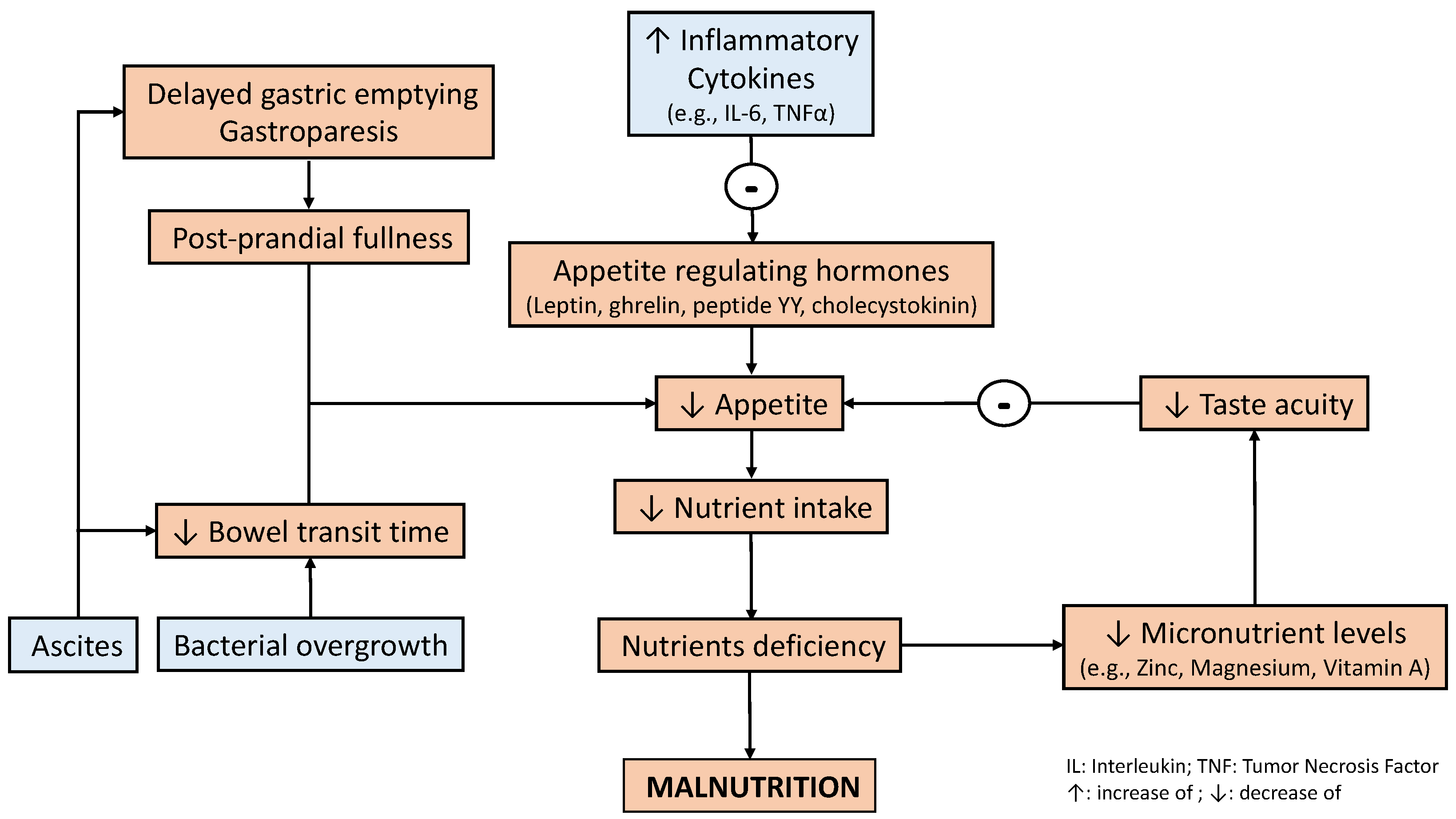
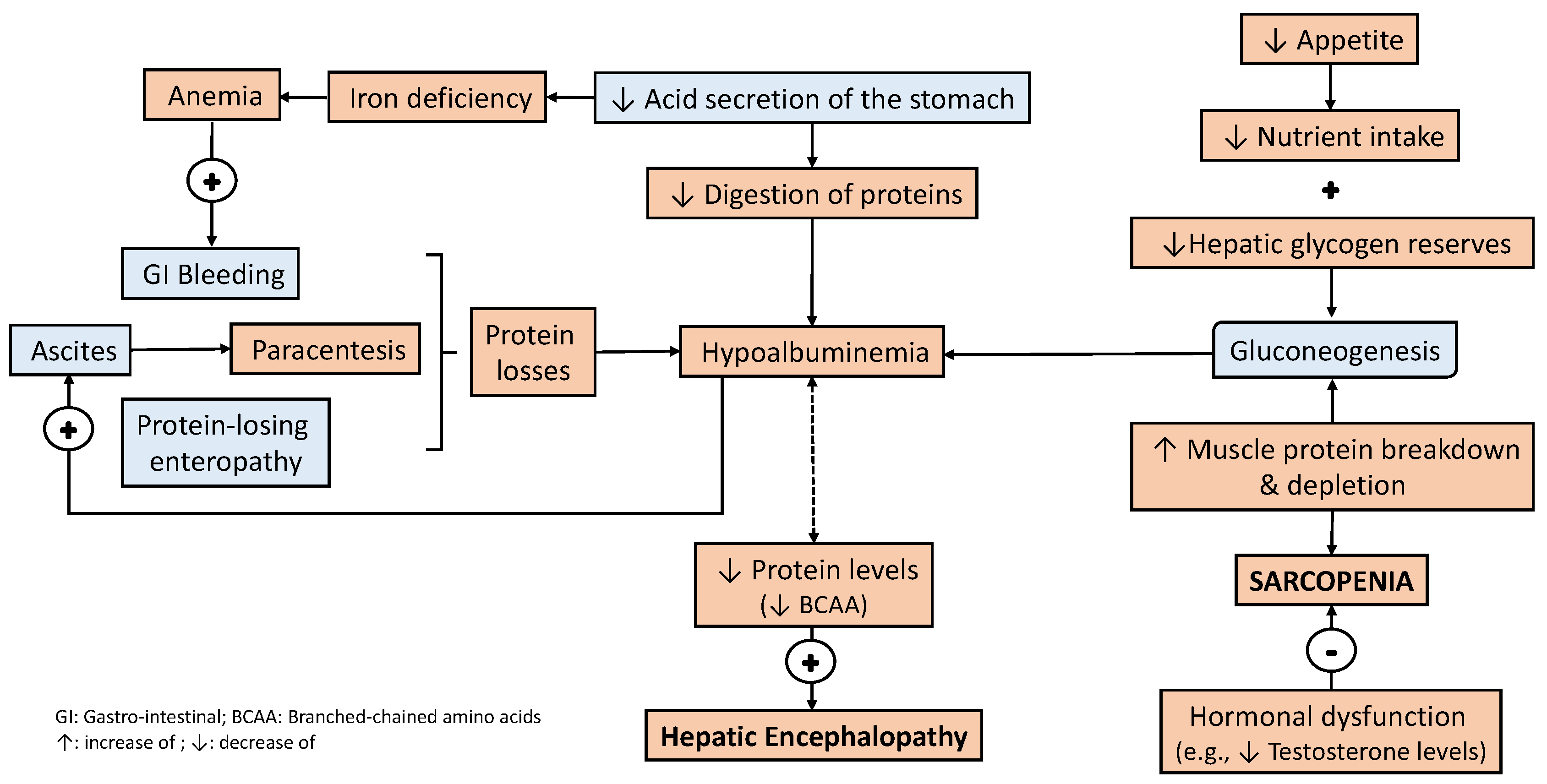
3. Bidirectional Relationship between Liver Disease and Nutritional Status

Liver disease negatively affects metabolism and the patient’s nutritional status and, simultaneously, a worsening of nutritional status could negatively affect the severity and progression of liver disease. Malnutrition is frequently caused by a variety of factors, including a decreased nutrient intake, gastrointestinal dysfunction leading to nutrient malabsorption, and increased protein catabolism leading to sarcopenia [1,2,3,4,5]. In addition, some cirrhotic patients (≈30%) have an increased resting energy expenditure (>120%) that negatively affects nutritional status [11]. It has been hypothesized that the altered hypermetabolism is caused by chronic inflammation (e.g., increased blood levels of interleukin-1 and interleukin-6 (IL-6)) [12]. Thus, the hypercatabolic status of critical illness together with this hypermetabolic status put the patient with liver disease at higher nutritional risk [5].
Patients with liver disease frequently experience symptoms that may contribute toward a reduced nutrient intake, such as gastrointestinal effects (e.g., increased gastric sensitivity to distension and delayed gut transit). Delayed gastric emptying has been reported in patients with liver disease and has been associated with post-prandial fullness and bloating [13]. These patients suffer from alterations in taste acuity, which has been associated with deficiencies in trace elements including zinc, magnesium, and vitamin A [14,15]. Appetite is also reduced due to increased inflammatory cytokines and alterations in appetite-regulating hormones (i.e., leptin, ghrelin, peptide YY, and cholecystokinin) [16]. A decreased dietary intake in patients with liver disease enhances anorexia, which is also affected by micronutrient deficiency (i.e., low zinc levels) and metabolic alterations, such as hyperglycemia, that may contribute to increased inflammatory cytokine production (e.g., Tumor Necrosis Factor (TNF-α) and IL-6) and leptin [17]. Functional problems such as gastroparesis and delayed bowel transit time, which may induce bacterial overgrowth, together with tense ascites, cause nausea and early satiety [18].
In addition, hormonal alterations and dysfunction negatively influence both nutrient intake and metabolism. Increased insulin levels, caused by hyperinsulinemia and insulin resistance, induce satiety [19]. Hyperglycemia may eventually contribute to the development of autonomic neuropathy, which modifies gustatory sensation and dietary intake and may also enhance the negative effect of liver disease on gastrointestinal functions, especially regarding motility (e.g., gastric emptying) [20]. Another endocrine alteration is the lack of response to high levels of ghrelin, a peripherally derived orexigenic hormone that normally increases appetite and food intake. Despite the presence of high ghrelin levels in cirrhotic patients, appetite is not increased [21].
In a patient with liver disease, the digestion, absorption, and metabolism of nutrients are all also negatively affected. Fat and fat-soluble vitamin malabsorption due to impaired bile acid metabolism is common [22]. A reduction in acid secretion in the stomach, or even achlorhydria, may be present in patients with liver cirrhosis, regardless of Helicobacter pylori infection, with a variable prevalence [18,23]. This may contribute to the impaired digestion of macronutrients (e.g., proteins) and some micronutrients (e.g., vitamin B12 and iron), which may ultimately worsen their absorption and increase their deficit. Consequently, this may also contribute to worsening clinical manifestations of nutrient deficiency (e.g., anemia) [24].
Alcohol abuse, which is the most frequent cause of liver disease, may contribute to the poor nutritional intake caused by the development of chronic pancreatitis, which is per se associated with fat and micronutrient malabsorption and metabolic alterations (e.g., hyperglycemia), which may produce liver damage [25]. Alcohol inhibits fatty acid oxidation, leading to triglyceride accumulation in the liver and the development of fatty liver disease or NAFLD [26].
The liver plays a central role in amino acid and protein metabolism, and a reduction in blood levels is not surprising (e.g., hypoalbuminemia). Indeed, a reduction in branched chained amino acid (BCAA) serum levels is associated with the occurrence of hepatic encephalopathy [27]. Protein losses are caused by gastrointestinal bleeding and frequent paracentesis, which may be further worsened by protein-losing enteropathy, contributing to the development of hypoalbuminemia [28]. It is important to highlight that alterations in carbohydrate metabolism also contribute to lower amino acid levels and protein deficit. A decreased hepatic glucose production and lower hepatic glycogen reserves increase gluconeogenesis from amino acids and even secondary protein breakdown from muscle [29]. All these effects are enhanced by hypermetabolism accompanied by a reduced food intake, leading to a negative caloric and protein balance, which is a vicious circle that worsens the malnutrition and clinical manifestations of liver disease [30].
Some data suggest that preservation of the body’s lean mass is important during the evolution of patients with liver disease since it is associated with fewer complications [31,32]. Muscle wasting or sarcopenia is the most objective feature of chronic protein malnutrition in cirrhosis and is also an important predictor of survival in decompensated liver disease, quality of life, and the ability to respond to stressors, such as surgery, prolonged ventilator support, longer ICU and hospital stays, higher risk of infections, mortality, and even lower survival chances during the perioperative course of liver transplantation [31,32,33,34,35]. Testosterone, which plays an important role in protein synthesis, is highly reduced (≈90% approximately) in cirrhosis and protein breakdown [36]. Despite malnutrition not being a formal contraindication for liver transplantation, it is well known that in candidates for liver transplantation, malnutrition adversely affects the perioperative course, and nutritional status should be improved before surgery.
Some metabolic and nutritional factors related to the occurrence of malnutrition in liver disease should be better elucidated; nevertheless, it is obvious that the metabolic and nutritional status strongly impacts liver disease and vice versa. These pathophysiological factors are common to all degrees of liver disease in patients admitted to the ICU and vary depending on the severity of the liver failure. However, a reduced nutritional intake (Figure 1) and protein depletion (Figure 2) both seem to play a key role in the occurrence of malnutrition and sarcopenia in these patients, which ultimately strongly influence outcomes.

4. Nutrition Management in Patients with Liver Disease

Evidence for malnutrition as a prognostic indicator in patients with decompensated liver disease has mostly been derived from the liver transplantation population, in which severe malnutrition has been associated with higher perioperative mortality [13]. However, common models for determining the prognosis and degree of liver disease (i.e., model of end-stage liver disease and the Child–Pugh score) do not evaluate nutritional status. Identifying the degree of malnutrition in patients with liver disease is the first step toward addressing nutrition therapy in these patients.

4.1. Evaluation of Nutritional Status in Patients with Liver Disease

Evaluating nutritional status and the presence of malnutrition involves different parameters, such as physical examination, anthropometric measurements, laboratory tests, and instrumental examination, along with the determination of the patient’s functional capacity [9]. These parameters may be modified due to the inherent alterations associated with critical illness and liver disease (e.g., fluid overload and alterations in protein metabolism) which make it difficult to determine the nutritional status.
Underweight (<18.5 kg/m2) and obesity (>30 kg/m2) are related to the presence of malnutrition and sarcopenia in liver disease [37]. However, these measurements may be affected by fluid overload, a frequent feature in these patients. Indeed, other anthropometric measurements, such as triceps skinfold and mid-arm muscle circumference, which assess subcutaneous fat and muscle mass, can also be affected by fluid overload when patients are admitted to an ICU [28]. Fluid overload may also affect both weight and laboratory biomarkers (e.g., albumin levels), which can be even lower due to impaired hepatic protein synthesis [38]. Despite this, laboratory markers may be helpful; serum protein levels (e.g., albumin, prealbumin, and transferrin), vitamin levels, and creatinine may be considered useful for nutritional assessment and can detect nutritional deficiencies in the absence of ideal biomarkers [5,7]. Prealbumin, also known as transthyretin, is a hepatic protein that correlates well with the body’s protein status and was found to be the best nutritional predictor of survival in liver transplant patients in comparison with serum albumin, cholesterol, and creatinine [39,40].
Regarding nutritional scores, some have been evaluated in patients with liver disease. The subjective global assessment (SGA) has been used as a reference for the validation of other malnutrition screening tools. It has been applied to this end in liver transplant candidates with an overall fair to good inter-observer reproducibility rate [41]. Four features of the medical history are elicited: weight loss in the previous six months, dietary intake in relation to a patient’s usual pattern, the presence of significant gastrointestinal symptoms (e.g., anorexia, nausea, vomiting, diarrhea, etc.), and patient’s functional capacity or energy level (e.g., bedridden to full capacity) [42]. Patients are classified as well-nourished or with mild, moderate, or severe malnutrition. Muscle wasting and fat depletion have both been strongly associated with SGA scoring [41]. This test has shown a high specificity (96%) with a very low sensitivity (22%) for diagnosing malnutrition in these patients, especially in those with alcoholic liver disease [43]. Despite the limitations of the SGA, it has been the reference screening tool for comparison with others in different studies.
The Mini-Nutritional Assessment (MNA) may also be an appropriate tool for nutritional screening assessment in cirrhotic patients of any etiology [44]. It consists of six preliminary questions with a total score of up to 30 points. Cases are defined as “malnourished” (0–16 points), “at risk of malnutrition” (17–23.5 points), and “normal nutritional status” (24–30 points). The MNA also correlates with representative nutrition-related anthropometric parameters (e.g., percentage arm circumference, triceps skinfold, maximum grasp strength, and arm muscle circumference) and serum blood chemistry data (i.e., albumin) [44]. The European Nutritional Risk Screening (NRS)-2002 is a suitable screening tool for patients with early and mild liver disease; however, it is less accurate in end-stage liver disease since it may promote false positives [45]. It identifies a nutritional risk group with a score of three points or more and it was the first score to include disease severity as a component of nutritional screening [45,46]. The Controlling Nutrition Status (CONUT) score is an objective tool extensively used to evaluate nutritional status in various diseases, which is based on three laboratory parameters (i.e., albumin, cholesterol, and lymphocytes). It has been evaluated in adults with hepatitis C virus-related liver cirrhosis reflecting liver functional reserves, which may potentially make the CONUT score a useful marker of the presence of malnutrition in these patients [47]. The Malnutrition Universal Screening Tool (MUST) was evaluated in cirrhotic patients. It evaluates body mass index (BMI), percentage body weight loss, and acute or chronic disease. It is divided into three stages, where patients scoring two points or more are classified as the high-risk group; it is a relatively quick and easy screening tool. Unfortunately, MUST is less accurate in patients with ascites/fluid retention, which makes it unreliable for identifying malnutrition in patients with cirrhosis [48].
The Liver Disease Undernutrition Screening Tool (LDUST) has been validated as a screening tool for malnutrition in ambulatory patients with liver disease [49]. It is based on six parameters (i.e., recent oral intake, weight loss over 12 months, body fat loss, muscle loss, fluid retention/ascites, and effects on daily activities) chosen by consensus and recognized by the American Society for Parenteral and Enteral Nutrition (ASPEN) and the Academy of Nutrition and Dietetics (AND) [50]. LDUST theoretically overcomes the ascites/fluid retention limitations of MUST. Another specific screening tool validated for malnutrition assessment in patients with liver cirrhosis, the Royal Free Hospital–Nutritional Prioritizing Tool (RFH–NPT), also considers ascites/fluid retention [51]. It is included as part of the algorithm to assess malnutrition in the clinical practice guidelines of the European Association for the Study of the Liver (EASL) [52]. The RFH–NPT and LDUST are screening tools that proved to be accurate in detecting malnutrition in cirrhotic patients [53].
Theoretically, a more objective way to diagnose malnutrition is using body composition assessment. Numerous indirect methods have been used to quantify body composition in liver transplantation candidates, including total body electrical conductivity, bioelectrical impedance analysis (BIA), dual-energy X-ray absorptiometry, air displacement plethysmography, or magnetic resonance imaging (MRI) and computed tomography (CT) scans [54]. Unfortunately, like body weight and BMI, the accuracy of some body composition assessment techniques is limited due to the presence of ascites/fluid retention, and their current utility should be elucidated.
The annual rate of muscle loss increases with worsening liver reserves, and patients with liver disease are more likely to present complications with sarcopenia. Sarcopenia is prevalent in cirrhosis patients, with an incidence ranging from 30% to 70% [55]. This wide range is related to the lack of standard diagnostic criteria for sarcopenia in cirrhosis, the wide variability in etiology, the presence of comorbidities, the degree of liver dysfunction, and patient characteristics across different studies [54]. Low muscle mass or muscle function have been identified as independent predictors of a poor quality of life associated with a higher incidence of hepatic decompensation (i.e., ascites, hepatic encephalopathy, and variceal bleeding) and mortality in patients with cirrhosis evaluated for liver transplantation. The negative impact that sarcopenia has on patients with cirrhosis has been demonstrated in several studies (Table 1).
Evaluation of physical activity in patients with liver disease may be mandatory as it is both a mainstay of therapy for improving muscle mass and evaluating functional status in patients with cirrhosis and liver disease. There are now several trials, predominantly enrolling patients with compensated cirrhosis, which have evaluated the effects of exercise therapy, reporting improvements in health-related quality of life (HRQoL), muscle mass, exercise capacity, and even reductions in the hepatic venous pressure gradient [64,65,66]. However, it is important to underline that physical activity has been poorly studied until recently because earlier studies associated physical activity with an increase in portal pressure and, therefore, the risk of variceal bleeding [67]. However, these drawbacks have not been detected in contemporary studies [64,65,66].
Deconditioning and fatigue are major issues for patients with cirrhosis, even those with compensated liver disease, which may represent a significant barrier to physical activity. Physical activity regimens, therefore, need to be adjusted to the patient’s level of baseline function and carried out at their perceived level of effort [64]. For example, some very deconditioned patients may achieve a 7–8 out of 10 perceived exertions with just slow walking. Despite there being no formal guidelines available for physical activity in cirrhosis, recognizing the need to individualize prescribed physical activity in cirrhotic patients should be mandatory due to the potential benefits that exercise entails. Further research is needed regarding the optimal characteristics of physical activity/exercise (i.e., dose, duration, frequency, intensity, type, etc.), not just for these patients but the entire ICU population.
As there is no gold standard, we recommend using a combination of different methods to evaluate and determine the extent of malnutrition in patients with liver disease. Simultaneous evaluation of BMI and ascites/fluid retention together with the degree of liver disease (i.e., the Child–Turcotte–Pugh score and Model for End-Stage Liver Disease score) may be helpful for this purpose. The SGA, LDUST, and the RFH–NPT seem to be the most suitable tools for screening malnutrition in these patients. Evaluating sarcopenia and physical activity should be mandatory since these are considered surrogate markers of nutritional and functional status irrespective of the degree of liver disease. Despite there not being any gold standard or ideal tool in these patients, the presence of sarcopenia needs to be evaluated (e.g., by CT scans, MRI, BIA, etc.). A summary detailing the nutritional parameters for evaluating the metabolic and nutritional status in patients with liver disease is provided in Table 2.

4.2. What Do Clinical Practice Guidelines Recommend?

It is important to remark that most recommendations for nutrition therapy in patients with liver disease arise from a pathophysiological basis and expert opinion or consensus; the degree of evidence is moderate due to the lack of nutrition studies in this specific population of ICU patients. As a result, there are few main contemporary international guidelines available specifically addressing nutrition therapy in liver disease [5].
The EASL has published clinical practice guidelines on nutrition in advanced liver disease, including how to recognize and assess nutritional problems and what the consequences of malnutrition are and its correction, in addition to addressing different clinical scenarios, such as hepatic encephalopathy and before/after transplantation [52]. The ESPEN guidelines on clinical nutrition in liver disease include recommendations on different issues of nutritional management in various forms of liver disease (e.g., ALF, ACLF, cirrhosis, major liver surgery, or transplantation) [68]. Indeed, a recent ESPEN review underlined the importance of sarcopenia in cirrhosis and made some recommendations for clinical practice [69]. Table 3 summarizes these ESPEN guideline recommendations for nutrition therapy in patients with liver disease.
It is important to note that none of these guidelines specifically address the population admitted to the ICU with liver disease; the recommendations are selected and adapted based on the characteristics of liver disease patients with critical illness. In consequence, there are some concerns related to nutrition therapy and metabolic status in these patients that are not emphasized in the guidelines and require careful attention. First, these patients have major vitamin and trace element deficits, and identifying these deficits (if possible) may be of crucial importance [24]. The routine administration of vitamins and trace element supplementation may be considered when these patients are admitted to an ICU.
Second, some patients develop a hypercatabolic status based on the metabolic characteristics that liver disease entails; however, they are unable to tolerate full enteral nutrition (EN) since they are critically ill (e.g., severe or prolonged shock) [10]. The hypercatabolic status combined with the stress response of their critical status rapidly leads to malnutrition because of the difficulty in achieving nutritional and metabolic requirements. Thus, nutrition therapy should be individualized based on these considerations; nutrition therapy should follow the recommendations for ICU patients (i.e., early and progressive administration) and the specifications described above (e.g., reduce protein administration when hepatic encephalopathy is present).
Third, enteral is the main administration route to maintain the integrity of the gastric mucosa and gut barrier, yet patients frequently experience difficulties in achieving the entire nutrition therapy volume because liver disease and ICU admission both entail gastrointestinal dysfunction (e.g., delayed gastric emptying) [70]. We should follow prompt strategies to optimize the enteral route during the first 72 h of ICU admission, such as early use of prokinetics, and consider post-pyloric administration, especially if we suspect previous malnutrition or chronic liver disease. In addition, the use of PN should be considered early on (i.e., day 3–4 of ICU admission) to achieve nutritional targets and avoid the occurrence or development of malnutrition [24]. Older patients (i.e., >50 years old) under PN more frequently have ascites and a positive fluid balance; however, these patients usually exhibit a higher risk of malnutrition compared with patients on EN [71]. Physicians should be aware of the detrimental effects that a positive fluid balance caused by PN may have on gastrointestinal function, which ultimately are related to worse outcomes [72]. EN, even with minimal delivery, should be considered in combination with PN to maintain gut structure and minimize the adverse effects (i.e., induced gut and liver inflammation) that PN may entail [73]. It is important to remark that PN with a higher proportion of omega-3 polyunsaturated fatty acids (ω3-PUFA) may help minimize this adverse effect [74].
Finally, appropriate glycemic management may be an important metabolic target to control. Glycemia may be monitored every 2 h with several objectives: to maintain glucose levels between 150 and 180 mg/dL, avoid hypoglycemia, and detect hyperglycemia or high glycemic variability during admission. It is important to remember that hyperglycemia may exacerbate intracranial pressure when patients develop intracranial hypertension in the setting of hepatic encephalopathy [75]. All these concerns should be considered for a practical approach to nutrition therapy in these patients.

4.3. A Practical Approach to Nutrition Therapy in Patients with Liver Disease

We previously underlined that patients with cirrhosis may have a higher energy requirement due to increased protein catabolism; however, we must balance this phenomenon with nutrition tolerance, which is lower in the setting of critical illness and worsening liver disease [5,33,52,68]. It is clear that an adequate protein intake is important to prevent protein–calorie malnutrition and avoid sarcopenia [69]. The energy supply should be mixed (i.e., carbohydrates and lipids) but limiting the lipid intake is recommended due to the increased risk of hepatic steatosis or worsening of hypertriglyceridemia. The higher risk of vitamin deficiencies due to intestinal malabsorption, higher energy demands, and previous deficits, mostly involving vitamins A, D, E, and K, as well as iron and zinc, should make their routine administration necessary. According to general and specific guidelines for nutrition therapy in ICU patients, Table 4 summarizes the main points to consider for nutrition therapy in patients with liver disease admitted to an ICU [5,33,52,54,65,68,69,76,77].
Some of the aforementioned recommendations may apply to patients with liver disease who are not admitted to an ICU because the pathophysiological basis of liver disease applies to all patients. However, the nutrition guidance provided cannot apply to all patients with liver disease as ICU patients suffer from alterations that prevent adequate nutrition therapy.

4.4. Considerations in the Perioperative Patient

Nutrition therapy improves outcomes (i.e., mortality, length of hospital stays, and postoperative complications) in all types of perioperative scenarios in patients with liver disease (i.e., liver transplantation, liver resections, and hepatocellular carcinoma) [70]. The application of nutrition therapy in patients with severe nutritional risk (i.e., SGA C) is recommended for 10–14 days before major surgery to optimize their nutritional status, even if surgery must be delayed because of the presence of malnutrition [78]. This would definitively positively affect the outcome of the patient, especially in terms of a lower incidence of postoperative complications.
After surgery, especially after liver transplantation, enteral nutrient intake should be started early in the postoperative period, even though transpyloric access and the composition of the nutritional formula should be adapted to the patient’s metabolic stress. The prevalence of sarcopenia does not seem to decrease, and although patients gain weight, sarcopenic obesity may co-exist [79]. A greater food intake and physical inactivity are responsible for the positive energy balance, which is observed in up to 88% of patients after liver transplantation [80]. Several metabolic complications related to weight gain and immunosuppression are developed in the long-term post-transplant. The risk of arterial hypertension, dyslipidemia, and diabetes mellitus increases after surgery and impacts outcomes as well as survival [81]. This set of metabolic disorders yields an increased risk of metabolic syndrome, described in approximately half of liver transplant recipients [82]. Despite weight gain seeming positive for nutritional status after liver transplantation, this is an additional risk factor for developing metabolic syndrome, which makes a nutritional follow-up necessary.

5. Conclusions

In summary, nutrition therapy in critically ill patients with liver disease remains difficult due to the pathophysiological alterations involved in these patients. Physicians face both the nutritional risk that critically ill patients represent and changes in gastrointestinal function associated with liver disease. Nutrition therapy clearly improves the survival of these patients, especially in liver transplantation, and evaluation of the nutritional status is mandatory to develop an individualized nutrition plan in the ICU. The delivery of nutrition therapy should focus on avoiding the occurrence or development of malnutrition, and strategies to optimize nutrition should be applied early on. PN during ICU admission should be strongly considered to achieve nutrition delivery. Monitoring and detecting nutrition-therapy-related and liver-related complications that may influence the tolerance of nutrition therapy during an ICU stay are of notable importance. Adequate protein delivery and physiotherapy may play a key role in the prevention and recovery of sarcopenia in these patients.

Author Contributions

M.Á.H.-B., J.C.L.-D. and J.A.S.-G. conceived the present review and performed a critical and concise review of the data retrieved from the selected manuscripts during the review process. J.C.L.-D. put together the first draft of the manuscript but all authors were involved in the writing of different versions of the manuscript. All the authors revised the manuscript and approved the final version. All authors have read and agreed to the published version of the manuscript.

Funding

This research received no external funding.

Institutional Review Board Statement

Not applicable.

Informed Consent Statement

Not applicable.

Data Availability Statement

All data reported in the present review comes from the manuscripts cited among the text. There is not any specific dataset or specific data available.

Acknowledgments

We would like to thank the University of Barcelona and CERCA program of Generalitat de Catalunya for institutional support.

Conflicts of Interest

The authors declare no conflict of interest.

References

  1. Patton, H.M. Nutritional assessment of patients with chronic liver disease. Gastroenterol. Hepatol. 2012, 8, 687–690. [Google Scholar]
  2. Sevastianos, V.A.; Dourakis, S.P. Malnutrition and Sarcopenia in Advanced Liver Disease. J. Nutr. Food Sci. 2016, 6, 487. [Google Scholar]
  3. Huynh, D.K.; Selvanderan, S.P.; Harley, H.A.; Holloway, R.H.; Nguyen, N.Q. Nutritional care in hospitalized patients with chronic liver disease. World J. Gastroenterol. 2015, 21, 12835–12842. [Google Scholar] [CrossRef] [PubMed]
  4. Periyalwar, P.; Dasarathy, S. Malnutrition in cirrhosis: Contribution and consequences of sarcopenia on metabolic and clinical responses. Clin. Liver Dis. 2012, 16, 95–131. [Google Scholar] [CrossRef]
  5. Lorencio, C.; Bonet Sarís, A.; Navas Moya, E. Recommendations for specialized nutritional-metabolic treatment of the critical patient: Nonsurgical abdominal disease. Metabolism and Nutrition Working Group of the Spanish Society of Intensive and Critical Care Medicine and Coronary Units (SEMICYUC). Med. Intensiva. 2020, 44 (Suppl. S1), 60–64. [Google Scholar] [CrossRef] [PubMed]
  6. Perez Ruiz de Garibay, A.; Kortgen, A.; Leonhardt, J.; Zipprich, A.; Bauer, M. Critical care hepatology: Definitions, incidence, prognosis and role of liver failure in critically ill patients. Crit. Care 2022, 26, 289. [Google Scholar] [CrossRef]
  7. Shergill, R.; Syed, W.; Rizvi, S.A.; Singh, I. Nutritional support in chronic liver disease and cirrhotics. World J. Hepatol. 2018, 10, 685–694. [Google Scholar] [CrossRef]
  8. Saunders, J.; Brian, A.; Wright, M.; Stroud, M. Malnutrition and nutrition support in patients with liver disease. Frontline Gastroenterol. 2010, 1, 105–111. [Google Scholar] [CrossRef]
  9. Hammad, A.; Kaido, T.; Aliyev, V.; Mandato, C.; Uemoto, S. Nutritional Therapy in liver transplantation. Nutrients 2017, 9, 1126. [Google Scholar] [CrossRef]
  10. Chang, Y.; Liu, Q.Y.; Zhang, Q.; Rong, Y.M.; Lu, C.Z.; Li, H. Role of nutritional status and nutritional support in outcome of hepatitis B virus-associated acute-on-chronic liver failure. World J. Gastroenterol. 2020, 26, 4288–4301. [Google Scholar] [CrossRef]
  11. Prieto-Frias, C.; Conchillo, M.; Payeras, M.; Inarrairaegui, M.; Davola, D.; Fruhbeck, G.; Salvador, J.; Rodriguez, M.; Richter, J.A.; Mugueta, C.; et al. Factors related to increased resting energy expenditure in men with liver cirrhosis. Eur. J. Gastroenterol. Hepatol. 2016, 28, 139–145. [Google Scholar] [CrossRef] [PubMed]
  12. Lin, S.Y.; Wang, Y.Y.; Sheu, W.H. Increased serum leptin concentrations correlate with soluble tumour necrosis factor receptor levels in patients with cirrhosis. Clin. Endocrinol. 2002, 57, 805–811. [Google Scholar] [CrossRef] [PubMed]
  13. Kalaitzakis, E. Gastrointestinal dysfunction in liver cirrhosis. World J. Gastroenterol. 2014, 20, 14686–14695. [Google Scholar] [CrossRef] [PubMed]
  14. Nicoll, R.; Gerasimidis, K.; Forrest, E. The Role of Micronutrients in the Pathogenesis of Alcohol-Related Liver Disease. Alcohol Alcohol. 2022, 57, 275–282. [Google Scholar] [CrossRef]
  15. Saeed, A.; Dullaart, R.P.F.; Schreuder, T.C.M.A.; Blokzijl, H.; Faber, K.N. Disturbed Vitamin A Metabolism in Non-Alcoholic Fatty Liver Disease (NAFLD). Nutrients 2017, 10, 29. [Google Scholar] [CrossRef] [PubMed]
  16. Perry, B.; Wang, Y. Appetite regulation and weight control: The role of gut hormones. Nutr. Diabetes 2012, 2, e26. [Google Scholar] [CrossRef]
  17. Thuluvath, P.J.; Triger, D.R. Autonomic neuropathy and chronic liver disease. Q. J. Med. 1989, 72, 737–747. [Google Scholar]
  18. Shindo, K.; Machida, M.; Miyakawa, K.; Fukumura, M. A syndrome of cirrhosis, achlorhydria, small intestinal bacterial overgrowth, and fat malabsorption. Am. J. Gastroenterol. 1993, 88, 2084–2091. [Google Scholar]
  19. Lee, J.H.; Kwon, Y.J.; Park, K.; Lee, H.S.; Park, H.K.; Han, J.H.; Ahn, S.B. Metabolic Score for Insulin Resistance Is Inversely Related to Incident Advanced Liver Fibrosis in Patients with Non-Alcoholic Fatty Liver Disease. Nutrients 2022, 14, 3039. [Google Scholar] [CrossRef]
  20. Liu, K.; Yang, L.; Wang, G.; Liu, J.; Zhao, X.; Wang, Y.; Li, J.; Yang, J. Metabolic stress drives sympathetic neuropathy within the liver. Cell Metab. 2021, 33, 666–675.e4. [Google Scholar] [CrossRef]
  21. Marchesini, G.; Bianchi, G.; Lucidi, P.; Villanova, N.; Zoli, M.; De Feo, P. Plasma ghrelin concentrations, food intake, and anorexia in liver failure. J. Clin. Endocrinol. Metab. 2004, 89, 2136–2141. [Google Scholar] [CrossRef] [PubMed]
  22. Hofmann, A.F. The continuing importance of bile acids in liver and intestinal disease. Arch. Intern. Med. 1999, 159, 2647–2658. [Google Scholar] [CrossRef] [PubMed]
  23. Lodato, F.; Azzaroli, F.; Di Girolamo, M.; Feletti, V.; Cecinato, P.; Lisotti, A.; Festi, D.; Roda, E.; Mazzella, G. Proton pump inhibitors in cirrhosis: Tradition or evidence-based practice? World J. Gastroenterol. 2008, 14, 2980–2985. [Google Scholar] [CrossRef] [PubMed]
  24. Kappus, M.R. Acute Hepatic Failure and Nutrition. Nutr. Clin. Pract. 2020, 35, 30–35. [Google Scholar] [CrossRef]
  25. Li, B.R.; Pan, J.; Du, T.T.; Liao, Z.; Ye, B.; Zou, W.B.; Chen, H.; Ji, J.T.; Zheng, Z.H.; Wang, D.; et al. Risk Factors for Steatorrhea in Chronic Pancreatitis: A Cohort of 2153 Patients. Sci. Rep. 2016, 6, 21381. [Google Scholar] [CrossRef]
  26. Correnti, J.M.; Gottshall, L.; Lin, A.; Williams, B.; Oranu, A.; Beck, J.; Chen, J.; Bennett, M.J.; Carr, R.M. Ethanol and C2 ceramide activate fatty acid oxidation in human hepatoma cells. Sci. Rep. 2018, 8, 12923. [Google Scholar] [CrossRef]
  27. Kinny-Koster, B.; Bartels, M.; Becker, S.; Scholz, M.; Thiery, J.; Ceglarek, U.; Kaiser, T. Plasma Amino Acid Concentrations Predict Mortality in Patients with End-Stage Liver Disease. PLoS ONE 2016, 11, e0159205. [Google Scholar] [CrossRef]
  28. Anastácio, L.R.; Davisson Correia, M.I. Nutrition therapy: Integral part of liver transplant care. World J. Gastroenterol. 2016, 22, 1513–1522. [Google Scholar] [CrossRef]
  29. Petersen, K.F.; Krssak, M.; Navarro, V.; Chandramouli, V.; Hundal, R.; Schumann, W.C.; Landau, B.R.; Shulman, G.I. Contributions of net hepatic glycogenolysis and gluconeogenesis to glucose production in cirrhosis. Am. J. Physiol. 1999, 276, E529–E535. [Google Scholar] [CrossRef]
  30. Ferreira, L.G.; Ferreira Martins, A.I.; Cunha, C.E.; Anastácio, L.R.; Lima, A.S.; Correia, M.I. Negative energy balance secondary to inadequate dietary intake of patients on the waiting list for liver transplantation. Nutrition 2013, 29, 1252–1258. [Google Scholar] [CrossRef]
  31. Ridola, L.; Gioia, S.; Faccioli, J.; Riggio, O.; Nardelli, S. Gut liver muscle brain axis: A comprehensive viewpoint on prognosis in cirrhosis. J. Hepatol. 2022, 77, 262–263. [Google Scholar] [CrossRef]
  32. Tantai, X.; Liu, Y.; Yeo, Y.H.; Praktiknjo, M.; Mauro, E.; Hamaguchi, Y.; Engelmann, C.; Zhang, P.; Jeong, J.Y.; van Vugt, J.L.A.; et al. Effect of sarcopenia on survival in patients with cirrhosis: A meta-analysis. J. Hepatol. 2022, 76, 588–599. [Google Scholar] [CrossRef] [PubMed]
  33. Martin, P.; DiMartini, A.; Feng, S.; Brown, R.; Fallon, M. Evaluation for liver transplantation in adults: 2013 practice guideline by the American Association for the Study of Liver Diseases and the American Society of Transplantation. Hepatology 2014, 59, 1144–1165. [Google Scholar] [CrossRef]
  34. Preiser, J.C.; van Zanten, A.R.; Berger, M.M.; Biolo, G.; Casaer, M.P.; Doig, G.S.; Griffiths, R.D.; Heyland, D.K.; Hiesmayr, M.; Iapichino, G.; et al. Metabolic and nutritional support of critically ill patients: Consensus and controversies. Crit. Care 2015, 19, 35. [Google Scholar] [CrossRef] [PubMed]
  35. Ebadi, M.; Bhanji, R.A.; Mazurak, V.C.; Montano-Loza, A.J. Sarcopenia in cirrhosis: From pathogenesis to interventions. J. Gastroenterol. 2019, 54, 845–859. [Google Scholar] [CrossRef] [PubMed]
  36. Grossmann, M.; Hoermann, R.; Gani, L.; Chan, I.; Cheung, A.; Gow, P.J.; Li, A.; Zajac, J.D.; Angus, P. Low testosterone levels as an independent predictor of mortality in men with chronic liver disease. Clin. Endocrinol. 2012, 77, 323–328. [Google Scholar] [CrossRef] [PubMed]
  37. Nishikawa, H.; Enomoto, H.; Nishiguchi, S.; Iijima, H. Sarcopenic Obesity in Liver Cirrhosis: Possible Mechanism and Clinical Impact. Int. J. Mol. Sci. 2021, 22, 1917. [Google Scholar] [CrossRef]
  38. Dasarathy, S.; Merli, M. Sarcopenia from mechanism to diagnosis and treatment in liver disease. J. Hepatol. 2016, 65, 1232–1244. [Google Scholar] [CrossRef]
  39. Sreedhara, R.; Avram, M.M.; Blanco, M.; Batish, R.; Avram, M.M.; Mittman, N. Prealbumin is the best nutritional predictor of survival in hemodialysis and peritoneal dialysis. Am. J. Kidney Dis. 1996, 28, 937–942. [Google Scholar] [CrossRef]
  40. Dağ, Z.; Köseoğlu, H.; Kekilli, M. The use of prealbumin as a predictor of malnutrition in cirrhotic patients and the effect of nutritional support in patients with low prealbumin levels. Turk. J. Med. Sci. 2020, 50, 398–404. [Google Scholar] [CrossRef]
  41. Hasse, J.; Strong, S.; Gorman, M.A.; Liepa, G. Subjective global assessment: Alternative nutrition-assessment technique for liver-transplant candidates. Nutrition 1993, 9, 339–343. [Google Scholar] [PubMed]
  42. Bakshi, N.; Singh, K. Nutrition assessment in patients undergoing liver transplant. Indian J. Crit. Care Med. 2014, 18, 672–681. [Google Scholar] [CrossRef] [PubMed]
  43. Hasse, J. Liver transplantation: The benefits of nutrition therapy in the liver transplant patient. In Recent Developments in Transplantation Medicine: Liver Transplantation, 1st ed.; Klintmalm, G., Ed.; Physicians and Scientists Publishing Co.: Glenview, IL, USA, 1996; Volume 3, pp. 81–100. [Google Scholar]
  44. Yasutake, K.; Koga, S.; Hokko, Y.; Ikemoto, M.; Yaguchi, Y.; Sakai, H.; Murata, Y.; Ohe, K.; Kohjima, M.; Nakamuta, M.; et al. Relevance of the Mini Nutritional Assessment in cirrhotic liver disease patients. Asia Pac. J. Clin. Nutr. 2018, 27, 300–305. [Google Scholar] [PubMed]
  45. Wu, Y.; Zhu, Y.; Feng, Y.; Wang, R.; Yao, N.; Zhang, M.; Liu, X.; Liu, H.; Shi, L.; Zhu, L.; et al. Royal Free Hospital-Nutritional Prioritizing Tool improves the prediction of malnutrition risk outcomes in liver cirrhosis patients compared with Nutritional Risk Screening 2002. Br. J. Nutr. 2020, 124, 1293–1302. [Google Scholar] [CrossRef] [PubMed]
  46. Kondrup, J.; Allison, S.P.; Elia, M.; Vellas, B. ESPEN guidelines for nutrition screening 2002. Clin. Nutr. 2003, 22, 415–421. [Google Scholar] [CrossRef]
  47. Nishikawa, H.; Yoh, K.; Enomoto, H.; Ishii, N.; Iwata, Y.; Takata, R.; Nishimura, T.; Aizawa, N.; Sakai, Y.; Ikeda, N.; et al. The Relationship between Controlling Nutritional (CONUT) Score and Clinical Markers among Adults with Hepatitis C Virus Related Liver Cirrhosis. Nutrients 2018, 10, 1185. [Google Scholar] [CrossRef]
  48. Casas Deza, D.; Betoré Glaria, M.E.; Sanz-París, A.; Lafuente Blasco, M.; Fernández Bonilla, E.M.; Bernal Monterde, V.; Arbonés Mainar, J.M.; Fuentes Olmo, J. Mini Nutritional Assessment—Short Form Is a Useful Malnutrition Screening Tool in Patients with Liver Cirrhosis, Using the Global Leadership Initiative for Malnutrition Criteria as the Gold Standard. Nutr. Clin. Pract. 2021, 36, 1003–1010. [Google Scholar] [CrossRef]
  49. Booi, A.N.; Menendez, J.; Norton, H.J.; Anderson, W.E.; Ellis, A.C. Validation of a screening tool to identify undernutrition in ambulatory patients with liver cirrhosis. Nutr. Clin. Pract. 2015, 30, 683–689. [Google Scholar] [CrossRef]
  50. White, J.V.; Guenter, P.; Jensen, G.; Malone, A.; Schofield, M. Consensus statement of the Academy of Nutrition and Dietetics/American Society for Parenteral and Enteral Nutrition: Characteristics recommended for the identification and documentation of adult malnutrition (undernutrition). JPEN J. Parenter. Enteral. Nutr. 2012, 36, 275–283. [Google Scholar] [CrossRef]
  51. Borhofen, S.M.; Gerner, C.; Lehmann, J.; Fimmers, R.; Gortzen, J.; Hey, B.; Geiser, F.; Strassburg, C.P.; Trebicka, J. The Royal Free Hospital-Nutritional Prioritizing Tool Is an Independent Predictor of Deterioration of Liver Function and Survival in Cirrhosis. Dig. Dis. Sci. 2016, 61, 1735–1743. [Google Scholar] [CrossRef]
  52. European Association for the Study of the Liver. EASL Clinical Practice Guidelines on nutrition in chronic liver disease. J. Hepatol. 2019, 70, 172–193. [Google Scholar] [CrossRef] [PubMed]
  53. Georgiou, A.; Papatheodoridis, G.V.; Alexopoulou, A.; Deutsch, M.; Vlachogiannakos, I.; Ioannidou, P.; Papageorgiou, M.V.; Papadopoulos, N.; Tsibouris, P.; Prapa, A.; et al. Evaluation of the effectiveness of eight screening tools in detecting risk of malnutrition in cirrhotic patients: The KIRRHOS study. Br. J. Nutr. 2019, 122, 1368–1376. [Google Scholar] [CrossRef] [PubMed]
  54. Mazurak, V.C.; Tandon, P.; Montano-Loza, A.J. Nutrition and the transplant candidate. Liver Transpl. 2017, 23, 1451–1464. [Google Scholar] [CrossRef] [PubMed]
  55. Kim, H.Y.; Jang, J.W. Sarcopenia in the prognosis of cirrhosis: Going beyond the MELD score. World J. Gastroenterol. 2015, 21, 7637–7647. [Google Scholar] [CrossRef]
  56. Cruz, R.J., Jr.; Dew, M.A.; Myaskovsky, L.; Goodpaster, B.; Fox, K.; Fontes, P.; DiMartini, A. Objective radiologic assessment of body composition in patients with end-stage liver disease: Going beyond the BMI. Transplantation 2013, 95, 617–622. [Google Scholar] [CrossRef] [PubMed]
  57. Englesbe, M.J.; Patel, S.P.; He, K.; Lynch, R.J.; Schaubel, D.E.; Harbaugh, C.; Holcombe, S.A.; Wang, S.C.; Segev, D.L.; Sonnenday, C.J. Sarcopenia and mortality after liver transplantation. J. Am. Coll. Surg. 2010, 211, 271–278. [Google Scholar] [CrossRef]
  58. Pirlich, M.; Schütz, T.; Spachos, T.; Ertl, S.; Weiss, M.L.; Lochs, H.; Plauth, M. Bioelectrical impedance analysis is a useful bedside technique to assess malnutrition in cirrhotic patients with and without ascites. Hepatology 2000, 32, 1208–1215. [Google Scholar] [CrossRef]
  59. Tandon, P.; Ney, M.; Irwin, I.; Ma, M.M.; Gramlich, L.; Bain, V.G.; Esfandiari, N.; Baracos, V.; Montano-Loza, A.J.; Myers, R.P. Severe muscle depletion in patients on the liver transplant wait list: Its prevalence and independent prognostic value. Liver Transpl. 2012, 18, 1209–1216. [Google Scholar] [CrossRef]
  60. Meza-Junco, J.; Montano-Loza, A.J.; Baracos, V.E.; Prado, C.M.; Bain, V.G.; Beaumont, C.; Esfandiari, N.; Lieffers, J.R.; Sawyer, M.B. Sarcopenia as a prognostic index of nutritional status in concurrent cirrhosis and hepatocellular carcinoma. J. Clin. Gastroenterol. 2013, 47, 861–870. [Google Scholar] [CrossRef]
  61. Krell, R.W.; Kaul, D.R.; Martin, A.R.; Englesbe, M.J.; Sonnenday, C.J.; Cai, S.; Malani, P.N. Association between sarcopenia and the risk of serious infection among adults undergoing liver transplantation. Liver Transpl. 2013, 19, 1396–1402. [Google Scholar] [CrossRef]
  62. Montano-Loza, A.J.; Duarte-Rojo, A.; Meza-Junco, J.; Baracos, V.E.; Sawyer, M.B.; Pang, J.X.; Beaumont, C.; Esfandiari, N.; Myers, R.P. Inclusion of sarcopenia within MELD (MELD-sarcopenia) and the prediction of mortality in patients with cirrhosis. Clin. Transl. Gastroenterol. 2015, 16, e102. [Google Scholar] [CrossRef] [PubMed]
  63. Masuda, T.; Shirabe, K.; Ikegami, T.; Harimoto, N.; Yoshizumi, T.; Soejima, Y.; Uchiyama, H.; Ikeda, T.; Baba, H.; Maehara, Y. Sarcopenia is a prognostic factor in living donor liver transplantation. Liver Transpl. 2014, 20, 401–407. [Google Scholar] [CrossRef] [PubMed]
  64. Macías-Rodríguez, R.U.; Ruiz-Margáin, A.; Román-Calleja, B.M.; Moreno-Tavarez, E.; Weber-Sangri, L.; González-Arellano, M.F.; Fernández-Del-Rivero, G.; Ramírez-Soto, K. Exercise prescription in patients with cirrhosis: Recommendations for clinical practice. Rev. Gastroenterol. Mex. 2019, 84, 326–343. [Google Scholar] [CrossRef]
  65. West, J.; Gow, P.J.; Testro, A.; Chapman, B.; Sinclair, M. Exercise physiology in cirrhosis and the potential benefits of exercise interventions: A review. J. Gastroenterol. Hepatol. 2021, 36, 2687–2705. [Google Scholar] [CrossRef]
  66. Tandon, P.; Ismond, K.P.; Riess, K.; Duarte-Rojo, A.; Al-Judaibi, B.; Dunn, M.A.; Holman, J.; Howes, N.; Haykowsky, M.J.F.; Josbeno, D.A.; et al. Exercise in cirrhosis: Translating evidence and experience to practice. J. Hepatol. 2018, 69, 1164–1177. [Google Scholar] [CrossRef]
  67. García-Pagàn, J.C.; Santos, C.; Barberá, J.A.; Luca, A.; Roca, J.; Rodriguez-Roisin, R.; Bosch, J.; Rodés, J. Physical exercise increases portal pressure in patients with cirrhosis and portal hypertension. Gastroenterology 1996, 111, 1300–1306. [Google Scholar] [CrossRef] [PubMed]
  68. Bischoff, S.C.; Bernal, W.; Dasarathy, S.; Merli, M.; Plank, L.D.; Schütz, T.; Plauth, M.; Burgos Peláez, R.; Rivera Irigoin, R. ESPEN Practical Guideline: Clinical nutrition in liver disease. Nutr. Hosp. 2022, 39, 434–472. [Google Scholar] [CrossRef]
  69. Vasques, J.; Guerreiro, C.S.; Sousa, J.; Pinto, M.; Cortez-Pinto, H. Nutritional support in cirrhotic patients with sarcopenia. Clin. Nutr. ESPEN 2019, 33, 12–17. [Google Scholar] [CrossRef]
  70. Koretz, R.L. Nutritional support in liver disease—An updated systematic review. Curr. Opin. Gastroenterol. 2023, 39, 115–124. [Google Scholar] [CrossRef]
  71. Aslam, M.; Farooq, S.; Rizwan, B.; Asghar, A. Assessment of nutritional status of the cirrhotic patients on enteral and parenteral feeding. Nutr. Health 2022, 28, 69–76. [Google Scholar] [CrossRef]
  72. Besen, B.A.; Gobatto, A.L.; Melro, L.M.; Maciel, A.T.; Park, M. Fluid and electrolyte overload in critically ill patients: An overview. World J. Crit. Care. Med. 2015, 4, 116–129. [Google Scholar] [CrossRef]
  73. Lucchinetti, E.; Lou, P.H.; Wawrzyniak, P.; Wawrzyniak, M.; Scharl, M.; Holtzhauer, G.A.; Krämer, S.D.; Hersberger, M.; Rogler, G.; Zaugg, M. Novel Strategies to Prevent Total Parenteral Nutrition-Induced Gut and Liver Inflammation, and Adverse Metabolic Outcomes. Mol. Nutr. Food Res. 2021, 65, e1901270. [Google Scholar] [CrossRef]
  74. Videla, L.A.; Hernandez-Rodas, M.C.; Metherel, A.H.; Valenzuela, R. Influence of the nutritional status and oxidative stress in the desaturation and elongation of n-3 and n-6 polyunsaturated fatty acids: Impact on non-alcoholic fatty liver disease. Prostaglandins Leukot. Essent Fatty Acids 2022, 181, 102441. [Google Scholar] [CrossRef]
  75. Fallahzadeh, M.A.; Rahimi, R.S. Hepatic Encephalopathy and Nutrition Influences: A Narrative Review. Nutr. Clin. Pract. 2020, 35, 36–48. [Google Scholar] [CrossRef] [PubMed]
  76. Singer, P.; Blaser, A.R.; Berger, M.M.; Alhazzani, W.; Calder, P.C.; Casaer, M.P.; Hiesmayr, M.; Mayer, K.; Montejo, J.C.; Pichard, C.; et al. ESPEN guideline on clinical nutrition in the intensive care unit. Clin. Nutr. 2019, 38, 48–79. [Google Scholar] [CrossRef] [PubMed]
  77. McClave, S.A.; Taylor, B.E.; Martindale, R.G.; Warren, M.M.; Johnson, D.R.; Braunschweig, C.; McCarthy, M.S.; Davanos, E.; Rice, T.W.; Cresci, G.A.; et al. Guidelines for the Provision and Assessment of Nutrition Support Therapy in the Adult Critically Ill Patient: Society of Critical Care Medicine (SCCM) and American Society for Parenteral and Enteral Nutrition (A.S.P.E.N.). JPEN J. Parenter. Enteral. Nutr. 2016, 40, 159–211. [Google Scholar] [CrossRef]
  78. Plauth, M.; Cabré, E.; Riggio, O.; Assis-Camilo, M.; Pirlich, M.; Kondrup, J.; DGEM (German Society for Nutritional Medicine); Ferenci, P.; Holm, E.; Vom Dahl, S.; et al. ESPEN Guidelines on Enteral Nutrition: Liver disease. Clin. Nutr. 2006, 25, 285–294. [Google Scholar] [CrossRef] [PubMed]
  79. Kouz, J.; Vincent, C.; Leong, A.; Dorais, M.; Räkel, A. Weight gain after orthotopic liver transplantation: Is nonalcoholic fatty liver disease cirrhosis a risk factor for greater weight gain? Liver Transpl. 2014, 20, 1266–1274. [Google Scholar] [CrossRef] [PubMed]
  80. Choudhary, N.S.; Saigal, S.; Saraf, N.; Mohanka, R.; Rastogi, A.; Goja, S.; Menon, P.B.; Mishra, S.; Mittal, A.; Soin, A.S. Sarcopenic obesity with metabolic syndrome: A newly recognized entity following living donor liver transplantation. Clin. Transpl. 2015, 29, 211–215. [Google Scholar] [CrossRef]
  81. Parekh, J.; Corley, D.A.; Feng, S. Diabetes, hypertension and hyperlipidemia: Prevalence over time and impact on long-term survival after liver transplantation. Am. J. Transpl. 2012, 12, 2181–2187. [Google Scholar] [CrossRef]
  82. Anastácio, L.R.; Ferreira, L.G.; de Sena Ribeiro, H.; Liboredo, J.C.; Lima, A.S.; Correia, M.I.T.D. Metabolic syndrome after liver transplantation: Prevalence and predictive factors. Nutrition 2011, 27, 931–937. [Google Scholar] [CrossRef] [PubMed]
Figure 1. Factors associated with reduced nutrient intake in liver disease. Inflammatory factors negatively influence appetite-regulating hormones. Simultaneously, both a lower taste acuity and some liver-related complications also negatively influence appetite. The latter is related to reduced motility (i.e., gastric emptying and bowel transit time), which may impact appetite and contribute to a reduced nutrient intake [13,14,15,16,17,18,19,20,21].
Livers 03 00036 g001
Figure 2. Factors related to protein depletion in liver disease. Protein depletion and the development of sarcopenia are closely related to a reduced nutrient intake and the activation of mechanisms to guarantee the metabolic demands of carbohydrates. Liver-related insensitive protein losses and impaired protein digestion both negatively affect protein depletion, which also enhances the development of liver-related complications (e.g., hepatic encephalopathy) [31,32,33,34,35,36].
Livers 03 00036 g002
Table 1. Studies describing the association of sarcopenia with outcomes in patients with liver disease.
Studies
(Year)
nIncidence of SarcopeniaMain Results Related to the Occurrence of Sarcopenia
Merli et al. (2010) [56]3853%Increased LOS in the hospital and ICU and increased incidence of perioperative infections in patients with sarcopenia
Englesbe et al. (2010) [57]16325%Higher mortality after LT
Montano-Loza et al. (2012) [58]11240%Sarcopenia was independently associated with mortality
Tandon et al. (2012) [59]14241%Sarcopenia was associated with increased mortality on the LT waiting list
Meza-Junco et al. (2013) [60]11630%Sarcopenia was independently associated with mortality
Krell et al. (2013) [61]20733%Higher risk of infectious complications and mortality after LT
DiMartini et al. (2013) [62]33868%Muscle mass predicted longer LOS in ICU, total LOS, and a longer time on mechanical ventilation
Masuda et al. (2014) [63]20447%Sarcopenia was an independent prognostic factor for post-LT mortality and postoperative sepsis
LOS: length of stay; ICU: Intensive Care Unit; LT: liver transplantation.
Table 2. Parameters to evaluate the metabolic and nutritional status in patients with liver disease [36,37,41,42,43,49,50,51,52,53,54,64,65,66,67].
Type of ParameterValue Associated with the Presence of Malnutrition
Anthropometric
measurement
BMI<18.5 kg/m2 and >30 kg/m2
Laboratory biomarkersAlbumin <3 g/dL2
Prealbumin<160 mg/dL2
Vitamin levels Consider malnutrition in the presence of low levels
Nutrition ScoresSGASGA B (mild) or C (severe)
LDUST≥2 boxes in columns B or C
RFH–NPTScores 1 and 2–7 correspond to moderate and high risk
Liver ScoresCTPB (mild) or C (severe)
MELD>15
Muscle massBIAEvaluate muscle mass and the presence of sarcopenia
CT scan
MRI
Physical activityHRQoL>3 reflects a poor quality of life
BMI: body mass index; SGA: Subjective Global Assessment; LDUST: Liver Disease Undernutrition Screening Tool; RFH–NPT: Royal Free Hospital–Nutritional Prioritizing Tool; CTP: Child–Turcotte–Pugh score; MELD: Model for End-Stage Liver Disease; BIA: bioelectrical impedance analysis; MRI: magnetic resonance imaging; CT: computed tomography; HRQoL: health-related quality of life questionnaire.
Table 3. Summary of recommendations for nutrition therapy in patients with liver disease in an ICU [52,68,69].
Nutritional and metabolic assessment
Should be performed regularly in all patients to detect malnutrition, especially those with acute liver failureEvaluation should include measurement of weight, height, body mass index (BMI), arm circumference, and serum albumin/prealbumin
Evaluate the occurrence of hypoglycemia in the setting of acute liver failure
Screening of alcohol consumptionAlcohol worsens liver disease and increases its associated complications
Dietary advice
Patients at risk of or diagnosed with malnutritionIndividualized dietary advice from a registered dietitian/nutritionist.
Nutrition medical therapy and nutritional considerations
Protein intakeHigh protein intake to avoid muscle loss and sarcopenia
Consider frequent monitoring of ammonia levels (i.e., 24–48 h) to modulate protein intake
Energy intake Adequate to maintain the patient’s nutritional status. In some cases, it may be necessary to increase energy intake to compensate for increased energy expenditure due to acute complications or malnutrition
Carbohydrates as the main source of energy
Fats should be limited to avoid liver damage from dyslipidemia
Consider nutritional supplements To improve nutritional status or achieve nutritional requirements
Consumption of a snack at night (if possible oral route) with at least 50 g of carbohydrates
Careful sodium administrationAdequate to avoid ascites/fluid retention
Metabolic disturbances
Careful control of acid–base balancePrevent complications such as lactic acidosis
Correction of electrolyte disturbancesHyponatremia, hypokalemia, and hypomagnesemia
Route of administration
Oral feedingRoute of choice in the absence of severe HE
Enteral feeding by nasogastric tubeInsufficient oral intake or not tolerated due to complications such as ascites or HE
Consider post-pyloric tube if high risk of bronchial aspiration
Consider parenteral nutrition Total or supplementary PN if they cannot tolerate enteral feedings or if they have compromised intestinal absorption.
Prevention and treatment of sarcopenia
Protein supplementation combined with physiotherapy (e.g., resistance exercise training)Avoid muscle depletion
L-leucine Reverse the decrease in muscle protein balance due to hyperammonemia
Dietary recommendationsHigh protein diet (≈1.5 g/kg/day) with 30–40 kcal/kg/day
Consider nutritional supplements
Specific recommendations
Hepatic encephalopathyReduce protein intake to avoid worsening of hepatic encephalopathy
Consider supplementation with BCAAs
Caution with the enteral route due to the high risk of bronchial aspiration (i.e., HE III–IV)
Bleeding complicationsPrevent (i.e., frequent screening of coagulation disorder) and treat coagulopathy (i.e., vitamin K administration)
Liver-disease-related complications (i.e., portal hypertension, hepatic encephalopathy, and ascites)Prompt drug treatment to prevent or treat complications that ultimately may affect the nutritional status
Vitamin DCorrection of deficiency
HE: hepatic encephalopathy; PN: parenteral nutrition; BCAA: branched-chain amino acids.
Table 4. Recommendations for nutrition status screening, management, and strategies to optimize nutrition therapy in patients with liver disease admitted to an ICU [5,33,52,54,65,68,69,76,77].
Screening of nutrition status
  • Evaluate the degree of liver disease (i.e., MELD and CTP scores).
  • Measure BMI.
  • Evaluate fluid status (e.g., ascites).
  • Evaluate sarcopenia and functional status.
  • Use a screening tool (i.e., SGA, LDUST, and/or RFH-NPS) to identify the occurrence of malnutrition or if the patient is malnourished.
General considerations for the delivery of nutrition therapy
  • Consider EN as the first choice for nutrition therapy if the oral route is not possible.
  • Consider early PN (total or supplemental) when EN does not achieve the energy (<60%) and protein requirements.
  • Consider nutritional supplements if the oral route does not achieve all the energy and protein requirements.
  • More aggressive approach (e.g., early use of PN) in the presence of suspected malnutrition or nutritional risk (i.e., previous liver disease or alcohol consumption).
Nutrition therapy route
EN
  • Initiate early, within 24–48 h after ICU admission, when the patient is hemodynamically stable.
  • EN should be initiated after correct resuscitation (i.e., stable dosage of vasopressors, normal or stable arterial lactate levels, catecholamines, and/or mechanical circulatory support) in the presence of shock or hemodynamic failure and in the absence of contraindications.
PNConsider in the presence of absolute or relative contraindications for EN:
  • Active gastrointestinal hemorrhage (absolute).
  • Uncontrolled shock or hemodynamic failure (e.g., increasing dosage of vasopressors and/or greater need for circulatory support), uncontrolled hypoxemia, hypercapnia, or acidosis (absolute).
  • GRV > 500 mL every 6 h or inability to achieve energy requirements by the enteral route (relative).
  • Suspected or diagnosed acute mesenteric ischemia, intestinal obstruction, or abdominal compartmental syndrome (absolute).
Nutritional requirements
Energy20–25 Kcal/Kg/day
Protein1.2–2 g/Kg/day
Slowly increase EN rate considering clinical state and EN tolerance for 48–72 h
Strategies to optimize nutrition therapy and avoid complications
Recommended strategyObjective
Bed position > 30–45°Avoid regurgitation, vomiting, and aspiration
Prokinetic agents (i.e., Erythromycin and/or Metoclopramide)
  • Consider in the presence of gastric intolerance (GRV > 500 mL), regurgitation, nausea, or vomiting.
  • Optimize delivery of nutrition therapy.
Post-pyloric NG tube placement
Optimize drugs that interfere with gastrointestinal function (e.g., opioids)
  • Avoid and prevent ileus and gastrointestinal failure.
  • Optimize delivery of nutrition therapy.
Avoid fluid overload
Early mobilization and physiotherapyAvoid progression or development of sarcopenia
Vitamin and trace element supplementationAvoid micronutrient deficiency
BMI: body mass index; SGA: Subjective Global Assessment; LDUST: Liver Disease Undernutrition Screening Tool; RFH–NPT: Royal Free Hospital–Nutritional Prioritizing Tool; PN: parenteral nutrition; ICU: Intensive Care Unit; EN: enteral nutrition; GRV: gastric residual volume; NG: nasogastric.
Disclaimer/Publisher’s Note: The statements, opinions and data contained in all publications are solely those of the individual author(s) and contributor(s) and not of MDPI and/or the editor(s). MDPI and/or the editor(s) disclaim responsibility for any injury to people or property resulting from any ideas, methods, instructions or products referred to in the content.

