Review

Liver Fibrosis, Liver Cancer, and Advances in Therapeutic Approaches

by
Indu Rajapaksha
1,2
1
Department of Basic Sciences, Faculty of Health Sciences, University of Sri Jayewardenepura, Nugegoda 10250, Sri Lanka
2
Department of Medicine, Faculty of Medicine, Dentistry and Health Sciences, The University of Melbourne, Parkville, VIC 3010, Australia
Livers 2022, 2(4), 372-386; https://doi.org/10.3390/livers2040028
Submission received: 26 July 2022 / Revised: 19 October 2022 / Accepted: 20 October 2022 / Published: 2 November 2022
(This article belongs to the Topic Liver Fibrosis and Cirrhosis)

Abstract

:
Chronic liver diseases (CLDs) that lead to hepatic fibrosis, cirrhosis, and/or hepatocellular carcinoma (HCC) have become a major cause of illness and death worldwide. The main causative factors for CLDs are chronic viral infections, excessive alcohol consumption, non-alcoholic fatty liver disease (NAFLD), and cholestatic diseases. The primary approach to managing cirrhosis should be removing the causative agent, and the secondary approach should address fibrogenesis. Liver cancer is also a leading cause of death worldwide, and many therapeutic approaches exist to treat the disease. However, liver transplantation remains the last treatment option for cirrhosis and liver cancer. Thus, this review discusses the pathophysiology of liver fibrosis, its progression to cirrhosis and HCC, and current therapeutic options available to treat the diseases with potential therapeutic options that will be available in the near future.

1. Liver Fibrosis

Hepatic fibrosis is the result of the liver’s wound-healing response to repeated injury. After an acute liver injury, parenchymal cells regenerate and replace the necrotic or apoptotic cells. The most common etiologies for chronic liver disease (CLD) include chronic viral infections (e.g., hepatitis B and C), excessive alcohol consumption, non-alcoholic fatty liver disease (NAFLD), and cholestatic diseases, such as primary biliary cholangitis and primary sclerosing cholangitis [1]. Although the pathologies are rare, inherited liver diseases (e.g., Wilson disease, hereditary hemochromatosis, glycogen storage disease type IV), which are caused by metabolic and genetic defects, are also predisposed to fibrosis and cirrhosis [2]. As injury persists, regardless of the initial cause, liver tissue responds by depositing the fibrillar extracellular matrix (ECM) [3]. This phenomenon is known as the wound-healing response. Usually, the ECM consists of glycoproteins, collagens, glycosaminoglycans, and proteoglycans that provide mechanical strength to the tissue [4]. In addition, ECM synthesis is considered an effort of the liver tissue to localize the injury by encapsulating the area of injury. Even though it is an essential part of the wound healing process, once it is deregulated, the condition progresses to ‘liver fibrosis’, which becomes an inefficient attempt at repairing liver tissue [5,6]. Thus, liver fibrosis is mainly characterized by the excessive accumulation of the ECM in the liver parenchyma, replacing the functional hepatic tissue [7].
However, if the hepatic injury persists, the balance between parenchymal cell regeneration and wound healing response is shifted toward impaired regenerative pathways. Over time, hepatocytes are substituted with abundant ECM, eventually leading to the accumulation of excess fibrotic scar tissue [8]. Thus, in advanced stages of fibrosis, the liver contains approximately six times more ECM than normal, including collagens, fibronectin, undulin, elastin, laminin, hyaluronan, and proteoglycans. Under these conditions, not only is an increased synthesis of ECM prominent, but it is also associated with decreased degradation of ECM [9,10]. The result of liver fibrosis is cirrhosis, where there is no specific medical treatment [1].
Interestingly, the microenvironment in the liver is an organized multidirectional interaction complex (cell–matrix–cell). It delivers the molecular signals crucial for normal liver homeostasis. In this process, each cell type in the liver, including hepatocytes, hepatic stellate cells (HSCs), Kupffer cells (KCs) and liver sinusoidal endothelial cells (LSECs), has their roles to play while talking to each other. The cell-to-cell communication process is referred to as the cellular “crosstalk”, [11] and it involves a number of signaling pathways, including TGF-β signaling pathway, MiRNA signaling pathways, TLR pathway, STAT-3, Wnt/β-catenin, and NF-кB signaling pathways [12].

1.1. Hepatic Stellate Cells

Hepatic stellate cells (HSCs) reside in the space of Disse, lying in between the hepatocytes and the sinusoidal epithelium. These cells were also called Ito cells and inhabit one-third of the non-parenchymal cells of the liver [13]. They possess round cell bodies with numerous cellular processes in different thicknesses and sizes [14]. HSCs are the major sites for vitamin A storage in the normal liver.
Most importantly, quiescent HSCs are activated following chronic liver injury and/or transdifferentiated into myofibroblast-like cells. The significant stimuli for HSC activation are the chemokines, cytokines, and reactive oxygen species (ROS) that are released from the apoptosis of hepatocytes. The activated HSCs migrate to damaged sites, proliferate and acquire a myofibroblastic phenotype with a capacity to express and secrete extracellular matrix proteins, such as collagens, cytokines, chemokines, matrix metalloproteinases (MMPs), and their respective tissue inhibitors of metalloproteinases (TIMPs) [15]. The cytokines and chemokines secreted by activated HSCs are also involved in the inflammatory process [16,17]. Therefore, HSCs are considered a group of passive cells reacting to ROS and cytokines secreted by neighboring cells (e.g., Kupffer cells, hepatocytes), as well as a group of active cells producing cytokines, chemokines, and ROS [18]. The activated HSCs can be recognized by specific myogenic markers, such as α-smooth muscle actin (α-SMA), c-myb, and myocyte enhancer factor-2 [19].
Importantly, Battler [20] and others [21] have demonstrated that there is an increased expression of angiotensin II type 1 receptor (AT1-R) on the activated HSCs suggesting that these cells are an important target of angiotensin II (Ang II). Ang II is a well-known pro-oxidant and fibrogenic cytokine significantly upregulated in a liver injury [22,23]. Thus, Ang II further regulates contractility and proliferation of activated HSCs leading to chronic liver fibrosis.
In addition, TGF-β signaling plays an important role in HSC activation, proliferation, and migration. TGF-β is the most potent fibrogenic cytokine that activates the HSCs and fibroblasts to express fibrosis-related genes, including alpha smooth muscle actin (α-SMA) and α1 type I collagen (COL1A1), and consequently promotes hepatic fibrogenesis [24]. TGF-β activates the TGF-β receptor complex and phosphorylation of the intracellular signal mediators in the downstream signaling pathways (e.g., Smad2/3). In addition to an increase in the expression of COL1A1 [24,25], activation of Smad2/3 regulates the expression of other fibrosis-related genes, including plasminogen activator inhibitor-1 [26], various proteoglycans [27], integrins [28], connective tissue growth factor [29], and matrix metalloproteases [30,31]. In liver fibrosis, TGF-β-mediated connective tissue growth factor (CTGF) expression in HSC depends on the signaling pathways, including Erk, JNK, p38, or Stat3, whereas CTGF also modulates extra cellular matrix production. Another role of TGF-β in liver fibrosis is the upregulation of NADPH oxidase expression, which mediates HSC activation [32]. Therefore, targeting TGF-β signaling is also an emerging option as a therapeutic target for liver fibrosis.
There are two major intercellular crosstalk pathways, which involve during liver injury that contributes to HSC activation [11,33], and they are:
  • Capillarization of LSECs;
  • Apoptosis of hepatocytes.

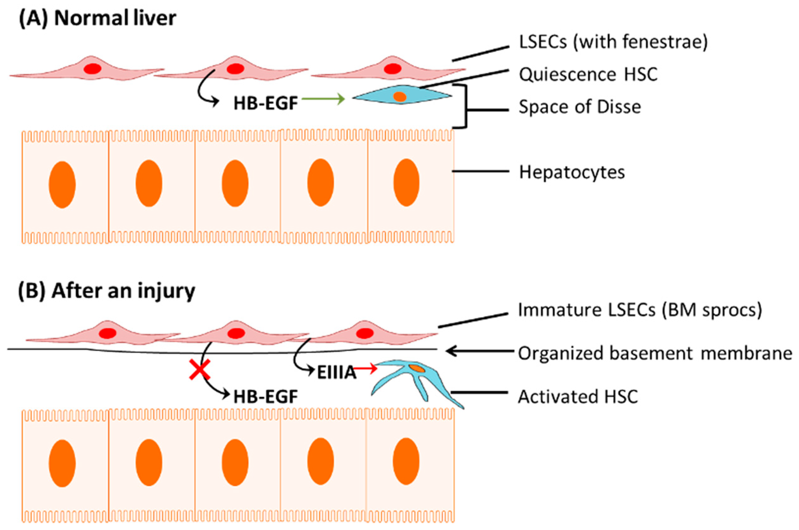
1.1.1. Capillarization of LSECs

The normal liver sinusoids consist of fenestrae that facilitate the solutes to transfer between the sinusoidal blood and the space of Disse. During liver injury, these LSECs are also damaged along with the other cells in the tissue. However, they are replaced by bone marrow (BM)-derived sinusoidal endothelial cell progenitor cells (sprocs) to compensate for the loss. These BM sprocs, which lack LSEC fenestrae, arrange along the sinusoid, and an organized basement membrane is laid down. This de-differentiation process of injured LSECs leads to “capillarization” of the sinusoids [34].
Normal LSECs are responsible for the production of a protein called heparin-binding epidermal growth factor (HB-EGF), which maintains HSC quiescence (Figure 1A). However, the immature LSECs have less or reduced capacity to release HB-EGF, which effect on quiescence nature of HSCs causing activation of those cells [34,35].
While there is a reduction in HB-EGF, de-differentiated LSECs express EIIIA fibronectin isoform, which influences quiescence HSCs to change their phenotype to the activated form [11,36]. Thus, LSECs perform critical crosstalk in the HSCs activation process in hepatic fibrosis by a simultaneous reduction in HB-EGF and expression of EIIIA (Figure 1B).

1.1.2. Apoptosis of Hepatocytes

Hepatocytes play a significant role in HSC activation parallel to LSECs crosstalk during a liver injury. A persistent injury modifies the hepatocyte transcriptional programmes toward apoptosis, a process known as programmed cell death [11]. The apoptotic bodies from hepatocytes stimulate HSC differentiation to the activated HSC phenotype with contractile, proliferative, and fibrogenic properties [11,37].
It has been shown that Kupffer cells are also involved in the cellular crosstalk during the process of fibrosis [11]. KCs are the resident macrophages in the liver and engulf apoptotic bodies arising from the apoptotic hepatocytes [11,38]. After engulfing those apoptotic bodies, KCs become activated and start to express death ligands, such as Fas, TNF-α, and TNF-related apoptosis-inducing ligand (TRAIL), which induce the apoptosis of hepatocytes in a feed-forward loop [38].
Simultaneously, activated KCs release cytokines and reactive oxygen species (ROS) and stimulate HSCs in a paracrine manner [13].

1.2. Cells That Contribute to Extracellular Matrix Synthesis (ECM) Other Than Hepatic Stellate Cells

HSCs are not the only cell type that has the potential to synthesize ECM. Usually, HSCs are found to be the main fibrogenic cells in the pericentral areas of the injured liver [19]. Some myofibroblasts predominate around the portal tracts, particularly in cholestatic liver injuries. These myofibroblasts are derived from small portal vessels in response to cholestasis and proliferate around biliary tracts. After that, they are also involved in collagen synthesis, performing a similar role to HSCs [19,39].
There is evidence that mast cells are also involved in liver fibrosis as a response to an injury. A mast cell is a white blood cell in the circulation containing histamine and heparin granules. It has been shown that mast cell infiltration is prominent during liver fibrosis in several rat models, including bile duct ligation [23,40]. Mast cell involvement during liver fibrosis can be explained by three major mechanisms (Figure 2).
1.
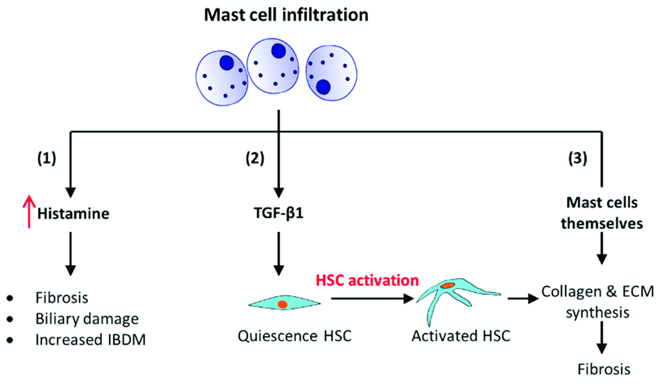
It has been shown using a mouse model with progressive biliary fibrosis, Mdr2-KO; those mast cells infiltrate into the liver during the progression of biliary fibrosis. The presence of mast cells increases the local levels of histamine, which is a pro-fibrogenic and proliferative factor. It induces intrahepatic bile duct mass (IBDM) and proliferation leading to fibrosis [41].
2.
The second concept of mast cells during biliary fibrosis is that they produce transforming growth factor β-1 (TGF-β1), a key pro-fibrotic cytokine in the liver, which activates the quiescence HSCs and stimulates the ECM synthesis and fibrosis [23,42,43].
3.
In addition, mast cells could induce the production of ECM components by overproduction of the basement membrane, which induces fibroblast attachment, spreading and proliferation [23,44].
After all, when the injury persists, an excessive accumulation of fibrous scar leads to the subsequent formation of nodules distorting the liver architecture and leading to cirrhosis.

1.3. Cirrhosis

Cirrhosis is the most advanced stage of liver fibrosis. It is characterized by the abnormal continuation of fibrogenesis (fibrous tissue formation) and distortion of hepatic vasculature by neo-angiogenesis (new sinusoid formation). In this advanced stage of fibrogenesis, there is a collective ECM synthesis from activated HSCs, myofibroblasts derived from bone marrow, portal fibroblasts, and mast cells together with neo-angiogenesis and capillarization (with an organized basement membrane) [6].
Angiogenesis belongs to the process of the wound healing response. The sinusoids in a cirrhotic liver can arise from either pre-existing liver sinusoids in the fibrotic areas and/or via neo-angiogenesis. These new sinusoids undergo sinusoidal capillarization as described previously. As a result of capillarization, they lose their fenestrae and shunt the portal and arterial blood supply directly into the central veins without exchanging the components between hepatic sinusoids and the adjacent liver parenchyma [46]. Therefore, the nutrient and oxygen requirements of hepatocytes are diminished, leading to hepatocyte dysfunction and cell death [47].
Two major mechanisms stimulate the angiogenesis process. The first is the over-expression of pro-fibrogenic and pro-angiogenic cytokines, MMPs, and growth factors, such as platelet-derived growth factor (PDGF), TGF-β1, fibroblast growth factor (FGF), and vascular endothelial growth factor (VEGF). The second mechanism is the progressive increase in tissue hypoxia due to a continuous capillarization of sinusoids and loss of fenestrae. This mechanism stimulates subsequent upregulation of pro-angiogenic pathways and then promotes angiogenesis and fibrogenesis. However, fibrosis and hypoxia aggravate each other in the presence of persistent parenchymal injury [6]. As a result, cirrhosis is histologically characterized by vascularized fibrotic septa that link portal tracts with each other and with central veins, forming clusters of hepatocyte islands surrounded by fibrotic septa [46]. Thus, the cirrhotic liver is characterized by diffuse fibrosis, regenerative nodules, altered lobular architecture, and establishment of intrahepatic vascular shunts between afferent (portal vein and hepatic artery) and efferent (hepatic vein) vessels of the liver [6,48]. The significant clinical consequences of all these distortions are loss of liver function, portal hypertension (PHT), variceal bleeding, ascites, renal failure, hepatic encephalopathy, and hepatocellular carcinoma [46].
The capillarization of the sinusoids contributes to dysfunction in LSECs. LSEC dysfunction is mainly characterized by the insufficient release of vasodilators (e.g., nitric oxide) and concomitantly increased levels of vasoconstrictors (e.g., due to adrenergic stimulation and thromboxane A2, activation of the renin-angiotensin system (RAS), antidiuretic hormone, and endothelin) [49,50,51]. Vasoconstriction increases hepatic resistance to incoming portal blood flow and contributes to portal hypertension and simultaneous hepatocellular dysfunction [6,19].
Generally, the normal hepatic vein pressure gradient (HVPG) in a healthy liver is equal to or less than 5 mmHg, whereas HVPG of more than 6 mm Hg is considered portal hypertension. As cirrhosis is advanced and portal hypertension is clinically significant (HVPG > 10 mmHg), the formation of portosystemic shunts and splanchnic vasodilatation appeared as adaptive mechanisms to correct the portal inflow [49,50]. Unfortunately, extensive portosystemic shunts lead to gastro-esophageal varices and variceal bleeding, increasing the mortality rate in cirrhotic patients [48]. In addition, in advanced cirrhosis, the portosystemic shunts bypass the blood into the systemic circulation without being screened by the liver, allowing many harmful substances, such as toxins, bacteria, and drugs, to escape into the systemic circulation [48,50,52].
Intense splanchnic vasodilatation associated with hyperdynamic circulation is the second prominent complication seen in advanced cirrhosis and portal hypertension. This complication is mainly characterized by increased production of vasodilators (e.g., NO and vasodilatory prostacyclins) and resistance to vasoconstrictors. At the same time, the formation of new splanchnic vessels contributes to the increased compensatory portal venous inflow [48,53].
Splanchnic and systemic vasodilatation (due to increased production of vasodilators and reduced response to vasoconstrictors), results in decreased effective blood volume (effective hypovolemia) in the circulation despite the increased circulating blood volume. Subsequently, to counteract the systemic hypotension caused by systemic vasodilatation, sodium and water retention and cardiac output are increased [49]. Simultaneously, effective hypovolemia leads to the development of hyperdynamic circulation.
Ascites is a common complication in cirrhotic patients due to elevated sinusoidal hydrostatic pressure [54], splanchnic vasodilatation, and sodium and water retention [50].
Infections are another complication associated with cirrhosis, which increases the mortality in patients. Among those, the most common infections are spontaneous bacterial peritonitis (SBP), which occurs due to the translocation of intestinal bacteria into mesenteric lymph nodes [6], pneumonia, and urinary tract infections [52]. Cirrhotic cardiomyopathy associated with increased cardiac output is another life-threatening condition in cirrhotic patients. Sequestration of corpuscular elements in the blood resulting from splenomegaly in portal hypertension causes thrombocytopenia, leucopenia, and anemia in those patients. In addition, hepatocyte functional failure causes reduced thrombopoietin levels, which exacerbate thrombocytopenia as another complication in advanced cirrhosis [50,52].

2. Treatment for Liver Fibrosis

Surprisingly, as the 13th leading cause of death worldwide, cirrhosis does not have a cure except for managing the complications associated with the disease. The causal therapy, which targets the cause of cirrhosis (e.g., hepatitis C or B, alcoholic, NASH, etc.) is the first step in the cirrhosis treatment strategy. However, causal therapy itself cannot stop the disease progression in a chronic condition as there are systemic complications that deteriorate the functions of the body. Therefore, those complications, such as portal hypertension, excessive variceal bleeding, ascites, and infections, are addressed and clinically managed simultaneously to rescue the patients’ lives [46,55].

2.1. Non-Selective Beta-Blockers (NSBB)

NSBBs (e.g., propranolol, and nadolol) reduce the portal pressure in two major pathways. The first is β1 adrenergic receptor blockade, which reduces the cardiac output. The second pathway is β2 adrenergic receptor blockade that reduces portal blood inflow to the liver through splanchnic vasoconstriction. However, the latter effect is the most important in portal hypertension than its cardiac effect, and therefore, selective β1-blockers (e.g., atenolol, metoprolol) are considered less effective in cirrhosis. Another NSBB, carvedilol, has intrinsic anti-alpha adrenergic vasodilatory effects and has a capacity to enhance nitric oxide release [56]. Thereby, carvedilol reduces hepatic venous-portal gradient. Thus, carvedilol is preferred over traditional NSBBs in compensated cirrhosis, due to its benefits including preventing decompensation, better tolerance than traditional NSBBs and improve survival [56,57,58].
There are both advantages and disadvantages associated with NSBBs. They can prevent bleeding from the sources associated with pHs, such as esophageal varices, gastric varices, and portal hypertension gastropathy. In addition, they can potentially reduce the incidence of SBP in cirrhotic patients by increasing intestinal transit while decreasing the rates of bacterial overgrowth and translocation [55,59].
On the contrary, NSBBs are associated with a high rate of side effects, such as bradycardia (<50 beats/min), fatigue, wheezing, and shortness of breath. Out of all the side effects, the cardiac effects become more important as they might even lead to heart failure. Notably, a sudden cessation of short-acting beta-blockers, such as propranolol, could result in accelerated angina, myocardial infarction, and sudden death in cirrhotic patients, even though they do not have a history of coronary artery disease [60,61]. Those severe side effects are thought to be the rebound sympathetic activity that appears in a hyperadrenergic state created with the non-selective beta-blockade. Additionally, there can be claudication, cold extremities, absence of pulses, cyanosis, and impending gangrene in patients who receive NSBBs due to the reduction in cardiac output and β-2-adrenergic skeletal muscle vasodilation [61]. Additionally, the use of NSBBs, such as propranolol in cirrhotic patients with insulin-dependent diabetes mellitus, is not highly recommended as there could be prolonged hypoglycemia in those patients as their glucose recovery is dependent on epinephrine-mediated beta-adrenergic mechanisms [61,62]. Thus, NSBBs are not muchly favorable due to their side effects and inefficacy in advanced cirrhosis.

2.2. Renin-Angiotensin System Inhibitors

The renin-angiotensin system (RAS) modulation during cirrhosis is another promising therapeutic option, as this system is deliberately active during cirrhosis. Therefore, ACE inhibitors (ACEi), such as captopril and enalapril, and ARBs, such as losartan, irbesartan, and candesartan, are already in clinical use as RAS inhibitors. ACEi blocks the angiotensin I (Ang I) conversion into Ang II, the major effector peptide of the classical arm of the RAS, which stimulates inflammation, proliferation, vasoconstriction, and fibrosis during a liver injury. ARBs antagonize Ang II by binding to angiotensin II type 1 receptor (AT1-R) and inhibiting the peptide’s effects, exerting similar effects to ACEi. Thus, both these treatment options can reduce portal pressure and the progression of fibrosis. Additionally, these drugs can be used as alternative drugs for patients who are either intolerant or non-responsive to NSBBs [63,64], although they are less effective in hepatic fibrosis [43].

2.3. Statins

Statins are another group of drugs used in patients with cirrhosis to reduce portal hypertension and fibrosis. Studies show that simvastatin improves sinusoidal endothelial dysfunction in rats [65], increases hepatic nitric oxide production, and decreases hepatic vascular resistance in cirrhotic patients [66,67]. In addition, a large population-based cohort study has demonstrated that statin therapy with a higher dose and a longer duration of use could be a potential therapy against the development of cirrhosis and its decompensation in chronic hepatitis B infection [67,68]. At the same time, statins are profoundly used in non-alcoholic fatty liver disease as a lipid-altering agent [69].
The other complications in cirrhosis, such as SBP, ascites and hepatic encephalopathy are managed with antibiotics, diuretics, and lactulose, respectively [55].
However, no standard antifibrotic therapy has been discovered for hepatic fibrosis until today. Although numerous therapeutic agents have been tested using animal models, the efficacy of those drugs has not yet been proven in humans [19]. Among those successful therapies in-vivo, anti-inflammatory therapy to prevent inflammation, antioxidants to attenuate HSC activation, disruption of TGF-β1 synthesis or signaling pathways, and administration of growth factors, such as hepatocyte growth factor are a few treatment options documented to ameliorate liver fibrosis. Although those agents are beneficial in animal models, they may not be the same in humans and may produce severe adverse events, such as carcinoma development [19]. Therefore, they should be expected to undergo several other screening experiments before use in translational studies and humans, suggesting that implementing these therapeutic options for clinical use will likely take more time.

2.4. Antifibrotic Therapies

Antifibrotic therapies hold promise in treating liver fibrosis, irrespective of the cause of the disease. These drugs can be used to prevent the formation of excessive ECM by inhibiting the activation of the myofibroblastic cell population or stimulating ECM degradation. Two antifibrotic agents are used in ongoing clinical trials by targeting fibrogenesis during NAFLD progression.
One of those antifibrotic agents is simtuzumab. Simtuzumab (GS-6624) is being used to target a key matrix enzyme, lysyl oxidase-like 2 (LOXL2), which is highly expressed in the liver and involved in collagen formation. Current clinical trials aim to evaluate the safety and efficacy of simtuzumab in adults with compensated cirrhosis due to NASH. The other agent is a galectin-3 inhibitor, GR-MD-02, which can inhibit galectin-3, an essential protein in fibrogenesis [69]. The objective of an ongoing clinical trial using this drug is to characterize the safety, tolerability, and dose-limiting toxicities in biopsy-proven NASH patients with advanced liver fibrosis. The efficacy of these antifibrotic drugs in treating liver fibrosis is yet to be determined.
Interestingly, RAS is shown to be a potential target for developing antifibrotic therapies. Previous studies using short-term and long-term mouse models of liver disease have shown that manipulation of the alternate RAS is a successful antifibrotic therapeutic approach [70,71].
Thus, the unavailability of an efficacious antifibrotic therapy with minimum or no side effects is the main hurdle in the search for a treatment for liver fibrosis. As a result, liver transplantation has inevitably become the only option in patients with hepatic fibrosis. At present, the increasing incidence of chronic liver disease, lack of donor organs, post-transplantation complications, and the high cost of liver transplantation make the current situation worse; therefore, there is a major need to discover and formulate specific, effective, safe, and inexpensive novel therapy.

3. Hepatocellular Carcinoma (HCC)

There are two types of liver cancer: primary and secondary liver cancer. Primary liver cancer arises in the liver, whereas secondary liver cancers are metastatic, form in other parts of the body, and then spread to the liver. Primary liver cancer includes hepatocellular carcinoma (HCC), cholangiocarcinoma, and hepatoblastoma, one of the most severe complications of hepatic fibrosis and cirrhosis, ranking as the seventh most common cancer worldwide and the third leading cause of cancer-related deaths [72,73]. Thus, there is an urgent need for research to improve our ability to diagnose, prevent, and treat this disease. As known, most liver cancer cases occur in patients with long-term liver disease. Approximately 80% to 90% of them have severe liver scarring, and most of the remainder have moderate to advanced liver scarring.
Among the primary liver cancers, HCC is the fifth most common cancer and the second leading cause of cancer-related mortalities in the world [74]. It is characterized by a low detection rate for the curable stages, ineffective therapeutic options, and a high relapse rate. Statistics indicate that the survival rate of patients after the removal of parts of the liver with cancer growth is 30% to 40% at 5 years [75].
HCC is a heterogeneous disease in terms of cellular morphology and clinical outcome and results in complex pathophysiology [76]. Several factors can contribute to developing HCC in humans, including genetic predisposition, reciprocal interactions between viral and non-viral risk factors, the cellular microenvironment and various immune cells, and underlying chronic liver diseases [77]. Ultimately, HCC can develop into cirrhosis regardless of the initial cause [50]. Recent findings have shown that hepatitis C and B infections, chronic alcohol consumption, and diabetes or fatty liver disease are the most common risk factors that predispose HCC development [77]. However, occasionally, HCC may develop in the normal liver after the transformation of the hepatocellular adenoma [72].

3.1. Liver Stem Cells and Mature Hepatocytes

Liver cancers are heterogeneous in morphology. Some HCCs have stem cell features [78], whereas some have cells with a phenotype of hepatocytes (e.g., CK19-positive cells, cytokeratin 19-, A6-, and α-fetoprotein-positive cells) [79].
Both liver stem cells and mature hepatocytes can give rise to HCC [80]. Several experimental studies provide evidence that hepatic progenitor cells/stem cells originate liver cancer [81,82]. On the contrary, many experimental studies have shown that mature hepatocytes give rise to HCC [79,83]. However, regardless of their cell of origin, liver cancers with stem cell features present with an aggressive clinical behavior and a worse prognosis when compared to liver cancers with mature hepatocytes features [84,85].

3.2. Genetic Mutations and Oncogenic Genes

In HCC, cancer-driver gene alterations are one of the frequent findings [77]. The most common alterations are telomerase activation via TERT promoter mutations, viral insertions, chromosome translocation, or gene amplification, whereas activation of the Wnt–β- catenin signaling pathway can be found in about 30–50% of the HCC cases [77,86,87]. In addition, other cancer-driver gene mutations have been identified in patients, which involve altering cell cycle control. Those gene alterations are found in TP53, RB1, CCNA2, CCNE1, PTEN, ARID1A, ARID2, RPS6KA3, or NFE2L2 s [77].
Viral infection-associated molecular alterations also contribute to patients’ genetic mutations [77]. For example, Hepatitis B virus-mediated insertional mutagenesis is located within the TERT promoter, which leads to an overexpression of telomerase [88] and activation of potent oncogenes, including CCNA2 or CCNE1, involved in cell cycle control [89].
Oncogenic genes also play a crucial part in liver cancer development. YAP is a gene known to be involved in the development and progression of multiple cancers as a transcriptional regulator of the Hippo signaling pathway. YAP activation is an early event in HCC development [90]. There are other signaling pathways involved in controlling oncogenic YAP in liver cancer (e.g., Wnt and Notch signaling) [91]. The main mechanisms of YAP activation are unchecked proliferation and deregulated cell cycle control, which induces tumor development.

3.3. Immune Response

The liver consists of the most significant number of immune cells in the body and has its unique immune state. Thus, the immune microenvironment, which includes the changes in the extracellular matrix of the liver, signaling between parenchymal and non-parenchymal cells, and immune dysfunction, can contribute to liver cancer progression as well as limit liver cancer progression [80]. The experimental studies revealed that the presence of immune infiltrates in HCC is associated with a better prognosis [92,93], and certain immune signals, such as IL-6, lymphotoxin-α and TNF, can accelerate tumor formation and affect tumor aggressiveness in mice with HCC [77,94]. In chronic liver diseases, multiple cell types in the liver, including macrophages, stellate cells, endothelial cells, and different lymphocyte subtypes interact with hepatocytes [95]. The animal studies have shown that out of those different cell types, immune cell types have both pro-tumor (e.g., NF-κB and JAK–STAT pathways) and anti-tumor (e.g., anti-tumor effector molecules expressed by cytotoxic T (CD8+) cells) [96] roles.

3.4. Treatments for Liver Cancer

Liver cancer is one of the most challenging cancers to treat. The conventional treatment options to treat liver cancer at early stages remain surgery, chemotherapy, and liver transplantation [97,98]. However, at the advanced stage of hepatocellular carcinoma, most patients are not eligible for curative treatments and even chemotherapy shows less efficacy [99].
Over several decades, novel drugs have developed to regulate specific steps of the mechanisms in hepatocarcinogenesis and its progression. Systemic therapeutic options that have been developed for advanced hepatocellular carcinoma include molecular-targeted agents (multi-tyrosine kinase inhibitor (TKI)), cytotoxic chemotherapy, immunotherapy, and combination therapy [100].

3.4.1. Molecular Targeted Agents

Several molecular signaling pathways are involved in HCC development and progression. The most manipulated signaling pathway so far is tyrosine kinase–related signaling, which is associated with cell survival, proliferation, differentiation, migration, and angiogenesis. It involves receptor functions, such as VEGF receptor (VEGFR), fibroblast growth factor receptor (FGFR), platelet-derived growth factor receptor (PDGFR), epidermal growth factor receptor, hepatocyte growth factor receptor (HGFR), and also the downstream signaling pathways, including Ras/Raf/MEK/ERK [101,102]. Sorafenib, an oral tyrosine kinase inhibitor (TKi), is used as first-line therapy in HCC treatment considering its beneficial effects on anti-proliferative and anti-angiogenic effects by blocking Raf/MEK/ERK and JAK/STAT and inhibiting tyrosine kinase receptors, such as VEGFRs, PDGFR-β [103]. Lenvatinib is another first-line oral multi-TKi, which targets receptors, such as VEGFR1-3 and FGFR1-4 [100]. In addition, cabozantinib [104] (an oral multi-TKI) and ramucirumab (an intravenous recombinant monoclonal antibody against VEGFR-2) [105] are other molecular targeted therapeutic options for HCC, which are in current use.
However, these drugs have major disadvantages, such as multiple adverse effects on patients (rash, fatigue, proteinuria, anemia, dyspnoea, hypertension) and cost [106].

3.4.2. Cytotoxic Chemotherapy

Cytotoxic chemotherapy agents are commonly used in HCC patients, especially in developing countries, and clinical trials have shown their beneficial effects on HCC patients with significant survival rates [107]. Doxorubicin alone or in combination therapy, such as fluorouracil, leucovorin, and oxaliplatin (FOLFOX4 regimen), improve the survival of patients with advanced HCC. However, systemic chemotherapy is not well tolerated by patients with significant liver damage, and therefore, it is recommended to use cytotoxic chemotherapy in patients with adequate hepatic function [97]. The major disadvantage of cytotoxic chemotherapy is non-specificity, causing the destruction of normal cells [97,100].

3.4.3. Immune Therapy

Immune therapy is another treatment option for HCC, which aims to target tumor cells selectively. This strategy targets to induce or boost the existing tumor-specific immune response [108]. Immunotherapy is based on using immune checkpoint inhibitors (ICIs) to block the cellular pathways that inhibit the activity of T lymphocytes. It has been shown that cytokines, such as IL-1, IL-4, and IL-5 are upregulated in advanced stage of HCC, leading to a higher ratio of CD4+ to CD8+ T cells and a lower expression of MHC-1 [109]. This prevents tumor-associated antigens from being recognized by cytotoxic T cells, which facilitates the immune escape of tumor cells [100,110]. Nivolumab and pembrolizumab are monoclonal antibodies used in second-line therapy to treat advanced HCC [100].

3.4.4. Nanomedical Approaches

Nanocarriers (NCs) are colloidal systems, which consist of structures below a particle/droplet size of 500 nm [111]. There are several types of drug delivery NCs with different sizes, molecules, conformations, and surface physicochemical characteristics to deliver drugs. Those NCs show high stability, biocompatibility, enhanced permeability, reduced toxicity, and retention effect, which favors use in cancer-targeted therapies [112].
The different types of NCs include liposomes, micelles, dendrimers, metal oxide nanoparticles, nanocrystals, carbon nanotubes, magnetic nanoparticles, and nanogels. Both organic and inorganic NCs carry physical properties, including optical absorption, fluorescence, and magnetic moment [98]. A recent study by Zang et al. has shown that multifunctional calcium phosphate nanoparticles with chemotherapeutic agent doxorubicin and magnetic resonance imaging contrast agent diethylenetriaminepentaacetic acid gadolinium as an effective treatment and real-time monitoring of hepatic cancer. The study revealed that diethylenetriaminepentaacetic acid gadolinium had higher distribution and longer retention time in tumor tissue, whereas antitumor efficacy was also significantly improved along with lower toxicity [113].
Thus, it has been predicted that nano carrier-based drug delivery systems (DDS) will be a leading method of DDS for liver diseases, such as hepatitis, liver fibrosis, and hepatocellular carcinoma, due to a number of advantages, such as few side effects, low drug distribution in normal cells, and high drug distribution in target tumor cells.

4. Conclusions

Cirrhosis is the end result of chronic liver diseases where the hepatic tissue is replaced by fibrous tissue, altering the liver functions. The fibrosis or scarring of the liver is initiated after an injury to the liver parenchyma as a part of the wound-healing response to encapsulate the injury. Once the injury is sustained, it leads to cirrhosis and results in more scar formation, distortion of liver parenchyma by septae and nodular formation, alterations in blood flow, and finally, ending up with liver failure (Friedman, 2008; Schuppan and Afdhal, 2008). The ultimate therapy for cirrhosis is liver transplantation, which is not very promising due to the lack of donor livers and also the post-transplantation complications (Schuppan and Afdhal, 2008). Therefore, there is a significant necessity for antifibrotic therapies to ameliorate or reverse the fibrogenic process in the liver. The primary approach to managing liver fibrosis should be the removal of the causative agent while managing the complications during the disease process. The major complication of cirrhosis is portal hypertension, which leads to variceal bleeding and ascites, usually managed using antihypertensive drugs, such as β-adrenergic blockers [114]. The secondary approach should be to address fibrogenesis. However, no successful antifibrotic or anticancer therapy has yet been discovered for hepatic fibrosis or liver cancer. However, novel treatment options, such as nanomedical approaches will be promising targets in future to aid precision therapy with fewer side effects but more efficacy than the remedies available.

Funding

This research received no external funding.

Institutional Review Board Statement

Not applicable.

Informed Consent Statement

Not applicable.

Conflicts of Interest

The author declares no conflict of interest.

References

  1. Rajapaksha, I.; Gunarathne, L.; Angus, P.; Herath, C. Update on New Aspects of the Renin-Angiotensin System in Hepatic Fibrosis and Portal Hypertension: Implications for Novel Therapeutic Options. J. Clin. Med. 2021, 10, 702. [Google Scholar] [CrossRef] [PubMed]
  2. Scorza, M.; Elce, A.; Zarrilli, F.; Liguori, R.; Amato, F.; Castaldo, G. Genetic Diseases That Predispose to Early Liver Cirrhosis. Int. J. Hepatol. 2014, 2014, 713754. [Google Scholar] [CrossRef] [Green Version]
  3. Minton, K. Extracellular matrix: Preconditioning the ECM for fibrosis. Nat. Rev. Mol. Cell Biol. 2014, 15, 766–767. [Google Scholar] [CrossRef] [PubMed]
  4. Kim, S.-H.; Turnbull, J.; Guimond, S. Extracellular matrix and cell signalling: The dynamic cooperation of integrin, proteoglycan and growth factor receptor. J. Endocrinol. 2011, 209, 139–151. [Google Scholar] [CrossRef] [PubMed] [Green Version]
  5. Albanis, E.; Friedman, S.L. HEPATIC FIBROSIS: Pathogenesis and Principles of Therapy. Clin. Liver Dis. 2001, 5, 315–334. [Google Scholar] [CrossRef]
  6. Pinzani, M.; Rosselli, M.; Zuckermann, M. Liver cirrhosis. Best Pract. Res. Clin. Gastroenterol. 2011, 25, 281–290. [Google Scholar] [CrossRef]
  7. Liang, S.; Kisseleva, T.; Brenner, D.A. The Role of NADPH Oxidases (NOXs) in Liver Fibrosis and the Activation of Myofibroblasts. Front. Physiol. 2016, 7, 17. [Google Scholar] [CrossRef] [Green Version]
  8. Benyon, R.C.; Iredale, J.P. Is liver fibrosis reversible? Gut 2000, 46, 443–446. [Google Scholar] [CrossRef] [Green Version]
  9. Arthur, M.J.P. Fibrogenesis II. Metalloproteinases and their inhibitors in liver fibrosis. Am. J. Physiol. Gastrointest. Liver Physiol. 2000, 279, G245–G249. [Google Scholar]
  10. Tu, T.; Calabro, S.R.; Lee, A.; Maczurek, A.E.; Budzinska, M.A.; Warner, F.J.; McLennan, S.V.; Shackel, N.A. Hepatocytes in liver injury: Victim, bystander, or accomplice in progressive fibrosis? J. Gastroenterol. Hepatol. 2015, 30, 1696–1704. [Google Scholar] [CrossRef]
  11. Marrone, G.; Shah, V.H.; Gracia-Sancho, J. Sinusoidal communication in liver fibrosis and regeneration. J. Hepatol. 2016, 65, 608–617. [Google Scholar] [CrossRef] [PubMed]
  12. Zhangdi, H.-J.; Su, S.-B.; Wang, F.; Liang, Z.-Y.; Yan, Y.-D.; Qin, S.-Y.; Jiang, H.-X. Crosstalk network among multiple inflammatory mediators in liver fibrosis. World J. Gastroenterol. 2019, 25, 4835–4849. [Google Scholar] [CrossRef] [PubMed]
  13. Rockey, D.C.; Friedman, S.L. Chapter 6—Hepatic Fibrosis and Cirrhosis A2. In Zakim and Boyer’s Hepatology, 5th ed.; Boyer, T.D., Terrault, N.A., Zakim, D., Saunders, W.B., Eds.; Elsevier: Edinburgh, UK, 2006; pp. 87–109. [Google Scholar]
  14. Takahashi-Iwanaga, H.; Fujita, T. The three-dimensional fine structure of Ito cells and hepatocytes studied by a maceration method. In Biopathology of the Liver; Springer: Dordrecht, The Netherlands, 1988; pp. 59–68. [Google Scholar] [CrossRef]
  15. de Macêdo, S.M.; Guimarães, T.A.; Feltenberger, J.D.; Santos, S.H.S. The role of renin-angiotensin system modulation on treatment and prevention of liver diseases. Peptides 2014, 62, 189–196. [Google Scholar] [CrossRef] [PubMed]
  16. Friedman, S.L. Hepatic Stellate Cells: Protean, Multifunctional, and Enigmatic Cells of the Liver. Physiol. Rev. 2008, 88, 125–172. [Google Scholar] [CrossRef]
  17. Godoy, P.; Hewitt, N.J.; Albrecht, U.; Andersen, M.E.; Ansari, N.; Bhattacharya, S.; Bode, J.G.; Bolleyn, J.; Borner, C.; Böttger, J.; et al. Recent advances in 2D and 3D in vitro systems using primary hepatocytes, alternative hepatocyte sources and non-parenchymal liver cells and their use in investigating mechanisms of hepatotoxicity, cell signaling and ADME. Arch. Toxicol. 2013, 87, 1315–1530. [Google Scholar] [CrossRef] [Green Version]
  18. Bataller, R.; Sancho-Bru, P.; Ginès, P.; Lora, J.M.; Al-Garawi, A.; Solé, M.; Colmenero, J.; Nicolás, J.M.; Jiménez, W.; Weich, N.; et al. Activated human hepatic stellate cells express the renin-angiotensin system and synthesize angiotensin II. Gastroenterology 2003, 125, 117–125. [Google Scholar] [CrossRef]
  19. Bataller, R.; Brenner, D.A. Liver fibrosis. J. Clin. Investig. 2005, 115, 209–218. [Google Scholar] [CrossRef]
  20. Bataller, R.; Ginès, P.; Nicolás, J.M.; Görbig, M.; Garcia–Ramallo, E.; Gasull, X.; Bosch, J.; Arroyo, V.; Rodés, J. Angiotensin II induces contraction and proliferation of human hepatic stellate cells. Gastroenterology 2000, 118, 1149–1156. [Google Scholar] [CrossRef]
  21. Wei, H.S.; Lu, H.M.; Li, D.G.; Zhan, Y.T.; Wang, Z.R.; Huang, X.; Cheng, J.L.; Xu, Q.F. The regulatory role of AT1 receptor on activated HSCs in hepatic fibrogenesis: Effects of RAS inhibitors on hepatic fibrosis induced by CCl4. World J. Gastroenterol. 2000, 6, 824–828. [Google Scholar] [CrossRef]
  22. Friedman, S.L. The answer: Angiotensin II. The question: What do inflammation, oxidant stress and fibrogenesis have in common? J. Hepatol. 2004, 40, 1050–1052. [Google Scholar] [CrossRef]
  23. Paizis, G.; Cooper, M.E.; Schembri, J.M.; Tikellis, C.; Burrell, L.M.; Angus, P.W. Up-regulation of components of the renin-angiotensin system in the bile duct–ligated rat liver. Gastroenterology 2002, 123, 1667–1676. [Google Scholar] [CrossRef] [PubMed]
  24. Fabregat, I.; Moreno-Càceres, J.; Sánchez, A.; Dooley, S.; Dewidar, B.; Giannelli, G.; Ten Dijke, P.; IT-LIVER Consortium. TGF-β signalling and liver disease. FEBS J. 2016, 283, 2219–2232. [Google Scholar] [CrossRef] [Green Version]
  25. Verrecchia, F.; Chu, M.-L.; Mauviel, A. Identification of Novel TGF-β/Smad Gene Targets in Dermal Fibroblasts using a Combined cDNA Microarray/Promoter Transactivation Approach. J. Biol. Chem. 2001, 276, 17058–17062. [Google Scholar] [CrossRef] [PubMed] [Green Version]
  26. Dennler, S.; Itoh, S.; Vivien, D.; ten Dijke, P.; Huet, S.; Gauthier, J.M. Direct binding of Smad3 and Smad4 to critical TGFβ-inducible elements in the promoter of human plasminogen activator inhibitor-type 1 gene. EMBO J. 1998, 17, 3091–3100. [Google Scholar]
  27. Dadlani, H.; Ballinger, M.L.; Osman, N.; Getachew, R.; Little, P.J. Smad and p38 MAP Kinase-mediated Signaling of Proteoglycan Synthesis in Vascular Smooth Muscle. J. Biol. Chem. 2008, 283, 7844–7852. [Google Scholar] [CrossRef] [Green Version]
  28. Margadant, C.; Sonnenberg, A. Integrin–TGF-β crosstalk in fibrosis, cancer and wound healing. EMBO Rep. 2010, 11, 97–105. [Google Scholar] [PubMed] [Green Version]
  29. Hen, Y.; Blom, I.E.; Sa, S.; Goldschmeding, R.; Abraham, D.J.; Leask, A. CTGF expression in mesangial cells: Involvement of SMADs, MAP kinase, and PKC. Kidney Int. 2002, 62, 1149–1159. [Google Scholar]
  30. Yuan, W.; Varga, J. Transforming Growth Factor-β Repression of Matrix Metalloproteinase-1 in Dermal Fibroblasts Involves Smad3. J. Biol. Chem. 2001, 276, 38502–38510. [Google Scholar] [CrossRef] [Green Version]
  31. Walton, K.L.; Johnson, K.E.; Harrison, C.A. Targeting TGF-β Mediated SMAD Signaling for the Prevention of Fibrosis. Front. Pharmacol. 2017, 8, 461. [Google Scholar] [CrossRef] [Green Version]
  32. Dewidar, B.; Soukupova, J.; Fabregat, I.; Dooley, S. TGF-β in Hepatic Stellate Cell Activation and Liver Fibrogenesis: Updated. Curr. Pathobiol. Rep. 2015, 3, 291–305. [Google Scholar] [CrossRef] [Green Version]
  33. Tsuchida, T.; Friedman, S.L. Mechanisms of hepatic stellate cell activation. Nat. Rev. Gastroenterol. Hepatol. 2017, 14, 397–411. [Google Scholar] [CrossRef] [PubMed]
  34. DeLeve, L.D. Liver sinusoidal endothelial cells in hepatic fibrosis. Hepatology 2014, 61, 1740–1746. [Google Scholar] [CrossRef] [PubMed]
  35. Maretti-Mira, A.C.; Wang, X.; Wang, L.; DeLeve, L.D. 1667 Role of incomplete stem cell maturation in hepatic fibrosis. Hepatology 2016, 64, 811–1050. [Google Scholar] [CrossRef]
  36. Jarnagin, W.R.; Rockey, D.C.; Koteliansky, V.E.; Wang, S.S.; Bissell, D.M. Expression of variant fibronectins in wound healing: Cellular source and biological activity of the EIIIA segment in rat hepatic fibrogenesis. J. Cell Biol. 1994, 127, 2037–2048. [Google Scholar] [CrossRef]
  37. Jiang, J.X.; Mikami, K.; Venugopal, S.; Li, Y.; Török, N.J. Apoptotic body engulfment by hepatic stellate cells promotes their survival by the JAK/STAT and Akt/NF-κB-dependent pathways. J. Hepatol. 2009, 51, 139–148. [Google Scholar] [CrossRef] [PubMed] [Green Version]
  38. Canbay, A.; Feldstein, A.E.; Higuchi, H.; Werneburg, N.; Grambihler, A.; Bronk, S.F.; Gores, G.J.G. Kupffer cell engulfment of apoptotic bodies stimulates death ligand and cytokine expression. Hepatology 2003, 38, 1188–1198. [Google Scholar] [CrossRef] [PubMed]
  39. Kinnman, N.; Housset, C. Peribiliary myofibroblasts in biliary type liver fibrosis. Front. Biosci.-Landmark 2002, 7, 496–503. [Google Scholar] [CrossRef] [Green Version]
  40. Rioux, K.P.; Sharkey, K.A.; Wallace, J.L.; Swain, M.G. Hepatic mucosal mast cell hyperplasia in rats with secondary biliary cirrhosis. Hepatology 1996, 23, 888–895. [Google Scholar] [CrossRef]
  41. Jennifer, D.; Hargrove, L.; Kennedy, L.; Jarido, V.; Francis, H.L. 181 Knockout of the HDC/histamine axis and reduction of mast cell number/function rescues Mdr2-KO mice from PSC-related biliary proliferation and fibrosis. Hepatology 2016, 64, 1–136. [Google Scholar] [CrossRef]
  42. Grizzi, F.; Di Caro, G.; Laghi, L.; Hermonat, P.; Mazzola, P.; Nguyen, D.D.; Radhi, S.; A Figueroa, J.; Cobos, E.; Annoni, G.; et al. Mast cells and the liver aging process. Immun. Ageing 2013, 10, 9–10. [Google Scholar] [CrossRef] [Green Version]
  43. Paizis, G.; E Gilbert, R.; E Cooper, M.; Murthi, P.; Schembri, J.M.; Wu, L.L.; Rumble, J.R.; Kelly, D.J.; Tikellis, C.; Cox, A.; et al. Effect of angiotensin II type 1 receptor blockade on experimental hepatic fibrogenesis. J. Hepatol. 2001, 35, 376–385. [Google Scholar] [CrossRef]
  44. Thompson, H.L.; Burbelo, P.D.; Gabriel, G.; Yamada, Y.; Metcalfe, D.D. Murine mast cells synthesize basement membrane components. A potential role in early fibrosis. J. Clin. Investig. 1991, 87, 619–623. [Google Scholar] [CrossRef] [PubMed] [Green Version]
  45. Rajapaksha, I.G.; Angus, P.W.; Herath, C.B. Current therapies and novel approaches for biliary diseases. World J. Gastrointest. Pathophysiol. 2019, 10, 1–10. [Google Scholar] [CrossRef] [PubMed]
  46. Schuppan, D. Liver fibrosis: Common mechanisms and antifibrotic therapies. Clin. Res. Hepatol. Gastroenterol. 2015, 39, S51–S59. [Google Scholar] [CrossRef] [PubMed]
  47. Deleve, L.D. The Hepatic Sinusoidal Endothelial Cell: Morphology, Function, and Pathobiology. Liver Biol. Pathobiol. 2009, 371–388. [Google Scholar] [CrossRef]
  48. Fernández, M.; Semela, D.; Bruix, J.; Colle, I.; Pinzani, M.; Bosch, J. Angiogenesis in liver disease. J. Hepatol. 2009, 50, 604–620. [Google Scholar] [CrossRef]
  49. García-Pagán, J.-C.; Gracia-Sancho, J.; Bosch, J. Functional aspects on the pathophysiology of portal hypertension in cirrhosis. J. Hepatol. 2012, 57, 458–461. [Google Scholar] [CrossRef]
  50. Tsochatzis, E.A.; Bosch, J.; Burroughs, A.K. Liver cirrhosis. Lancet 2014, 383, 1749–1761. [Google Scholar] [CrossRef]
  51. Herath, C.B.; A Grace, J.; Angus, P.W. Therapeutic potential of targeting the renin angiotensin system in portal hypertension. World J. Gastrointest. Pathophysiol. 2013, 4, 1–11. [Google Scholar] [CrossRef]
  52. Bosch, J.; Berzigotti, A.; Garcia-Pagan, J.C.; Gonzalez-Abraldes, J. The management of portal hypertension: Rational basis, available treatments and future options. J. Hepatol. 2008, 48, S68–S92. [Google Scholar] [CrossRef] [Green Version]
  53. Bosch, J.; Abraldes, J.G.; Fernández, M.; García-Pagán, J.C. Hepatic endothelial dysfunction and abnormal angiogenesis: New targets in the treatment of portal hypertension. J. Hepatol. 2010, 53, 558–567. [Google Scholar] [CrossRef] [PubMed] [Green Version]
  54. Garcia-Tsao, G. Ascites. In Textbook of Clinical Gastroenterology and Hepatology, 2nd ed.; Blackwell Publishing: Hoboken, NJ, USA, 2012; pp. 103–106. [Google Scholar]
  55. Garcia-Tsao, G.; Veterans Affairs Hepatitis C Resource Center Program; Lim, J.K. Management and Treatment of Patients with Cirrhosis and Portal Hypertension: Recommendations from the Department of Veterans Affairs Hepatitis C Resource Center Program and the National Hepatitis C Program. Am. J. Gastroenterol. 2009, 104, 1802–1829. [Google Scholar] [CrossRef] [PubMed]
  56. Villanueva, C.; Torres, F.; Sarin, S.K.; Shah, H.A.; Tripathi, D.; Brujats, A.; Rodrigues, S.G.; Bhardwaj, A.; Azam, Z.; Hayes, P.C.; et al. Carvedilol reduces the risk of decompensation and mortality in patients with compensated cirrhosis in a competing-risk meta-analysis. J. Hepatol. 2022, 77, 1014–1025. [Google Scholar] [CrossRef] [PubMed]
  57. Herath, C.B.; Grace, J.A.; Angus, P.W. Baveno VII—Renewing consensus in portal hypertension. J. Hepatol. 2022, 76, 959–974. [Google Scholar]
  58. Wong, S.Y.; Lee, J.; Anile Sule, A. Is carvedilol better than propranolol in portal hypertension? AME Med. J. 2017, 2, 1–3. [Google Scholar] [CrossRef]
  59. Turnes, J.; Garcia-Pagan, J.C.; Abraldes, J.G.; Hernandez-Guerra, M.; Dell’Era, A.; Bosch, J. Pharmacological Reduction of Portal Pressure and Long-Term Risk of First Variceal Bleeding in Patients with Cirrhosis. Am. J. Gastroenterol. 2006, 101, 506–512. [Google Scholar] [CrossRef] [PubMed]
  60. Miller, R.R.; Olson, H.G.; Amsterdam, E.A.; Mason, D.T. Propranolol-Withdrawal Rebound Phenomenon. N. Engl. J. Med. 1975, 293, 416–418. [Google Scholar] [CrossRef] [PubMed]
  61. Ge, P.S.; Runyon, B.A. The changing role of beta-blocker therapy in patients with cirrhosis. J. Hepatol. 2014, 60, 643–653. [Google Scholar] [CrossRef] [Green Version]
  62. Popp, D.A.; Tse, T.F.; Shah, S.D.; Clutter, W.E.; Cryer, P.E. Oral Propranolol and Metoprolol Both Impair Glucose Recovery from Insulin-induced Hypoglycemia in Insulin-dependent Diabetes Mellitus. Diabetes Care 1984, 7, 243–247. [Google Scholar] [CrossRef]
  63. Grace, J.A.; Herath, C.B.; Mak, K.Y.; Burrell, L.M.; Angus, P.W. Update on new aspects of the renin–angiotensin system in liver disease: Clinical implications and new therapeutic options. Clin. Sci. 2012, 123, 225–239. [Google Scholar] [CrossRef] [Green Version]
  64. Lubel, J.S.; Herath, C.B.; Burrell, L.M.; Angus, P.W. Liver disease and the renin-angiotensin system: Recent discoveries and clinical implications. J. Gastroenterol. Hepatol. 2008, 23, 1327–1338. [Google Scholar] [CrossRef] [PubMed]
  65. Abraldes, J.G.; Rodríguez-Vilarrupla, A.; Graupera, M.; Zafra, C.; García-Calderó, H.; García-Pagán, J.C.; Bosch, J. Simvastatin treatment improves liver sinusoidal endothelial dysfunction in CCl4 cirrhotic rats. J. Hepatol. 2007, 46, 1040–1046. [Google Scholar] [CrossRef] [PubMed]
  66. Zafra, C.; Abraldes, J.G.; Turnes, J.; Berzigotti, A.; Fernández, M.; García-Pagán, J.C.; Rodés, J.; Bosch, J. Simvastatin enhances hepatic nitric oxide production and decreases the hepatic vascular tone in patients with cirrhosis. Gastroenterology 2004, 126, 749–755. [Google Scholar] [CrossRef] [PubMed]
  67. Huang, Y.-W.; Lee, C.-L.; Yang, S.-S.; Fu, S.-C.; Chen, Y.-Y.; Wang, T.-C.; Hu, J.-T.; Chen, D.-S. Statins Reduce the Risk of Cirrhosis and Its Decompensation in Chronic Hepatitis B Patients: A Nationwide Cohort Study. Am. J. Gastroenterol. 2016, 111, 976–985. [Google Scholar] [CrossRef]
  68. Ge, P.S.; Runyon, B.A. Treatment of Patients with Cirrhosis. N. Engl. J. Med. 2016, 375, 767–777. [Google Scholar] [CrossRef] [Green Version]
  69. Oseini, A.M.; Sanyal, A.J. Therapies in non-alcoholic steatohepatitis (NASH). Liver Int. 2017, 37 (Suppl. 1), 97–103. [Google Scholar] [CrossRef] [Green Version]
  70. Mak, K.Y.; Chin, R.; Cunningham, S.C.; Habib, M.R.; Torresi, J.; Sharland, A.F.; E Alexander, I.; Angus, P.W.; Herath, C.B. ACE2 Therapy Using Adeno-associated Viral Vector Inhibits Liver Fibrosis in Mice. Mol. Ther. 2015, 23, 1434–1443. [Google Scholar] [CrossRef] [Green Version]
  71. Rajapaksha, I.G.; Gunarathne, L.S.; Asadi, K.; Cunningham, S.C.; Sharland, A.; Alexander, I.E.; Angus, P.W.; Herath, C.B. Liver-Targeted Angiotensin Converting Enzyme 2 Therapy Inhibits Chronic Biliary Fibrosis in Multiple Drug-Resistant Gene 2-Knockout Mice. Hepatol. Commun. 2019, 3, 1656–1673. [Google Scholar] [CrossRef] [Green Version]
  72. Paradis, V. Histopathology of Hepatocellular Carcinoma. World J. Gastroenterol. 2012, 190, 21–32. [Google Scholar] [CrossRef]
  73. Yang, J.D.; Roberts, L.R. Hepatocellular carcinoma: A global view. Nat. Rev. Gastroenterol. Hepatol. 2010, 7, 448–458. [Google Scholar] [CrossRef] [Green Version]
  74. Global Health Estimates 2014 Summary Tables: Deaths by Cause, Age and Sex, 2000–2012; WHO: Geneva, Switzerland, 2014.
  75. Choti, M.A.; Sitzmann, J.V.; Tiburi, M.F.; Sumetchotimetha, W.; Rangsin, R.; Schulick, R.D.; Lillemoe, K.D.; Yeo, C.J.; Cameron, J.L. Trends in Long-Term Survival Following Liver Resection for Hepatic Colorectal Metastases. Ann. Surg. 2002, 235, 759–766. [Google Scholar] [CrossRef] [PubMed]
  76. Meloche, M.L.S. YES, a novel therapeutic target in hepatocellular carcinoma. Mol. Cell. Oncol. 2022, 9, 2069993. [Google Scholar]
  77. Llovet, J.M.; Kelley, R.K.; Villanueva, A.; Singal, A.G.; Pikarsky, E.; Roayaie, S.; Lencioni, R.; Koike, K.; Zucman-Rossi, J.; Finn, R.S. Hepatocellular carcinoma. Nat. Rev. Dis. Prim. 2021, 7, 6. [Google Scholar] [CrossRef] [PubMed]
  78. Roskams, T.A.; Libbrecht, L.; Desmet, V.J. Progenitor Cells in Diseased Human Liver. Semin. Liver Dis. 2003, 23, 385–396. [Google Scholar] [CrossRef]
  79. Mu, X.; Español-Suñer, R.; Mederacke, I.; Affò, S.; Manco, R.; Sempoux, C.; Lemaigre, F.P.; Adili, A.; Yuan, D.; Weber, A.; et al. Hepatocellular carcinoma originates from hepatocytes and not from the progenitor/biliary compartment. J. Clin. Investig. 2015, 125, 3891–3903. [Google Scholar] [CrossRef] [PubMed] [Green Version]
  80. Sia, D.; Villanueva, A.; Friedman, S.L.; Llovet, J.M. Liver Cancer Cell of Origin, Molecular Class, and Effects on Patient Prognosis. Gastroenterology 2017, 152, 745–761. [Google Scholar] [CrossRef] [Green Version]
  81. Benhamouche, S.; Curto, M.; Saotome, I.; Gladden, A.B.; Liu, C.H.; Giovannini, M.; McClatchey, A.I. Nf2/Merlin controls progenitor homeostasis and tumorigenesis in the liver. Genes Dev. 2010, 24, 1718–1730. [Google Scholar] [CrossRef] [Green Version]
  82. Lee, K.P.; Lee, J.H.; Kim, T.S.; Kim, T.H.; Park, H.D.; Byun, J.S.; Kim, M.C.; Jeong, W.I.; Calvisi, D.F.; Kim, J.M.; et al. The Hippo-Salvador pathway restrains hepatic oval cell proliferation, liver size, and liver tumorigenesis. Proc. Natl. Acad. Sci. USA 2010, 107, 8248–8253. [Google Scholar] [CrossRef] [Green Version]
  83. Shin, S.; Wangensteen, K.J.; Teta-Bissett, M.; Wang, Y.J.; Mosleh-Shirazi, E.; Buza, E.L.; Greenbaum, L.E.; Kaestner, K.H. Genetic lineage tracing analysis of the cell of origin of hepatotoxin-induced liver tumors in mice. Hepatology 2016, 64, 1163–1177. [Google Scholar] [CrossRef] [Green Version]
  84. Lee, J.-S.; Heo, J.; Libbrecht, L.; Chu, I.-S.; Kaposi-Novak, P.; Calvisi, D.F.; Mikaelyan, A.; Roberts, L.R.; Demetris, A.J.; Sun, Z.; et al. A novel prognostic subtype of human hepatocellular carcinoma derived from hepatic progenitor cells. Nat. Med. 2006, 12, 410–416. [Google Scholar] [CrossRef]
  85. Roskams, T. Liver stem cells and their implication in hepatocellular and cholangiocarcinoma. Oncogene 2006, 25, 3818–3822. [Google Scholar] [CrossRef] [PubMed] [Green Version]
  86. Guichard, C.; Amaddeo, G.; Imbeaud, S.; Ladeiro, Y.; Pelletier, L.; Maad, I.B.; Calderaro, J.; Bioulac-Sage, P.; Letexier, M.; Degos, F.; et al. Integrated analysis of somatic mutations and focal copy-number changes identifies key genes and pathways in hepatocellular carcinoma. Nat. Genet. 2012, 44, 694–698. [Google Scholar] [CrossRef] [PubMed]
  87. Schulze, K.; Imbeaud, S.; Letouzé, E.; Alexandrov, L.B.; Calderaro, J.; Rebouissou, S.; Couchy, G.; Meiller, C.; Shinde, J.; Soysouvanh, F.; et al. Exome sequencing of hepatocellular carcinomas identifies new mutational signatures and potential therapeutic targets. Nat. Genet. 2015, 47, 505–511. [Google Scholar] [CrossRef] [PubMed] [Green Version]
  88. Paterlini-Bréchot, P.; Saigo, K.; Murakami, Y.; Chami, M.; Gozuacik, D.; Mugnier, C.; Lagorce, D.; Bréchot, C. Hepatitis B virus-related insertional mutagenesis occurs frequently in human liver cancers and recurrently targets human telomerase gene. Oncogene 2003, 22, 3911–3916. [Google Scholar] [CrossRef] [PubMed]
  89. Bayard, Q.; Meunier, L.; Peneau, C.; Renault, V.; Shinde, J.; Nault, J.C.; Mami, I.; Couchy, G.; Amaddeo, G.; Tubacher, E.; et al. Cyclin A2/E1 activation defines a hepatocellular carcinoma subclass with a rearrangement signature of replication stress. Nat. Commun. 2018, 9, 5235. [Google Scholar] [CrossRef] [Green Version]
  90. Perra, A.; Kowalik, M.A.; Ghiso, E.; Ledda-Columbano, G.M.; Di Tommaso, L.; Angioni, M.M.; Raschioni, C.; Testore, E.; Roncalli, M.; Giordano, S.; et al. YAP activation is an early event and a potential therapeutic target in liver cancer development. J. Hepatol. 2014, 61, 1088–1096. [Google Scholar] [CrossRef] [Green Version]
  91. Manmadhan, S.; Ehmer, U. Hippo Signaling in the Liver—A Long and Ever-Expanding Story. Front. Cell Dev. Biol. 2019, 7, 33. [Google Scholar] [CrossRef]
  92. Sia, D.; Jiao, Y.; Martinez-Quetglas, I.; Kuchuk, O.; Villacorta-Martin, C.; de Moura, M.C.; Putra, J.; Campreciós, G.; Bassaganyas, L.; Akers, N.; et al. Identification of an Immune-specific Class of Hepatocellular Carcinoma, Based on Molecular Features. Gastroenterology 2017, 153, 812–826. [Google Scholar] [CrossRef] [Green Version]
  93. Wada, Y.; Nakashima, O.; Kutami, R.; Yamamoto, O.; Kojiro, M. Clinicopathological study on hepatocellular carcinoma with lymphocytic infiltration. Hepatology 1998, 27, 407–414. [Google Scholar] [CrossRef]
  94. Seehawer, M.; Heinzmann, F.; D’Artista, L.; Harbig, J.; Roux, P.-F.; Hoenicke, L.; Dang, H.; Klotz, S.; Robinson, L.; Doré, G.; et al. Necroptosis microenvironment directs lineage commitment in liver cancer. Nature 2018, 562, 69–75. [Google Scholar] [CrossRef]
  95. Ringelhan, M.; Pfister, D.; O’Connor, T.; Pikarsky, E.; Heikenwalder, M. The immunology of hepatocellular carcinoma. Nat. Immunol. 2018, 19, 222–232. [Google Scholar] [CrossRef] [PubMed]
  96. Shalapour, S.; Lin, X.J.; Bastian, I.N.; Brain, J.; Burt, A.D.; Aksenov, A.A.; Vrbanac, A.F.; Li, W.; Perkins, A.; Matsutani, T.; et al. Inflammation-induced IgA+ cells dismantle anti-liver cancer immunity. Nature 2017, 551, 340–345. [Google Scholar] [CrossRef] [PubMed] [Green Version]
  97. Liu, C.Y.; Chen, K.F.; Chen, P.J. Treatment of Liver Cancer. Cold Spring Harb. Perspect. Med. 2015, 5, a021535. [Google Scholar] [PubMed] [Green Version]
  98. Ruman, U.; Fakurazi, S.; Masarudin, M.J.; Hussein, M.Z. Nanocarrier-Based Therapeutics and Theranostics Drug Delivery Systems for Next Generation of Liver Cancer Nanodrug Modalities. Int. J. Nanomed. 2020, 15, 1437–1456. [Google Scholar] [CrossRef]
  99. Verslype, C.; Van Cutsem, E.; Dicato, M.; Arber, N.; Berlin, J.D.; Cunningham, D.; De Gramont, A.; Diaz-Rubio, E.; Ducreux, M.; Gruenberger, T.; et al. The management of hepatocellular carcinoma. Current expert opinion and recommendations derived from the 10th World Congress on Gastrointestinal Cancer, Barcelona, 2008. Ann. Oncol. 2009, 20, vii1–vii6. [Google Scholar] [CrossRef]
  100. Fan, Y.; Xue, H.; Zheng, H. Systemic Therapy for Hepatocellular Carcinoma: Current Updates and Outlook. J. Hepatocell. Carcinoma 2022, 9, 233–263. [Google Scholar] [CrossRef]
  101. Regad, T. Targeting RTK Signaling Pathways in Cancer. Cancers 2015, 7, 1758–1784. [Google Scholar] [CrossRef]
  102. Jiao, Q.; Bi, L.; Ren, Y.; Song, S.; Wang, Q.; Wang, Y.-S. Advances in studies of tyrosine kinase inhibitors and their acquired resistance. Mol. Cancer 2018, 17, 36. [Google Scholar] [CrossRef]
  103. Qin, S.; Li, A.; Yi, M.; Yu, S.; Zhang, M.; Wu, K. Recent advances on anti-angiogenesis receptor tyrosine kinase inhibitors in cancer therapy. J. Hematol. Oncol. 2019, 12, 27. [Google Scholar] [CrossRef] [Green Version]
  104. Xiang, Q.; Chen, W.; Ren, M.; Wang, J.; Zhang, H.; Deng, D.Y.; Zhang, L.; Shang, C.; Chen, Y. Cabozantinib Suppresses Tumor Growth and Metastasis in Hepatocellular Carcinoma by a Dual Blockade of VEGFR2 and MET. Clin. Cancer Res. 2014, 20, 2959–2970. [Google Scholar] [CrossRef] [Green Version]
  105. Syed, Y.Y. Ramucirumab: A Review in Hepatocellular Carcinoma. Drugs 2020, 80, 315–322. [Google Scholar] [CrossRef] [PubMed]
  106. Kudo, M.; Finn, R.S.; Qin, S.; Han, K.-H.; Ikeda, K.; Piscaglia, F.; Baron, A.; Park, J.-W.; Han, G.; Jassem, J.; et al. Lenvatinib versus sorafenib in first-line treatment of patients with unresectable hepatocellular carcinoma: A randomised phase 3 non-inferiority trial. Lancet 2018, 391, 1163–1173. [Google Scholar] [CrossRef] [Green Version]
  107. Johnson, P.J. Are there indications for chemotherapy in hepatocellular carcinoma? Surg. Oncol. Clin. N. Am. 2003, 12, 127–134. [Google Scholar] [CrossRef]
  108. Breous, E.; Thimme, R. Potential of immunotherapy for hepatocellular carcinoma. J. Hepatol. 2011, 54, 830–834. [Google Scholar] [CrossRef] [PubMed]
  109. Budhu, A.E.A. Prediction of venous metastases, recurrence, and prognosis in hepatocellular carcinoma based on a unique immune response signature of the liver microenvironment. Cancer Cell 2006, 10, 99–111. [Google Scholar] [CrossRef] [PubMed] [Green Version]
  110. Fu, J.; Xu, D.; Liu, Z.; Shi, M.; Zhao, P.; Fu, B.; Zhang, Z.; Yang, H.; Zhang, H.; Zhou, C.; et al. Increased Regulatory T Cells Correlate with CD8 T-Cell Impairment and Poor Survival in Hepatocellular Carcinoma Patients. Gastroenterology 2007, 132, 2328–2339. [Google Scholar] [CrossRef]
  111. Neubert, R.H. Potentials of new nanocarriers for dermal and transdermal drug delivery. Eur. J. Pharm. Biopharm. 2011, 77, 1–2. [Google Scholar] [CrossRef]
  112. Kaushik, N.; Borkar, S.B.; Nandanwar, S.K.; Panda, P.K.; Choi, E.H.; Kaushik, N.K. Nanocarrier cancer therapeutics with functional stimuli-responsive mechanisms. J. Nanobiotechnol. 2022, 20, 152. [Google Scholar]
  113. Zhang, N.-N.; Yu, R.-S.; Xu, M.; Cheng, X.-Y.; Chen, C.-M.; Xu, X.-L.; Lu, C.-Y.; Lu, K.-J.; Chen, M.-J.; Zhu, M.-L.; et al. Visual targeted therapy of hepatic cancer using homing peptide modified calcium phosphate nanoparticles loading doxorubicin guided by T1 weighted MRI. Nanomed. Nanotechnol. Biol. Med. 2018, 14, 2167–2178. [Google Scholar] [CrossRef]
  114. Garcia–Tsao, G. Current management of the complications of cirrhosis and portal hypertension: Variceal hemorrhage, ascites, and spontaneous bacterial peritonitis. Gastroenterology 2001, 120, 726–748. [Google Scholar] [CrossRef]
Figure 1. Reduced HB-EGF levels and the expression of fibronectin isoform EIIIA by immature LSECs, which activate HSCs [1].
Livers 02 00028 g001
Figure 2. Mast cell infiltration and its role in biliary fibrosis [45].
Livers 02 00028 g002
Publisher’s Note: MDPI stays neutral with regard to jurisdictional claims in published maps and institutional affiliations.

