Article

Breath Analysis in Children with Ketogenic Glycogen Storage Diseases

by
Mohammad Nasser Kabbany
1,*,
Praveen Kumar Conjeevaram Selvakumar
1,
Xiaozhen Han
2,
Xiaofeng Wang
2,
David Grove
3,
Adriano R. Tonelli
3,4,
Raed A. Dweik
3,4,
Laurie Minarich
5,
Kadakkal Radhakrishnan
1 and
Naim Alkhouri
1,6
1
Department of Pediatric Gastroenterology, Hepatology and Nutrition, Cleveland Clinic, Cleveland, OH 44195, USA
2
Department of Quantitative Health Sciences, Cleveland Clinic, Cleveland, OH 44195, USA
3
Department of Inflammation and Immunity, Learner Research Institute, Cleveland Clinic, Cleveland, OH 44195, USA
4
Department of Pulmonary and Critical Care Medicine, Respiratory Institute, Cleveland Clinic, Cleveland, OH 44195, USA
5
Department of Pediatric Endocrinology, Cleveland Clinic, Cleveland, OH 44195, USA
6
Arizona Liver Health, Phoenix, AZ 85224, USA
*
Author to whom correspondence should be addressed.
Livers 2022, 2(4), 336-343; https://doi.org/10.3390/livers2040025
Submission received: 24 August 2022 / Revised: 13 October 2022 / Accepted: 18 October 2022 / Published: 21 October 2022

Abstract

:
(1) Background: The treatment goal of ketogenic glycogen storage diseases (GSDs) is appropriate control of hypoglycemia and other disturbances such as dyslipidemia. Monitoring and treatment of ketosis are known to improve outcomes. We used breath analysis to identify volatile organic compounds (VOCs) that correlate with serum ketones in order to provide a non-invasive method of monitoring ketosis. (2) Methods: Consecutive children with ketogenic GSDs were recruited from a single center during routine admission to monitor serum glucose and ketone levels. Five breath samples were collected from every patient at the same time of blood draws. SIFT-mass spectrometry was used to analyze breath samples. Univariate linear mixed-effects regression models for 22 known VOCs and either serum ketones or glucose were performed. (3) Results: Our cohort included 20 patients aged 5–15 years with a mean BMI of 20 kg/m2 (72% tile). Most patients had GSD type 0 (35%), while 25% had type IX. VOCs that showed a significant correlation with serum ketone levels included acetone (p < 0.0001), trimethylamine (p < 0.0001), pentane (p = 0.0001), 3-methylhexane (p = 0.0047), and carbon disulfide (p = 0.0499). No correlation was found between serum glucose and any VOC. (4) Conclusions: Breath analysis is a promising noninvasive tool that can be used to predict ketone serum levels in patients with GSD.
Keywords:
GSD; VOC; breath analysis

1. Introduction

Glycogen storage diseases (GSDs) are a heterogeneous group of inherited diseases that involve carbohydrate metabolism. Several types have been described [1]. Ketogenic GSD include types 0, III, VI, and IX. These disorders have an intact ketogenesis mechanism and typically present with ketotic hypoglycemia. They also manifest with varying degrees of hepatomegaly, hyperlipidemia, and growth retardation [2,3]. The primary goal of treatment is good glycemic control and improving other metabolic disturbances, such as hyperlactatemia and ketosis. Aggressive monitoring for ketosis and treatment with uncooked corn starch and protein are known to improve outcomes [4]. Treatment monitoring is best achieved by having the patient stay overnight in the hospital during which a peripheral IV is inserted and hourly labs are drawn. This titration process may be performed every few months, which can be particularly challenging in young children, besides the interruption of normal daily activities for older patients and their families. Available home urine dipstick is not a reliable method to predict serum ketone levels [5]. Having a non-invasive method that can accurately determine ketone levels in patients with GSD can lead to significant improvements in their quality of life and a lower cost of care.
Breath analysis is now being used for evaluation of health and disease states [6]. Recent advancements in mass spectrometry and improved technology such as selected-ion flow-tube mass spectrometry (SIFT-MS) have allowed us to discover many substances in the human breath called volatile organic compounds (VOCs) in the parts-per-trillion range [7].
We hypothesize that certain VOCs identified in breath analysis in GSD correlate with serum ketone levels, providing an outpatient technology for ketosis monitoring which will allow for therapy adjustment if needed without the need for hospital admission.

2. Materials and Methods

This study is designed as a pilot prospective study at a single tertiary center. The Cleveland Clinic has a multidisciplinary GSD program where patients are admitted periodically for 24 h during which clinicians monitor serum glucose, ketones, and lactate levels hourly in order to titrate their treatment accordingly. Our inclusion criteria included pediatric patients (<18 years of age) with ketogenic GSD including types 0, III, VI, and IX. We excluded patients with other forms of GSD and patients with significant alcohol consumption. An informed consent was obtained from each patient or their parents (if younger than 18 years). Assent was obtained from all children participating in the study. Demographic and clinical data were collected, including age, height, weight, BMI, BMI percentile, medical history, and laboratory results. Exhaled breath was collected and analyzed per protocol at baseline, 2 h, 4 h, 8 h, and 24 h after admission. Breath collection was performed at the same time when serum glucose, lactate, and ketones were drawn. The times of sample collection were selected to coincide with scheduled clinical blood draws for the patients and to allow for timely analysis of the breath samples given the lab’s operation hours. The Cleveland Clinic Institutional Review Board approved the study under protocol number 15–1151.

2.1. Exhaled Breath Collection and Analysis

Prior to breath sample collection, each participant carried out a mouth rinse to minimize contamination from VOCs produced in the mouth. Exhaled gas was collected by having the child inhale to TLC then exhaling into a collection bag at a constant flow against 10 cm of water pressure. The collection bag was then capped and stored in an incubator at 37° Celsius until analysis. Analysis was performed within 4 h of sample collection, with bags flushed with nitrogen between patients. Exhaled breath samples were analyzed with selected-ion-flow-tube mass spectrometry (SIFT-MS) using a VOICE200 SIFT-MS machine (Syft Technologies Ltd., Christchurch, New Zealand). Charged precursor ions H3O+, O2+, and NO+ were used. Table 1 shows the significant molecules measured, the precursor ions used to transfer the charge, and the resulting product ion monitored for that molecule. The VOC concentration is reported in parts per billion (PPB).

2.2. Statistical Analysis

Patients’ data were described using mean (range) for all continuous variables and counts and percentages for all categorical variables. Linear mixed-effects regression models with random intercept were applied separately to model: (1) the relationship between the serum glucose level and each VOC; (2) the relationship between the serum ketone level and each VOC; (3) the relationship between the serum lactate level and each VOC in breath analysis. All analyses were two-tailed and were performed at a significance level of 0.05. R version 3.3.2 (The R Foundation for Statistical Computing, Vienna, Austria) and SAS 9.3 software (SAS Institute, Cary, NC, USA) was used for all analyses.

3. Results

3.1. Participant Characteristics

Table 2 summarizes patient demographics. We enrolled 20 patients in the study (15 males). Age ranged between 5–15 years. There were 7 patients (7/20) with GSD type 0 and 5 patients had type IX, whereas types III and VI were found in only 1 patient each. Due to similar clinical presentation and lack of genetic confirmation, 6 patients were classified as having either GSD type VI or IX. Palpable hepatomegaly was noted on exam in 6 patients, while liver enzyme ALT had a wide range of 10–409 U/L with a mean of 46.1 U/L.

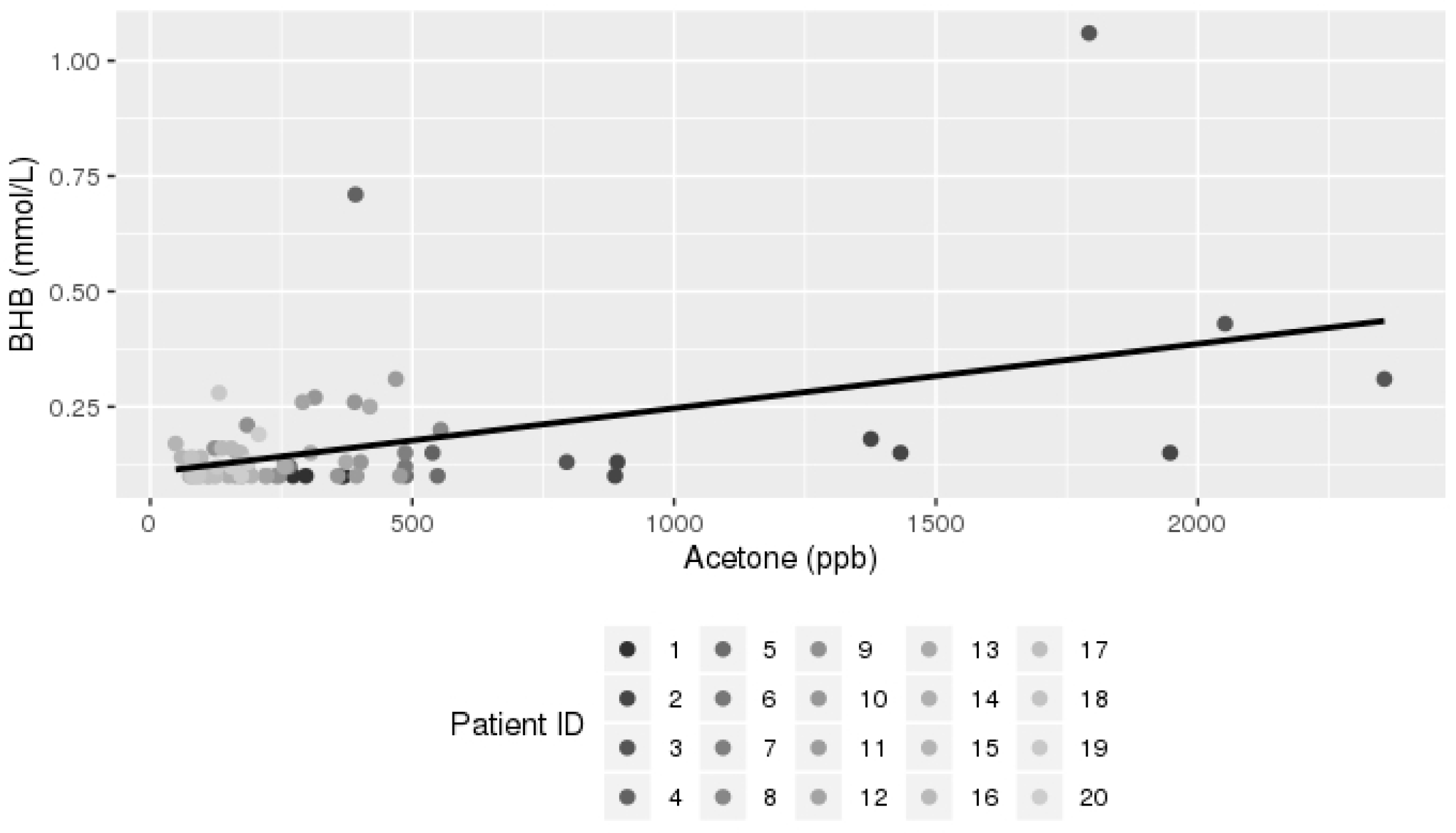
3.2. Breath Analysis Results

Among the 100 samples projected, only 75 breath samples had blood drawn simultaneously and those 75 samples were analyzed.
The following VOCs showed significant positive correlation with serum ketone levels: acetone (p < 0.0001), trimethylamine (p < 0.0001), pentane (p = 0.0001), 3-methylhexane (p = 0.0047), and carbon disulfide (p = 0.0499) (Table 3). We plotted those VOCs vs. serum BHB, as shown in Figure 1, Figure 2, Figure 3 and Figure 4. No significant correlation was found between serum glucose and any VOC.

4. Discussion

This is the first breath analysis study conducted in pediatric GSD patients. The major findings are: (1) breath analysis is a noninvasive modality that can be used to estimate serum ketone levels in GSD patients and (2) breath acetone, pentane, TMA, 3-methylhexane, and carbon disulfide positively correlate with serum BHB levels.
In normal individuals and during the fasting state, the liver keeps the serum glucose level stable through glycogenolysis and later through gluconeogenesis. When glycogen stores are depleted, ketone bodies production is upregulated via a process called ketogenesis [8,9]. Ketogenesis involves beta oxidation of fatty acids which produces two water soluble ketones (BHB, acetoacetic acid) and one volatile ketone (acetone). Acetone is produced via a non-enzymatic decarboxylation of acetoacetic acid. It then crosses to the alveoli and is exhaled in breath [10].
Ketogenesis represents a major source of acetone followed by oxidation of isopropanol [11]. In GSD patients, either glycogenolysis or gluconeogenesis is affected, leading to varying degrees of hypoglycemia. In ketogenic forms of GSD where the ketogenesis process is intact, ketone bodies are produced as an alternative source of energy. Table 4 shows enzymes affected in ketogenic GSD forms. Several studies showed significant correlation between serum ketones and breath acetone level in other diseases. Musa-Veloso et al. conducted breath analysis in children with refractory seizures on ketogenic diet and found a significant correlation between breath acetone and plasma acetone (r2 = 0.99, p < 0.0001), plasma acetoacetate (r2 = 0.89, p < 0.0001), and plasma β hydroxybutyrate (r2 = 0.94, p <0.0001) [12]. A similar correlation between breath acetone and serum ketones was observed in patients with diabetic ketoacidosis [13], healthy volunteers undergoing an oral glucose tolerance test [14], and fasted obese individuals [15]. Some studies have found a strong correlation between breath acetone and serum glucose in diabetic patients [13,16], a finding our study failed to prove likely due to multiple factors including the relatively low number of serum samples with low glucose or high ketones in our cohort. In other words, too many samples had relatively normal or close-to-normal glucose levels which correlated with mild ketogenesis. This is likely attributed to the small sample size and the fact that all patients were already on an established dose of corn starch and protein regimen. As a rule, the serum glucose level will inversely correlate with breath acetone level in GSD patients due to the different pathogenesis compared to diabetes.
Trimethylamine (TMA) results from the metabolism of dietary choline and carnitine by the gut flora. It is metabolized via a human hepatic enzyme called flavin-containing monooxygenase 3 (FMO3) to trimethylamine-N-oxide (TMAO) which plays a rule in atherosclerosis and cardiovascular disease in humans [17]. In an animal model, Shih et al. noted that using antisense oligonucleotides (ASO) against FMO3 was associated with lower serum BHB, indicating an effect of FMO3 on ketogenesis [18]. We hypothesize that elevated serum ketones may have a negative feedback effect on FMO3 activity, which subsequently causes an increase in its substrate TMA. It is important to note that this correlation could also be confounded by microbiome differences between patients as well as the dietary content of choline and carnitine which our study did not control for.
Pentane is a byproduct of lipid peroxidation of polysaturated fatty acids in membranes and is caused by free radicals [19]. It is an indicator of oxidative stress seen in critically ill patients and other conditions including reperfusion of ischemic liver, after abdominal ischemia in ischemic heart disease, and following cardiopulmonary bypass [20]. There is in vitro evidence that ketone bodies, particularly acetoacetate, can generate oxygen radicals and subsequently cause lipid peroxidation in human endothelial cells [21]. This likely explains the correlation we found between breath pentane level and serum ketones. Our group found that 3-methylhexane, a branched alkane, was elevated in the breath of children with chronic liver disease compared to healthy controls [22]. Liver cytochrome P450 converts hydrocarbons into alcohols in a normal healthy state. Hydrocarbons include alkanes (such as methylhexane) and alkenes. Hepatic dysfunction leads to an increased level of hydrocarbons in the serum and subsequently in the breath. We hypothesize that the hypoglycemic state in GSD patients is associated with hepatic metabolic dysfunction including the metabolism of hydrocarbons. It is also important to mention that 3-methylhexane has an exogenous source from carpets, paints, and plastics that acquire 3-methylhexane and other VOCs during manufacturing and slowly emit them over time [23]. Carbon disulfide is considered a volatile sulfide compound that is mainly produced from oral flora. Tanda et al. failed to find a correlation between breath and serum ketones and breath volatile sulfide compounds in healthy volunteer undergoing an oral glucose tolerance test [14]. The observed correlation between carbon sulfide and serum ketones in our study lacks physiologic explanation given its presumed oral origin.
This study has several limitations: (1) small sample size. (2) We did not control for microbiome differences as well as dietary carnitine and choline differences between patients. (3) No room air samples were collected to rule out other confounding VOCs. (4) Limited number of high or abnormal serum ketone levels. This limitation can be overcome by conducting the study on newly diagnosed patients with GSD who are undergoing initial evaluation and before treatment is implemented. Those patients will likely have higher serum ketone levels. (5) We did not control for corn starch and protein supplement dose or the timing of meals. Controlling for those factors can give us a clearer idea about serum ketone and VOC patterns during therapy. This can be addressed in a larger future study. (6) Finally, although several VOCs showed a correlation with serum ketones, similar studies are needed on a larger scale to further confirm this correlation.

5. Conclusions

This study supported our hypothesis that few VOCs detected with breath analysis can predict ketone serum levels in GSD. This is a pilot study and further validation is required.

Author Contributions

Conceptualization, M.N.K., P.K.C.S., D.G., A.R.T., R.A.D., L.M., K.R. and N.A.; design, M.N.K., P.K.C.S., D.G., A.R.T., R.A.D., L.M., K.R. and N.A.; data acquisition, M.N.K., P.K.C.S., D.G., A.R.T., R.A.D., L.M., K.R. and N.A.; statistical analysis, M.N.K., P.K.C.S., D.G., A.R.T., R.A.D., L.M., K.R., N.A., X.H. and X.W.; data interpretation, M.N.K., P.K.C.S., D.G., A.R.T., R.A.D., L.M., K.R. and N.A.; writing—original draft preparation, M.N.K., P.K.C.S., D.G., A.R.T., R.A.D., L.M., K.R. and N.A.; writing—review and editing, M.N.K., P.K.C.S., D.G., A.R.T., R.A.D., L.M., K.R., N.A., X.H. and X.W. All authors have read and agreed to the published version of the manuscript.

Funding

This work was supported by the American College of Gastroenterology through the Junior Faculty Development Award 2013–2016 to Naim Alkhouri Grant number 662014.

Institutional Review Board Statement

The study was conducted in accordance with the Declaration of Helsinki and approved by the Cleveland Clinic Institutional Review Board, protocol number 15-1151.

Informed Consent Statement

Informed consent was obtained from all subjects involved in the study.

Data Availability Statement

Data are available on request due to restrictions, e.g., privacy. The data presented in this study are available on request from the corresponding author.

Conflicts of Interest

The authors declare no conflict of interest. The funders had no role in the design of the study; in the collection, analyses, or interpretation of data; in the writing of the manuscript; or in the decision to publish the results.

References

  1. Hendriksz, C.J.; Gissen, P. Glycogen storage disease. Paediatr. Child Health 2015, 25, 139–144. [Google Scholar] [CrossRef]
  2. Ozen, H. Glycogen storage diseases: New perspectives. World J. Gastroenterol. 2007, 13, 2541–2553. [Google Scholar] [CrossRef] [PubMed]
  3. Chen, M.A.; Weinstein, D.A. Glycogen storage diseases: Diagnosis, treatment and outcome. Transl. Sci. Rare Dis. 2016, 1, 45–72. [Google Scholar] [CrossRef] [Green Version]
  4. Tsilianidis, L.A.; Fiske, L.M.; Siegel, S.; Lumpkin, C.; Hoyt, K.; Wasserstein, M.; Weinstein, D.A. Aggressive therapy improves cirrhosis in glycogen storage disease type, I.X. Mol. Genet. Metab. 2013, 109, 179–182. [Google Scholar] [CrossRef] [PubMed] [Green Version]
  5. Kuru, B.; Sever, M.; Aksay, E.; Dogan, T.; Yalcin, N.; Eren, E.S.; Ustuner, F. Comparing finger-stick β-hydroxybutyrate with dipstick urine tests in the detection of ketone bodies. Turkiye Acil Tip Derg. 2014, 14, 47–52. [Google Scholar] [CrossRef] [Green Version]
  6. Shirasu, M.; Touhara, K. The scent of disease: Volatile organic compounds of the human body related to disease and disorder. J. Biochem. 2011, 150, 257–266. [Google Scholar] [CrossRef] [PubMed] [Green Version]
  7. Paschke, K.M.; Mashir, A.; Dweik, R.A. Clinical applications of breath testing. F1000 Med. Rep. 2010, 2, 56. [Google Scholar] [CrossRef] [PubMed]
  8. Dhillon, K.K.; Gupta, S. Biochemistry, Ketogenesis; StatPearls: Treasure Island, FL, USA, 2018; p. 493179. [Google Scholar]
  9. Ercal, B.; Crawford, P.A. 37—Ketone Body Metabolism in the Neonate, 5th ed.; Elsevier Inc.: Amsterdam, The Netherlands, 2017. [Google Scholar] [CrossRef]
  10. Pasquel, F.J.; Umpierrez, G.E. Hyperglycemic Crises, 7th ed.; Elsevier: Amsterdam, The Netherlands, 2016. [Google Scholar] [CrossRef]
  11. Ruzsányi, V.; Kalapos, M.P. Breath acetone as a potential marker in clinical practice. J. Breath Res. 2017, 11, 024002. [Google Scholar] [CrossRef] [PubMed]
  12. Musa-Veloso, K.; Likhodii, S.S.; Rarama, E.; Benoit, S.; Liu, Y.M.C.; Chartrand, D.; Curtis, R.; Carmant, L.; Lortie, A.; Comeau, F.J.E.; et al. Breath acetone predicts plasma ketone bodies in children with epilepsy on a ketogenic diet. Nutrition 2006, 22, 1–8. [Google Scholar] [CrossRef] [PubMed]
  13. Qiao, Y.; Gao, Z.; Liu, Y.; Cheng, Y.; Yu, M.; Zhao, L.; Duan, Y.; Liu, Y. Breath ketone testing: A new biomarker for diagnosis and therapeutic monitoring of diabetic ketosis. BioMed Res. Int. 2014, 2014, 869186. [Google Scholar] [CrossRef] [PubMed] [Green Version]
  14. Tanda, N.; Hinokio, Y.; Washio, J.; Takahashi, N. Analysis of ketone bodies in exhaled breath and blood of ten healthy Japanese at OGTT using a portable gas chromatograph. J. Breath Res. 2014, 8, 046008. [Google Scholar] [CrossRef] [PubMed]
  15. Fraser, T.R.; Gilmour, A.; Prentice, E. Tension is a more certain form of treatment on both measurements in diabetes conclusion tion with a secondary disorder of coagulation in the the fibrinoid lesion and red-blood-cell fragmentation patients overnight fasting levels of both breath acetone P-hydroxybutyrate sampled during prolonged fasting (10–36 days). Forty-Five 1969, 1282, 1282–1286. [Google Scholar]
  16. Wang, C.; Mbi, A.; Shepherd, M. A Study on Breath Acetone in Diabetic Patients Using a Cavity Ringdown Breath Analyzer: Exploring Correlations of Breath Acetone with Blood Glucose and Glycohemoglobin A1C. IEEE Sens. J. 2010, 10, 54–63. [Google Scholar] [CrossRef]
  17. Brown, J.M.; Hazen, S.L. The Gut Microbial Endocrine Organ: Bacterially Derived Signals Driving Cardiometabolic Diseases. Annu. Rev. Med. 2015, 66, 343–359. [Google Scholar] [CrossRef] [PubMed] [Green Version]
  18. Shih, D.M.; Wang, Z.; Lee, R.; Meng, Y.; Che, N.; Charugundla, S.; Qi, H.; Wu, J.; Pan, C.; Brown, J.M.; et al. Flavin containing monooxygenase 3 exerts broad effects on glucose and lipid metabolism and atherosclerosis. J. Lipid Res. 2015, 56, 22–37. [Google Scholar] [CrossRef] [PubMed] [Green Version]
  19. Kohlmüller, D.; Kochen, W. Is n-pentane really an index of lipid peroxidation in humans and animals? A methodological reevaluation. Anal. Biochem. 1993, 210, 268–276. [Google Scholar] [CrossRef]
  20. Amann, A.; de Lacy Costello, B.; Miekisch, W.; Schubert, J.; Buszewski, B.; Pleil, J.; Ratcliffe, N.; Risby, T. The human volatilome: Volatile organic compounds (VOCs) in exhaled breath, skin emanations, urine, feces and saliva. J. Breath Res. 2014, 8, 034001. [Google Scholar] [CrossRef] [PubMed]
  21. Jain, S.K.; Kannan, K.; Lim, G. Ketosis (acetoacetate) can generate oxygen radicals and cause increased lipid peroxidation and growth inhibition in human endothelial cells. Free Radic. Biol. Med. 1998, 25, 1083–1088. [Google Scholar] [CrossRef]
  22. Eng, K.; Alkhouri, N.; Cikach, F.; Patel, N.; Yan, C.; Grove, D.; Lopez, R.; Rome, E.; Dweik, R.A. Analysis of breath volatile organic compounds in children with chronic liver disease compared to healthy controls. J. Breath Res. 2015, 9, 26002. [Google Scholar] [CrossRef] [PubMed]
  23. You, K.W.; Ge, Y.S.; Qian, Y.X.; Liu, W.; Feng, B.; Zhang, Y.N.; Ning, Z.W.; Hu, B.; Zhao, S.T. Volatile organic compounds concentrations and sources inside new air-conditioned bus. Huan Jing Ke Xue = Huanjing Kexue 2008, 29, 1436–1440. Available online: http://www.ncbi.nlm.nih.gov/pubmed/18624220 (accessed on 21 June 2018). [PubMed]
Figure 1. Scatter plot for breath acetone vs. serum ketones (BHB). Dots with the same color shade belong to the same patient.
Livers 02 00025 g001
Figure 2. Scatter plot for breath TMA vs. serum ketones (BHB). Dots with the same color shade belong to the same patient.
Livers 02 00025 g002
Figure 3. Scatter plot for breath pentane vs. serum ketones (BHB). Dots with the same color shade belong to the same patient.
Livers 02 00025 g003
Figure 4. Scatter plot for breath 3-methylhexane vs. serum ketones (BHB). Dots with the same color shade belong to the same patient.
Livers 02 00025 g004
Table 1. Precursor and product mass for each measured VOC.
MoleculePrecursorProduct Mass
AcetoneH3O+59
NO+88
PentaneO2+42
72
3-methylhexaneNO+99
O2+71
Carbon DisulfideO2+76
TrimethylamineH3O+58
60
NO+59
O2+58
59
Table 2. Patient demographic and laboratory data.
Total (N = 20)
Mean (Range)
Age (year)8.9 (5.7–15.7)
BMI (kg/m2)20 (14.7–31.7)
BMI percentile72.4 (20.4–99.8)
WBC (k/µL)5.5 (3.3–8.5)
Hemoglobin (g/dL)12.7 (9.9–14.0)
MCV (fL)81.3 (69.9–88.6)
Platelets (k/µL)249.2 (176.0–398.0)
AST (U/L)54.4 (14–487)
ALT(U/L)46.1 (10.0–409.0)
ALK. Phosphatase (U/L)224.3 (110.0–407.0)
Total Bilirubin (mg/dL)0.3 (0.2–0.7)
Albumin (g/dL)4.2 (3.6–4.9)
Total Cholesterol (mg/dL)143.6 (105.0–216.0)
LDL (mg/dL)78.9 (38.0–157.0)
HDL (mg/dL)49.9 (21.0–74.0)
TG (mg/dL)76.4 (27.0–222.0)
CK (U/L)269.3(41.0–3040.0)
ESR (mm/h)7.6 (2.0–26.0)
Ferritin (ng/mL)35.1 (6.8–122.1)
TIBC (µg/dL)348.6 (251.0–514.0)
Prealbumin (mg/dL)17.6 (10.0–27.0)
Table 3. VOCs that correlate with serum ketone levels.
Breath ElementUnivariate Mixed Model EstimateStandard Errorp Value
Acetone0.0001410.000032<0.0001
Trimethylamine (TMA)0.0076350.001357<0.0001
Pentane0.0032480.0007770.0001
3-methylhexane0.0065020.0022030.0047
Carbon disulfide0.037990.018940.0499
Table 4. Enzymes involved in ketogenic glycogen storage disease.
GSD TypeEponymEnzyme Involved
0 Glycogen synthetase
IIICoriDebranchig enzyme (Amylo-1,6 glucosidase)
VIHersLiver phosphorylase
IX Phosphorylase kinase
Publisher’s Note: MDPI stays neutral with regard to jurisdictional claims in published maps and institutional affiliations.

