Review

Is There a Place for Somatostatin Analogues for the Systemic Treatment of Hepatocellular Carcinoma in the Immunotherapy Era?

by
Elias Kouroumalis
1,*,
Ioannis Tsomidis
2,3 and
Argryro Voumvouraki
2
1
Dept of Gastroenterology and Liver Research Laboratory, University of Crete Medical School, 71110 Heraklion, Greece
2
1st Department of Internal Medicine, AHEPA University Hospital, 54636 Thessaloniki, Greece
3
Research Fellow, University of Crete Medical School, 71110 Heraklion, Greece
*
Author to whom correspondence should be addressed.
Livers 2022, 2(4), 315-335; https://doi.org/10.3390/livers2040024
Submission received: 1 October 2022 / Accepted: 17 October 2022 / Published: 18 October 2022

Abstract

:
Patients with advanced hepatocellular carcinoma (HCC) have a very limited survival rate even after the recent inclusion of kinase inhibitors or immune checkpoint inhibitors in the therapeutic armamentarium. A significant problem with the current proposed therapies is the considerable cost of treatment that may be a serious obstacle in low- and middle-income countries. Implementation of somatostatin analogues (SSAs) has the potential to overcome this obstacle, but due to some negative studies their extensive evaluation came to a halt. However, experimental evidence, both in vitro and in vivo, has revealed various mechanisms of the anti-tumor effects of these analogues, including inhibition of cancer cell proliferation and angiogenesis and induction of apoptosis. Favorable indirect effects such as inhibition of liver inflammation and fibrosis and influence on macrophage-mediated innate immunity have also been noted and are presented in this review. Furthermore, the clinical application of SSAs is both presented and compared with clinical trials of kinase and immune checkpoint inhibitors (ICIs). No direct trials have been performed to compare survival in the same cohort of patients, but the cost of treatment with SSAs is a fraction compared to the other modalities and with significantly less serious side effects. As in immunotherapy, patients with viral HCC (excluding alcoholics), as well as Barcelona stage B or C and Child A patients, are the best candidates, since they usually have a survival prospect of at least 6 months, necessary for optimum results. Reasons for treatment failures are also discussed and further research is proposed.

1. Introduction

Primary liver cancer is the seventh most prevalent cancer and the fourth cause of cancer-related deaths worldwide [1].
Deaths from hepatocellular carcinoma are rising faster than deaths from any other type of cancer in the United States [2]. Current treatment of advanced HCC is rather disappointing, as its benefits in terms of survival are measured in months rather than years even after introduction of multi-kinase inhibitors and immunotherapy [3].
The HCC world incidence varies. It usually-but not always-follows the prevalence of viral hepatitis either B or C. China, Eastern Asia and Africa report the highest HCC rates, with the lowest figures in Northern Europe and the USA. Eastern European and Mediterranean countries show intermediate figures [4].
The greatest obstacle in setting up a therapeutic trial or in comparing the results of different trials is the heterogeneity of HCC together with the fact that in most instances (over 80%) there are two diseases (cirrhosis and cancer), each affecting the natural course of the other. To overcome this problem, many classifications have been proposed. The most extensively used, but by no means perfect, is the Barcelona Clinic Liver Cancer (BCLC) staging. HCC is classified into very early stage (BCLC stage 0), early stage (BCLC A), intermediate stage (BCLC B), advanced stage (BCLC C) and terminal stage (BCLC D) [3].
The use of deep genome sequencing, transcriptome, epigenome, proteome and single-cell analysis tests started to unveil tumor heterogeneity, which may lead to a change in the classification of HCC. These technological changes are important not only for immunotherapy but also for re-evaluation of previous treatments [5].
Kinase inhibitors and immune checkpoint inhibitors (ICI) were recently introduced as systemic treatment of HCC. Favorable results showed promising efficacy in early-phase trials with single agents, but this was not confirmed in phase III trials. The combination of atezolizumab (an anti-PD-L1 ICI) with bevacizumab (an anti-VEGF antibody) was accepted as first-line therapy in 2020, but again the results are still not that impressive [6].
Soon after its discovery in 1968, somatostatin (SST) was shown to have many potential therapeutic implications and a host of synthetic somatostatin analogues (SSAs) with longer half-lives (i.e., octreotide, vapreotide, lanreotide and pasireotide) available for clinical use ensued [7].
We summarize the current place of SSAs in the era of kinase and immune checkpoint inhibitors (ICIs) based on two important recent observations: (1) depending on HCC etiology, different results were noted. ICIs performed the worst in NAFLD-associated HCC and the best in viral-associated HCC. (2) The best results were seen in Child Pugh A patients and the B and C stages of the BCLC classification.

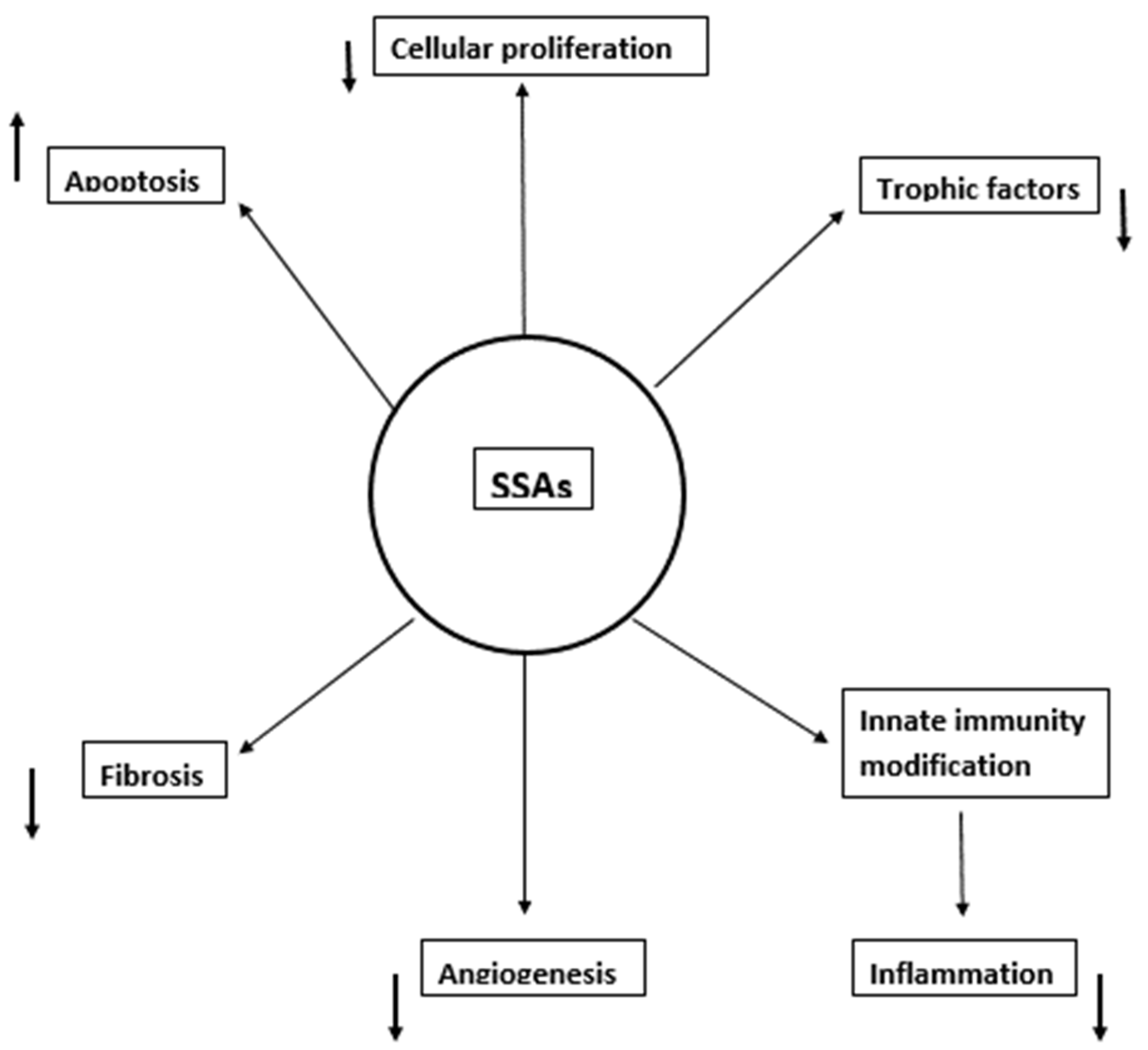
2. Mechanisms of SSAs’ Activity in HCC

Experimental data have provided convincing evidence that SSAs have a beneficial effect on HCC. Binding of SST and SSAs to their respective receptors activates various intracellular pathways leading to several antineoplastic effects through cell proliferation, apoptosis and possibly autophagy [8]. Data on SST receptor expression in both hepatocytes and liver sinusoidal cells and a detailed review of the antineoplastic mechanisms have recently been published [9].
Results on cellular proliferation are not uniform. Most studies indicate that octreotide induces cell cycle arrest. This process is regulated by the presence of specific SSTR subtypes [10,11] as well as the concentration of the peptide and the time of exposure. Thus, the administration of SSAs had a dual effect: low concentrations reduced proliferation while higher concentrations increased proliferation [12]. In Hepatoblastoma G2 (HepG2) cells, inhibition of proliferation was obtained at a concentration of 10−8 mol/L [13]. Short-term octreotide treatment could lead to SSTR2 desensitization and reduced HCC response. However, long-term octreotide administration inhibited the growth of HCC effectively–possibly through the re-sensitization and upregulation of SSTR2 [14].
The anti-tumoral action of SSAs is further mediated through the enhancement of apoptosis [12,13,14].
Autophagy is a double-edged sword in hepatocellular carcinoma and may either be protective or detrimental [15]. In normal cells and at the initial stages of HCC, autophagy is a tumor suppressor and maintains genomic stability. However, once HCC is established, autophagy will promote cell survival and tumor growth [16]. The effects of SSAs on autophagy are very few. Octreotide increased autophagy markers and hepatoprotection in a rat model of ischemia-reperfusion liver injury [17].
Additional mechanisms of the anti-tumor effect of SSAs have been proposed. They suppress several trophic factors implicated in the migration and tumor progression, such as insulin-like growth factors 1 and 2 (IGF1 and IGF2) [18]. Other potential tumor trophic factors such as gastrin, glucagon and insulin are also inhibited by SSAs, but their exact role in HCC has not been clarified yet [19].
HCC-induced angiogenesis is also inhibited by octreotide [20], either through direct binding to the endothelium or indirectly through the inhibition of the vascular endothelial growth factor (VEGF) [21,22]. This is particularly important, as a combination of SSAs with a multi-kinase inhibitor seems therapeutically rational.
Modification of innate immunity response is an additional antineoplastic mechanism [23]. Octreotide pre-incubation of Kupffer cells inhibited tumor necrosis factor a (TNFa) and nitric oxide (NO) secretion, but it also reduced the production of the pro-inflammatory cytokine interleukin-12 (IL-12), whereas the anti-inflammatory IL-13 secretion was increased [24]. HCC, like most neoplasms, is associated with inflammation; therefore, reduction in inflammation may be an indirect antineoplastic mechanism.
Liver and tumor-associated macrophages (TAMs) are also implicated in HCC progression. At the initial stages of HCC, Kupffer cells and TAMs overproduce transforming growth factor beta 1 (TGFb1) and IL-6, favoring a tumor-promoting M2 macrophage polarization [25]. Octreotide inhibited the production of TGFb1 by isolated Kupffer cells [26]. It is conceivable then that M2 polarization will also be reduced. In addition, a reduction in the apoptosis of cultured Kupffer cells incubated with octreotide has been described which may strengthen the defense against HCC [27].
Most HCCs develop in a cirrhotic background, and octreotide has effectively attenuated liver fibrosis in a rat fibrosis model [28]. The CCL2 (monocyte chemoattractant protein-1 or MCP-1) chemokine is also implicated in the process of liver fibrosis [29]. Stimulation of Kupffer cells with lipopolysaccharide (LPS) led to the production of significantly increased amounts of both CC chemokines (MCP-1, Rantes) and CXC chemokines (IL-8, macrophage inflammatory protein-2 or MIP-2). Octreotide considerably reduced CC chemokines but not CXC chemokine production [30]. Moreover, octreotide, synergistically with LPS, stimulated the production of increased amounts of collagenase (MMP1) by Kupffer cells and decreased several pro-fibrotic factors in unstimulated Kupffer cells, including TGFb. Both these effects eventually led to attenuation of liver fibrosis [26].
Apart from Kupffer cells, hepatic stellate cells (HSCs) are also involved in liver fibrosis by producing the components of extracellular matrix. HSCs permit tumor growth by producing hepatocyte growth factor [31]. Somatostatin had no effect on HSCs proliferation but significantly inhibited collagen production and strongly repressed platelet-derived growth factor (PDFG) and TGFb1-dependent procollagen production [32,33], thus attenuating fibrosis (Figure 1).
HSCs are implicated in the development of inflammation even though they do not belong to the innate immune system, as they produce several pro-inflammatory molecules [34]. Secretion of the pro-inflammatory cytokines IL-1beta and IL-8 by rat HSCs is inhibited by somatostatin [35,36].
Animal models add further support to the antineoplastic effects of SSAs in HCC. Octreotide inhibited HCC development after implantation of Morris hepatoma cells [37]. Studies from China in the nude mice model of HCC showed that tumor growth and lung metastases were decreased after octreotide administration and survival was considerably prolonged [38]. The combination of an SSA with a cyclooxygenase (COX 2) inhibitor suppressed the metastasis of HCC in nude mice [39]. The long-acting lanreotide was effective in chemically-induced HCCs, inhibiting cell proliferation and reducing angiogenic factor levels, while apoptosis was increased [40].
In view of the above preclinical data, it was only logical that SSAs were tested in clinical studies for the treatment of HCC.

3. Somatostatin Analogues as a Systemic Treatment of HCC

3.1. Positive Studies (Table 1)

Subcutaneous octreotide was initially used in a randomized controlled study of Okuda II and III HCC cirrhotic patients. Survival rates almost doubled and there was a significant reduction in the risk of death. These results were confirmed with long-acting analogues in a non-randomized trial where the relative risk of death of the control patients was 2.7 higher. Interestingly, 10% of tumors regressed and 30% were stable, a result closely approaching the expression of SSTRs reported before. Despite radiologic progression of tumors, patients had no significant weight loss and retained their appetite. HCC and cirrhosis in both trials were due to either HBV or HCV in approximately 90% of cases [41,42]. The somatostatin survival benefit became apparent in the Kaplan–Meier curves only after 6 months of treatment. The worst response was observed in alcoholic cirrhosis, particularly in active drinkers [43].
Lanreotide also showed a 43% response rate, in an uncontrolled study. It should be noted that octreotide scintigraphy was negative for receptor expression in all patients; 24% of patients had a significant reduction in serum-a fetoprotein (AFP) levels [44].
Two additional studies from other Greek centers also demonstrated favorable results. The first, with viral hepatitis HCC cases, showed prolonged survival only in non-cirrhotic patients. However, 40% of their cirrhotics were Child Pugh C and many died before the critical time of 6 months [45].
A second randomized controlled study of viral-related hepatitis patients also demonstrated that octreotide doubled the survival of those with detectable SSTRs on 111 Indium octreotide scintigraphy. No survival benefit was observed in SSTR-negative patients and again, the benefit was significant after approximately 6 months of therapy [46].
Twenty patients with advanced HCC were treated with octreotide LAR, while forty patients with comparable staging and liver function were retrospectively selected as controls. Results were published as an abstract. The treated patients not only had a significant survival gain compared to controls but also exhibited significant correlation with SSTR2 expression in the tumor [47].
Tamoxifen and octreotide in combination were used in a controlled trial from China. Survival was doubled compared to conventional chemotherapy with a response rate of 43% in the octreotide arm [48].
A Pakistani controlled trial showed regression of HCC in 45.4% of patients with significant survival benefit and improved quality of life. A parallel decrease in a-fetoprotein in 50% of patients was also observed [49].
A small observational study of advanced HCC from the USA reported a median survival of only 4.5 months. Yet, 27% of their patients lived for more than 10 months. Those were patients of Asian descent infected with the Hepatitis B virus [50].
Total regression of HCC after SSA treatment was shown in a patient with viral cirrhosis [51]. An additional case report presented a patient with HBV-related HCC and lung and mediastinal metastases. Treatment with lanreotide reduced the mediastinal nodes considerably, while the lung nodes regressed completely. Remission lasted for 42 months. SSTR2 overexpression was identified in tumor tissue [52].
The importance of SSTR2 and SSTR5 expression was clearly demonstrated in two large Chinese studies of operable early-stage liver tumors after the surgical removal of HCC. In the first trial [53], patients were divided and treated according to the presence or absence of SSTRs in the tumor tissue. Survival was significantly longer if receptors were present. Similarly, in the second study of HBV-associated HCC, patients survived longer when a high expression of SSTR2 was present [54].
SSAs have been directly compared to transarterial chemoembolization (TACE) and radiofrequency ablation (RAF). Non-randomly selected patients were treated with octreotide and a group of TACE-treated patients served as the control in a German trial. The survival of approximately 19 months was similar in both groups [55]. A larger randomized trial from the same country confirmed these results. Schoniger-Hekele et al. retrospectively studied 95 BCLC stage A and B, matched HCC patients given octreotide, TACE, multimodal therapy or only palliative care. Median survival was similar in all treatment modalities and significantly higher than palliative treatment. Results were similar irrespective of the BCLC stage [56].
In a slightly different trial, 147 HCC non-randomized patients were treated with 2–4 TACE applications. One group received heparin and octreotide as an adjunct therapy and was compared with heparin-only controls. After a one-year follow-up there was a statistically significant lower incidence of metastatic tumors in the octreotide group [57].
In a similarly designed Chinese study, patients with BCLC stages B or C were randomly assigned to be treated with either TACE or TACE plus celecoxib and octreotide. After 3 years of follow-up, a median survival of 15 months was reported in the combination group: two times higher than the 7.5 months of the TACE-only group. The survival benefit was very significant for both BCLC stage B and C. Quality of life was improved in the octreotide group, where the incidence of the post-embolization syndrome was significantly lower. It should be noted that most patients were infected with either HBV or HCV [58].
RAF followed by octreotide was used in an observational study of Child A and Child B patients with viral cirrhosis with multiple liver HCC nodules. Mean survival was 31.4 months; 80% of patients had a clinical response while 14% had complete or partial tumor regression associated with serum VEGF levels [59].
A study of pasireotide in advanced HCC of viral etiology was recently published. The best response was stable disease in nine patients (45%). Median time to tumor progression was 3 months, and median survival was 9 months, even though 75% had BCLC stage C and 55% had metastatic disease. It should be noted that all patients were treatment failures: 60% had prior TACE treatment and 70% had received sorafenib before [60].
Table 1. Summary of the positive studies about the role of somatostatin analogues in the treatment of advanced HCC.
AuthorsType of StudyNo. of PatientsSST Receptors AssessmentInterventionControlOutcomes
Kouroumalis E et al., 1998 [41]RCT58 patients with advanced HCCHistology in all casesOctreotide 250 μg s.c. b.i.d in 28 patients No treatment in 30 patients
  • Increased median survival (13 mos. vs 4 mos.) in the treated group
  • Increased cumulative survival rate at 6 mos. (75% vs 37%) and 12 mos. (56% vs 13%) in the treated group
Samonakis DN et al., 2002 [42]Comparative observational study59 patients with inoperable HCCN/ALong-acting somatostatin analogues in 32 patientsNo treatment in 27 patients
  • Improved OS in the treated group (15 mos. vs 8 mos.)
  • HCC remained stable or regressed in 40% of the treated patients
Raderer M et al., 2000 [44]Uncontrolled study21 patients with inoperable HCC Scintigraphy in 15 patients Lanreotide 30 mg i.m. every 14 days None
  • 24% had a decrease in serum AFP levels by 30%
  • 5% showed a partial response to treatment
  • 38% had stable disease
Patsanas T et al., 2004 [45]Comparative observational study 30 patients with unresectable HCC (group A: cirrhosis group B: non-cirrhosis)N/AOctreotide 500 μg s.c. b.i.dNone
  • Octreotide improved significantly the survival time in non-cirrhotic patients with HCC compared to cirrhotic patients (mean survival time 8.3 mos. vs 5 mos.)
  • Octreotide administration stopped gradual elevation of serum AFP levels
Dimitroulopoulos D et al., 2007 [46]RCT127 cirrhotic patients, stages A–B, due to chronic viral infections with advanced HCC, 61 patients with positive SSTR were randomizedScintigraphy in all cases31 patients with SSTR positive were treated with octreotide 0.5 mg s.c. every 8 h for 6 wk., at the end of wk. 4–8 octreotide LAR 20 mg i.m. and at the end of wk. 12 and every 4 wks. octreotide LAR 30 mg i.m.Oral placebo in 30 patients
  • A significantly higher survival time was observed for the octreotide group (49+/−6 wk.) as compared to the control group (28+/−1 wk.) and to the SSTR negative group (28+/−2 wk.)
Pan DY et al., 2003 [48]RCT39 patients with inoperable HCCN/A24 patients were treated with TAM + OCT15 patients were treated with regular chemotherapy
  • TAM + OCT group lowered AFP levels
  • TAM + OCT increased median survival compared to chemotherapy (12.8 mos. vs 5.5 mos.)
Gill ML et al., 2005 [49]Comparative observational study42 patients with inoperable HCCN/A22 patients were treated with long-acting octreotide 100mcg s.c. t.i.d for 2 wks. followed by 20mg i.m. monthly20 patients with no treatment due to socioeconomic issues
  • Tumor size regression in 45.5% of the patients treated
  • Decrease of serum AFP levels in 50% of the patients treated
  • Octreotide LAR increased survival in 6 mos. follow up compared to no treatment group (64% vs 50%)
Shah U et al., 2009 [50]Observational study22 patients with advanced inoperable HCC and CLIP score of 3 or higherN/A22 patients were treated with long-acting octreotide 30 mg i.m. (or 20 mg) monthlyNoneTreatment with long-acting octreotide showed:
  • Median TTP of 5.7 mos.
  • Median PFS time of 3 mo
  • Median OS time of 4.5 mo
  • 6 out of 22 patients achieved an OS time of greater than 10 mos.
Siveke JT et al., 2003 [51]Case report1 patient with advanced HCCScintigraphyOctreotide 250 μg b.i.d followed by long-acting octreotide 10mg monthlyNone
  • Tumor size reduction by 50–70% after 4 mos.
  • Complete tumor regression after 10 mos.
Borbath I et al., 2012 [52]Case report1 patient with metastatic HCCHistology and scintigraphyLanreotide 30 mg twice monthlyNone
  • Tumor size regression and remission for 42 mos.
Li S et al., 2012 [53]Observational study76 patients with inoperable HCCHistology, 2 groups based on high or low expression of SSTR-2 and 5 in tumor tissueOctreotideNone
  • Mean survival time was longer in the high SSTR-2/5 expression group
Liu Y et al., 2013 [54]Comparative observational study99 patients with HCC and cirrhosis who underwent curative liver resection HistologyAt one day post-surgery, all the patients were administered 20 mg octreotide LAR i.m. monthly for 12 mos. None
  • Tumor recurrence rate was significantly lower in the high expression group compared with that of the low expression group (63.83% vs. 82.69%)
  • The survival time of the members of the high expression group was longer compared with that of the low expression group
  • SSTR mRNA expression correlated with survival in patients with early-stage hepatitis B virus (HBV)-related HCC who were treated with octreotide LAR following surgery
  • The inhibitory effects of somatostatin analogues on tumor growth may be mediated by SSTR expression
Plentz RR et al., 2005 [55]Observational study41 patients with advanced HCC and cirrhosisN/AOctreotide 250 μg t.i.d followed by long-acting octreotide 30 mg i.m. monthlyNone
  • Tumor size was stable in 63% of the patients treated after a median follow-up of 21 mos.
Schöniger-Hekele M et al., 2009 [56]Retrospective observational study95 patients with HCC (BCLC stage A or B)N/AOctreotide TACE, multimodal therapy, palliative care
  • Survival under octreotide treatment was not different compared to TACE or multimodal therapy
Jia W et al., 2012 [57]RCT147 patients with HCC receiving 2–4 TACE treatments N/A84 patients were treated with heparin + octreotide for 12 mos.No treatment in 63 patients
  • A significant decrease in the incidence of tumor metastasis in patients receiving the combination treatment post-TACE for up to 1 year with no significant toxic or adverse effects
Tong H et al., 2017 [58]RCT71 patients with inoperable HCCN/A36 patients were treated with TACE + C + L35 patients were treated with TACEDuring a 3-year follow-up period:
  • The median OS of the TACE + C + L group was doubled compared to that of TACE group (15 mos. vs 7.5 mos.)
  • Disease control rate of the TACE + C + L group was significantly higher than that of the TACE group either at 6 mos. (72.2% vs 42.9%) or at 12 mos. (61.1% vs 28.6%)
  • TACE + C + L prolonged OS, enhanced tumor response, reduced post-embolization syndrome and was well-tolerable in the patients with unresectable HCC
Montella L et al., 2008 [59]Observational study35 patients with advanced HCC and Child A or B cirrhosisN/A35 patients treated with RFA + octreotideNone
  • Mean OS was 31.4 mos.
  • 80% of the patients had a clinical response
  • 14.2% of the patients had complete or partial tumor regression associated with serum VEGF levels
Feun LG et al., 2018 [60]Observational study20 patients with advanced or metastatic HCC (BCLC stage C or D)N/A20 patients were treated with pasireotide LAR 60 mg i.m. monthlyNone
  • 45% of the patients had stable disease
  • Median TTP was 3 mos.
  • Median survival was 9 mos.
  • 90% of the patients had prior therapy that failed
Abbrev.: RCT, randomized controlled trial; HCC, hepatocellular carcinoma; s.c., subcutaneous; i.m., intramuscular; b.i.d., twice a day; mo(s)., month(s); N/A, not applicable; OS, overall survival; AFP, a-fetoprotein; SSTR, somatostatin receptors; wk(s)., week(s); TAM, tamoxifen; OCT, octreotide; t.i.d., three times a day; TTP, time to tumor progression; PFS, progression free survival; TACE, transarterial chemoembolization; C, celecoxib; L, lanreotide; RFA, radiofrequency ablation.

3.2. Negative Studies (Table 2)

There have been several negative studies that demonstrate no survival benefit from the administration of SSAs. One is a retrospective observational study of 63 patients, almost half of which also presented alcoholic cirrhosis [61].
Yuen et al. published a randomized trial with no survival benefit [62]. This study has been criticized by us and many others due to the extremely low median survival of 1.9 months of the controls compared to only 2 months in the long-acting SSA group, suggesting that most patients were obviously moribund, as they received only one octreotide injection [63].
It should be noted that the BCLC new stratification advocates that systemic treatment should be given only at stages B and C but not D (primarily moribund patients with expected survival of up to 3 months), further disproving the value of most negative SSA trials which mainly focused on BCLC D patients [64].
A limited survival benefit of octreotide treatment was also reported by a non-randomized study [65]. Again, 6% of cases had a short survival due to the rapid progression of the disease, so in fact they had received no treatment, but they were nevertheless analyzed in the final report. An additional 56% received four doses or less, likely indicating that their stage was also BCLC D. Even so, 50% of patients who were treated for at least three months were considered to be stable, in agreement with previous studies [63,66].
An open trial (with 22% alcoholic cirrhosis patients) showed a small anti-tumor effect of SSAs with a median survival of 8 months. However, treatment was discontinued not because of death but because of either toxicity or disease progress. Therefore, actual survival could not really be assessed [67].
Another open-label trial (88% BCLC stage C and more than 60% alcoholics) reported a median survival of only 6.7 months. The same problems were evident, as treatment stopped not because of death but rather because of disease progression. It is further noteworthy that only a median of two 28-day cycles was administered [68].
A randomized controlled study compared treatment with tamoxifen plus octreotide against a control group that received only tamoxifen: 52.4% of the patients were alcoholics. No survival gain was demonstrated for the tamoxifen plus octreotide group. Yet again, combination patients survived for a median of only 3 months and almost half of them received one to three injections. In addition, Child Pugh A patients had a very low median survival of only 6 months [69].
A double-blind, placebo-controlled study (HECTOR) also reported no survival gain for octreotide (median survival of 4.7 months compared to 5.3 months of the controls) without an effect on quality of life: 52% of the treatment group had alcoholic cirrhosis and only 40% survived for the critical 6-month period [70].
Another multicenter randomized placebo-controlled study also reported negative results. Again, almost 50% of the participating patients suffered from alcoholic cirrhosis [71].
A Chinese meta-analysis of nine studies with almost 800 patients may possibly explain the conflicting results. The survival gain was clearly in favor of octreotide treatment during a 6- and 12-month follow-up. Interestingly, the survival gain was not significant when only Western studies were included [72]. This report clearly demonstrates a discordance between China (and Greece) and the West.
There are many potential explanations for the negative results from primarily Western studies. The desensitization of SST receptors after short exposure to octreotide may be one of them, but if researchers had persevered, there is evidence that re-sensitization may occur after prolonged administration of octreotide [13]. This may also explain why a survival gain is significant only in patients surviving for more than 6 months, proving that SSAs are by no means rescue drugs.
An additional explanation is the variability of SSTRs’ expression profile among tumors, as many of the aforementioned studies clearly show that the treatment response rates often depend on SSTRs’ expression.
A critical factor for tumor response may be the serum concentration of SSAs. This is possible as octreotide reduction in cellular proliferation was observed only at a concentration of 10−8 mol/L. Therefore, monitoring drug serum levels may be mandatory, at least in clinical studies [14].
The negative results of some studies might be explained by these observations, but they still fail to explain the discrepancies between the conclusions reached by Eastern countries (and Greece) and the West. A possible reason for the discrepancies may be patient selection, since a large number of alcoholic cirrhotics participated in Western trials (up to 60%), reflecting the etiological stratification of their cirrhotic population, while studies from the East have mostly recruited patients with viral cirrhosis. China has mostly HBV-related HCC in contrast to Europe’s proportionally larger ethanol-related HCC [73]. Moreover, in many countries including Japan and North America, HCV-related HCC is very common, as well as steatosis. This was certainly the case in the initial Greek trials which were also reflecting the etiological background of cirrhosis at that time, a situation that has since changed [74]. We pointed out that the few alcoholics in our study had a poor response to SSAs, especially those that continued drinking. This very crucial observation, however, has not been commented upon by researchers in any of the negative trials [43].
There may be an additional explanation for the poor response of alcoholics. A German study found very low levels of weak expression of SSTRs in tissue from alcohol-related HCC patients (only two of their patients had viral HCC) [75].
However, it was the recent observation in a meta-analysis of three phase III immunotherapy trials that provided further support to our ignored suggestions that SSAs may in fact be more effective in viral HCC and not so effective when steatosis is present (as in alcoholic cirrhosis). Indeed, in this study, immune therapy did not improve survival in NASH-driven HCC patients compared to other etiologies [76].
Kinase inhibitors are probably equally effective in NAFLD-HCC compared to viral HCC. Although current treatment guidelines for HCC do not address etiology as a factor for concern, the time has come for modifications of treatment according to HCC etiology.
Table 2. Summary of the negative studies about the role of somatostatin analogues in the treatment of advanced HCC [77].
AuthorsType of StudyNo.of PatientsSST Receptors AssessmentInterventionControlOutcomes
Rabe C et al., 2002 [61]Retrospective multicenter cohort study63 patients with unresectable HCC and cirrhosis Child A–C)N/AInitially: 43 cases received long-acting octreotide 20–30 mg/mo.
20 cases received octreotide 50 t.i.d-300 ug t.i.d. s.c.
Later: 11 patients of the s.c. group were converted to long-acting octreotide
NoneAt 3 mos.:
  • 2 patients showed partial remission
  • 22 tumors showed no change
  • 26 tumors showed progression
At 6 mons:
  • 11 tumors showed no change
  • 15 tumors progressed
  • Median survival was 9 mos.
Yuen MF et al., 2002 [62]RCT70 patients with advanced HCCN/A 35 patients received a 2-week course of 250 μg short-acting octreotide twice daily followed by Sandostatin LAR 30 mg injection once every 4 wks. for 6 doses 35 patients received placebo
  • Long-acting octreotide showed no survival benefit
  • No difference in the cumulative survival between the 2 groups
  • No tumor regression and no reduction of serum AFP levels in patients receiving Sandostatin LAR
Slijkhuis WA et al., 2005 [65]Prospective cohort study30 patients with advanced HCCN/A Long-acting octreotide 30 mg i.m. every 4 to 6 wks. None
  • Median TTP was 3.6 mos.
  • Median survival was 5.1 mos.
  • Overall, limited beneficial response in terms of TTP and survival
Cebon J et al., 2006 [67]Observational study63 patients with advanced HCCScintigraphyLong-acting octreotide 20 mg i.m. monthlyNone
  • No major changes in QoL
  • 2% of the patients had partial tumor response
  • 6% of the patients showed a decrease a more than 50% decrease of serum AFP levels
  • Median survival was 8 mos.
Sanoff HK et al., 2015 [68]Multicenter observational study24 patients with advanced HCC (46% metastatic disease)N/APasireotide LAR 60 mg i.m. monthly + everolimus 7.5 mg per os dailyNone
  • Median TTP was 3.5 mos.
  • Median survival was 6.7 mos.
  • 10 patients had stable disease as the best response.
Verset G et al., 2007 [69]RCT109 patients with advanced HCCN/A56 patients were treated with octreotide LAR + TAM53 patients were treated with TAMNo difference between the groups concerning:
  • Tumor regression
  • AFP decrease
  • Improvement of QoL
  • survival
Becker G et al., 2007 [70]RCT120 patients with advanced HCCN/A60 patients were treated with long-acting octreotide 30 mg i.m. monthly59 patients were treated with placebo
  • No difference in cumulative survival
Barbare JC et al., 2009 [71]RCT272 patients with untreatable or relapsed HCCN/A135 patients were treated with long-acting octreotide 30 mg i.m. monthly for 2 years137 patients were treated with placebo
  • The median time until definitive global health score deterioration was 2.3 mos. in the octreotide and 4 mos. in the placebo group
  • Median OS was 6.53 mos. for octreotide versus 7.03 mos. for placebo
  • PFS did not differ significantly between the two treatment groups
  • No objective responses were achieved in the octreotide group but 33% of patients achieved disease stabilization for a mean time of 5.5 mos.
Abbrev.: RCT, randomized controlled trial; HCC, hepatocellular carcinoma; s.c., subcutaneous; i.m., intramuscular; b.i.d., twice a day; mo(s)., month(s); N/A, not applicable; OS, overall survival; AFP, a-fetoprotein; wk(s)., week(s); TAM, tamoxifen; t.i.d., three times a day; TTP, time to tumor progression; PFS, progression free survival; QoL, quality of life.

4. Is There a Place for SSAs in the Treatment of HCC Today?

A comparison of the survival results between SSAs and kinase inhibitors or the recently introduced checkpoint inhibitors could provide an answer to this question. As secondary parameters, side effects and cost of treatment should also be examined. However, it should be kept in mind that only indirect conclusions can be drawn, as there are no direct, head-to-head comparisons available.
Parameters of progression such as time to progression and progression-free survival may not represent treatment failure, as they are not in fact a surrogate marker of survival. The real endpoints in clinical trials should be overall survival [78] and to a lesser extent, quality of life.
HCC tumors are characterized by high vascularization and several local abnormalities of the immune reactions. Increased Treg expression is associated with poor prognosis and metastatic disease [79], as well as overexpression of the checkpoint inhibitors programmed cell death protein 1 (PD-1) and programmed cell death protein ligand-1 (PD-L1) [80]. Clinically, PD-L1 expression is associated with increased tumor recurrence risk and poor prognosis [81]. Therefore, it is only logical that anti-angiogenic kinase inhibitors and immune checkpoint inhibitors (ICI) are the two treatment modalities that have shown efficacy in HCC. Combination of anti-angiogenics and ICI seems to be synergistic, because vascular endothelial growth factor (VEGF) inhibition improves the survival of cytotoxic T lymphocytes and decreases Tregs infiltration, thus providing a favorable immune microenvironment for the ICI anti-tumoral effect [82,83]. An elegant review on the mechanisms of action of immune checkpoint inhibitors has recently been published [82].
In 2007, the FDA approved sorafenib for the treatment of advanced HCC, despite the rather limited survival gain from 7.9 to 10.7 months [84]. The response rates were consistently low, probably due to the tumor heterogeneity. In one trial, the median survival gain was again only 2–3 months in Asian and Caucasian patients [85]. It is pertinent in our comparisons that large non-interventional observational studies have shown that the survival of patients with Child B cirrhosis on sorafenib is significantly shorter when compared to Child A patients. No conclusive evidence for a benefit in Child B patients was provided. Therefore, it is suggested that the use of sorafenib should be limited to patients with well compensated stages of cirrhosis [86].
Lenvatinib is a multi-kinase inhibitor approved in 2018 with a better survival benefit for HBV-related HCC than sorafenib. Since then, three second-line drugs, Regorafenib, Cabozantinib and Ramucirumab (a VEGF-2 receptor inhibitor), were approved for patients not responding to sorafenib. However, none improved the overall survival, the progression-free survival or life quality significantly [87]. Specifically, the overall survival was reported to be 13.6 months for Lenvatinib, 10.6 months for Regorafenib, 10.2 months for Cabozantinib and only 8.5 months for Ramucirumab [3,83,88]. These survival data are very similar (if not lower) than those reported in many SSA trials, with the exception of those selecting moribund cirrhotics or trials selecting a substantial number of patients with alcoholic cirrhosis or are also active drinkers.
The relative success of immunotherapy in several neoplasms prompted its application in HCC.
Two recombinant monoclonal human immunoglobulin IgG4 antibodies specific for human PD-1, nivolumab and pembrolizumab, received accelerated FDA approval as second-line therapies for advanced HCC based on very recently updated data from phase II studies [89]. However, in a phase III trial the median overall survival was 13.9 months in the pembrolizumab arm compared to 10.6 months of the placebo arm [90], and a similar overall survival of 16.4 months versus 14.7 months with sorafenib was reported for nivolumab [91]. These results are again comparable to SSA trials. Given the not convincing data of phase III trials, the European Medicines Agency (EMA) has not approved either agent for treatment of patients with HCC [92].
Currently, the holy grail of ICI is the search for effective combinations of a kinase or VEGF inhibitor with an ICI [93]. The only combination that has reached a phase III trial is that of atezolizumab plus bevacizumab with patients on sorafenib being the control arm [94].
In the first randomized study, mostly patients with Barcelona stage C were included (15% stage B, 2% stage A), all of them Child A with 5 and 6 points. It is important to note that 70% of recruits in both arms were viral-related cirrhotics. The latest report on overall survival presents 22 months of survival for the combination and 13.4 months for the sorafenib group [95]. A total of 336 patients received atezolizumab plus bevacizumab and 165 received sorafenib with a median follow up of 15.6 months. The median OS was 19.2 months with the combination and 13.4 months with sorafenib (p < 0.001). However, the complete response was very low for both regimens. Better survival, as expected, was observed in Barcelona B than C (both regimens) and much better in viral-related HCC than in non-viral HCC. Based on these results, the combination is currently the recommended first-line treatment of advanced HCC. However, the increased efficacy was obtained at the cost of a high incidence of grade 3–5 toxicity.
Moreover, a smaller real-world trial reported a median overall survival of 15.0 months for Child A patients and only 6.0 months for Child B patients [96].
Extensive overviews of immune checkpoint inhibitors were very recently published [97,98,99,100], describing mechanisms of drug resistance as well [101,102].
A second combination trial of Lenvatinib and pembrolizumab (KEYNOTE-524) also reported an overall survival of 22 months, but these were findings of a phase 1b study. Results of a phase III trial are pending [103]. Admittedly, the reported survival of 22 months is the best one yet, but it remains to be confirmed in real-world studies. Moreover, a combination trial with an SSA as one of the two components would be a very interesting proposition in view not only of survival results and the anti-angiogenic effects of SSAs, but also of serious side effects reported in the combination trials. Synergistic immunomodulation is often accompanied by synergistic toxicity, as exemplified in KEYNOTE-524, in which the combination of Lenvatinib with pembrolizumab shows an overall response rate of 36% but also a 67% incidence of treatment-related side effects of grade 3 or worse [103]. By contrast, SSAs’ side effects are limited to moderate diarrhea and some degree of hyperglycemia easily controlled.
New trials of immunotherapy are currently underway [104].
The cost of the new treatments, either monotherapies or combinations, is considerably higher than SSA treatment and certainly pose a serious burden in weak economies particularly in the era of the SARS-CoV-2 pandemic. Thus, treatment with atezolizumab plus bevacizumab offered only 0.530 quality-adjusted life years (QALYs) and an incremental cost of USD 89,807 over sorafenib, which had an incremental cost–utility ratio of USD 169,223 per QALY gained. Atezolizumab plus bevacizumab treatment is therefore not a cost-effective alternative when compared with sorafenib [105].
Finally, even in the different setting of recurrence prevention after a curative hepatectomy, the administration of a different approach immunotherapy based on CIK (cytokine-induced killer cells) has produced conflicting results. A phase III study reported a median time of recurrence-free survival of 44.0 months in the immunotherapy group compared to 30.0 months of the control group [106], but another phase III trial reported different results, with a recurrence-free survival of 13.6 months for the treatment group and 7.8 months for the controls [107]. Since no data for a similar use of SSAs exist, no comparisons can be made.
There is an important point that all reported trials of new drugs have in common: they all included B or C BCLC stage (with occasional A stage) patients who were also almost 99% Child Pugh A classification, in contrast to most SSA-negative trials that rarely included Child Pugh A patients. Moreover, numerous exclusion criteria and adverse effects were applied which were not applied in SSAs treatment trials. Thus, Child Pugh B and C patients are consistently excluded, even those with moderate ascites and HIV co-infection (4). BCLC new stratification recommends systemic treatment only at stages B and C and not D (moribund with expected survival of only 3 months), as in most octreotide-negative trials [64].

5. Future Research

Many molecular mechanisms have been clarified in the pathogenesis of HCC, including HBV gene integration, genomic instability caused by mutation and activation of cancer-promoting signaling pathways. New mechanisms, such as epigenetics, exosomes, autophagy, metabolic regulation and immune suppression, are continually under intense investigation [108]. A recent study using a decision tree analysis described eight HCC phenotypes with variable cancer risks [109]. Four immunovascular subtypes were recently described [110].
Trials based on newer molecular and genetic classifications of HCC may lead to better selection of patients suitable for SSA treatment. Results on HCC related to non-alcoholic liver disease should also be investigated. Since octreotide (and possibly other SSAs as well) inhibits cell proliferation in certain concentrations, it is advisable that serum levels are frequently monitored in future trials.
Most importantly, direct comparisons with kinase inhibitors or immune checkpoint inhibitors should be performed, since indirect comparison suggested a potentially relevant antineoplastic activity, at a low cost and with very few side effects.
Combination treatment modalities should also be performed. Moreover, a clinical trial investigating a possible role of SSAs as adjunct therapy after radiofrequency ablation (RFA), transarterial chemoembolization (TACE) or surgery in patients with high risk of tumor relapse could be very interesting, as small, mostly uncontrolled trials have been very promising in these settings.

6. Conclusions and Recommendations

Somatostatin analogues are by no means magic bullets. It is totally inappropriate to give treatment to moribund patients, as many negative studies have performed. Conflicting results could also depend on the heterogeneous population enrolled in different studies. Indeed, positive studies had a high proportion of HBV- and HCV-related HCC and a low proportion of alcohol-related HCC. Patients with alcoholic-cirrhosis-related HCC do not respond equally well. This may explain the better results in patients from Greece and the East. China has mostly HBV-related HCC compared to the higher percentage of ethanol-related HCC in Europe. Moreover, in many countries including Japan and North America, HCV HCC is common [73] and it is usually associated with steatosis that may affect results.
In all probability, SSAs are not inferior to multiple-kinase or VEGF inhibitors and not inferior to the monotherapy with ICIs. Therefore, direct comparison trials are justified. The combination therapy seems to be superior to SSAs, but combinations with SSAs may be equally effective at a fraction of the cost and with much less toxicity.
In future trials of HCC systemic therapy, patient selection is mandatory. Patient cohorts should be carefully matched for underlying etiology and disease stage. Current evidence indicates that SSAs are suitable for patients with virally induced cirrhosis, ideally after identification of the presence of SSTR2 and SSTR5 with scintigraphy or, even better, on tumor tissue after a liver biopsy. Candidates are those classified as BCLC stage B or C, Child Pugh A, which is the same indication for TACE and kinase inhibitors or ICI therapy. If a long-acting somatostatin analogue is used, it would be reasonable to add a short-acting analogue during the first two months in order to ensure high enough plasma levels from the initiation of the treatment [111]. Quality of life is an important end point in this kind of trial, but direct trials have to be conducted to compare SSAs with kinase inhibitors or ICIs in that respect. Classification of HCC is becoming more accurate, and new molecular classification based on single-cell analysis is under way [112]. Homogeneity of clinical trials will therefore be improved, and better selection of patients is to be expected with the new and promising very simple CRAFITY classification [113,114]. It is hoped that new classifications based on HCC different molecular abnormalities will further improve the validity of trials. In the end, however, every trial should be focused on the hard final point, which is survival [78].
In conclusion, we believe that SSAs may have a place even today, although more detailed studies based on careful patient selection are definitely required. After careful selection of patients according to the recent BCLC strategy [64], approximately 40% of HCC patients respond to the administration of SSAs with a significant survival gain and improvement in their quality of life, but this is mostly based on observational studies. Some groups of patients, such as the Octreoscan-positive or those with viral-related HCC, are better candidates for this treatment.

Author Contributions

E.K. designed the study and revised the final manuscript; I.T. performed data acquisition and wrote the first draft; A.V. All authors have read and agreed to the published version of the manuscript.

Funding

This research received no external funding.

Institutional Review Board Statement

Not applicable.

Informed Consent Statement

Not applicable.

Data Availability Statement

Not applicable.

Conflicts of Interest

All authors declared that there are no conflicts of interest.

References

  1. Sung, H.; Ferlay, J.; Siegel, R.L.; Laversanne, M.; Soerjomatarm, I.; Jemal, A.; Bray, F. Global Cancer Statistics 2020: GLOBOCAN Estimates of Incidence and Mortality Worldwide for 36 Cancers in 185 Countries. CA Cancer. J. Clin. 2021, 71, 209–249. [Google Scholar] [CrossRef]
  2. Ryerson, A.B.; Eheman, C.R.; Altekruse, S.F.; Ward, J.W.; Jemal, A.; Sherman, R.L.; Henley, S.J.; Holtzman, D.; Lake, A.; Noone, A.M.; et al. Annual report to the nation on the status of cancer, 1975–2012, featuring the increasing incidence of liver cancer. Cancer 2016, 122, 1312–1337. [Google Scholar] [CrossRef] [PubMed] [Green Version]
  3. Bruix, J.; Chan, S.L.; Galle, P.R.; Rimassa, L.; Sangro, B. Systemic treatment of hepatocellular carcinoma: An EASL position paper. J. Hepatol. 2021, 75, 960–974. [Google Scholar] [CrossRef]
  4. Petruzziello, A. Epidemiology of hepatitis B virus (HBV) and hepatitis C virus (HCV) related hepatocellular carcinoma. Open Virol. J. 2018, 12, 26–32. [Google Scholar] [CrossRef] [PubMed] [Green Version]
  5. Dhanasekaran, R. Deciphering Tumor Heterogeneity in Hepatocellular Carcinoma (HCC)-Multi-Omic and Singulomic Approaches. Semin. Liver Dis. 2021, 41, 9–18. [Google Scholar] [CrossRef] [PubMed]
  6. Sperandio, R.C.; Pestana, R.C.; Miyamura, B.V.; Kaseb, A.O. Hepatocellular Carcinoma Immunotherapy. Annu. Rev. Med. 2022, 73, 267–278. [Google Scholar] [CrossRef] [PubMed]
  7. Dasgupta, P. Somatostatin analogues: Multiple roles in cellular proliferation, neoplasia, and angiogenesis. Pharmacol. Ther. 2004, 102, 61–85. [Google Scholar] [CrossRef] [PubMed]
  8. Hasskarl, J.; Kaufmann, M.; Schmid, H.A. Somatostatin receptors in non-neuroendocrine malignancies: The potential role of somatostatin analogs in solid tumors. Future Oncol. 2011, 7, 895–913. [Google Scholar] [CrossRef]
  9. Kouroumalis, E.; Samonakis, D.; Notas, G. Somatostatin in hepatocellular carcinoma: Experimental and therapeutic implications. Hepatoma Res. 2018, 4, 34. [Google Scholar] [CrossRef] [Green Version]
  10. Lahlou, H.; Saint-Laurent, N.; Estève, J.-P.; Eychene, A.; Pradayrol, L.; Pyronnet, S.; Susini, C. sst2 Somatostatin receptor inhibits cell proliferation through Ras-, Rap1-, and B-Raf-dependent ERK2 activation. J. Biol. Chem. 2003, 278, 39356–39371. [Google Scholar] [CrossRef]
  11. Grant, M.; Alturaihi, H.; Jaquet, P.; Collier, B.; Ujendra, U. Cell growth inhibition and functioning of human somatostatin receptor type 2 are modulated by receptor heterodimerization. Mol. Endocrinol. 2008, 22, 2278–2292. [Google Scholar] [CrossRef] [Green Version]
  12. War, S.A.; Kumar, U. Co-expression of human somatostatin receptor-2 (SSTR2) and SSTR3 modulates antiproliferative signaling and apoptosis. J. Mol. Signal. 2012, 7, 5. [Google Scholar] [CrossRef] [Green Version]
  13. Hua, Y.-P.; Yin, X.-Y.; Peng, B.-G.; Li, S.-Q.; Lai, J.-M.; Liang, H.-Z.; Liang, L.-J. Mechanisms and influence of octreotide-induced regulation of somatostatin receptor 2 on hepatocellular carcinoma. Chemotherapy 2009, 55, 312–320. [Google Scholar] [CrossRef]
  14. Tsagarakis, N.J.; Drygiannakis, I.; Batistakis, A.G.; Kolios, G.; Kouroumalis, E.A. Octreotide induces caspase activation and apoptosis in human hepatoma HepG2 cells. World J. Gastroenterol. 2011, 17, 313–321. [Google Scholar] [CrossRef]
  15. Kouroumalis, E.; Voumvouraki, A.; Augoustaki, A.; Samonakis, D.N. Autophagy in liver diseases. World J. Hepatol. 2021, 13, 6–65. [Google Scholar] [CrossRef]
  16. Yang, S.; Yang, L.; Li, X.; Li, B.; Li, Y.; Zhang, X.; Ma, Y.; Peng, X.; Jin, H.; Li, H. New insights into autophagy in hepatocellular carcinoma: Mechanisms and therapeutic strategies. Am. J. Cancer Res. 2019, 9, 1329–1353. [Google Scholar]
  17. Zou, S.; Sun, H.; Candiotti, K.A.; Peng, Y.; Zhang, Q.; Xiao, W.; Zhao, S.; Wu, L.; Yang, J. Octreotide protects against hepatic ischemia/reperfusion injury via HO-1- mediated autophagy. Acta Biochim. Biophys. Sin. Shanghai 2018, 50, 316–318. [Google Scholar] [CrossRef] [Green Version]
  18. Pivonello, C.; de Martino, M.C.; Negri, M.; Cuomo, G.; Cariati, F.; Izzo, F.; Colao, A.; Pivonello, R. The GH-IGF-SST system in hepatocellular carcinoma: Biological and molecular pathogenetic mechanisms and therapeutic targets. Infect. Agent Cancer 2014, 9, 27. [Google Scholar] [CrossRef]
  19. Barbieri, F.; Bajetto, A.; Pattarozzi, A.; Gatti, M.; Würth, R.; Thellung, S.; Corsaro, A.; Villa, V.; Nizzari, M.; Florio, T. Peptide receptor targeting in cancer: The somatostatin paradigm. Int. J. Pept. 2013, 2013, 926295. [Google Scholar] [CrossRef] [Green Version]
  20. Wang, J.; Li, J.-S.; Zhai, Z.-M.; Xue, Q.; Jia, W.-D.; Xu, G.-L.; Xu, R.-N.; Sun, H.-C.; Wang, L.; Yu, J.-H. Octreotide acts as an antitumor angiogenesis compound and suppresses tumor growth in nude mice bearing human hepatocellular carcinoma xenografts. J. Cancer Res. Clin. Oncol. 2003, 129, 327–334. [Google Scholar] [CrossRef]
  21. de la Torre, N.G.; Wass, J.A.; Turner, H.E. Antiangiogenic effects of somatostatin analogues. Clin. Endocrinol. 2002, 57, 425–441. [Google Scholar] [CrossRef] [PubMed]
  22. Gao, J.H.; Wen, S.L.; Feng, S.; Yang, W.J.; Lu, Y.Y.; Tong, H.; Liu, R.; Tang, S.H.; Huang, Z.Y.; Tang, Y.M.; et al. Celecoxib and octreotide synergistically ameliorate portal hypertension via inhibition of angiogenesis in cirrhotic rats. Angiogenesis 2016, 19, 501–511. [Google Scholar] [CrossRef] [PubMed] [Green Version]
  23. Dalm, V.A.S.H.; Hofland, L.J.; Lamberts, S.W. Future clinical prospects in somatostatin/cortistatin/somatostatin receptor field. Mol. Cell Endocrinol. 2008, 286, 262–277. [Google Scholar] [CrossRef] [PubMed] [Green Version]
  24. Valatas, V.; Kolios, G.; Manoussou, P.; Xidakis, C.; Notas, G.; Ljumovic, D.; Kouroumalis, E. Secretion of inflammatory mediators by isolated rat Kupffer cells: The effect of octreotide. Reg. Pept. 2004, 120, 215–225. [Google Scholar] [CrossRef]
  25. Wan, S.; Zhao, E.; Kryczek, I.; Vatan, L.; Sadovskaya, A.; Ludema, G.; Simeone, D.M.; Zou, W.; Welling, T.H. Tumor associated macrophages produce interleukin 6 and signal via STAT3 to promote expansion of human hepatocellular carcinoma stem cells. Gastroenterology 2014, 147, 1393–1404. [Google Scholar] [CrossRef] [Green Version]
  26. Xidakis, C.; Ljumovic, D.; Manousou, P.; Notas, G.; Valatas, V.; Kolios, G.; Kouroumalis, E. Production of pro- and anti-fibrotic agents by rat Kupffer cells: The effect of octreotide. Dig. Dis. Sci. 2005, 50, 935–941. [Google Scholar] [CrossRef]
  27. Xidakis, C.; Kolios, G.; Valatas, V.; Notas, G.; Mouzas, I.; Kouroumalis, E. Effect of octreotide on apoptosis-related proteins in rat Kupffer cells: A possible anti-tumour mechanism. Anticancer Res. 2004, 24, 833–841. [Google Scholar]
  28. Zhang, C.; An, R.; Bao, Y.-W.; Meng, X.-M.; Wang, T.-Q.; Sun, H.-N.; Pan, F.-M.; Zhang, C. Inhibitory effects of octreotide on the progression of hepatic fibrosis via the regulation of Bcl-2/Bax and PI3K/AKT signaling pathways. Int. Immunopharmacol. 2019, 73, 515–526. [Google Scholar] [CrossRef]
  29. Baeck, C.; Wei, X.; Bartneck, M.; Fech, V.; Heymann, F.; Gassler, N.; Hittatiya, K.; Eulberg, D.; Luedde, T.; Trautwein, C.; et al. Pharmacological inhibition of the chemokine CCL2 (MCP-1) accelerates liver fibrosis regression by suppressing Ly-6C+ macrophage infiltration. Hepatology 2014, 59, 1060–1072. [Google Scholar] [CrossRef]
  30. Valatas, V.; Kolios, G.; Manousou, P.; Notas, G.; Xidakis, C.; Diamantis, I.; Kouroumalis, E. Octreotide regulates CC but not CXC LPS-induced chemokine secretion in rat Kupffer cells. Br. J. Pharmacol. 2004, 141, 477–487. [Google Scholar] [CrossRef]
  31. Neaud, V.; Faouzi, S.; Guirouilh, J.; Le Bail, B.; Balabaud, C.; Bioulac-Sage, P.; Rosenbaum, J. Human hepatic myofibroblasts increase invasiveness of hepatocellular carcinoma cells: Evidence for a role of hepatocyte growth factor. Hepatology 1997, 26, 1458–1466. [Google Scholar] [CrossRef]
  32. Reynaert, H.; Rombouts, K.; Jia, Y.; Urbain, D.; Chatterjee, N.; Uyama, N.; Geerts, A. Somatostatin at nanomolar concentration reduces collagen I and III synthesis by, but not proliferation of activated rat hepatic stellate cells. Br. J. Pharmacol. 2005, 146, 77–88. [Google Scholar] [CrossRef]
  33. Klironomos, S.; Notas, G.; Sfakianaki, O.; Kiagiadaki, F.; Xidakis, C.; Kouroumalis, E. Octreotide modulates the effects on fibrosis of TNF-α, TGF-β and PDGF in activated rat hepatic stellate cells. Regul Pept. 2014, 188, 5–12. [Google Scholar] [CrossRef]
  34. Fujita, T.; Narumiya, S. Roles of hepatic stellate cells in liver inflammation: A new perspective. Inflamm. Regen. 2016, 36, 1. [Google Scholar] [CrossRef] [Green Version]
  35. Lang, A.; Sakhnini, E.; Fidder, H.H.; Maor, Y.; Bar-Meir, S.; Chowers, Y. Somatostatin inhibits pro-inflammatory cytokine secretion from rat hepatic stellate cells. Liver Int. 2005, 25, 808–816. [Google Scholar] [CrossRef]
  36. Aziz, N.M.; Ragy, M.M.; Ahmed, S.M. Somatostatin analogue, Octreotide, improves restraint stress-induced liver injury by ameliorating oxidative stress, inflammatory response, and activation of hepatic stellate cells. Cell Stress Chaperones. 2018, 23, 1237–1245. [Google Scholar] [CrossRef]
  37. Schindel, D.T.; Grosfeld, J.L. Hepatic resection enhances growth of residual intrahepatic and subcutaneous hepatoma, which is inhibited by octreotide. J. Pediatr. Surg. 1997, 32, 995–997. [Google Scholar] [CrossRef]
  38. Jia, W.-D.; Xu, G.-L.; Wang, W.; Wang, Z.-H.; Li, J.-S.; Ma, J.-L.; Ren, W.-H.; Ge, Y.-S.; Yu, J.-H.; Liu, W.-B. A somatostatin analogue, octreotide, inhibits the occurrence of second primary tumors and lung metastasis after resection of hepatocellular carcinoma in mice. Tohoku J. Exp. Med. 2009, 218, 155–160. [Google Scholar] [CrossRef] [Green Version]
  39. Xie, Y.; Chen, S.; Wang, C.-H.; Tang, C.-W. SOM230 combined with celecoxib prolongs survival in nude mice with HepG-2 xenografts. Cancer Biol. Ther. 2011, 12, 86–92. [Google Scholar] [CrossRef] [Green Version]
  40. Borbath, I.; Leclercq, I.A.; Sempoux, C.; Abarca-Quinones, J.; Desaeger, C.; Horsmans, Y. Efficacy of lanreotide in preventing the occurrence of chemically induced hepatocellular carcinoma in rats. Chem. Biol. Interact. 2010, 183, 238–248. [Google Scholar] [CrossRef]
  41. Kouroumalis, E.; Skordilis, P.; Thermos, K.; Vasilaki, A.; Moschandrea, J.; Manousos, O.N. Treatment of hepatocellular carcinoma with octreotide: A randomised controlled study. Gut 1998, 42, 442–447. [Google Scholar] [CrossRef] [Green Version]
  42. Samonakis, D.N.; Moschandreas, J.; Arnaoutis, T.; Skordilis, P.; Leontidis, C.; Vafiades, I.; Kouroumalis, E. Treatment of hepatocellular carcinoma with long acting somatostatin analogues. Oncol. Rep. 2002, 9, 903–907. [Google Scholar] [CrossRef]
  43. Samonakis, D.N.; Kouroumalis, E.A. Systemic treatment for hepatocellular carcinoma: Still unmet expectations. World J. Hepatol. 2017, 9, 80–90. [Google Scholar] [CrossRef]
  44. Raderer, M.; Hejna, M.H.; Müller, C.; Kornek, G.V.; Kurtaran, A.; Virgolini, I.; Fiebieger, W.; Hamilton, G.; Scheithauer, W. Treatment of hepatocellular cancer with the long- acting somatostatin analog lanreotide in vitro and in vivo. Int. J. Oncol. 2000, 16, 1197–1201. [Google Scholar] [CrossRef]
  45. Patsanas, T.; Kapetanos, D.; Ilias, A.; Gessiou, C.; Tzarou, V.; Kokozidis, G.; Kitis, G. Octreotide in the treatment of inoperable hepatocellular carcinoma. Ann. Gastroenterol. 2004, 17, 69–74. [Google Scholar]
  46. Dimitroulopoulos, D.; Xinopoulos, D.; Tsamakidis, K.; Zisimopoulos, A.; Andriotis, E.; Panagiotakos, D.; Fotopoulou, A.; Chrysohoou, C.; Bazinis, A.; Daskalopoulou, D.; et al. Long acting octreotide in the treatment of advanced hepatocellular cancer and overexpression of somatostatin receptors: Randomized placebo-controlled trial. World J. Gastroenterol. 2007, 13, 3164–3170. [Google Scholar] [CrossRef]
  47. Pivonello, R.; Sarno, A.D.; Vitale, G.; Ferraioli, G.; Guerra, E.; de Stefano, G. Somatostatin analogs in the treatment of hepatocellular carcinoma: Correlation between sst2 receptor expression and patients’ survival. Endocr. Rev. 2006, 34, 917–924. [Google Scholar]
  48. Pan, D.-Y.; Qiao, J.-G.; Chen, J.-W.; Huo, Y.-C.; Zhou, Y.-K.; Shi, H.-A. Tamoxifen combined with octreotide or regular chemotherapeutic agents in treatment of primary liver cancer: A randomized controlled trial. Hepatobiliary Pancreat. Dis. Int. 2003, 2, 211–215. [Google Scholar]
  49. Gill, M.L.; Atiq, M.; Sattar, S.; Khokhar, N. Treatment outcomes with long- acting octreotide in inoperable hepatocellular carcinoma: A local experience and review of literature. J. Pak. Med. Assoc. 2005, 55, 135–138. [Google Scholar]
  50. Shah, U.; O’Neil, B.; Allen, J.; Goldberg, R.M.; Bernard, S.; Moore, D.; Venook, A.P.; Morse, M.M. A phase II study of long-acting octreotide in patients with advanced hepatocellular carcinoma and CLIP score of 3 or higher. Gastrointest Cancer Res. 2009, 3, 45–48. [Google Scholar] [PubMed]
  51. Siveke, J.T.; Folwaczny, C.; Herberhold, C. Complete regression of advanced HCC with long- acting octreotide. Gut 2003, 52, 1531. [Google Scholar] [CrossRef] [PubMed] [Green Version]
  52. Borbath, I.; Lhommel, R.; Guiot, Y.; Coche, E.; Sempoux, C. Lanreotide treatment of metastatic hepatocellular carcinoma resulting in partial regression and more than 3 years of progression-free survival. Acta Gastroenterol. Belg. 2012, 75, 270–273. [Google Scholar] [PubMed]
  53. Li, S.; Liu, Y.; Shen, Z. Characterization of somatostatin receptor 2 and 5 expression in operable hepatocellular carcinomas. Hepatogastroenterology 2012, 59, 2054–2058. [Google Scholar] [CrossRef] [PubMed]
  54. Liu, Y.; Jiang, L.; Mu, Y. Somatostatin receptor subtypes 2 and 5 are associated with better survival in operable hepatitis B-related hepatocellular carcinoma following octreotide long-acting release treatment. Oncol. Lett. 2013, 6, 821–828. [Google Scholar] [CrossRef] [PubMed] [Green Version]
  55. Plentz, R.R.; Tillmann, H.L.; Kubicka, S.; Bleck, J.S.; Gebel, M.; Manns, M.P.; Rudolph, K.L. Hepatocellular carcinoma and octreotide: Treatment results in prospectively assigned patients with advanced tumor and cirrhosis stage. J. Gastroenterol. Hepatol. 2005, 20, 1422–1428. [Google Scholar] [CrossRef]
  56. Schöniger-Hekele, M.; Kettenbach, J.; Peck-Radosavljevic, M.; Müller, C. Octreotide treatment of patients with hepatocellular carcinoma–a retrospective single centre -controlled study. J. Exp. Clin. Cancer Res. 2009, 28, 142. [Google Scholar] [CrossRef]
  57. Jia, W.; Feng, K.; Fan, P.; Fan, G.; Yang, S.; Zhang, T.; Wei, Q.; Qian, L. Post-TACE combination therapy of heparin and octreotide results in decreased tumor metastasis in extrahepatic tumorigenesis. Cell Biochem Biophys. 2012, 62, 35–40. [Google Scholar] [CrossRef]
  58. Tong, H.; Wei, B.; Chen, S.; Xie, Y.-M.; Zhang, M.-G.; Zhang, L.-H.; Huang, Z.-Y.; Tang, C.-W. Adjuvant celecoxib and lanreotide following transarterial chemoembolisation for unresectable hepatocellular carcinoma: A randomized pilot study. Oncotarget 2017, 8, 48303–48312. [Google Scholar] [CrossRef] [Green Version]
  59. Montella, L.; Addeo, R.; Caraglia, M.; Faiola, V.; Guarrasi, R.; Vincenzi, B.; Palmeri, A.; Capasso, E.; Nocera, V.; Tarantino, L.; et al. Vascular endothelial growth factor monitoring in advanced hepatocellular carcinoma patients treated with radiofrequency ablation plus octreotide: A single center experience. Oncol. Rep. 2008, 20, 385–390. [Google Scholar] [CrossRef] [Green Version]
  60. Feun, L.G.; Wangpaichitr, M.; Li, Y.-Y.; Kwon, D.; Richman, S.P.; Hosein, P.J.; Savaraj, N. Phase II trial of SOM230 (pasireotide LAR) in patients with unresectable hepatocellular carcinoma. J. Hepatocell. Carcinoma. 2018, 5, 9–15. [Google Scholar] [CrossRef] [Green Version]
  61. Rabe, C.; Pilz, T.; Allgaier, H.P.; Halm, U.; Straßer, C.; Wettstein, M.; Sauerbruch, T.; Caselmann, W.H. Clinical outcome of a cohort of 63 patients with hepatocellular carcinoma treated with octreotide. Z Gastroenterol. 2002, 40, 395–400. [Google Scholar] [CrossRef]
  62. Yuen, M.F.; Poon, R.T.; Lai, C.L.; Fan, S.T.; Lo, C.M.; Wong, K.W.; Wong, W.M.; Wong, B.C. A randomized placebo-controlled study of long-acting octreotide for treatment of advanced hepatocellular carcinoma. Hepatology 2002, 36, 687–691. [Google Scholar] [CrossRef]
  63. Kouroumalis, E.; Samonakis, D.; Skordilis, P. Octreotide treatment of hepatocellular carcinoma. Hepatology 2003, 37, 477. [Google Scholar] [CrossRef]
  64. Reig, M.; Forner, A.; Rimola, J.; Ferrer-Fàbrega, J.; Burrel, M.; Garcia-Criado, Á.; Kelley, R.K.; Galle, P.R.; Mazzaferro, V.; Salem, R.; et al. BCLC strategy for prognosis prediction and treatment recommendation: The 2022 update. J. Hepatol. 2022, 76, 681–693. [Google Scholar] [CrossRef]
  65. Slijkhuis, W.A.; Stadheim, L.; Hassoun, Z.M.; Nzeako, U.C.; Kremers, W.K.; Talwalkar, J.A.; Gores, G.J. Octreotide therapy for advanced hepatocellular carcinoma. J. Clin. Gastroenterol. 2005, 39, 333–338. [Google Scholar] [CrossRef]
  66. Samonakis, D.N.; Christodoulakis, N.S.; Kouroumalis, E.A. Octreotide for unresectable hepatocellular carcinoma: Beyond the first sight. J. Clin. Gastroenterol. 2006, 40, 86–87. [Google Scholar] [CrossRef]
  67. Cebon, J.; Findlay, M.; Hargreaves, C.; Stockler, M.; Thompson, P.; Boyer, M.; Roberts, S.; Poon, A.; Scott, A.M.; Kalf, F.V.; et al. Somatostatin receptor expression, tumour response, and quality of life in patients with advanced hepatocellular carcinoma treated with long-acting octreotide. Br. J. Cancer 2006, 95, 853–861. [Google Scholar] [CrossRef] [Green Version]
  68. Sanoff, H.K.; Kim, R.; Ivanova, A.; Alistar, A.; McRee, A.J.; O’Neil, B.H. Everolimus and pasireotide for advanced and metastatic hepatocellular carcinoma. Investig. New Drugs 2015, 33, 505–509. [Google Scholar] [CrossRef] [Green Version]
  69. Verset, G.; Verslype, C.; Reynaert, H.; Borbath, I.; Langlet, P.; Vandebroek, A.; Peeters, M.; Houbiers, G.; Francque, S.; Arvanitakis, M.; et al. Efficacy of the combination of long-acting release octreotide and tamoxifen in patients with advanced hepatocellular carcinoma: A randomised multicentre phase III study. Br. J. Cancer 2007, 97, 582–588. [Google Scholar] [CrossRef]
  70. Becker, G.; Allgaier, H.-P.; Olschewski, M.; Zähringer, A.; Blum, H.E.; HECTOR Study Group. Long-acting octreotide versus placebo for treatment of advanced HCC: A randomized controlled double-blind study. Hepatology 2007, 45, 9–15. [Google Scholar] [CrossRef]
  71. Barbare, J.-C.; Bouché, O.; Bonnetain, F.; Dahan, L.; Lombard-Bohas, C.; Faroux, R.; Raoul, J.-L.; Cattan, S.; Lemoine, A.; Blanc, J.-F.; et al. Treatment of advanced hepatocellular carcinoma with long-acting octreotide: A phase III multicentre, randomised, double blind placebo-controlled study. Eur. J. Cancer 2009, 45, 1788–1797. [Google Scholar] [CrossRef]
  72. Ji, X.-Q.; Ruan, X.-J.; Chen, H.; Chen, G.; Li, S.-Y.; Yu, B. Somatostatin analogues in advanced hepatocellular carcinoma: An updated systematic review and meta-analysis of randomized controlled trials. Med. Sci. Monit. 2011, 17, RA169–RA176. [Google Scholar] [CrossRef] [Green Version]
  73. Llovet, J.M.; Kelley, R.K.; Villanueva, A.; Singal, A.G.; Pikarsky, E.; Roayaie, S.; Lencioni, R.; Koike, K.; Zucman-Rossi, J.; Finn, R.S. Hepatocellular carcinoma. Nat. Rev. Dis. Primers 2021, 7, 6. [Google Scholar] [CrossRef]
  74. Karageorgos, S.A.; Stratakou, S.; Koulentaki, M.; Voumvouraki, A.; Mantaka, A.; Samonakis, D.; Notas, G.; Kouroumalis, E.A. Long-term change in incidence and risk factors of cirrhosis and hepatocellular carcinoma in Crete, Greece: A 25-year study. Ann. Gastroenterol. 2017, 30, 357–363. [Google Scholar] [CrossRef]
  75. Kaemmerer, D.; Schindler, R.; Mußbach, F.; Dahmen, U.; Altendorf-Hofmann, A.; Dirsch, O.; Sänger, J.; Schulz, S.; Lupp, A. Somatostatin and CXCR4 chemokine receptor expression in hepatocellular and cholangiocellular carcinomas: Tumor capillaries as promising targets. BMC Cancer 2017, 17, 896. [Google Scholar] [CrossRef] [Green Version]
  76. Pfister, D.; Núñez, N.G.; Pinyol, R.; Govaere, O.; Pinter, M.; Szydlowska, M.; Gupta, R.; Qiu, M.; Deczkowska, A.; Weiner, A.; et al. NASH limits anti-tumour surveillance in immunotherapy-treated HCC. Nature 2021, 592, 450–456. [Google Scholar] [CrossRef]
  77. Foerster, F.; Gairing, S.J.; Müller, L.; Galle, P.R. NAFLD-driven HCC: Safety and efficacy of current and emerging treatment options. J. Hepatol. 2022, 76, 446–457. [Google Scholar] [CrossRef]
  78. Bruix, J. Endpoints in clinical trials for liver cancer and their value in evidence-based clinical decision making: An unresolved Gordian knot. J. Hepatol. 2021, 74, 1483–1488. [Google Scholar] [CrossRef]
  79. Trehanpati, N.; Vyas, A.K. Immune Regulation by T Regulatory Cells in Hepatitis B Virus-Related Inflammation and Cancer. Scand. J. Immunol. 2017, 85, 175–181. [Google Scholar] [CrossRef] [Green Version]
  80. Shrestha, R.; Prithviraj, P.; Anaka, M.; Bridle, K.R.; Crawford, D.H.G.; Dhungel, B.; Steel, J.; Jayachandran, A. Monitoring immune checkpoint regulators as predictive biomarkers in hepatocellular carcinoma. Front. Oncol. 2018, 8, 269. [Google Scholar] [CrossRef] [Green Version]
  81. Langhans, B.; Nischalke, H.D.; Krämer, B.; Dold, L.; Lutz, P.; Mohr, R.; Vogt, A.; Toma, M.; Eis-Hübinger, A.M.; Nattermann, J.; et al. Role of regulatory T cells and checkpoint inhibition in hepatocellular carcinoma. Cancer Immunol. Immunother. 2019, 68, 2055–2066. [Google Scholar] [CrossRef]
  82. Sangro, B.; Sarobe, P.; Hervás-Stubbs, S.; Melero, I. Advances in immunotherapy for hepatocellular carcinoma. Nat. Rev. Gastroenterol. Hepatol. 2021, 18, 525–543. [Google Scholar] [CrossRef] [PubMed]
  83. Koulouris, A.; Tsagkaris, C.; Spyrou, V.; Pappa, E.; Troullinou, A.; Nikolaou, M. Hepatocellular Carcinoma: An Overview of the Changing Landscape of Treatment Options. J. Hepatocell. Carcinoma 2021, 8, 387–401. [Google Scholar] [CrossRef] [PubMed]
  84. Llovet, J.M.; Ricci, S.; Mazzaferro, V.; Hilgard, P.; Gane, E.; Blanc, J.F.; de Oliveira, A.C.; Santoro, A.; Raoul, J.L.; Forner, A.; et al. Sorafenib in advanced hepatocellular carcinoma. N. Engl. J. Med. 2008, 359, 378–390. [Google Scholar] [CrossRef] [PubMed] [Green Version]
  85. Cheng, A.L.; Kang, Y.K.; Chen, Z.; Tsao, C.J.; Qin, S.; Kim, J.S.; Luo, R.; Feng, J.; Ye, S.; Yang, T.S.; et al. Efficacy and safety of sorafenib in patients in the Asia-Pacific region with advanced hepatocellular carcinoma: A phase III randomised, double-blind, placebo-controlled trial. Lancet Oncol. 2009, 10, 25–34. [Google Scholar] [CrossRef]
  86. Marrero, J.A.; Kudo, M.; Venook, A.P.; Ye, S.-L.; Bronowicki, J.-P.; Chen, X.-P.; Dagher, L.; Furuse, J.; Geschwind, J.-F.H.; de Guevara, L.L.; et al. Observational registry of sorafenib use in clinical practice across Child-Pugh subgroups: The GIDEON study. J. Hepatol. 2016, 65, 1140–1147. [Google Scholar] [CrossRef] [PubMed]
  87. Kudo, M. Targeted and immune therapies for hepatocellular carcinoma: Predictions for 2019 and beyond. World J. Gastroenterol. 2019, 25, 789–807. [Google Scholar] [CrossRef] [PubMed]
  88. Vogel, A.; Saborowski, A. Current strategies for the treatment of intermediate and advanced hepatocellular carcinoma. Cancer Treat Rev. 2020, 82, 101946. [Google Scholar] [CrossRef] [Green Version]
  89. Kudo, M.; Finn, R.S.; Edeline, J.; Cattan, S.; Ogasawara, S.; Palmer, D.H.; Verslype, C.; Zagonel, V.; Fartoux, L.; Vogel, A.; et al. KEYNOTE-224 Investigators. Updated efficacy and safety of KEYNOTE-224, a phase II study of pembrolizumab in patients with advanced hepatocellular carcinoma previously treated with sorafenib. Eur. J. Cancer 2022, 167, 1–12. [Google Scholar] [CrossRef]
  90. Finn, R.S.; Ryoo, B.-Y.; Merle, P.; Kudo, M.; Bouattour, M.; Lim, H.Y.; Breder, V.; Edeline, J.; Chao, Y.; Ogasawara, S.; et al. Pembrolizumab as Second-Line Therapy in Patients with Advanced Hepatocellular Carcinoma in KEYNOTE-240, A Randomized, Double-Blind, Phase III Trial. J. Clin. Oncol. 2020, 38, 193–202. [Google Scholar] [CrossRef]
  91. Yau, T.; Park, J.W.; Finn, R.S.; Cheng, A.-L.; Mathurin, P.; Edeline, J.; Kudo, M.; Harding, J.J.; Merle, P.; Rosmorduc, O.; et al. Nivolumab versus sorafenib in advanced hepatocellular carcinoma (CheckMate 459): A randomised, multicentre, open-label phase 3 trial. Lancet Oncol 2022, 23, 77–90. [Google Scholar] [CrossRef]
  92. Bangaru, S.; Marrero, J.A.; Singal, A.G. Review article: New therapeutic interventions for advanced hepatocellular carcinoma. Aliment. Pharmacol. Ther. 2020, 51, 78–89. [Google Scholar] [CrossRef]
  93. Dyhl-Polk, A.; Mikkelsen, M.K.; Ladekarl, M.; Nielsen, D.L. Clinical Trials of Immune Checkpoint Inhibitors in Hepatocellular Carcinoma. J. Clin. Med. 2021, 10, 2662. [Google Scholar] [CrossRef]
  94. Jain, A.; Chitturi, S.; Peters, G.; Yip, D. Atezolizumab and bevacizumab as first line therapy in advanced hepatocellular carcinoma: Practical considerations in routine clinical practice. World J. Hepatol. 2021, 13, 1132–1142. [Google Scholar] [CrossRef]
  95. Cheng, A.-L.; Qin, S.; Ikeda, M.; Galle, P.R.; Ducreux, M.; Kim, T.-Y.; Lim, H.Y.; Kudo, M.; Breder, V.; Merle, P.; et al. Updated efficacy and safety data from IMbrave150, Atezolizumab plus bevacizumab vs. sorafenib for unresectable hepatocellular carcinoma. J. Hepatol. 2022, 76, 862–873. [Google Scholar] [CrossRef]
  96. de Castro, T.; Jochheim, L.S.; Bathon, M.; Welland, S.; Scheiner, B.; Shmanko, K.; Roessler, D.; Ben Khaled, N.; Jeschke, M.; Ludwig, J.M.; et al. Atezolizumab and bevacizumab in patients with advanced hepatocellular carcinoma with impaired liver function and prior systemic therapy: A real-world experience. Ther. Adv. Med. Oncol. 2022, 14, 17588359221080298. [Google Scholar] [CrossRef]
  97. Singh, A.; Beechinor, R.; Huynh, J.; Li, D.; Dayyani, F.; Valerin, J.; Hendifar, A.; Gong, J.; Cho, M. Immunotherapy Updates in Advanced Hepatocellular Carcinoma. Cancers 2021, 13, 2164. [Google Scholar] [CrossRef] [PubMed]
  98. van Doorn, D.J.; Takkenberg, R.B.; Klümpen, H.-J. Immune Checkpoint Inhibitors in Hepatocellular Carcinoma: An Overview. Pharmaceuticals 2020, 14, 3. [Google Scholar] [CrossRef] [PubMed]
  99. Vogel, A.; Saborowski, A. Medical therapy of HCC. J. Hepatol. 2022, 76, 208–210. [Google Scholar] [CrossRef] [PubMed]
  100. Llovet, J.M.; Pinyol, R.; Kelley, R.K.; El-Khoueiry, A.; Reeves, H.L.; Wang, X.W.; Gores, G.J.; Villanueva, A. Molecular pathogenesis and systemic therapies for hepatocellular carcinoma. Nat. Cancer 2022, 3, 386–401. [Google Scholar] [CrossRef]
  101. Sanceau, J.; Gougelet, A. Epigenetic mechanisms of liver tumor resistance to immunotherapy. World J. Hepatol. 2021, 13, 979–1002. [Google Scholar] [CrossRef]
  102. Rai, V.; Mukherjee, S. Targets of immunotherapy for hepatocellular carcinoma: An update. World J. Hepatol. 2022, 14, 140–157. [Google Scholar] [CrossRef]
  103. Finn, R.S.; Ikeda, M.; Zhu, A.X.; Sung, M.W.; Baron, A.D.; Kudo, M.; Okusaka, T.; Kobayashi, M.; Kumada, H.; Kaneko, S.; et al. Phase Ib Study of Lenvatinib Plus Pembrolizumab in Patients with Unresectable Hepatocellular Carcinoma. J. Clin. Oncol. 2020, 38, 2960–2970. [Google Scholar] [CrossRef]
  104. Machairas, N.; Tsilimigras, D.I.; Pawlik, T.M. Current Landscape of Immune Checkpoint Inhibitor Therapy for Hepatocellular Carcinoma. Cancers 2022, 14, 2018. [Google Scholar] [CrossRef]
  105. Su, D.; Wu, B.; Shi, L. Cost-effectiveness of Atezolizumab Plus Bevacizumab vs Sorafenib as First-Line Treatment of Unresectable Hepatocellular Carcinoma. JAMA Netw Open 2021, 4, e210037. [Google Scholar] [CrossRef]
  106. Lee, J.H.; Lee, J.H.; Lim, Y.S.; Yeon, J.E.; Song, T.J.; Yu, S.J.; Gwak, G.Y.; Kim, K.M.; Kim, Y.J.; Lee, J.W.; et al. Adjuvant immunotherapy with autologous cytokine- induced killer cells for hepatocellular carcinoma. Gastroenterology 2015, 148, 1383–1391e6. [Google Scholar] [CrossRef]
  107. Xu, L.; Wang, J.; Kim, Y.; Shuang, Z.-Y.; Zhang, Y.-J.; Lao, X.-M.; Li, Y.-Q.; Chen, M.-S.; Pawlik, T.M.; Xia, J.-C.; et al. A randomized controlled trial on patients with or without adjuvant autologous cytokine-induced killer cells after curative resection for hepatocellular carcinoma. Oncoimmunology 2015, 5, e1083671. [Google Scholar] [CrossRef] [Green Version]
  108. Jiang, Y.; Han, Q.; Zhao, H.; Zhang, J. The Mechanisms of HBV-Induced Hepatocellular Carcinoma. J. Hepatocell. Carcinoma 2021, 8, 435–450. [Google Scholar] [CrossRef]
  109. Audureau, E.; Carrat, F.; Layese, R.; Cagnot, C.; Asselah, T.; Guyader, D.; Larrey, D.; De Lédinghen, V.; Ouzan, D.; Zoulim, F.; et al. Personalized surveillance for hepatocellular carcinoma in cirrhosis—using machine learning adapted to HCV status. J. Hepatol. 2020, 73, 1434–1445. [Google Scholar] [CrossRef]
  110. Kurebayashi, Y.; Matsuda, K.; Ueno, A.; Tsujikawa, H.; Yamazaki, K.; Masugi, Y.; Kwa, W.T.; Effendi, K.; Hasegawa, Y.; Yagi, H.; et al. Immunovascular classification of HCC reflects reciprocal interaction between immune and angiogenic tumor microenvironments. Hepatology 2022, 75, 1139–1153. [Google Scholar] [CrossRef]
  111. Reynaert, H.; Colle, I. Treatment of Advanced Hepatocellular Carcinoma with Somatostatin Analogues: A Review of the Literature. Int. J. Mol. Sci. 2019, 20, 4811. [Google Scholar] [CrossRef] [Green Version]
  112. Heinrich, S.; Craig, A.J.; Ma, L.; Heinrich, B.; Greten, T.F.; Wang, X.W. Understanding tumour cell heterogeneity and its implication for immunotherapy in liver cancer using single-cell analysis. J. Hepatol. 2021, 74, 700–715. [Google Scholar] [CrossRef]
  113. Scheiner, B.; Pomej, K.; Kirstein, M.M.; Hucke, F.; Finkelmeier, F.; Waidmann, O.; Himmelsbach, V.; Schulze, K.; von Felden, J.; Fründt, T.W.; et al. Prognosis of patients with hepatocellular carcinoma treated with immunotherapy—Development and validation of the CRAFITY score. J. Hepatol. 2022, 76, 353–363. [Google Scholar] [CrossRef]
  114. Cabibbo, G.; Singal, A.G. The quest for precision oncology with immune checkpoint inhibitors for hepatocellular carcinoma. J. Hepatol. 2022, 76, 262–264. [Google Scholar] [CrossRef]
Figure 1. The several potential mechanisms implicated in HCC inhibition.
Livers 02 00024 g001
Publisher’s Note: MDPI stays neutral with regard to jurisdictional claims in published maps and institutional affiliations.

