Review

Role of Oxidative Stress in Liver Disorders

by
Laura Conde de la Rosa
1,2,3,†,
Leire Goicoechea
1,2,3,†,
Sandra Torres
1,2,3,
Carmen Garcia-Ruiz
1,2,3,4,* and
José C. Fernandez-Checa
1,2,3,4
1
Department of Cell Death and Proliferation, Institute of Biomedical Research of Barcelona (IIBB), CSIC, 08036 Barcelona, Spain
2
Liver Unit, Hospital Clinic i Provincial de Barcelona, Institut d’Investigacions Biomèdiques August Pi i Sunyer (IDIBAPS), 08036 Barcelona, Spain
3
Centro de Investigación Biomédica en Red (CIBEREHD), 08036 Barcelona, Spain
4
Research Center for ALPD, Keck School of Medicine, University of Southern California, Los Angeles, CA 90033, USA
*
Author to whom correspondence should be addressed.
These authors contributed equally to this work.
Livers 2022, 2(4), 283-314; https://doi.org/10.3390/livers2040023
Submission received: 24 August 2022 / Revised: 21 September 2022 / Accepted: 10 October 2022 / Published: 14 October 2022
(This article belongs to the Special Issue Oxidant Stress in Liver Diseases)

Abstract

:
Oxygen is vital for life as it is required for many different enzymatic reactions involved in intermediate metabolism and xenobiotic biotransformation. Moreover, oxygen consumption in the electron transport chain of mitochondria is used to drive the synthesis of ATP to meet the energetic demands of cells. However, toxic free radicals are generated as byproducts of molecular oxygen consumption. Oxidative stress ensues not only when the production of reactive oxygen species (ROS) exceeds the endogenous antioxidant defense mechanism of cells, but it can also occur as a consequence of an unbalance between antioxidant strategies. Given the important role of hepatocytes in the biotransformation and metabolism of xenobiotics, ROS production represents a critical event in liver physiology, and increasing evidence suggests that oxidative stress contributes to the development of many liver diseases. The present review, which is part of the special issue “Oxidant stress in Liver Diseases”, aims to provide an overview of the sources and targets of ROS in different liver diseases and highlights the pivotal role of oxidative stress in cell death. In addition, current antioxidant therapies as treatment options for such disorders and their limitations for future trial design are discussed.

1. Introduction

Oxidative stress reflects the imbalance between the excessive formation of reactive oxygen/nitrogen species (ROS/RNS) and limited antioxidant defenses. A direct consequence of excessive ROS production derives from its interaction with cellular biomolecules, such as DNA, lipids, and proteins, which are modified and may cause cell death. Besides exogenous sources, such as ionizing radiation, diet, metals, pesticides or other toxic compounds, ROS are also generated endogenously as byproducts of a variety of enzymatic reactions and metabolic pathways that require molecular oxygen and are likely involved in the pathogenesis of different human diseases including liver diseases (Figure 1). Liver is one of the most critical organs in the body due to its numerous functions. Importantly, the metabolism and detoxification of alcohol and drugs are its main tasks which produce ROS as byproducts [1]. In addition, it is responsible for the storage of vitamins (A, B, D, E, and K), glycogen, and minerals like iron and copper which are involved in ROS-generating reactions. Remarkably, all chronic hepatic disorders, independently from their etiology, share as a common feature a highly oxidative milieu that perpetuates cellular damage and contributes to the progression of fibrosis, cirrhosis, and ultimately hepatocellular carcinoma, meaning that all could benefit from the same therapeutic strategies aiming to boost the antioxidant defense system.

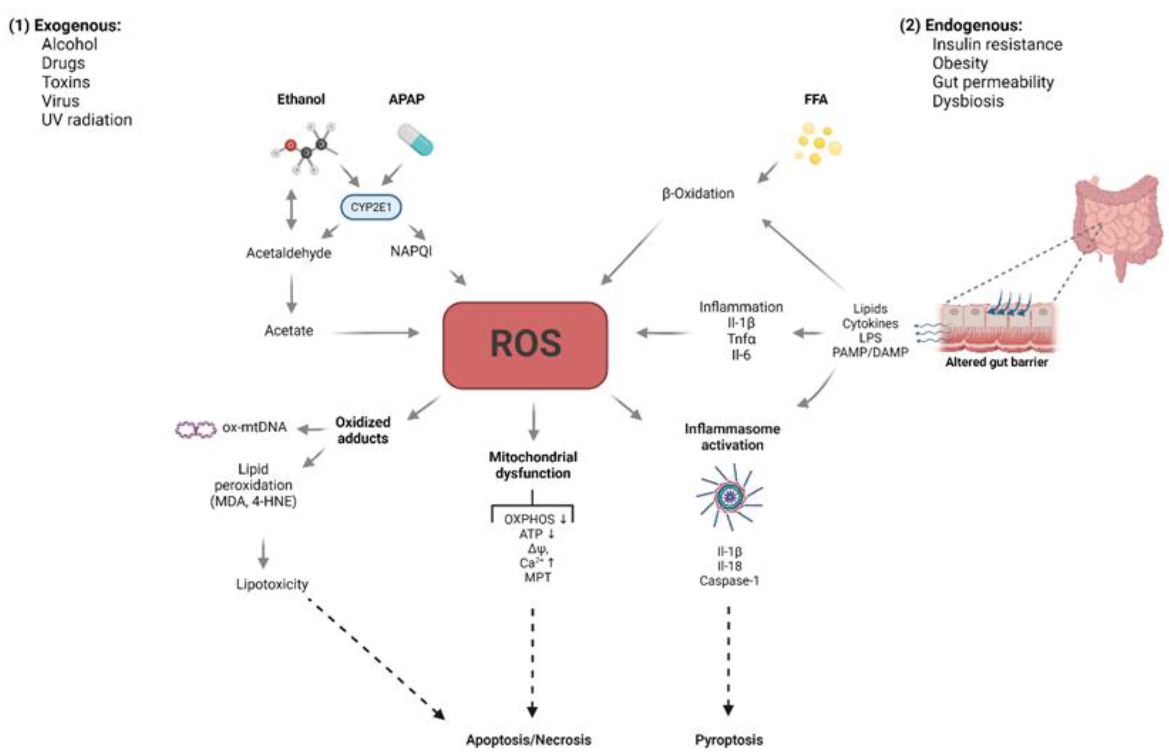
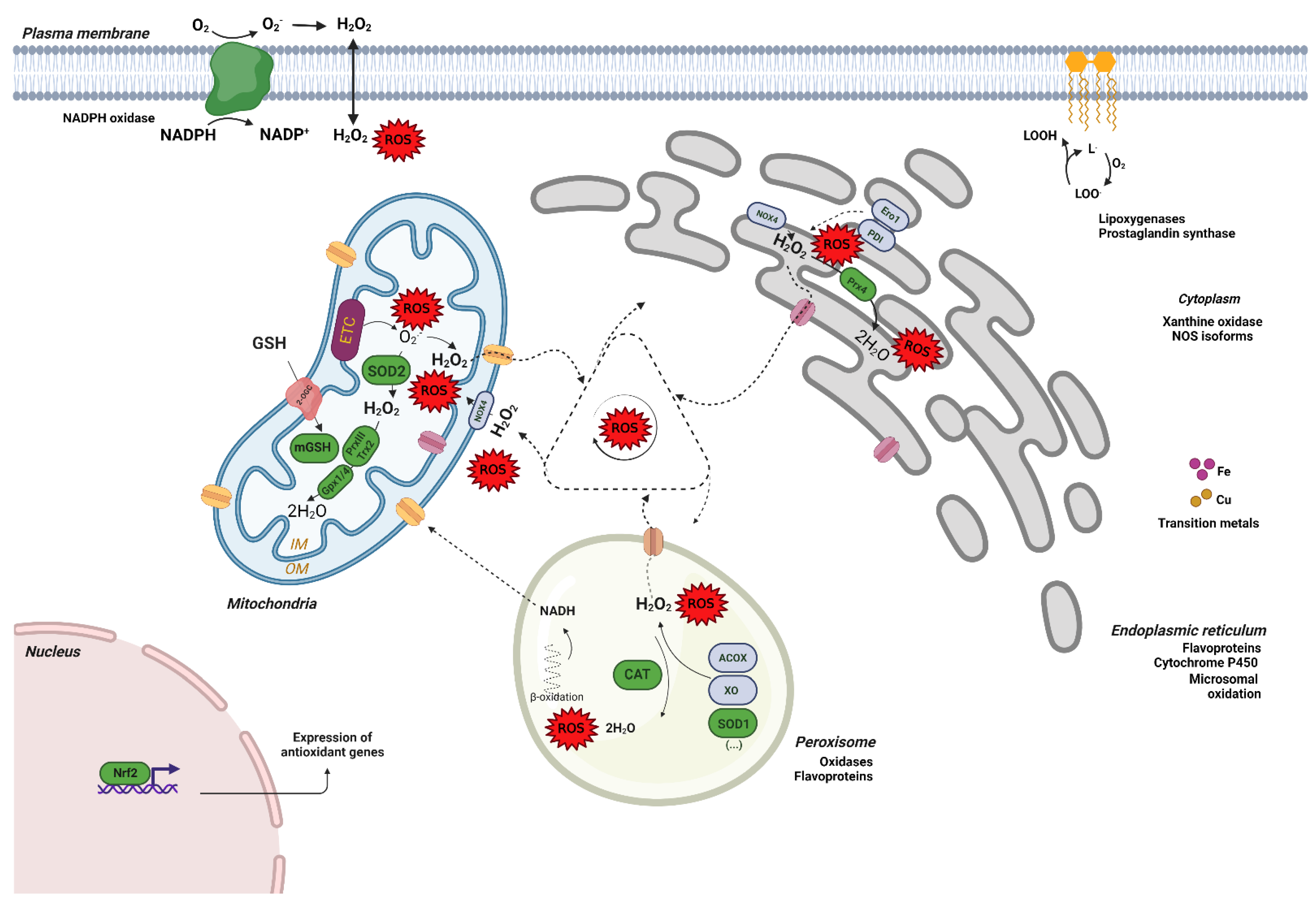
2. Free Radicals: Sources and Defense

ROS include a number of molecular species derived from oxygen, such as superoxide (O2•−) anion, the prototype of ROS which is produced in enzymatic reactions and in the electron transport chain (ETC) of mitochondria, as well as other oxidants like hydrogen peroxide (H2O2) or hydroxyl radicals (•OH). The majority of ROS is produced in mitochondria [2,3], where superoxide anion can be generated as a by-product of transfer of electrons to oxygen during the respiratory chain to produce ATP [4]. Extramitochondrial sources of ROS include membrane nicotinamide adenine dinucleotide phosphate (NADPH) oxidase, lipoxygenases, cyclooxygenases, peroxidases, other heme proteins, xanthine oxidase, peroxisomal fatty acid β-oxidation, and hepatic P-450 microsomal detoxification [5,6,7,8,9]. Although oxidative stress ensues through the alteration of the balance between oxidants (i.e., ROS) and antioxidants, a strict equilibrium between antioxidants (i.e., superoxide dismutase (SOD) and GSH redox cycle) is also necessary to circumvent the generation of oxidants such as hydrogen peroxide [10]. In this regard, the cell displays antioxidant mechanisms to combat ROS of different origins which comprise enzymatic and non-enzymatic components. The enzymatic system includes different enzymes that catalyze reactions involved in ROS detoxification. Some of the most relevant are SOD, catalase (CAT), glutathione peroxidase, and reductase (GSH-Prx or Gpx), peroxiredoxins, glutaredoxins, thioredoxins, and sulfiredoxins. To increase efficiency in ROS scavenging, some of these enzymes are located in specific sites where free radicals and ROS are generated within the cells to counteract them in a more direct fashion. As an example, two isoforms of the SOD enzyme exist, the cytosolic one (Cu/Zn–SOD) and the mitochondrial one (Mn-SOD), also known as SOD-1 and SOD-2. Non-enzymatic antioxidant components include small molecules, such as glutathione (GSH), ascorbic acid (vitamin C), retinol (vitamin A), and tocopherol (vitamin E) which provide protection against radical species by accepting electrons in their structure [11]. Another key factor is the oxidative stress-induced transcriptional machinery governed by the nuclear factor E2-related factor 2 (Nrf2) which regulates the expression of various antioxidant genes [12]. Indeed, pharmacologic activation of the Nrf2-dependent antioxidant signaling pathway has been shown to protect the liver in different oxidative stress models [13]. Figure 2 illustrates the contribution of each cellular compartment to oxidative stress and the antioxidant defense mechanism to overcome ROS generation.

3. Redox Control in the Liver

Due to the predominance of mitochondrial-origin ROS and the involvement of this organelle in liver diseases, attention will be focused in the strategies to attenuate mitochondrial ROS formation. Hepatocytes are well equipped with non-enzymatic and enzymatic antioxidant defense systems that neutralize free radicals. SOD enzyme is the “first line” defense in reducing superoxide radicals formed in the ETC to H2O2 and O2. CAT is a major H2O2 detoxifying enzyme. However, mitochondrial expression of CAT is null as the localization of this enzyme is mainly peroxisomal. Therefore, the degradation of mitochondrial hydrogen peroxide is carried out by other alternative antioxidant enzymes. Mitochondrial glutathione peroxidases (Gpx1,4) and various hydroperoxides (PrxIII, Trx2) catalyze the reduction of H2O2 in a chain reaction with GSH as the electron donor, and the subsequent conversion of GSH disulfide (GSSG) back to GSH by the NADPH-dependent glutathione reductase (GR). Hence, mitochondrial GSH (mGSH) protects mitochondria from the lack of catalase, implying that its availability is crucial for the appropriate redox maintenance in this organelle.
GSH is exclusively synthesized in the cytosol, thus specific carriers are required to distribute GSH to different compartments including mitochondria. In the liver, GSH is transported into mitochondria via the 2-oxoglutarate carrier (OGC; Slc25a11) which imports GSH in exchange for 2-oxoglutarate (2-OG). A relevant aspect of GSH transport in hepatic disorders is the dependence of the 2-OG carrier on membrane fluidity [14] which is determined by fatty acid composition and cholesterol content. Increment of the membrane rigidity impairs this transport system, limiting mGSH availability and antioxidant protection. In this sense, previous studies in various models of alcoholic (ASH) and non-alcoholic steatohepatitis (NASH) have reported increased mitochondrial cholesterol which resulted in mitochondrial dysfunction, reduced levels of mGSH and increased susceptibility to cell demise [15,16,17,18,19,20]. In addition, by controlling cardiolipin redox status, mGSH plays a key role in regulating cell death pathways [21], which is further described later in this review.
When free radical species elude cellular antioxidant defense mechanisms, they react with macromolecules generating toxic adducts that lead to the alteration of function of all cellular components preceding apoptosis [22]. In this aspect, elevated markers of oxidative stress such as DNA damage marker 8-hydroxy-2′-deoxyguanosine (8-OHdG) and lipid peroxidation products (MDA and 4-HNE) have been observed in hepatic tissue and plasma of patients diagnosed with different liver disorders (e.g., NASH, hepatitis C, liver fibrosis or HCC), serving as potential biomarkers for disease [23,24,25]. Likewise, excessive oxidation of lipids modifies the physical properties of cellular membranes and can contribute to the impairment of the mitochondrial membrane potential and Ca2+ buffering capacity. If prolonged in time, Ca2+ overload elicits mitochondrial permeability transition (MPT) opening which results in a secondary burst of ROS and ignites a cascade of detrimental effects that lead to hepatocellular death as ultimate consequence [26,27].

4. Cell Aging and Cell Death Regulation in the Liver

4.1. Cell Aging in the Liver

The aging process is characterized by the gradual decay of physiologic functions that occurs with time. Several hallmark features have been identified in aging such as genetic alterations, mitochondrial impairment, loss of proteostasis, dysregulation of nutrient sensing, altered intracellular communication, and telomere shortening. Major interest has been focused in the last years in this progressive condition due to its correlation with the onset of diseases including liver disorders. Indeed, aging is associated with the severity and poor prognosis of various liver diseases including nonalcoholic fatty liver disease, alcoholic liver disease, hepatitis C, and liver transplantation [28,29]. In addition, as this organ has the capacity for regeneration, process that declines with age, fibrotic response is also altered during aging [30,31].
In the young liver solutes such as lipoproteins, insulin, and carbohydrates are able to diffuse between the blood and hepatocytes via the liver sinusoid endothelial cell (LSECs) fenestrations which are critical for maintaining hepatic homeostasis. With age, multiple changes take place in each liver cell type (hepatocytes, liver sinusoidal endothelial cells, hepatic stellate cells, and Kupffer cells (KCs)), impairing many physiological functions. Aging-related changes in liver cells include volume changes, increased polyploidy and DNA damage, accumulation of lipofuscin, impaired mitochondrial oxidative capacity, elevated oxidative stress and ROS, and presence of senescent cells with senescence associated secretory phenotype (SASP) which promote the recruitment of inflammatory cells.
Concerning liver sinusoidal endothelial cells (LSECs), these cells show reduced fenestrations, cellular autophagy, and augmented levels of cell adhesion markers during aging. These traits have been described in several species including mice, rats, non-human primates and humans, and in mouse models of premature aging [32,33,34]. Loss of fenestrations has particular importance as it can lead to hyperlipidemia and hepatic insulin resistance by limiting the uptake of lipoproteins and insulin [35]. In addition, LSECs also play a key role in the removal of circulating macromolecules like collagen catabolism products, hyaluronic acid or antibodies. This endocytotic activity is mislaid with aging, contributing to the disproportionate accumulation of harmless molecules in the liver [36]. Moreover, aging LSECs display a reasonable pro-inflammatory profile as evidenced by the presence of leukocytes recruited by adhesion molecules and the high expression of interleukin 6 (Il-6) cytokine and CD68+-cells. Moreover, upregulation of p16 and downregulation of SIRT1 has been reported which might influence cellular senescence [37].
According to several studies, liver volume decreases by 20–40% as one gets older [28,38]. These changes are linked to a reduction of blood flow in the liver, as an approximately 35% reduction in blood volume of the liver is observed in individuals above 65 years of age compared with those aged less than 40 years [39]. These changes are associated with increases in portal pressure and vascular resistance leading to reduced hepatic blood flow due to downregulation of the vasodilatory pathways involving NO bioavailability in LSECs [40]. In addition, aged LSECs exhibit increased expression of intercellular adhesion molecule 1 (ICAM-1) which promotes leukocyte adhesion and contributes to further reduction of sinusoidal blood flow [40]. Regarding other hepatic cells, excessive lipid loading with marked increase in the number and size of lipid droplets and overproduction of collagen and laminin that are deposited in the basal lamina membrane account for the effect of aging on hepatic stellate cells (HSCs) [41]. Finally, KCs, major inflammatory cells in this organ, accumulate within the liver with age and attach to the adhesion markers expressed on LSECs, contributing to Il-6 release and extending inflammatory processes in the liver [37,42].
These molecular alterations lead to low-grade inflammation and facilitate multiple phenotypic changes in hepatocytes and other liver cells by the release of inflammatory signals, and will provoke cellular senescence and death as ultimate consequence. Notably, all these cellular events are observed in most types of liver disease including non-alcoholic fatty acid liver disease, alcoholic liver disease, primary biliary cholangitis, liver fibrosis, and hepatocellular carcinoma, which will be discussed in detail in the following section.
From the therapeutic point, prevention of age-associated alterations or inhibition/modulation of senescent cells might provide benefit to the health of patients with chronic liver diseases. As an example, several compounds have already shown positive effects by increasing fenestrations in LSECs isolated from old mice, including 7-ketocholesterol, sildenafil, amlodipine, cytochalasin D, bosentan, 2,5-dimethoxy-4-iodoamphetamine, and TNF-related apoptosis-inducing ligand. In addition, two drugs that delay aging by regulating the nutrient sensing pathways, nicotinamide mononucleotide and metformin, were also found to increase fenestrations in LSECs from aged mice [43,44]. Definitively, the identification of specific markers and senescence-related pathways will provide important knowledge to improve translational application of preclinical studies in human disease.

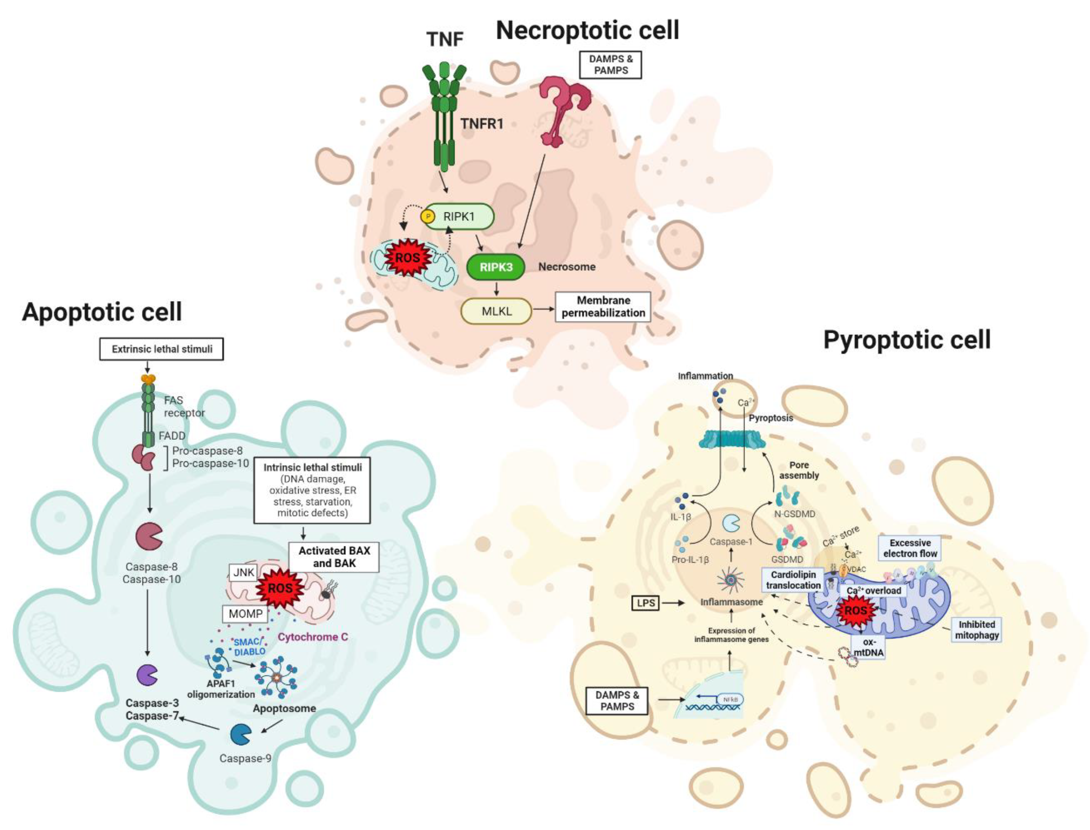
4.2. Cell Death Regulation in the Liver

Hepatocellular death participates in most types of acute and chronic liver disorders, being a crucial pathologic factor in disease progression. Despite the fact that it essentially occurs to accomplish the specific removal of injured cells as an adaptive reaction to diverse disturbances, it also takes place as a result of inability to cope with excessive stress, involvement in the promotion of inflammation, liver injury, fibrosis, cirrhosis, and hepatocellular carcinoma.
Depending on the nature of insult, trigger mechanisms and morphologic phenotype, cell death can be divided into different types. In this review, we will focus on apoptosis, necrosis, and pyroptosis types of cell death occurring in liver cells under various liver disorders.

4.2.1. Apoptosis

Apoptosis, or active programmed cell death, is deeply organized, caused by concrete signaling cascades and characterized by nuclear fragmentation, chromatin condensation, and cellular shrinkage. It commonly occurs to control cell populations in tissues during development and aging. Apoptosis is mainly dependent on caspase activation, although caspase-independent apoptosis also exits. Apoptosis can either be initiated via intrinsic mitochondrial or extrinsic death receptor-mediated pathways, both leading to the activation of the executioner caspases -3 and -7.
Intrinsic apoptosis is induced by different aberrant events such as DNA damage, oxidative stress, ER stress, starvation, and mitotic defects [45] which trigger BAX/BAK-induced mitochondrial outer membrane pore formation (MOMP) and the leakage of mitochondrial apoptotic components such as cytochrome c and SMAC/DIABLO which bind to APAF1 and pro-casp9 forming the apoptosome which activates CASP9. In turn, CASP9 induces the activation of executioner caspase-3 and caspase-7 [45,46,47]. MOMP is regulated by the balance between the pro-apoptotic and anti-apoptotic members of the B cell lymphoma-2 (Bcl-2) family [46]. In contrast, the binding of ligands to death receptors such as FAS and TNFR1 triggers the extrinsic death receptor apoptotic pathway, activating CASP8 via FADD and TRADD, and subsequently CASP3 and CASP7, and apoptosis [46].
ROS are powerful inducers of apoptosis and can trigger the intrinsic mitochondrial pathway, the extrinsic death receptor pathway and ER stress pathway [48], being mitochondria crucial to activating apoptosis in all those pathways [49]. Proteins are considered the principal targets of ROS and they are very frequently oxidized [50]. Indeed, increased ROS levels can induce reversible post-translational alteration of cysteine [51], selenocysteine [52], histidine [53], and methionine residues [54]. ROS drive to the oxidation of thiol groups of critical cysteine residues in many proteins including kinases, phosphatases, and transcription factors [55,56], achieving the regulation of cellular functions via the redox-balance of cysteine residues of redox-sensitive proteins. Thus, ROS regulate apoptosis via diverse mechanisms, including death receptor activation, caspase activation, Bcl-2 family proteins, and mitochondrial dysfunction. The activities of assorted protein kinases, including MAPK (mitogen-activated protein kinases), protein kinases-B/C, inhibitor of-I-kappaB kinases, and their equivalent phosphatases influence the apoptotic program depending on cellular context. In this line, it has been shown that ROS induce apoptosis via JNK activation [57,58] by intrinsic in the mitochondria or extrinsic apoptotic signaling by death receptor pathways [59,60,61,62]. Of note, a dual role of JNK has been demonstrated by early-transient activation of JNK by TNF promoting cell survival, whereas the long-sustained JNK activation following TNF administration in cells with blocked NF-kB-driven gene expression is associated with elevated ROS production which sustains JNK activity and triggers cell death [63,64,65]. In addition, JNK affects the activity of cytoplasmic proteins involved in the control of programmed cell death, such as Bim, Bid, and cFlip [66,67,68,69].
Additionally, cellular and organelle membranes are strongly susceptible to ROS damage. Oxidized phospholipids act as cell death signals triggering programmed cell death, activating both the intrinsic and extrinsic apoptotic signaling pathways [70]. It has been demonstrated that lipid peroxidation products form adducts with ERK, JNK, and p38, and activate caspase signals promoting apoptosis [71,72]. In addition, ROS may oxidize cardiolipin, a mitochondrion-specific inner membrane phospholipid, and thus activate the intrinsic apoptotic pathway [73]. Indeed, oxidized cardiolipin possesses lower affinity for cytochrome c which drives the detachment of cytochrome c from the MIM [21,74,75]. In addition, oxidized cardiolipin controls the permeabilization of MOMP via oligomerized Bax [21,74,76], supporting the hypothesis that cardiolipin is a critical upstream target in mitochondria-dependent apoptosis. Since mitochondrial ROS are involved in cardiolipin oxidation, mGSH arises as an important modulator of apoptotic cell death by indirectly controlling the redox state of cardiolipin [21,74]. In fact, apoptosis induced by ROS is linked to GSH oxidation and decreased GSH levels, increasing ROS production and the loss of redox homeostasis [77,78].
Apoptosis (both intrinsic and extrinsic) is involved in ASH, NASH, and cholestatic liver injury [79,80]. In ALD, metabolic, toxic, and inflammatory factors drive to mitochondrial dysfunction, ROS formation, Bax translocation to mitochondria, and caspase activation [81,82]. Alcohol and acetaldehyde are highly reactive and induce ROS generation and ER stress which can activate IRF3. In turn, IRF3 interacts with Bax leading to hepatocyte apoptosis [83]. In addition to ER and mitochondrial stress, alcohol also drives to apoptosis and inflammation via lysosomal malfunction raising lysosomal pH [84]. In addition, acute and chronic alcohol consumption modifies the intestinal permeability, driving to increased bacterial lipopolysaccharide (LPS) which activates Kupffer cells (KCs) and subsequently TNF generation [85]. High levels of circulating TNF and TNFR have been found in patients with alcoholic liver disease and ASH, while in animal models anti-TNF antibodies treatment protects against alcoholic liver injury [82,86]. In addition, apoptotic TUNEL positivity staining and CASP3 positive hepatocytes have been detected in liver biopsies of patients with alcoholic hepatitis with high bilirubin (>3 gr/dL) and elevated steatohepatitis [87]. Unfortunately, treatment with infliximab, an anti-TNF antibody, has been proven harmful in patients with alcoholic steatohepatitis because of the elevated infection and mortality rates, regardless of the evident link between TNF and alcoholic liver disease [88].
There are robust data indicating that hepatocyte cell death leads to inflammation and fibrosis in NASH [89]. Activation of caspase-3 and -7, elevated expression of FAS receptors and hepatocyte apoptosis are crucial events in experimental models and human NAFLD correlated with disease severity and progression [90,91]. Indeed, fragments of CK-18 produced by caspase 3 are used as indicators of NASH in patients with potential NAFLD [92,93]. In addition, ER stress provoked by free fatty acids (FFA) is related with the activation of JNK in human steatosis [94]. P-JNK and mitochondrial Sab (Sh3bp5) interaction drives to ROS generation, maintains JNK activation and apoptosis-related lipotoxicity, leading to NASH pathogenesis [95]. Indeed, JNK plays a critical role in lipotoxicity induced by palmitic acid [96,97,98]. Evidence supports a role for Src-dependent activation of the MAP3K, MLK3 [99,100]. The role of JNK in palmitic acid toxicity is associated with the induction and activation of PUMA and Bim [96,101], both pro-apoptotic Bcl2 family members involved in mitochondrial permeabilization. In addition, JNK phosphorylates Sab, an outer membrane mitochondrial protein containing C terminal JNK docking sites facing the cytoplasm, and directly drives to mitochondrial respiration impairment and the increase of ROS. This, in turn, sustains JNK activation, as ROS activate the MAPK pathways and further block mitochondrial function, inducing MOMP via regulation of Bcl2 proteins in TNF- and ER stress-induced apoptosis. Other possible players of palmitic acid toxicity might act upstream or downstream of JNK. These include NADPH oxidase [102,103] as a source of oxidative stress [104], induction or posttranslational regulation of Bcl2 family members, and lysosomal permeabilization [105]. Thus, highly elevated JNK1 signaling has been described in human NAFLD livers [106].
Moreover, in patients with NAFLD and NASH, hepatocytes undergoing apoptosis activate immune cells and hepatic stellate cells (HSCs), a crucial event during initiation and progression of fibrosis [107,108]. Accordingly, CASP3 and CASP8-deficient mice fed a methionine-choline deficient (MCD) diet were protected from apoptosis, and showed decreased inflammation and fibrosis [109,110]. In line with this, elevated lipid peroxidation, TUNEL positivity, augmented CASP3 and CASP8 activities, and fibrosis were found in mice fed a high fat diet (HFD) [111] which were significantly reduced using a pan-caspase inhibitor. Remarkably, presence of CASP6 was reported in NASH human livers. In NASH, inflammation-induced CASP3 and CASP7 activation leads to casp6 cleavage and activation which in turn cleaves Bid. Truncated Bid promotes the leakage of mitochondrial cytochrome c which activates CASP3 and 7 driving to a continuous apoptosis loop in hepatocytes [112]. Therefore, controlling hepatocyte cell death pathways might have therapeutic implications [113] and hence, assorted apoptosis inhibitors have been suggested as potential treatments for NASH [114,115,116,117,118].
Additionally, elevated FAS expression, cytoplasmic shrinkage, nuclear condensation, and TUNEL positivity of cholangiocytes have been described in liver biopsies of primary biliary cholangitis (PBC) patients [119]. Indeed, apoptosis blockage and enhanced survival was demonstrated in FAS KO mice [120]. Ursodeoxycholic acid (UDCA), a non-toxic bile acid used to treat cholestatic diseases, protects against toxic bile acid-induced apoptosis [121]. In addition, cleaved CASP3 and CASP8 have been found in PBC patients’ liver biopsies, indicating an activation of apoptosis [122]. Moreover, ameliorated AST/ALTs and reduced cleaved CASP3 and p-JNK were reported in liver-specific CASP8 KO mice after bile duct ligation (BDL) surgery, pointing at apoptosis as the presumable type of cell death in BDL mice. Nevertheless, cholestatic liver disease models promote areas of necrotic resembling cell death, as bile infarcts after BDL or in Mdr2 KO mice [79].
Despite apoptosis possessing a minor pro-inflammatory potential compared with necrosis, KCs phagocytosis of apoptotic bodies augments death ligands and cytokines in cholestatic liver damage in mice [123]. Thus, Fas-involved apoptosis of hepatocytes is linked with activation HSCs and liver fibrosis, connecting apoptosis to liver injury [124].

4.2.2. Necrosis

Traditionally, necrosis has been considered a passive and accidental form of cell death characterized by cell swelling, membrane disruption, and the leakage of inflammatory cellular components (DAMPs), leading to an inflammatory response. However, recent evidence indicates that necrosis is mostly mediated by MPT, characterized by the development of permeability transition pore at inner and outer mitochondria membranes, driving to a rapid disappearance of the membrane potential gradient, disruption of ATP synthesis, osmotic failure of both membranes, and cell death [46]. Thus, various regulated necrotic modalities exist, including necroptosis, pyroptosis and ferroptosis, that possess important morphologic characteristics as passive necrosis combined with specific and regulated causal mechanisms [46,125].
Necroptosis is a lytic form of regulated necrosis occurring usually after death receptor (RD) (TNFR1, CD95, TRAIL-R) and Toll-like receptor activation (TLRs, TLR3, and TLR4), but only when apoptotic signaling is inhibited. It is caspase-independent and initiated by activation of receptor interacting protein kinase (RIPK1/RIPK3) and the mixed lineage kinase domain like pseudokinase (MLKL) forming the necrosome [126], and increased the plasma membrane permeability which causes an important inflammatory response associated with most chronic liver diseases including viral hepatitis, autoimmune hepatitis, NASH, and ALD [80,127,128].
Several studies have demonstrated the relation between ROS production and necrosome signaling [129,130,131]. In some types of cells, mitochondrial ROS is critical for necroptosis by triggering RIPK1 autophosphorylation and RIPK3 recruitment which in turn mediate mitochondrial disruption and elevated ROS production in a feedforward mode, facilitating necrosome formation [129,130,132]. In addition, necroptosis is prevented by JNK inhibition, suggesting that JNK-induced mitochondrial dysfunction is a crucial trigger of necroptosis [133]. ROS can also induce ferroptosis, an iron-dependent form of cell death, different from apoptosis and necrosis [134] which requires redox cycling of Fe2+/Fe3+ that promotes membrane phospholipids peroxidation [135]. Free intracellular redox-active iron elevates ROS production via Fenton chemistry and/or raised lipoxygenase activity [136,137] which consecutively modify pores, integrity or curvature of membranes [138], demonstrating that redox signaling firmly regulates apoptosis and ferroptosis [139].
An important crosstalk between apoptosis and necroptosis pathways has been described [46,80]. The equilibrium between these two modalities of cell death not only regulates the degree of inflammation, but also influences the type of liver cancer caused. Recently it has been described that a necroptotic environment with an inflammatory cytokine component induced intrahepatic cholangiocarcinoma caused by genetic oncogenic activation, while an apoptotic background promoted HCC development [140]. Furthermore, the role of necroptosis is determined by the background, and a switch from necroptosis to apoptosis may be more inflammatory and fibrogenic in NAFLD than in an ALD model [113,128].

4.2.3. Pyroptosis

Pyroptosis is a modality of lytic regulated cell death which develops in response to intracellular pathogens or PAMPs, mainly LPS. Pyroptosis is associated with caspase-dependent pore formation in the plasma membrane, swelling, rupture of the cell, and pro-inflammatory IL-1β and IL-18 leakage [141]. In addition, the pore-making gasdermin D (GSDMD) has been recognized a necessary executioner of pyroptosis [142,143]. Indeed, pyroptosis is mediated by the inflammasome via the pro-inflammatory activity of GSDMD. Pyroptotic caspases include caspase-1, caspase-11 and its human homologs caspase-4 and 5 [144,145].
Cleave of GSDMD and subsequent pyroptosis occur through three different mechanisms: (1) caspase-1-dependent canonical inflammasome pathway activated by PAMPs, DAMPs, and cholesterol crystals [146], (2) caspases -4, -5 or -11-mediated non-canonical inflammasome mechanism [147], and via (3) caspase-8-mediated inflammasome-independent pathway [148].
The NLRP3 inflammasome activation driving pyroptosis has been linked with inflammation, fibrosis, and cell death in the liver [149]. Thus, the NLRP3 inflammasome is an important pathway for the release of pro-inflammatory cytokines in the liver and is strongly associated with the pathogenesis of liver fibrogenesis [150]. The activation of NF-κB is the first step in the canonical activation of NLRP3 inflammasome, and ROS strongly control the activity of the transcription factor NF-κB. Most canonical NLRP3 inducers elevate ROS generation via NADPH. The inflammasome activation is dependent on ROS accumulation upon inhibition of autophagy and mitophagy, where ROS production is controlled [151]. Moreover, GSDMD has a thiol group located in the cysteine residues that can be affected by oxidative stress to form disulphide bonds and stabilize pore formation in the plasma membrane [152]. In addition, antioxidants increase Nrf2 activity, reducing ROS generation and decreasing gasdermin D, TXNIP and NF-kB oxidation which is essential for inflammasome activation and pyroptosis. Indeed, treatment with antioxidants reduces ROS and consequently prevents inflammasome activation in a carbon tetrachloride-driven acute liver injury model [153,154], demonstrating the association between ROS generation and NLRP3 activation in liver disease.
Inflammatory caspases, including murine caspase-1 and -11, and human caspase-4/5, play critical roles as inflammation intermediaries [143], locating pyroptosis in NAFLD development to NASH progression [155,156]. Indeed, activation of IL-1β signaling, downstream of inflammasomes, drives liver inflammation, steatosis, and fibrosis in experimental NASH [157,158] and amplifies the response of other cytokines [159]. Thus, preventing inflammasome activation could be a promising therapy to hinder disease progression [160].
Since gut dysbiosis is frequently associated with ASH, NAFLD and NASH, it is reasonable that NASH might be connected to the pyroptotic GSDMD pathway activation [161]. Indeed, CASP11-GSDMD activation has been described in livers from AH patients [162], exacerbating neutrophilic infiltration via the leakage of DAMP, bacteria, PAMPs, IL-1β, and IL-18 from pyroptotic cells in AH. The involvement of NOD-like receptor family pyrin domain containing 3 (NLRP3) inflammasome in ALD has been demonstrated in several studies [163]. Indeed, an interaction has been recently described between activated NLRP3 inflammasome and mitochondrial dysfunction in the context of ASH and NASH [164].
The role of NLRP3 activation in ALD is supported by the protection of caspase-1, apoptosis-associated speck-like protein containing a caspase recruit domain (ASC), and IL-1β receptor global knockout mice which exhibit diminished steatosis and inflammation [165]. In addition, upregulation of caspase-1 and GSDMD processing were observed after using a Western diet combined with intragastric ethanol administration to induce ASH, indicating a critical role for pyroptosis in alcoholic hepatitis [162].
In addition, NASH is ameliorated in the GSDMD-knockout mice compared with WT mice which possess enhanced hepatic NLRP3 inflammasome expression [149,162]. Moreover, the NLRP3 inhibitor MCC950 prevented liver injury in a murine NASH model induced by feeding mice with a Western diet, decreasing transaminases, liver fibrosis, and cytokine levels compared with control mice [158]. Additionally, inflammasome-induced cholesterol crystallization in KCs is a critical factor of hepatic inflammation in NASH progression [166,167]. Sphingomyelin synthase 1 (SMS1) is involved in hepatocyte pyroptosis via a diacylglycerol (DAG)–protein kinase Cδ (PKCδ)–NLR family CARD domain-containing protein 4 (NLRC4) axis in NASH [168]. In addition, sphingosine 1 phosphate receptor 4 (S1PR4) mediates NLRP3 inflammasome activation in hepatic macrophages. SLB736, an antagonist for S1PR4 receptor, prevented NASH and hepatic fibrosis development, and could be considered a new therapeutic target in NASH development [169].
Remarkably, different cell death modalities can coincide simultaneously in pathological backgrounds, leading to the concept of PANoptosis as a novel form of inflammatory cell death during which the principal cell death pathways, apoptosis, necroptosis, and pyroptosis can be activated in parallel, driving to the formation of a large multiprotein complex named the PANoptosome [170].
Information summarizing the role of ROS in the various cell death types discussed above is included in Figure 3. Knowledge about the specific signaling pathways and cell death modalities involved in each disease will enable researchers to design treatments targeting specific molecules to ameliorate injury, inflammation and disease progression, opening novel therapy opportunities.

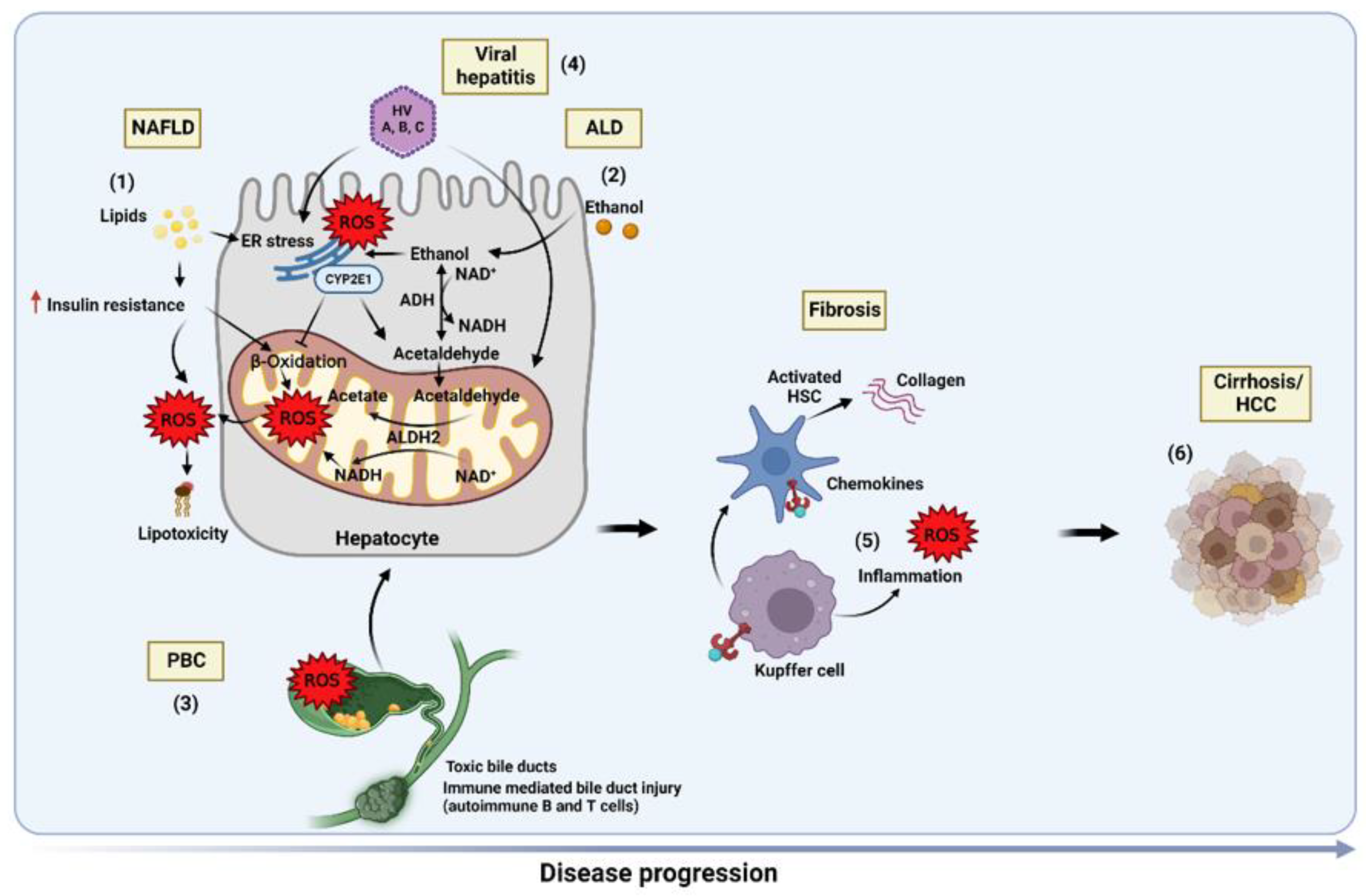
5. Role of Oxidative Stress in Liver Diseases

5.1. Non-Alcoholic Fatty Liver Disease (NAFLD)

NAFLD is an intricate and multifactorial disease associated with multiple genetic, epigenetic, and environmental factors that comprises a spectrum of alterations beginning with simple steatosis (non-alcoholic fatty liver, NAFL) which can progress to non-alcoholic steatohepatitis (NASH) that can culminate in cirrhosis and hepatocellular carcinoma (HCC) [171]. NAFL is characterized by macrovesicular steatosis (more than 5%) without the existence of hepatocellular injury, whereas NASH encompasses hepatic steatosis, plus inflammation, ballooning of hepatocytes and hepatocellular injury in the presence or absence of fibrosis. A “two-hit” hypothesis was originally proposed to define the pathogenesis of NAFLD. Insulin resistance (IR) acts as the first hit, promoting liver steatosis with augmented hepatic lipogenesis and defective degradation of FFA. The accumulation of fat sensitizes the liver to inflammation and cell death by a second hit that causes oxidative stress, concluding in NASH and fibrosis [172,173]. In the last years, the “multiple hit” theory, which includes different factors acting in parallel, has emerged to describe more accurately the pathogenesis of NAFLD. Oxidative stress is considered to be the main contributor of the “multiple hits” to liver injury and disease progression in NAFLD [174,175]. Currently, metabolic dysfunction-associated fatty liver disease (MAFLD) has been proposed to classify the heterogeneous population of patients with this disease. In this way, MAFLD includes patients with hepatic steatosis in combination with overweight/obesity, presence of type 2 diabetes, or evidence of metabolic dysregulation [176], excluding individuals with excessive alcohol consumption. The new nomenclature encloses coexisting toxic (e.g., alcohol) or viral factors that do not exclude the link to metabolic liver disease.
Liver steatosis, the first stage of MAFLD, although considered reversible, sensitizes to diverse secondary insults driving the development of NASH [177]. Different lipid species accumulate in the transition of MAFLD to NASH, including, among others, triglycerides, diacylglycerol, free fatty acids, ceramides, and cholesterol, underlining that the type rather than the amount of lipids is responsible for the progression from steatosis to NASH [15,178]. Therefore, the unbalance of lipid metabolism is the most direct etiology of MAFLD.
The mechanisms by which ROS contribute to MAFLD progression are multifactorial and involve both accumulative oxidative biomolecular damage and dysregulated redox signaling [179], since ROS induce oxidative alterations to macromolecules that lead to liver injury [172,173], and possess important signaling functions [180]. In the liver, the metabolism of lipids is regulated by nuclear receptors that activate diverse ligands and modulate important enzymes involved in lipogenesis and lipoxidation [181], acting as redox triggers that in turn affect several metabolic pathways due to particular ROS [182]. AMPK activation leads to the inhibition of lipogenesis [183], downregulating several nuclear receptors and enzymes implicated in lipid metabolism, such as acetyl-CoA carboxylase, PPARα, PPARγ, and SREBPs. Oxidative stress influences AMPK signaling, since both cytoplasmic and mitochondrial ROS can upregulate AMPK activity [184]. In addition, in MAFLD, an increment in the AMP/ATP ratio stimulates AMPK [185], and H2O2 can precisely modulate AMPK activity [186]. H2O2 also increases SREBP-1c expression, leading to an increment in fatty acid synthetase and promoting lipogenesis [187]. Remarkably, various antioxidants reduce hepatic steatosis through the downregulation of SREBP-1c expression [188]. Hence, ROS-mediated signaling may partially be responsible for the alterations in lipid metabolism homeostasis. Additionally, as an effort of liver cells to control the prooxidant effects of metabolic stress in MAFLD, stress-responsive transcription factors (such as AP-1, NF-κB and Nrf2) and their regulated genes are also redox sensitive [189]. Elevated Nrf2 expression is related to ROS pathological levels in liver biopsies of patients with NASH [190]. In diverse NASH models, using MCD and HFD diets, Nrf2-knockout mice display elevated susceptibility to NASH development [191,192]. Further, pharmacological activation of Nrf2 decreases IR, liver steatosis, and fibrosis in animals fed with a high-fat and high-fructose diet [193], linking antioxidant signaling and lipid metabolic pathways.
Excessive lipid accumulation produces an important hepatic metabolic stress which causes inflammatory pathways activation. After exposure to harmful stimuli, KCs are the principal effectors involved in ROS generation and the transcription of cytokines/growth factors/enzymes modulating redox balance, metabolism, inflammation, and fibrosis [194,195], functioning as a driving force in MAFLD progression at several levels [196,197,198]. Hence, the innate immune and ROS signaling collaborate and synergistically influence disease progression.
MAFLD is also linked to “mitochondrial dysfunction” which is shown by modified respiratory complex activity and fatty acids oxidation [199]. ETC activity gradually reduces during MAFLD progression, and livers with simple steatosis already show a mild deterioration of ETC activity. Besides targeting lipids, ROS generated in the mitochondria can also modify mitochondrial DNA, causing deletions and mutations [200]. Of note, an important reduction in mitochondrial DNA in MAFLD patients has been shown [201]. Thus, mitochondrial DNA damage and mutations induced by ROS may affect ETC and ATP generation which in turn activates mitochondrial ROS production through the mitochondrial MPT opening, causing a vicious cycle of damage amplification termed “ROS-induced ROS release” (RIRR) [202].
Oxidative stress biomarkers normally measured in clinical samples of MAFLD comprise DNA oxidation products (8-OH-dG), lipid damage products (thiobarbituric acid reactive substances or TBARS), MDA, 4-HNE, hydroperoxides, and 8-isoprostane), and protein oxidation products (protein carbonyl, nitrotyrosine). In general, the concentrations or activities of these biomarkers are elevated in NASH patients [24,203]. In addition, antioxidants commonly evaluated in MAFLD/NASH clinical samples include enzymatic antioxidants (e.g., catalase, SOD, and GPX) and nonenzymatic antioxidants (e.g., GSH, TrxR, α-tocopherol, and ubiquinone) which are reduced in most patients with MAFLD/NASH, although they are increased in a few exceptions [204,205]. Further, several lipid peroxidation circulating biomarkers have been detected in patients with MAFLD, their concentrations positively associated with the disease severity [206,207]. Overall, these data indicate that oxidative stress and mitochondrial antioxidant defense depletion plays an important role in disease progression in MAFLD/NASH.

5.2. Alcoholic Liver Disease (ALD)

ALD is a prevalent form of chronic liver disease caused by the persistent abuse of alcohol consumption. In 1967, Comporti and colleagues proposed for the first time the involvement of lipid peroxidation as a mechanism for ethanol-induced fatty liver [208]. The oxidative metabolism of ethanol triggers a complex array of mechanisms that contribute to the progression of ALD from its initial step of steatosis to alcoholic hepatitis, cirrhosis and eventually HCC. Unfortunately, the pathogenesis of ALD is not entirely understood, which has limited the availability of therapeutic opportunities. Alcohol-induced liver injury is linked to an exacerbated ROS generation resulting in the onset of oxidative stress in hepatocytes [209] due to the metabolism of alcohol in the liver which begins with the alcohol dehydrogenase (ADH) that metabolizes ethanol to acetaldehyde. Acetaldehyde is subsequently catabolized to acetate by the acetaldehyde dehydrogenase (ALDH). Unlike acetate, which is stable, acetaldehyde is very reactive and forms adducts with DNA, promoting tissue damage. Acetaldehyde and its derivative MDA form hybrid adducts which can be recognized by KCs, endothelial, and stellate cells that produce cytokines and activate an inflammatory response that contributes to the progression of ALD [210]. In addition, ethanol is also metabolized via the microsomal system cytochrome P450, CYP2E1, which converts alcohol to acetaldehyde during alcohol chronic consumption. CYP2E1 is inducible and becomes the preferred pathway of oxidative ethanol metabolism, due in part to the saturation of ADH for alcohol, resulting in the generation of ROS and the onset of oxidative stress. ROS can also sensitize hepatocytes to LPS released from the gut and tumor necrosis factor-alpha (TNFα) which in turn generates more ROS [211], lending further support for the interconnection between oxidative stress and inflammation during ALD.
Several pathways contribute to liver injury during ALD. ADH and ALDH enzymes reduce NAD+ to NADH. The modified NAD+/NADH ratio induces fatty liver through the inhibition of gluconeogenesis and fatty acid oxidation [212]. CYP2E1, increased in persistent alcohol intake and stabilized by alcohol itself, produces free radicals via the trans-formation of NADPH to NADP+ [213]. CYP2E1-produced ROS can peroxidase mitochondrial and peroxisomal enzymes involved in β-oxidation, resulting in the accumulation of fatty acids and hepatic steatosis development. Alcohol promotes a gradient of hypoxia from the portal vein to the central vein which contributes to hepatic injury [214]. Altered ROS metabolism increases hypoxia-inducible factor-1 alpha (HIF-1α) expression which increases TNFα secretion and impairs mitochondrial function, leading to an immune response that amplifies liver injury. Furthermore, oxidative stress induces hepatocyte apoptosis and mainly necrosis [57], a response that is accentuated due to the limited antioxidant defense that contributes to inflammation and disease severity described in patients with ALD [215]. Alcohol feeding induces mGSH depletion via cholesterol overload in the mitochondria [216,217] which is reversed after alcohol withdrawal [218]. Moreover, recent data described a mitochondrial cholesterol accumulation resulting in mGSH depletion and ASH in rats fed an ethanol-polyunsaturated fatty acid treatment, effects that are prevented by betaine cotreatment [219]. Remarkably, alcohol-induced endoplasmic reticulum (ER) stress elevates cholesterol synthesis and modifies its trafficking into mitochondria, thus indirectly affecting mGSH levels. In accordance, blocking ER stress by TUDCA restored the mGSH pool in ethanol-fed rats [216,220]. Finally, ROS contribute to the transformation of HSC into myofibroblasts and the activation of matrix metalloproteinases, leading to remodeling of the extracellular matrix in the liver, culminating in excessive liver fibrosis and cirrhosis during severe ALD. These effects are accentuated by impaired regenerative capacity of mature hepatocytes [221].

5.3. Primary Biliary Cholangitis (PBC)

PBC is a chronic and progressive cholestatic liver disease, in general affecting middle-aged women, which may lead to liver failure and transplantation [222,223]. PBC is associated with hypercholesterolemia and deficiencies of antioxidant vitamins. Patients with PBC display symptoms of endothelial dysfunction, inflammation and antioxidant deficiency [224]. Moreover, PBC is considered to possess an autoimmune etiology, since specific antimitochondrial antibodies are found in most patients. The abnormal immune response in PBC drives the induction of autoreactive T and B lymphocytes, and the generation of diverse inflammatory mediators [225,226] that subject the liver to several harmful factors including oxidative stress. Serum samples from PBC patients displayed increased oxidative stress biochemical marker levels such as lipid peroxidation and cholesterol self-oxidation products [218] which further increase the immune response. Indeed, PBC patients have increased IgGs levels against malondialdehyde adduct and human serum albumin [227]. It has been described that oxidative stress and steatosis are cofactors promoting liver injury in PBC. Besides altering lipid composition, oxidative stress in PBC also promotes the accumulation of protein oxidative products and alters GSH metabolism [228]. Hence, treating PBC with UDCA improves redox changes in serum and liver tissues [229,230,231].
As previously mentioned, Nrf2 induces many cytoprotective genes that influence xenobiotic metabolism, antioxidant and anti-inflammatory responses. It has been reported that Nrf2/Keap1 axis-mediated protection against oxidative stress is impaired in PBC. Thus, defects in the Nrf2/Keap1 integrity system may influence self-defense mechanisms against oxidative stress in PBC [232]. Moreover, recent observations demonstrated that the administration of S-Adenosyl-L-methionine (SAMe) may prevent autoimmune events in PBC patients via its antioxidant and S-glutathionylation properties, providing new awareness into the molecular events inducing PBC progression and pointing at the potential therapeutic use of SAMe in PBC [233]. Oxidative stress also induces apoptosis of bile duct cells in PBC, promoting biliary damage that is a consequence of reduced glutathione-S-transferase (GST) levels. Thus, the intracellular GSH reduction directly drives biliary epithelial cells to apoptosis, the modulation of these events crucial to decreasing the immune-mediated injury [229].

5.4. Viral Hepatitis

Worldwide, over half a billion of individuals are chronic carriers of viral hepatitis. The three dominant types of viral hepatitis are hepatitis A virus (HAV), HBV and HCV. The last two are increasing in prevalence and 12% of all cancers arise from them, including the hepatitis delta virus (HDV) which can lead to accelerated disease progression [234].
Oxidative stress also plays a pathogenic role in viral hepatitis. ROS and free radicals are generated in human diseases caused by viruses, among others. Viruses enter in to the cell by endocytosis and act as an intracellular parasite using the host cell synthetic processes for their replication [235]. These processes affect the physiological statement of the ER and the mitochondria producing ROS and antioxidant system depletion (GSH, GSHPx) [236]. HBV and HCV infections are characterized by the increased levels of a wide array of oxidative stress markers in liver and blood of infected patients. Such markers include MDA, lipid peroxides, protein carbonyl content, oxysterols, and thioredoxin, which are responsible of inflammatory pathways activation [237,238,239,240]. NADPH oxidases, CYP2E1 and Ero1a are also increased in HCV [241].
Furthermore, Zn deficiency occurs in HCV, causing lipid peroxidation, loss of mitochondrial energization and oxidative DNA damage. Its supplementation therapy has been shown to improve the prognosis of patients with HCV [242]. On the contrary, the antioxidant therapy is not always effective: antioxidants can become easily pro-oxidants if they are not used at the right dose and schedule [243]. Therefore, detailed investigation of the mechanisms by which viral proteins induce oxidative stress is needed to develop effective treatments.

5.5. Liver Fibrosis

Fibrosis is characterized by progressive and excessive deposition of extracellular matrix (ECM) between hepatocytes and sinusoids, impairing the physiological architecture of the liver. It is associated with disease progression in chronic inflammatory diseases [244]. In addition, chronic portal hypertension induced by liver fibrosis is the major cause of clinical complications, bleeding events and hepatic encephalopathy [245].
As described above, steatohepatitis from all etiologies is associated with an increase in ROS by vicious cycles encompassing steatosis, lipid peroxidation, ROS formation, anti-oxidants reduction, modified mitophagy, and mitochondrial danger signal-induced expression of inflammatory cytokines which cause apoptosis and necrosis of hepatocytes. The pro-fibrotic mediators such as superoxide, H2O2, and other hydroxyl radicals are generated in hepatocytes, HSCs, and macrophages [246]. Among the ROS production enzymes, the NAPDH oxidases (NOXs) mediate fibrogenic responses by inducing angiotensin II, PDGF, and TGFβ in HSCs and macrophages that activate collagen-producing myofibroblasts that account for excessive accumulation of ECM [108,247]. Necrotic hepatocytes release DAMPs that activate danger signals to neighboring cells (HSCs and KCs). In this regard, NF-κB plays a key role in the regulation of inflammation and is considered an important modulator of liver fibrosis progression. Its activity is linked with HSC proliferation [248]. Fibrosis development principally determines the quality of life and prognosis correlated with liver function, thus being a critical risk factor for HCC development [249].

5.6. Hepatocellular Carcinoma (HCC)

HCC is the second driving cause of cancer-associated deaths worldwide due to late diagnosis and poor therapeutic outcomes [250,251]. HCC is an inflammation-related cancer, as most of HCCs emerge in the background of hepatic inflammation [252,253]. HCC incidence is significantly associated with liver inflammation from exposure to different risk factors such as hepatitis B virus, hepatitis C virus, metabolic diseases, persistent alcohol drinking, obesity and type 2 diabetes [254,255]. Chronic liver inflammation promotes oxidative stress and lipid peroxidation, producing excess ROS and aldehydes which form promutagenic DNA adducts interacting with DNA bases. Therefore, oxidative stress acts as a critical factor promoting carcinogenesis. Further, blocking antioxidant defenses strongly elevated the rate of liver cancer [256]. Moreover, mild/excess iron deposition positively correlates with HCC and p53 mutations in patients with hemochromatosis, indicating a potential carcinogenic role of oxidative stress induced by iron via Fenton reactions [257,258].
Oxidative stress is a critical factor in NASH progression and NASH-driven HCC [174,259]. NASH-related HCC incidence is expected to increase worldwide due to its association with the obesity and type 2 diabetes mellitus epidemic. Recently we have provided evidence that cholesterol rather than steatosis per se plays a role in NASH-induced HCC development [260], giving further support to strategies aimed at targeting liver cholesterol homeostasis as potential therapeutic treatments with relevance in NASH-driven HCC. Indeed, recent findings indicated that ezetimibe [260] and atorvastatin [261] improved high-fat high cholesterol diet-induced HCC development, correlating with previous results in a subcutaneous HCC model [262]. Future research may explore whether combination therapy between ezetimibe and statins may be superior for the treatment of HCC. Moreover, our laboratory also uncovered an important role of StARD1 in non-alcoholic steatohepatitis-driven HCC, where it stimulates bile acid production in the mitochondrial acidic pathway, and, in turn, activates hepatocyte pluripotency and self-renewal as well as inflammation [263].
In this regard, as HCC is closely linked to elevated oxidative stress through viral proteins or persistent inflammation and lipids, modulating oxidative stress may emerge as a promising approach against progressive liver disease. However, the administration of antioxidant therapies as potential HCC treatment remains to be fully established and should be administered with attention to the fact that a strict antioxidant balance must be guaranteed to avoid the generation of unwanted prooxidants and free radicals.
Altogether, all this data indicate that ROS is present in many liver diseases and is a determinant hallmark contributing to the progression of diseases. Molecular mechanisms underlying the pathological events described in this section need to be further investigated. Whether the formation of ROS is in fact the cause or consequence in those pathologies is also an unanswered question. As a summary, Figure 4 illustrates the molecular events leading to the development of the liver diseases discussed in this section:

6. Antioxidant Therapies

Given the role of oxidative stress in liver disorders, boosting the pool of endogenous antioxidants or the intake of dietary antioxidants was proposed as an effective therapeutic strategy with many antioxidants undergoing clinical studies [264,265,266]. In vitro and animal model studies support the beneficial role of various antioxidant compounds in liver diseases. However, when translated to clinical trials, results were controversial or failed to be replicated in larger studies [267,268]. Potential reasons for these disappointing results include inaccurate dose schedules, inadequate choice of a certain antioxidant for a particular disease, and the redundancy of symptoms with other pathologies. Problems such as small sample size, short follow up, inappropriate endpoints, inefficacy to establish tissue delivery and antioxidant effect, and the heterogeneous character of antioxidants also complicate the analysis of results of clinical studies. Although in general the outcomes are promising, the precise cellular mechanisms of these compounds are still obscure evidencing the urge for additional research to study their potential as therapeutic option.
Non-enzymatic antioxidants currently undergoing clinical trials can be classified according to their origin: endogenous and exogenous, from diet or synthetic, antioxidants. As such, well-known endogenous antioxidants include melatonin, GSH, coenzyme Q10, lipoic acid, inosine, and carnosine. GSH is mainly produced in the liver and is one of the most important ROS scavengers in the organism. As described above, GSH replenishment has shown protective effects in liver disorders [269,270]. Antioxidant co-enzyme Q10 is localized in the inner membrane of the mitochondria, and thus represents a crucial target to prevent or minimize oxidative damage as it can directly act in the ETC. Lipoic acid is a dithiol compound produced in mitochondria that exerts a key function as it can recycle other oxidants including GSH and vitamins C and E. Importantly, both CoQ10 and lipoic acid are frequently given as adjuvant therapy in combination with other antioxidant compounds for treating degenerative diseases [271]. N-Acetyl cysteine (NAC) is a thiol-compound with antioxidant capacities as it modulates redox signaling and halts lipid peroxidation [272]. An important feature of NAC is that it is a GSH precursor by providing the rate-limiting amino acid cysteine. However, it is not able to replenish mGSH in the presence of mitochondrial cholesterol overload. Therefore, its ability to restore mGSH levels in NASH/ASH or other diseases in which cholesterol is accumulated in mitochondria is limited.
In some, if not all, pathological conditions, the endogenous pool of antioxidant defenses is not sufficient to combat the excess of ROS production. In this situation, the ad-ministration of dietary antioxidants as therapy or supplements has been proposed as it can be beneficial for improving the overall redox status and compensating the impaired endogenous antioxidant mechanisms. The list of dietary compounds with antioxidant properties includes selenium, carnitine, flavonoids, polyphenols (such as resveratrol, cur-cumin), carotenoids, lutein, and vitamins (A, C, E), among others. Vitamin E is considered the archetypal antioxidant vitamin and is the compound with the most encouraging results in the therapy of NASH so far [273]. Unluckily, the translation of these compounds from pre-clinical to clinical stages has several limitations largely related to pharmacokinetic and pharmacodynamic constraints, as many natural antioxidants do not fulfil the absorption, distribution, metabolism, and excretion (ADME) requirements of drugs [274].
The development of synthetic antioxidant compounds, small molecules with drug-like properties, has boomed in recent years. New benzoquinone-based antioxidants such as Mitoubiquinone (MitoQ), vatiquinone and idebenone have been developed and are included in several clinical trials for the treatment of liver disorders [275]. Another newly-designed synthetic drug with antioxidant capacity is despramipexole, a benzothiazole renowned for its effects on preserving mitochondrial function [276]. Moreover, it inhibits cytochrome P450 and confers protection against N-acetyl-p-benzoquinoneimine (NAPQI) [277], a highly reactive electrophilic intermediary produced in the liver after APAP overdose that is capable of depleting reduced GSH. To summarize the information on the latest advances in antioxidant therapy, antioxidant molecules currently undergoing clinical trials in the field of liver disorders are briefly described in Supplementary Table S1.

7. Conclusions

As it is well known, free radical species are physiological signaling molecules that, when produced in excess or accumulated due to an unbalance with antioxidants, trigger oxidative stress, producing paramount changes in cell function and even cell death. Given the strategic function of the liver in xenobiotic detoxification and protection against toxic radicals, growing evidence has reinforced the impact of ROS and oxidant species in the progression of many liver diseases. Here, we have reviewed the disease mechanism in a disparate group of liver disorders that share oxidative stress as a common entity. In addition, we addressed the most important pathogenic processes involved in the development of liver diseases, pointing out oxidative stress as a key player in its pathophysiology.
Despite the use of antioxidants for the treatment of liver diseases gaining attention in recent years, when the benefit of antioxidant compounds has been assessed in clinical trials, mixed and controversial results have been reported and many of the promising results obtained in animal models failed to be reproduced when translated into human disease. If the design of clinical trials is improved, the data extracted from them could provide valuable information promoting enthusiasm for the possible future of antioxidants in liver diseases.

Supplementary Materials

The following supporting information can be downloaded at: https://www.mdpi.com/article/10.3390/livers2040023/s1. Table S1. Antioxidant compounds under clinical trial for the treatment of liver disorders. Source: ClinicalTrials.gov (NIH). Only trials in “Recruiting”, “Enrolling by invitation”, “Active, not recruiting”, “Terminated” and “Completed” status are included.

Author Contributions

L.C.d.l.R., L.G., S.T., C.G.-R. and J.C.F.-C. revised the literature and discussed reported findings and conceived the focus of the review. L.G. conceived the figures. L.C.d.l.R., L.G., S.T., C.G.-R. and J.C.F.-C. wrote the manuscript. All authors have read and agreed to the published version of the manuscript.

Funding

We acknowledge the support from grants PID2019-111669RB-100 and PID2020-115055RB-I00 from Plan Nacional de I+D funded by the Agencia Estatal de Investigación (AEI) and the Fondo Europeo de Desarrollo Regional (FEDER) and from the CIBEREHD; the center grant P50AA011999 Southern California Research Center for ALPD and Cirrhosis funded by NIAAA/NIH; as well as support from AGAUR of the Generalitat de Catalunya SGR-2017- 1112, European Cooperation in Science & Technology (COST) ACTION CA17112, Prospective European Drug- Induced Liver Injury Network, the 2018-102799-T “Enfermedades Metabólicas y Cáncer” from the Red Nacional of the Spanish Health Ministry and the Project 201916/31 Contribution of mitochondrial oxysterol and bile acid metabolism to liver carcinogenesis 2019 by Fundació Marató TV3.

Acknowledgments

We thank our lab members and collaborators for carefully reading and contributing valuable inputs to improve the manuscript. Figures were created with BioRender.com.

Conflicts of Interest

The authors declare no conflict of interest.

Abbreviations

ADH alcohol dehydrogenase
ALDalcoholic liver disease
AMPK 5’ adenosine monophosphate-activated protein kinase
ATPadenosine triphosphate
ASCapoptosis-associated speck-like protein containing a caspase recruit domain
ASHalcoholic steatohepatitis
BDLbile duct ligation
Ca2+ calcium ion
DNA deoxyribonucleic acid
ER endoplasmic reticulum
ETC electron transport chain
GSH glutathione
GSSG oxidized glutathione
FADH2, flavin adenine dinucleotide (reduced form)
FFA free fatty acid
H2O2 hydrogen peroxide
HCC hepatocellular cancer
HFDhigh fat diet
HSC hepatic stellate cells
MAFLD metabolic-dysfunction-associated fatty liver disease
MCD methionine-choline deficient diet
MDA malondialdehyde
MMOs microsomal monooxygenases
MPT mitochondrial permeability transition
mDNA mitochondrial deoxyribonucleic acid
NAD+ nicotinamide adenine dinucleotide phosphate, (oxidized form)
NADPH nicotinamide adenine dinucleotide phosphate, (reduced form)
NAFLD non-alcoholic fatty liver disease
NASH non-alcoholic steatohepatitis
NLRP3NOD-like receptor family pyrin domain containing 3
NOX NAPDH oxidases
Nrf2 nuclear factor E2-related factor 2
O2•− superoxide anion
1O2singlet oxygen
•OH hydroxy radical
-OH hydroxy group
ONOO− peroxynitrite
OXPHOS oxidative phosphorylation system
PBC primary biliary cholangitis
Prx peroxiredoxin
ROS reactive oxygen species
SAMe S-(5-Adenosyl)-L-methionine
SOD1 superoxide dismutase 1
SOD2 superoxide dismutase 2
StARD1 steroidogenic acute regulatory protein
TNFα tumor necrosis factor alpha

References

  1. Chiang, J. Liver Physiology: MetaboLism and Detoxification. In Pathobiology of Human Disease; Academic Press: Oxford, UK, 2014; pp. 1770–1782. [Google Scholar]
  2. Halliwell, B. Biochemistry of oxidative stress. Biochem. Soc. Trans. 2007, 35, 1147–1150. [Google Scholar] [CrossRef] [PubMed]
  3. Bhattacharjee, S. Reactive Oxygen Species in Plant Biology; Springer: New Delhi, India, 2019; ISBN 978-81-322-3939-0. [Google Scholar]
  4. Murphy, M.P. How mitochondria produce reactive oxygen species. Biochem. J. 2009, 417, 1–13. [Google Scholar] [CrossRef] [PubMed] [Green Version]
  5. Starkov, A.A. The role of mitochondria in reactive oxygen species metabolism and signaling. Ann. N. Y. Acad. Sci. 2008, 1147, 37–52. [Google Scholar] [CrossRef] [PubMed] [Green Version]
  6. Gross, E.; Sevier, C.S.; Heldman, N.; Vitu, E.; Bentzur, M.; Kaiser, C.A.; Thorpe, C.; Fass, D. Generating disulfides enzymatically: Reaction products and electron acceptors of the endoplasmic reticulum thiol oxidase Ero1p. Proc. Natl. Acad. Sci. USA 2006, 103, 299–304. [Google Scholar] [CrossRef] [PubMed] [Green Version]
  7. Cho, K.-J.; Seo, J.-M.; Kim, J.-H. Bioactive lipoxygenase metabolites stimulation of NADPH oxidases and reactive oxygen species. Mol. Cells 2011, 32, 1–5. [Google Scholar] [CrossRef]
  8. McNally, J.S.; Davis, M.E.; Giddens, D.P.; Saha, A.; Hwang, J.; Dikalov, S.; Jo, H.; Harrison, D.G. Role of xanthine oxidoreductase and NAD(P)H oxidase in endothelial superoxide production in response to oscillatory shear stress. Am. J. Physiol. Circ. Physiol. 2003, 285, H2290–H2297. [Google Scholar] [CrossRef] [PubMed] [Green Version]
  9. Boveris, A.; Oshino, N.; Chance, B. The cellular production of hydrogen peroxide. Biochem. J. 1972, 128, 617–630. [Google Scholar] [CrossRef] [PubMed]
  10. de Haan, J.B.; Cristiano, F.; Iannello, R.; Bladier, C.; Kelner, M.J.; Kola, I. Elevation in the Ratio of Cu/Zn-Superoxide Dismutase to Glutathione Peroxidase Activity Induces Features of Cellular Senescence and This Effect Is Mediated by Hydrogen Peroxide. Hum. Mol. Genet. 1996, 5, 283–292. [Google Scholar] [CrossRef]
  11. Pamplona, R.; Costantini, D. Molecular and structural antioxidant defenses against oxidative stress in animals. Am. J. Physiol. Integr. Comp. Physiol. 2011, 301, R843–R863. [Google Scholar] [CrossRef] [PubMed] [Green Version]
  12. Hayes, J.D.; Dinkova-Kostova, A.T. The Nrf2 regulatory network provides an interface between redox and intermediary metabolism. Trends Biochem. Sci. 2014, 39, 199–218. [Google Scholar] [CrossRef] [PubMed]
  13. Chan, B.K.Y.; Elmasry, M.; Forootan, S.S.; Russomanno, G.; Bunday, T.M.; Zhang, F.; Brillant, N.; Starkey Lewis, P.J.; Aird, R.; Ricci, E.; et al. Pharmacological Activation of Nrf2 Enhances Functional Liver Regeneration. Hepatology 2021, 74, 973–986. [Google Scholar] [CrossRef]
  14. Lluis, J.M.; Colell, A.; García–Ruiz, C.; Kaplowitz, N.; Fernández–Checa, J.C. Acetaldehyde impairs mitochondrial glutathione transport in HepG2 cells through endoplasmic reticulum stress. Gastroenterology 2003, 124, 708–724. [Google Scholar] [CrossRef] [PubMed]
  15. Marí, M.; Caballero, F.; Colell, A.; Morales, A.; Caballeria, J.; Fernandez, A.; Enrich, C.; Fernandez-Checa, J.C.; García-Ruiz, C. Mitochondrial free cholesterol loading sensitizes to TNF- and Fas-mediated steatohepatitis. Cell Metab. 2006, 4, 185–198. [Google Scholar] [CrossRef] [PubMed]
  16. Fernandez, A.; Matias, N.; Fucho, R.; Ribas, V.; Von Montfort, C.; Nuño, N.; Baulies, A.; Martinez, L.; Tarrats, N.; Mari, M.; et al. ASMase is required for chronic alcohol induced hepatic endoplasmic reticulum stress and mitochondrial cholesterol loading. J. Hepatol. 2013, 59, 805–813. [Google Scholar] [CrossRef] [PubMed] [Green Version]
  17. Josekutty, J.; Iqbal, J.; Iwawaki, T.; Kohno, K.; Hussain, M.M. Microsomal Triglyceride Transfer Protein Inhibition Induces Endoplasmic Reticulum Stress and Increases Gene Transcription via Ire1α/cJun to Enhance Plasma ALT/AST. J. Biol. Chem. 2013, 288, 14372–14383. [Google Scholar] [CrossRef] [PubMed] [Green Version]
  18. García-Ruiz, C.; Morales, A.; Ballesta, A.; Rodés, J.; Kaplowitz, N.; Fernández-Checa, J.C. Effect of chronic ethanol feeding on glutathione and functional integrity of mitochondria in periportal and perivenous rat hepatocytes. J. Clin. Investig. 1994, 94, 193–201. [Google Scholar] [CrossRef] [Green Version]
  19. Colell, A.; Garcia-Ruiz, C.; Morales, A.; Ballesta, A.; Ookhtens, M.; Rodes, J.; Kaplowitz, N.; Fernandez-Checa, J.C. Transport of reduced glutathione in hepatic mitochondria and mitoplasts from ethanol-treated rats: Effect of membrane physical properties and S-adenosyl-L-methionine. Hepatology 1997, 26, 699–708. [Google Scholar] [CrossRef]
  20. Colell, A.; García-Ruiz, C.; Miranda, M.; Ardite, E.; Marí, M.; Morales, A.; Corrales, F.; Kaplowitz, N.; Fernández-Checa, J.C. Selective glutathione depletion of mitochondria by ethanol sensitizes hepatocytes to tumor necrosis factor. Gastroenterology 1998, 115, 1541–1551. [Google Scholar] [CrossRef]
  21. Marí, M.; Colell, A.; Morales, A.; Caballero, F.; Moles, A.; Fernández, A.; Terrones, O.; Basañez, G.; Antonsson, B.; García–Ruiz, C.; et al. Mechanism of Mitochondrial Glutathione-Dependent Hepatocellular Susceptibility to TNF Despite NF-κB Activation. Gastroenterology 2008, 134, 1507–1520. [Google Scholar] [CrossRef] [PubMed]
  22. Ciriolo, M.R. Redox Control of Apoptosis. Antioxid. Redox Signal. 2005, 7, 432–435. [Google Scholar] [CrossRef]
  23. Mahmood, S.; Kawanaka, M.; Kamei, A.; Izumi, A.; Nakata, K.; Niiyama, G.; Ikeda, H.; Hanano, S.; Suehiro, M.; Togawa, K.; et al. Immunohistochemical Evaluation of Oxidative Stress Markers in Chronic Hepatitis C. Antioxid. Redox Signal. 2004, 6, 19–24. [Google Scholar] [CrossRef]
  24. Stiuso, P.; Scognamiglio, I.; Murolo, M.; Ferranti, P.; De Simone, C.; Rizzo, M.R.; Tuccillo, C.; Caraglia, M.; Loguercio, C.; Federico, A. Serum Oxidative Stress Markers and Lipidomic Profile to Detect NASH Patients Responsive to an Antioxidant Treatment: A Pilot Study. Oxid. Med. Cell. Longev. 2014, 2014, 169216. [Google Scholar] [CrossRef] [PubMed]
  25. Chung, F.-L.; Nath, R.; Ocando, J.; Nishikawa, A.; Zhang, L. Deoxyguanosine adducts of t-4-Hydroxy-2-nonenal are endogenous DNA lesions in rodents and humans: Detection and potential sources. Cancer Res. 2000, 60, 1507–1511. [Google Scholar] [PubMed]
  26. Boyman, L.; Coleman, A.K.; Zhao, G.; Wescott, A.P.; Joca, H.C.; Greiser, B.M.; Karbowski, M.; Ward, C.W.; Lederer, W.J. Dynamics of the mitochondrial permeability transition pore: Transient and permanent opening events. Arch. Biochem. Biophys. 2019, 666, 31–39. [Google Scholar] [CrossRef] [PubMed]
  27. Maciel, E.N.; Vercesi, A.E.; Castilho, R.F. Oxidative stress in Ca2+-induced membrane permeability transition in brain mitochondria. J. Neurochem. 2001, 79, 1237–1245. [Google Scholar] [CrossRef] [PubMed]
  28. Schmucker, D.L. Age-related changes in liver structure and function: Implications for disease? Exp. Gerontol. 2005, 40, 650–659. [Google Scholar] [CrossRef]
  29. Sheedfar, F.; Biase, S.D.; Koonen, D.; Vinciguerra, M. Liver diseases and aging: Friends or foes? Aging Cell 2013, 12, 950–954. [Google Scholar] [CrossRef] [PubMed]
  30. Aravinthan, A.; Pietrosi, G.; Hoare, M.; Jupp, J.; Marshall, A.; Verrill, C.; Davies, S.; Bateman, A.; Sheron, N.; Allison, M.; et al. Hepatocyte Expression of the Senescence Marker p21 Is Linked to Fibrosis and an Adverse Liver-Related Outcome in Alcohol-Related Liver Disease. PLoS ONE 2013, 8, e72904. [Google Scholar] [CrossRef] [Green Version]
  31. Ramirez, T.; Li, Y.-M.; Yin, S.; Xu, M.-J.; Feng, D.; Zhou, Z.; Zang, M.; Mukhopadhyay, P.; Varga, Z.V.; Pacher, P.; et al. Aging aggravates alcoholic liver injury and fibrosis in mice by downregulating sirtuin 1 expression. J. Hepatol. 2017, 66, 601–609. [Google Scholar] [CrossRef] [PubMed] [Green Version]
  32. Cogger, V.C.; Svistounov, D.; Warren, A.; Zykova, S.; Melvin, R.G.; Solon-Biet, S.M.; O’Reilly, J.N.; McMahon, A.C.; Ballard, J.W.O.; De Cabo, R.; et al. Liver Aging and Pseudocapillarization in a Werner Syndrome Mouse Model. J. Gerontol. Ser. A 2014, 69, 1076–1086. [Google Scholar] [CrossRef] [PubMed]
  33. Koliaki, C.; Szendroedi, J.; Kaul, K.; Jelenik, T.; Nowotny, P.; Jankowiak, F.; Herder, C.; Carstensen, M.; Krausch, M.; Knoefel, W.T.; et al. Adaptation of Hepatic Mitochondrial Function in Humans with Non-Alcoholic Fatty Liver Is Lost in Steatohepatitis. Cell Metab. 2015, 21, 739–746. [Google Scholar] [CrossRef] [PubMed] [Green Version]
  34. Cogger, V.C.; Warren, A.; Fraser, R.; Ngu, M.; McLean, A.J.; Le Couteur, D.G. Hepatic sinusoidal pseudocapillarization with aging in the non-human primate. Exp. Gerontol. 2003, 38, 1101–1107. [Google Scholar] [CrossRef]
  35. Mohamad, M.; Mitchell, S.J.; Wu, L.E.; White, M.Y.; Cordwell, S.J.; Mach, J.; Solon-Biet, S.M.; Boyer, D.; Nines, D.; Das, A.; et al. Ultrastructure of the liver microcirculation influences hepatic and systemic insulin activity and provides a mechanism for age-related insulin resistance. Aging Cell 2016, 15, 706–715. [Google Scholar] [CrossRef] [PubMed]
  36. Simon-Santamaria, J.; Malovic, I.; Warren, A.; Oteiza, A.; Le Couteur, D.; Smedsrød, B.; McCourt, P.; Sørensen, K.K. Age-Related Changes in Scavenger Receptor–Mediated Endocytosis in Rat Liver Sinusoidal Endothelial Cells. J. Gerontol. Ser. A 2010, 65A, 951–960. [Google Scholar] [CrossRef] [PubMed] [Green Version]
  37. Maeso-Díaz, R.; Ortega-Ribera, M.; Fernández-Iglesias, A.; Hide, D.; Muñoz, L.; Hessheimer, A.J.; Vila, S.; Francés, R.; Fondevila, C.; Albillos, A.; et al. Effects of aging on liver microcirculatory function and sinusoidal phenotype. Aging Cell 2018, 17, e12829. [Google Scholar] [CrossRef]
  38. Wynne, H.A.; Cope, L.H.; Mutch, E.; Rawlins, M.D.; Woodhouse, K.W.; James, O.F.W. The effect of age upon liver volume and apparent liver blood flow in healthy man. Hepatology 1989, 9, 297–301. [Google Scholar] [CrossRef] [PubMed]
  39. Zoli, M.; Magalotti, D.; Bianchi, G.; Gueli, C.; Orlandini, C.; Grimaldi, M.; Marchesini, G. Total and functional hepatic blood flow decrease in parallel with ageing. Age Ageing 1999, 28, 29–33. [Google Scholar] [CrossRef] [PubMed] [Green Version]
  40. Ito, Y.; Sørensen, K.K.; Bethea, N.W.; Svistounov, D.; McCuskey, M.K.; Smedsrød, B.H.; McCuskey, R.S. Age-related changes in the hepatic microcirculation in mice. Exp. Gerontol. 2007, 42, 789–797. [Google Scholar] [CrossRef] [PubMed] [Green Version]
  41. Warren, A.; Cogger, V.C.; Fraser, R.; DeLeve, L.D.; McCuskey, R.S.; Le Couteur, D.G. The Effects of Old Age on Hepatic Stellate Cells. Curr. Gerontol. Geriatr. Res. 2011, 2011, 439835. [Google Scholar] [CrossRef] [Green Version]
  42. Stahl, E.; LoPresti, S.; Delgado, E.; Alencastro, F.; Wilkinson, P.; Duncan, A.W.; Brown, B.N. Age-induced Hepatic Steatosis and Inflammation of Murine Livers is Influenced by MCP-1. FASEB J. 2018, 32, 150.5. [Google Scholar] [CrossRef]
  43. Hunt, N.J.; Lockwood, G.P.; Kang, S.W.; Pulpitel, T.; Clark, X.; Mao, H.; McCourt, P.A.G.; Cooney, G.J.; Wali, J.A.; Le Couteur, F.H.; et al. The Effects of Metformin on Age-Related Changes in the Liver Sinusoidal Endothelial Cell. J. Gerontol. Ser. A 2020, 75, 278–285. [Google Scholar] [CrossRef] [PubMed]
  44. Hunt, N.J.; Lockwood, G.P.; Warren, A.; Mao, H.; McCourt, P.A.G.; Le Couteur, D.G.; Cogger, V.C. Manipulating fenestrations in young and old liver sinusoidal endothelial cells. Am. J. Physiol. Liver Physiol. 2018, 316, G144–G154. [Google Scholar] [CrossRef] [PubMed]
  45. Elmore, S. Apoptosis: A Review of Programmed Cell Death. Toxicol. Pathol. 2007, 35, 495–516. [Google Scholar] [CrossRef] [PubMed]
  46. Galluzzi, L.; Vitale, I.; Aaronson, S.A.; Abrams, J.M.; Adam, D.; Agostinis, P.; Alnemri, E.S.; Altucci, L.; Amelio, I.; Andrews, D.W.; et al. Molecular mechanisms of cell death: Recommendations of the Nomenclature Committee on Cell Death 2018. Cell Death Differ. 2018, 25, 486–541. [Google Scholar] [CrossRef]
  47. Wang, C.; Youle, R.J. The Role of Mitochondria in Apoptosis. Annu. Rev. Genet. 2009, 43, 95–118. [Google Scholar] [CrossRef] [PubMed] [Green Version]
  48. Redza-Dutordoir, M.; Averill-Bates, D.A. Activation of apoptosis signalling pathways by reactive oxygen species. Biochim. Biophys. Acta-Mol. Cell Res. 2016, 1863, 2977–2992. [Google Scholar] [CrossRef]
  49. Orrenius, S.; Gogvadze, V.; Zhivotovsky, B. Calcium and mitochondria in the regulation of cell death. Biochem. Biophys. Res. Commun. 2015, 460, 72–81. [Google Scholar] [CrossRef]
  50. Davies, M.J. The oxidative environment and protein damage. Biochim. Biophys. Acta-Proteins Proteom. 2005, 1703, 93–109. [Google Scholar] [CrossRef]
  51. Miki, H.; Funato, Y. Regulation of intracellular signalling through cysteine oxidation by reactive oxygen species. J. Biochem. 2012, 151, 255–261. [Google Scholar] [CrossRef] [Green Version]
  52. Hawkes, W.; Alkan, Z. Regulation of Redox Signaling by Selenoproteins. Biol. Trace Element Res. 2010, 134, 235–251. [Google Scholar] [CrossRef]
  53. Lee, J.-W.; Helmann, J.D. The PerR transcription factor senses H2O2 by metal-catalysed histidine oxidation. Nature 2006, 440, 363–367. [Google Scholar] [CrossRef] [PubMed]
  54. Hoshi, T.; Heinemann, S.H. Regulation of cell function by methionine oxidation and reduction. J. Physiol. 2001, 531, 1–11. [Google Scholar] [CrossRef] [PubMed]
  55. Groeger, G.; Quiney, C.; Cotter, T.G. Hydrogen Peroxide as a Cell-Survival Signaling Molecule. Antioxid. Redox Signal. 2009, 11, 2655–2671. [Google Scholar] [CrossRef] [PubMed]
  56. Veal, E.A.; Day, A.M.; Morgan, B.A. Hydrogen Peroxide Sensing and Signaling. Mol. Cell 2007, 26, 1–14. [Google Scholar] [CrossRef] [PubMed]
  57. Conde de la Rosa, L.; Schoemaker, M.H.; Vrenken, T.E.; Buist-Homan, M.; Havinga, R.; Jansen, P.L.M.; Moshage, H. Superoxide anions and hydrogen peroxide induce hepatocyte death by different mechanisms: Involvement of JNK and ERK MAP kinases. J. Hepatol. 2006, 44, 918–929. [Google Scholar] [CrossRef]
  58. Conde de la Rosa, L.; Vrenken, T.E.; Buist-Homan, M.; Faber, K.N.; Moshage, H. Metformin protects primary rat hepatocytes against oxidative stress-induced apoptosis. Pharmacol. Res. Perspect. 2015, 3, e00125. [Google Scholar] [CrossRef]
  59. Carmody, R.J.; Cotter, T.G. Signalling apoptosis: A radical approach. Redox Rep. 2001, 6, 77–90. [Google Scholar] [CrossRef]
  60. Dhanasekaran, D.N.; Reddy, E.P. JNK signaling in apoptosis. Oncogene 2008, 27, 6245–6251. [Google Scholar] [CrossRef] [Green Version]
  61. Pelicano, H.; Feng, L.; Zhou, Y.; Carew, J.S.; Hileman, E.O.; Plunkett, W.; Keating, M.J.; Huang, P. Inhibition of mitochondrial respiration: A novel strategy to enhance drug-induced apoptosis in human leukemia cells by a reactive oxygen species-mediated mechanism. J. Biol. Chem. 2003, 278, 37832–37839. [Google Scholar] [CrossRef] [Green Version]
  62. Sastre, J.; Pallardó, F.V.; Viña, J. Mitochondrial Oxidative Stress Plays a Key Role in Aging and Apoptosis. IUBMB Life 2000, 49, 427–435. [Google Scholar] [CrossRef]
  63. Ventura, J.-J.; Hübner, A.; Zhang, C.; Flavell, R.A.; Shokat, K.M.; Davis, R.J. Chemical Genetic Analysis of the Time Course of Signal Transduction by JNK. Mol. Cell 2006, 21, 701–710. [Google Scholar] [CrossRef] [PubMed]
  64. Kamata, H.; Honda, S.; Maeda, S.; Chang, L.; Hirata, H.; Karin, M. Reactive Oxygen Species Promote TNFα-Induced Death and Sustained JNK Activation by Inhibiting MAP Kinase Phosphatases. Cell 2005, 120, 649–661. [Google Scholar] [CrossRef] [PubMed] [Green Version]
  65. Li, R.; Gunarta, K.; Suzuki, R.; Jambaldorj, B.; Nakazato, R.; Yuliana, D.; Davaakhuu, G.; Tsendsuren, O.; Takamatsu, N.; Kobayashi, M.; et al. JLP-JNK signaling protects cancer cells from reactive oxygen species-induced cell death. Biochem. Biophys. Res. Commun. 2018, 501, 724–730. [Google Scholar] [CrossRef] [PubMed]
  66. Putcha, G.V.; Le, S.; Frank, S.; Besirli, C.G.; Clark, K.; Chu, B.; Alix, S.; Youle, R.J.; LaMarche, A.; Maroney, A.C.; et al. JNK-Mediated BIM Phosphorylation Potentiates BAX-Dependent Apoptosis. Neuron 2003, 38, 899–914. [Google Scholar] [CrossRef] [Green Version]
  67. Lei, K.; Davis, R.J. JNK phosphorylation of Bim-related members of the Bcl2 family induces Bax-dependent apoptosis. Proc. Natl. Acad. Sci. USA 2003, 100, 2432–2437. [Google Scholar] [CrossRef] [Green Version]
  68. Deng, Y.; Ren, X.; Yang, L.; Lin, Y.; Wu, X. A JNK-Dependent Pathway Is Required for TNFα-Induced Apoptosis. Cell 2003, 115, 61–70. [Google Scholar] [CrossRef] [Green Version]
  69. Chang, L.; Kamata, H.; Solinas, G.; Luo, J.-L.; Maeda, S.; Venuprasad, K.; Liu, Y.-C.; Karin, M. The E3 Ubiquitin Ligase Itch Couples JNK Activation to TNFα-induced Cell Death by Inducing c-FLIPL Turnover. Cell 2006, 124, 601–613. [Google Scholar] [CrossRef] [Green Version]
  70. Chaudhary, P.; Sharma, R.; Sharma, A.; Vatsyayan, R.; Yadav, S.; Singhal, S.S.; Rauniyar, N.; Prokai, L.; Awasthi, S.; Awasthi, Y.C. Mechanisms of 4-Hydroxy-2-nonenal Induced Pro- and Anti-Apoptotic Signaling. Biochemistry 2010, 49, 6263–6275. [Google Scholar] [CrossRef] [Green Version]
  71. Forman, H.J.; Dickinson, D.A.; Iles, K.E. HNE––signaling pathways leading to its elimination. Mol. Asp. Med. 2003, 24, 189–194. [Google Scholar] [CrossRef]
  72. McElhanon, K.; Bose, C.; Sharma, R.; Wu, L.; Awasthi, Y.; Singh, S. Gsta 4 Null Mouse Embryonic Fibroblasts Exhibit Enhanced Sensitivity to Oxidants: Role of 4-Hydroxynonenal in Oxidant Toxicity. Open J. Apoptosis 2013, 2, 1–11. [Google Scholar] [CrossRef]
  73. Zhong, H.; Xiao, M.; Zarkovic, K.; Zhu, M.; Sa, R.; Lu, J.; Tao, Y.; Chen, Q.; Xia, L.; Cheng, S.; et al. Mitochondrial control of apoptosis through modulation of cardiolipin oxidation in hepatocellular carcinoma: A novel link between oxidative stress and cancer. Free Radic. Biol. Med. 2017, 102, 67–76. [Google Scholar] [CrossRef] [PubMed]
  74. Montero, J.; Mari, M.; Colell, A.; Morales, A.; Basañez, G.; Garcia-Ruiz, C.; Fernández-Checa, J.C. Cholesterol and peroxidized cardiolipin in mitochondrial membrane properties, permeabilization and cell death. Biochim. Biophys. Acta-Bioenerg. 2010, 1797, 1217–1224. [Google Scholar] [CrossRef] [PubMed] [Green Version]
  75. Schug, Z.T.; Gottlieb, E. Cardiolipin acts as a mitochondrial signalling platform to launch apoptosis. Biochim. Biophys. Acta-Biomembr. 2009, 1788, 2022–2031. [Google Scholar] [CrossRef] [Green Version]
  76. Landeta, O.; Landajuela, A.; Gil-Carton, D.; Taneva, S.; Primo, C.; Sot, B.; Valle, M.; Frolov, V.; Basañez, G. Reconstitution of Proapoptotic BAK Function in Liposomes Reveals a Dual Role for Mitochondrial Lipids in the BAK-driven Membrane Permeabilization Process. J. Biol. Chem. 2011, 286, 8213–8230. [Google Scholar] [CrossRef] [Green Version]
  77. D’Alessio, M.; Cerella, C.; De Nicola, M.; Bergamaschi, A.; Magrini, A.; Gualandi, G.; Alfonsi, A.M.; Ghibelli, L. Apoptotic GSH Extrusion Is Associated with Free Radical Generation. Ann. N. Y. Acad. Sci. 2003, 1010, 449–452. [Google Scholar] [CrossRef] [PubMed]
  78. Nicole, A.; Santiard-Baron, D.; Ceballos-Picot, I. Direct evidence for glutathione as mediator of apoptosis in neuronal cells. Biomed. Pharmacother. 1998, 52, 349–355. [Google Scholar] [CrossRef]
  79. Luedde, T.; Kaplowitz, N.; Schwabe, R.F. Cell Death and Cell Death Responses in Liver Disease: Mechanisms and Clinical Relevance. Gastroenterology 2014, 147, 765–783.e4. [Google Scholar] [CrossRef] [Green Version]
  80. Schwabe, R.F.; Luedde, T. Apoptosis and necroptosis in the liver: A matter of life and death. Nat. Rev. Gastroenterol. Hepatol. 2018, 15, 738–752. [Google Scholar] [CrossRef]
  81. Sahai, A.; Malladi, P.; Pan, X.; Paul, R.; Melin-Aldana, H.; Green, R.M.; Whitington, P.F. Obese and diabetic db/db mice develop marked liver fibrosis in a model of nonalcoholic steatohepatitis: Role of short-form leptin receptors and osteopontin. Am. J. Physiol. Liver Physiol. 2004, 287, G1035–G1043. [Google Scholar] [CrossRef] [Green Version]
  82. Malhi, H.; Gores, G.J. Cellular and Molecular Mechanisms of Liver Injury. Gastroenterology 2008, 134, 1641–1654. [Google Scholar] [CrossRef]
  83. Petrasek, J.; Iracheta-Vellve, A.; Csak, T.; Satishchandran, A.; Kodys, K.; Kurt-Jones, E.A.; Fitzgerald, K.A.; Szabo, G. STING-IRF3 pathway links endoplasmic reticulum stress with hepatocyte apoptosis in early alcoholic liver disease. Proc. Natl. Acad. Sci. 2013, 110, 16544–16549. [Google Scholar] [CrossRef] [PubMed] [Green Version]
  84. Wang, S.; Pacher, P.; De Lisle, R.C.; Huang, H.; Ding, W.-X. A Mechanistic Review of Cell Death in Alcohol-Induced Liver Injury. Alcohol. Clin. Exp. Res. 2016, 40, 1215–1223. [Google Scholar] [CrossRef] [PubMed] [Green Version]
  85. Hartmann, P.; Seebauer, C.T.; Schnabl, B. Alcoholic Liver Disease: The Gut Microbiome and Liver Cross Talk. Alcohol. Clin. Exp. Res. 2015, 39, 763–775. [Google Scholar] [CrossRef] [PubMed] [Green Version]
  86. Naveau, S.; Emilie, D.; Balian, A.; Grangeot-Keros, L.; Borotto, E.; Portier, A.; Giraud, V.; Capron, F.; Galanaud, P.; Chaput, J.-C. Plasma levels of soluble tumor necrosis factor receptors p55 and p75 in patients with alcoholic liver disease of increasing severity. J. Hepatol. 1998, 28, 778–784. [Google Scholar] [CrossRef]
  87. Natori, S.; Rust, C.; Stadheim, L.M.; Srinivasan, A.; Burgart, L.J.; Gores, G.J. Hepatocyte apoptosis is a pathologic feature of human alcoholic hepatitis. J. Hepatol. 2001, 34, 248–253. [Google Scholar] [CrossRef]
  88. Naveau, S.; Chollet-Martin, S.; Dharancy, S.; Mathurin, P.; Jouet, P.; Piquet, M.-A.; Davion, T.; Oberti, F.; Broët, P.; Emilie, D.; et al. A double-blind randomized controlled trial of infliximab associated with prednisolone in acute alcoholic hepatitis. Hepatology 2004, 39, 1390–1397. [Google Scholar] [CrossRef]
  89. Cazanave, S.; Gores, G. Mechanisms and clinical implications of hepatocyte lipoapoptosis. Clin. Lipidol. 2010, 5, 71–85. [Google Scholar] [CrossRef] [Green Version]
  90. Ferreira, D.M.S.; Castro, R.E.; Machado, M.V.; Evangelista, T.; Silvestre, A.; Costa, A.; Coutinho, J.; Carepa, F.; Cortez-Pinto, H.; Rodrigues, C.M.P. Apoptosis and insulin resistance in liver and peripheral tissues of morbidly obese patients is associated with different stages of non-alcoholic fatty liver disease. Diabetologia 2011, 54, 1788–1798. [Google Scholar] [CrossRef]
  91. Feldstein, A.E.; Canbay, A.; Angulo, P.; Taniai, M.; Burgart, L.J.; Lindor, K.D.; Gores, G.J. Hepatocyte apoptosis and fas expression are prominent features of human nonalcoholic steatohepatitis. Gastroenterology 2003, 125, 437–443. [Google Scholar] [CrossRef]
  92. Feldstein, A.E.; Wieckowska, A.; Lopez, A.R.; Liu, Y.-C.; Zein, N.N.; McCullough, A.J. Cytokeratin-18 fragment levels as noninvasive biomarkers for nonalcoholic steatohepatitis: A multicenter validation study. Hepatology 2009, 50, 1072–1078. [Google Scholar] [CrossRef]
  93. Wieckowska, A.; Zein, N.N.; Yerian, L.M.; Lopez, A.R.; McCullough, A.J.; Feldstein, A.E. In vivo assessment of liver cell apoptosis as a novel biomarker of disease severity in nonalcoholic fatty liver disease. Hepatology 2006, 44, 27–33. [Google Scholar] [CrossRef] [PubMed]
  94. Ibrahim, S.H.; Akazawa, Y.; Cazanave, S.C.; Bronk, S.F.; Elmi, N.A.; Werneburg, N.W.; Billadeau, D.D.; Gores, G.J. Glycogen synthase kinase-3 (GSK-3) inhibition attenuates hepatocyte lipoapoptosis. J. Hepatol. 2011, 54, 765–772. [Google Scholar] [CrossRef] [PubMed] [Green Version]
  95. Win, S.; Than, T.A.; Le, B.H.A.; García-Ruiz, C.; Fernandez-Checa, J.C.; Kaplowitz, N. Sab (Sh3bp5) dependence of JNK mediated inhibition of mitochondrial respiration in palmitic acid induced hepatocyte lipotoxicity. J. Hepatol. 2015, 62, 1367–1374. [Google Scholar] [CrossRef] [PubMed] [Green Version]
  96. Akazawa, Y.; Cazanave, S.; Mott, J.L.; Elmi, N.; Bronk, S.F.; Kohno, S.; Charlton, M.R.; Gores, G.J. Palmitoleate attenuates palmitate-induced Bim and PUMA up-regulation and hepatocyte lipoapoptosis. J. Hepatol. 2010, 52, 586–593. [Google Scholar] [CrossRef] [Green Version]
  97. Ibrahim, S.H.; Gores, G.J. Who pulls the trigger: JNK activation in liver lipotoxicity? J. Hepatol. 2012, 56, 17–19. [Google Scholar] [CrossRef] [Green Version]
  98. Kakisaka, K.; Cazanave, S.C.; Fingas, C.D.; Guicciardi, M.E.; Bronk, S.F.; Werneburg, N.W.; Mott, J.L.; Gores, G.J. Mechanisms of lysophosphatidylcholine-induced hepatocyte lipoapoptosis. Am. J. Physiol. Liver Physiol. 2011, 302, G77–G84. [Google Scholar] [CrossRef] [Green Version]
  99. Kant, S.; Swat, W.; Zhang, S.; Zhang, Z.-Y.; Neel, B.G.; Flavell, R.A.; Davis, R.J. TNF-stimulated MAP kinase activation mediated by a Rho family GTPase signaling pathway. Genes Dev. 2011, 25, 2069–2078. [Google Scholar] [CrossRef] [Green Version]
  100. Sharma, M.; Gadang, V.; Jaeschke, A. Critical Role for Mixed-Lineage Kinase 3 in Acetaminophen-Induced Hepatotoxicity. Mol. Pharmacol. 2012, 82, 1001–1007. [Google Scholar] [CrossRef] [Green Version]
  101. Cazanave, S.C.; Wang, X.; Zhou, H.; Rahmani, M.; Grant, S.; Durrant, D.E.; Klaassen, C.D.; Yamamoto, M.; Sanyal, A.J. Degradation of Keap1 activates BH3-only proteins Bim and PUMA during hepatocyte lipoapoptosis. Cell Death Differ. 2014, 21, 1303–1312. [Google Scholar] [CrossRef] [Green Version]
  102. Dikalov, S. Cross talk between mitochondria and NADPH oxidases. Free Radic. Biol. Med. 2011, 51, 1289–1301. [Google Scholar] [CrossRef]
  103. Jiang, F.; Zhang, Y.; Dusting, G.J. NADPH Oxidase-Mediated Redox Signaling: Roles in Cellular Stress Response, Stress Tolerance, and Tissue Repair. Pharmacol. Rev. 2011, 63, 218–242. [Google Scholar] [CrossRef] [PubMed] [Green Version]
  104. Malhotra, J.D.; Miao, H.; Zhang, K.; Wolfson, A.; Pennathur, S.; Pipe, S.W.; Kaufman, R.J. Antioxidants reduce endoplasmic reticulum stress and improve protein secretion. Proc. Natl. Acad. Sci. 2008, 105, 18525–18530. [Google Scholar] [CrossRef] [PubMed] [Green Version]
  105. Feldstein, A.E.; Werneburg, N.W.; Li, Z.; Bronk, S.F.; Gores, G.J. Bax inhibition protects against free fatty acid-induced lysosomal permeabilization. Am. J. Physiol. Liver Physiol. 2006, 290, G1339–G1346. [Google Scholar] [CrossRef] [PubMed] [Green Version]
  106. Zhang, Q.-Y.; Zhao, L.-P.; Tian, X.-X.; Yan, C.-H.; Li, Y.; Liu, Y.-X.; Wang, P.-X.; Zhang, X.-J.; Han, Y.-L. The novel intracellular protein CREG inhibits hepatic steatosis, obesity, and insulin resistance. Hepatology 2017, 66, 834–854. [Google Scholar] [CrossRef] [PubMed] [Green Version]
  107. Pulli, B.; Ali, M.; Iwamoto, Y.; Zeller, M.; Schob, S.; Linnoila, J.; Chen, J. Myeloperoxidase–Hepatocyte–Stellate Cell Crosstalk Promotes Hepatocyte Injury and Fibrosis in Experimental NASH. Antioxid. Redox Signal. 2015, 23, 1255–1269. [Google Scholar] [CrossRef] [Green Version]
  108. Liang, S.; Kisseleva, T.; Brenner, D.A. The Role of NADPH Oxidases (NOXs) in Liver Fibrosis and the Activation of Myofibroblasts. Front. Physiol. 2016, 7, 17. [Google Scholar] [CrossRef] [Green Version]
  109. Hatting, M.; Zhao, G.; Schumacher, F.; Sellge, G.; Al Masaoudi, M.; Gaβler, N.; Boekschoten, M.; Müller, M.; Liedtke, C.; Cubero, F.J.; et al. Hepatocyte caspase-8 is an essential modulator of steatohepatitis in rodents. Hepatology 2013, 57, 2189–2201. [Google Scholar] [CrossRef]
  110. Thapaliya, S.; Wree, A.; Povero, D.; Inzaugarat, M.E.; Berk, M.; Dixon, L.; Papouchado, B.G.; Feldstein, A.E. Caspase 3 Inactivation Protects Against Hepatic Cell Death and Ameliorates Fibrogenesis in a Diet-Induced NASH Model. Dig. Dis. Sci. 2014, 59, 1197–1206. [Google Scholar] [CrossRef] [Green Version]
  111. Barreyro, F.J.; Holod, S.; Finocchietto, P.V.; Camino, A.M.; Aquino, J.B.; Avagnina, A.; Carreras, M.C.; Poderoso, J.J.; Gores, G.J. The pan-caspase inhibitor Emricasan (IDN-6556) decreases liver injury and fibrosis in a murine model of non-alcoholic steatohepatitis. Liver Int. 2015, 35, 953–966. [Google Scholar] [CrossRef] [Green Version]
  112. Zhao, P.; Sun, X.; Chaggan, C.; Liao, Z.; Wong, K.I.; He, F.; Singh, S.; Loomba, R.; Karin, M.; Witztum, J.L.; et al. An AMPK–caspase-6 axis controls liver damage in nonalcoholic steatohepatitis. Science 2020, 367, 652–660. [Google Scholar] [CrossRef]
  113. Roychowdhury, S.; McCullough, R.L.; Sanz-Garcia, C.; Saikia, P.; Alkhouri, N.; Matloob, A.; Pollard, K.A.; McMullen, M.R.; Croniger, C.M.; Nagy, L.E. Receptor interacting protein 3 protects mice from high-fat diet-induced liver injury. Hepatology 2016, 64, 1518–1533. [Google Scholar] [CrossRef] [Green Version]
  114. Kanda, T.; Matsuoka, S.; Yamazaki, M.; Shibata, T.; Nirei, K.; Takahashi, H.; Kaneko, T.; Fujisawa, M.; Higuchi, T.; Nakamura, H.; et al. Apoptosis and non-alcoholic fatty liver diseases. World J. Gastroenterol. 2018, 24, 2661–2672. [Google Scholar] [CrossRef]
  115. Ou-Yang, Q.; Xuan, C.; Wang, X.; Luo, H.; Liu, J.-E.; Wang, L.; Li, T.; Chen, Y.; Liu, J. 3-Acetyl-oleanolic acid ameliorates non-alcoholic fatty liver disease in high fat diet-treated rats by activating AMPK-related pathways. Acta Pharmacol. Sin. 2018, 39, 1284–1293. [Google Scholar] [CrossRef]
  116. Shi, T.; Yang, X.; Zhou, H.; Xi, J.; Sun, J.; Ke, Y.; Zhang, J.; Shao, Y.; Jiang, X.; Pan, X.; et al. Activated carbon N-acetylcysteine microcapsule protects against nonalcoholic fatty liver disease in young rats via activating telomerase and inhibiting apoptosis. PLoS ONE 2018, 13, e0189856. [Google Scholar] [CrossRef] [PubMed] [Green Version]
  117. Zhang, J.; Zhang, H.; Deng, X.; Zhang, N.; Liu, B.; Xin, S.; Li, G.; Xu, K. Baicalin attenuates non-alcoholic steatohepatitis by suppressing key regulators of lipid metabolism, inflammation and fibrosis in mice. Life Sci. 2018, 192, 46–54. [Google Scholar] [CrossRef]
  118. Kim, S.H.; Kim, G.; Han, D.H.; Lee, M.; Kim, I.; Kim, B.; Kim, K.H.; Song, Y.-M.; Yoo, J.E.; Wang, H.J.; et al. Ezetimibe ameliorates steatohepatitis via AMP activated protein kinase-TFEB-mediated activation of autophagy and NLRP3 inflammasome inhibition. Autophagy 2017, 13, 1767–1781. [Google Scholar] [CrossRef] [PubMed]
  119. Harada, K.; Ozaki, S.; Gershwin, M.E.; Nakanuma, Y. Enhanced apoptosis relates to bile duct loss in primary biliary cirrhosis. Hepatology 1997, 26, 1399–1405. [Google Scholar] [CrossRef]
  120. Miyoshi, H.; Rust, C.; Roberts, P.J.; Burgart, L.J.; Gores, G.J. Hepatocyte apoptosis after bile duct ligation in the mouse involves Fas. Gastroenterology 1999, 117, 669–677. [Google Scholar] [CrossRef]
  121. Schoemaker, M.H.; Conde de la Rosa, L.; Buist-Homan, M.; Vrenken, T.E.; Havinga, R.; Poelstra, K.; Haisma, H.J.; Jansen, P.L.M.; Moshage, H. Tauroursodeoxycholic acid protects rat hepatocytes from bile acid-induced apoptosis via activation of survival pathways. Hepatology 2004, 39, 1563–1573. [Google Scholar] [CrossRef] [PubMed]
  122. Cubero, F.J.; Peng, J.; Liao, L.; Su, H.; Zhao, G.; Zoubek, M.E.; Macías-Rodríguez, R.; Ruiz-Margain, A.; Reißing, J.; Zimmermann, H.W.; et al. Inactivation of caspase 8 in liver parenchymal cells confers protection against murine obstructive cholestasis. J. Hepatol. 2018, 69, 1326–1334. [Google Scholar] [CrossRef]
  123. Canbay, A.; Feldstein, A.E.; Higuchi, H.; Werneburg, N.; Grambihler, A.; Bronk, S.F.; Gores, G.J. Kupffer cell engulfment of apoptotic bodies stimulates death ligand and cytokine expression. Hepatology 2003, 38, 1188–1198. [Google Scholar] [CrossRef]
  124. Canbay, A.; Higuchi, H.; Bronk, S.F.; Taniai, M.; Sebo, T.J.; Gores, G.J. Fas enhances fibrogenesis in the bile duct ligated mouse: A link between apoptosis and fibrosis. Gastroenterology 2002, 123, 1323–1330. [Google Scholar] [CrossRef]
  125. Berghe, T.V.; Linkermann, A.; Jouan-Lanhouet, S.; Walczak, H.; Vandenabeele, P. Regulated necrosis: The expanding network of non-apoptotic cell death pathways. Nat. Rev. Mol. Cell Biol. 2014, 15, 135–147. [Google Scholar] [CrossRef] [PubMed]
  126. Tang, D.; Kang, R.; Berghe, T.V.; Vandenabeele, P.; Kroemer, G. The molecular machinery of regulated cell death. Cell Res. 2019, 29, 347–364. [Google Scholar] [CrossRef] [PubMed] [Green Version]
  127. Weinlich, R.; Oberst, A.; Beere, H.M.; Green, D.R. Necroptosis in development, inflammation and disease. Nat. Rev. Mol. Cell Biol. 2017, 18, 127–136. [Google Scholar] [CrossRef] [PubMed]
  128. Roychowdhury, S.; McMullen, M.R.; Pisano, S.G.; Liu, X.; Nagy, L.E. Absence of receptor interacting protein kinase 3 prevents ethanol-induced liver injury. Hepatology 2013, 57, 1773–1783. [Google Scholar] [CrossRef] [Green Version]
  129. Schenk, B.; Fulda, S. Reactive oxygen species regulate Smac mimetic/TNFα-induced necroptotic signaling and cell death. Oncogene 2015, 34, 5796–5806. [Google Scholar] [CrossRef] [PubMed]
  130. Zhang, Y.; Su, S.S.; Zhao, S.; Yang, Z.; Zhong, C.-Q.; Chen, X.; Cai, Q.; Yang, Z.-H.; Huang, D.; Wu, R.; et al. RIP1 autophosphorylation is promoted by mitochondrial ROS and is essential for RIP3 recruitment into necrosome. Nat. Commun. 2017, 8, 14329. [Google Scholar] [CrossRef] [PubMed] [Green Version]
  131. Yang, Z.; Wang, Y.; Zhang, Y.; He, X.; Zhong, C.-Q.; Ni, H.; Chen, X.; Liang, Y.; Wu, J.; Zhao, S.; et al. RIP3 targets pyruvate dehydrogenase complex to increase aerobic respiration in TNF-induced necroptosis. Nat. Cell Biol. 2018, 20, 186–197. [Google Scholar] [CrossRef] [PubMed]
  132. Chen, S.; Lv, X.; Hu, B.; Zhao, L.; Li, S.; Li, Z.; Qing, X.; Liu, H.; Xu, J.; Shao, Z. Critical contribution of RIPK1 mediated mitochondrial dysfunction and oxidative stress to compression-induced rat nucleus pulposus cells necroptosis and apoptosis. Apoptosis 2018, 23, 299–313. [Google Scholar] [CrossRef] [PubMed]
  133. Heslop, K.A.; Rovini, A.; Hunt, E.G.; Fang, D.; Morris, M.E.; Christie, C.F.; Gooz, M.B.; DeHart, D.N.; Dang, Y.; Lemasters, J.J.; et al. JNK activation and translocation to mitochondria mediates mitochondrial dysfunction and cell death induced by VDAC opening and sorafenib in hepatocarcinoma cells. Biochem. Pharmacol. 2020, 171, 113728. [Google Scholar] [CrossRef] [PubMed]
  134. Dixon, S.J.; Lemberg, K.M.; Lamprecht, M.R.; Skouta, R.; Zaitsev, E.M.; Gleason, C.E.; Patel, D.N.; Bauer, A.J.; Cantley, A.M.; Yang, W.S.; et al. Ferroptosis: An Iron-Dependent Form of Nonapoptotic Cell Death. Cell 2012, 149, 1060–1072. [Google Scholar] [CrossRef] [PubMed] [Green Version]
  135. Yang, W.S.; SriRamaratnam, R.; Welsch, M.E.; Shimada, K.; Skouta, R.; Viswanathan, V.S.; Cheah, J.H.; Clemons, P.A.; Shamji, A.F.; Clish, C.B.; et al. Regulation of Ferroptotic Cancer Cell Death by GPX4. Cell 2014, 156, 317–331. [Google Scholar] [CrossRef] [Green Version]
  136. Stockwell, B.R.; Friedmann Angeli, J.P.; Bayir, H.; Bush, A.I.; Conrad, M.; Dixon, S.J.; Fulda, S.; Gascón, S.; Hatzios, S.K.; Kagan, V.E.; et al. Ferroptosis: A Regulated Cell Death Nexus Linking Metabolism, Redox Biology, and Disease. Cell 2017, 171, 273–285. [Google Scholar] [CrossRef] [PubMed] [Green Version]
  137. Yang, W.S.; Kim, K.J.; Gaschler, M.M.; Patel, M.; Shchepinov, M.S.; Stockwell, B.R. Peroxidation of polyunsaturated fatty acids by lipoxygenases drives ferroptosis. Proc. Natl. Acad. Sci. 2016, 113, E4966–E4975. [Google Scholar] [CrossRef] [PubMed] [Green Version]
  138. Dodson, M.; Castro-Portuguez, R.; Zhang, D. NRF2 plays a critical role in mitigating lipid peroxidation and ferroptosis. Redox Biol. 2019, 23, 101107. [Google Scholar] [CrossRef] [PubMed]
  139. Benhar, M. Oxidants, Antioxidants and Thiol Redox Switches in the Control of Regulated Cell Death Pathways. Antioxidants 2020, 9, 309. [Google Scholar] [CrossRef] [PubMed] [Green Version]
  140. Seehawer, M.; Heinzmann, F.; D’Artista, L.; Harbig, J.; Roux, P.-F.; Hoenicke, L.; Dang, H.; Klotz, S.; Robinson, L.; Doré, G.; et al. Necroptosis microenvironment directs lineage commitment in liver cancer. Nature 2018, 562, 69–75. [Google Scholar] [CrossRef]
  141. Bergsbaken, T.; Fink, S.L.; Cookson, B.T. Pyroptosis: Host cell death and inflammation. Nat. Rev. Microbiol. 2009, 7, 99–109. [Google Scholar] [CrossRef] [Green Version]
  142. Kayagaki, N.; Stowe, I.B.; Lee, B.L.; O’Rourke, K.; Anderson, K.; Warming, S.; Cuellar, T.; Haley, B.; Roose-Girma, M.; Phung, Q.T.; et al. Caspase-11 cleaves gasdermin D for non-canonical inflammasome signalling. Nature 2015, 526, 666–671. [Google Scholar] [CrossRef] [PubMed]
  143. Shi, J.; Zhao, Y.; Wang, K.; Shi, X.; Wang, Y.; Huang, H.; Zhuang, Y.; Cai, T.; Wang, F.; Shao, F. Cleavage of GSDMD by inflammatory caspases determines pyroptotic cell death. Nature 2015, 526, 660–665. [Google Scholar] [CrossRef] [PubMed]
  144. Geng, S.; Chen, K.; Yuan, R.; Peng, L.; Maitra, U.; Diao, N.; Chen, C.; Zhang, Y.; Hu, Y.; Qi, C.-F.; et al. The persistence of low-grade inflammatory monocytes contributes to aggravated atherosclerosis. Nat. Commun. 2016, 7, 13436. [Google Scholar] [CrossRef] [PubMed] [Green Version]
  145. Kayagaki, N.; Warming, S.; Lamkanfi, M.; Walle, L.V.; Louie, S.; Dong, J.; Newton, K.; Qu, Y.; Liu, J.; Heldens, S.; et al. Non-canonical inflammasome activation targets caspase-11. Nature 2011, 479, 117–121. [Google Scholar] [CrossRef] [PubMed]
  146. Kelley, N.; Jeltema, D.; Duan, Y.; He, Y. The NLRP3 Inflammasome: An Overview of Mechanisms of Activation and Regulation. Int. J. Mol. Sci. 2019, 20, 3328. [Google Scholar] [CrossRef] [PubMed] [Green Version]
  147. Kayagaki, N.; Dixit, V.M. Rescue from a fiery death: A therapeutic endeavor. Science 2019, 366, 688–689. [Google Scholar] [CrossRef]
  148. Orning, P.; Lien, E.; Fitzgerald, K.A. Gasdermins and their role in immunity and inflammation. J. Exp. Med. 2019, 216, 2453–2465. [Google Scholar] [CrossRef] [Green Version]
  149. Al Mamun, A.; Akter, A.; Hossain, S.; Sarker, T.; Safa, S.A.; Mustafa, Q.G.; Muhammad, S.A.; Munir, F. Role of NLRP3 inflammasome in liver disease. J. Dig. Dis. 2020, 21, 430–436. [Google Scholar] [CrossRef] [PubMed]
  150. Hurtado-Navarro, L.; Angosto-Bazarra, D.; Pelegrín, P.; Baroja-Mazo, A.; Cuevas, S. NLRP3 Inflammasome and Pyroptosis in Liver Pathophysiology: The Emerging Relevance of Nrf2 Inducers. Antioxidants 2022, 11, 870. [Google Scholar] [PubMed]
  151. Zhou, R.; Yazdi, A.S.; Menu, P.; Tschopp, J. A role for mitochondria in NLRP3 inflammasome activation. Nature 2011, 469, 221–225. [Google Scholar] [CrossRef]
  152. Hu, J.J.; Liu, X.; Xia, S.; Zhang, Z.; Zhang, Y.; Zhao, J.; Ruan, J.; Luo, X.; Lou, X.; Bai, Y.; et al. FDA-approved disulfiram inhibits pyroptosis by blocking gasdermin D pore formation. Nat. Immunol. 2020, 21, 736–745. [Google Scholar] [CrossRef]
  153. Ishida, K.; Kaji, K.; Sato, S.; Ogawa, H.; Takagi, H.; Takaya, H.; Kawaratani, H.; Moriya, K.; Namisaki, T.; Akahane, T.; et al. Sulforaphane ameliorates ethanol plus carbon tetrachloride-induced liver fibrosis in mice through the Nrf2-mediated antioxidant response and acetaldehyde metabolization with inhibition of the LPS/TLR4 signaling pathway. J. Nutr. Biochem. 2021, 89, 108573. [Google Scholar] [CrossRef]
  154. Zhu, Y.; Yang, Q.; Liu, H.; Song, Z.; Chen, W. Phytochemical compounds targeting on Nrf2 for chemoprevention in colorectal cancer. Eur. J. Pharmacol. 2020, 887, 173588. [Google Scholar] [CrossRef]
  155. Henao-Mejia, J.; Elinav, E.; Jin, C.; Hao, L.; Mehal, W.Z.; Strowig, T.; Thaiss, C.A.; Kau, A.L.; Eisenbarth, S.C.; Jurczak, M.J.; et al. Inflammasome-mediated dysbiosis regulates progression of NAFLD and obesity. Nature 2012, 482, 179–185. [Google Scholar] [CrossRef] [PubMed] [Green Version]
  156. Xu, B.; Jiang, M.; Chu, Y.; Wang, W.; Chen, D.; Li, X.; Zhang, Z.; Zhang, D.; Fan, D.; Nie, Y.; et al. Gasdermin D plays a key role as a pyroptosis executor of non-alcoholic steatohepatitis in humans and mice. J. Hepatol. 2018, 68, 773–782. [Google Scholar] [CrossRef] [PubMed]
  157. Torres, S.; Brol, M.J.; Magdaleno, F.; Schierwagen, R.; Uschner, F.E.; Klein, S.; Ortiz, C.; Tyc, O.; Bachtler, N.; Stunden, J.; et al. The Specific NLRP3 Antagonist IFM-514 Decreases Fibrosis and Inflammation in Experimental Murine Non-Alcoholic Steatohepatitis. Front. Mol. Biosci. 2021, 8. [Google Scholar] [CrossRef] [PubMed]
  158. Mridha, A.R.; Wree, A.; Robertson, A.A.B.; Yeh, M.M.; Johnson, C.D.; Van Rooyen, D.M.; Haczeyni, F.; Teoh, N.C.-H.; Savard, C.; Ioannou, G.N.; et al. NLRP3 inflammasome blockade reduces liver inflammation and fibrosis in experimental NASH in mice. J. Hepatol. 2017, 66, 1037–1046. [Google Scholar] [CrossRef]
  159. Tilg, H.; Moschen, A.R.; Szabo, G. Interleukin-1 and inflammasomes in alcoholic liver disease/acute alcoholic hepatitis and nonalcoholic fatty liver disease/nonalcoholic steatohepatitis. Hepatology 2016, 64, 955–965. [Google Scholar] [CrossRef] [PubMed] [Green Version]
  160. Colak, Y.; Hasan, B.; Erkalma, B.; Tandon, K.; Zervos, X.; Menzo, E.L.; Erim, T. Pathogenetic mechanisms of nonalcoholic fatty liver disease and inhibition of the inflammasome as a new therapeutic target. Clin. Res. Hepatol. Gastroenterol. 2021, 45, 101710. [Google Scholar] [CrossRef] [PubMed]
  161. Overman, M.J.; Lonardi, S.; Wong, K.Y.M.; Lenz, H.-J.; Gelsomino, F.; Aglietta, M.; Morse, M.A.; Van Cutsem, E.; McDermott, R.; Hill, A.; et al. Durable Clinical Benefit With Nivolumab Plus Ipilimumab in DNA Mismatch Repair–Deficient/Microsatellite Instability–High Metastatic Colorectal Cancer. J. Clin. Oncol. 2018, 36, 773–779. [Google Scholar] [CrossRef] [PubMed]
  162. Khanova, E.; Wu, R.; Wang, W.; Yan, R.; Chen, Y.; French, S.W.; Llorente, C.; Pan, S.Q.; Yang, Q.; Li, Y.; et al. Pyroptosis by caspase11/4-gasdermin-D pathway in alcoholic hepatitis in mice and patients. Hepatology 2018, 67, 1737–1753. [Google Scholar] [CrossRef] [PubMed]
  163. Knorr, J.; Wree, A.; Tacke, F.; Feldstein, A. The NLRP3 Inflammasome in Alcoholic and Nonalcoholic Steatohepatitis. Semin. Liver Dis. 2020, 40, 298–306. [Google Scholar] [CrossRef]
  164. Torres, S.; Segalés, P.; García-Ruiz, C.; Fernández-Checa, J.C. Mitochondria and the NLRP3 Inflammasome in Alcoholic and Nonalcoholic Steatohepatitis. Cells 2022, 11, 1475. [Google Scholar] [CrossRef]
  165. Petrasek, J.; Bala, S.; Csak, T.; Lippai, D.; Kodys, K.; Menashy, V.; Barrieau, M.; Min, S.-Y.; Kurt-Jones, E.A.; Szabo, G. IL-1 receptor antagonist ameliorates inflammasome-dependent alcoholic steatohepatitis in mice. J. Clin. Investig. 2012, 122, 3476–3489. [Google Scholar] [CrossRef] [Green Version]
  166. Hendrikx, T.; Bieghs, V.; Walenbergh, S.M.A.; van Gorp, P.J.; Verheyen, F.; Jeurissen, M.L.J.; Steinbusch, M.M.F.; Vaes, N.; Binder, C.J.; Koek, G.H.; et al. Macrophage Specific Caspase-1/11 Deficiency Protects against Cholesterol Crystallization and Hepatic Inflammation in Hyperlipidemic Mice. PLoS ONE 2013, 8, e78792. [Google Scholar] [CrossRef] [Green Version]
  167. Ioannou, G.N.; Haigh, W.G.; Thorning, D.; Savard, C. Hepatic cholesterol crystals and crown-like structures distinguish NASH from simple steatosis. J. Lipid Res. 2013, 54, 1326–1334. [Google Scholar] [CrossRef] [Green Version]
  168. Koh, E.H.; Yoon, J.E.; Ko, M.S.; Leem, J.; Yun, J.-Y.; Hong, C.H.; Cho, Y.K.; Lee, S.E.; Jang, J.E.; Baek, J.Y.; et al. Sphingomyelin synthase 1 mediates hepatocyte pyroptosis to trigger non-alcoholic steatohepatitis. Gut 2021, 70, 1954–1964. [Google Scholar] [CrossRef]
  169. Hong, C.H.; Ko, M.S.; Kim, J.H.; Cho, H.; Lee, C.-H.; Yoon, J.E.; Yun, J.-Y.; Baek, I.-J.; Jang, J.E.; Lee, S.E.; et al. Sphingosine 1-Phosphate Receptor 4 Promotes Nonalcoholic Steatohepatitis by Activating NLRP3 Inflammasome. Cell. Mol. Gastroenterol. Hepatol. 2022, 13, 925–947. [Google Scholar] [CrossRef]
  170. Samir, P.; Malireddi, R.K.S.; Kanneganti, T.-D. The PANoptosome: A Deadly Protein Complex Driving Pyroptosis, Apoptosis, and Necroptosis (PANoptosis). Front. Cell. Infect. Microbiol. 2020, 10, 238. [Google Scholar] [CrossRef]
  171. Chalasani, N.; Younossi, Z.; Lavine, J.E.; Charlton, M.; Cusi, K.; Rinella, M.; Harrison, S.A.; Brunt, E.M.; Sanyal, A.J. The diagnosis and management of nonalcoholic fatty liver disease: Practice guidance from the American Association for the Study of Liver Diseases. Hepatology 2018, 67, 328–357. [Google Scholar] [CrossRef] [Green Version]
  172. Rolo, A.P.; Teodoro, J.S.; Palmeira, C.M. Role of oxidative stress in the pathogenesis of nonalcoholic steatohepatitis. Free Radic. Biol. Med. 2012, 52, 59–69. [Google Scholar] [CrossRef]
  173. Serviddio, G.; Bellanti, F.; Vendemiale, G. Free radical biology for medicine: Learning from nonalcoholic fatty liver disease. Free Radic. Biol. Med. 2013, 65, 952–968. [Google Scholar] [CrossRef] [Green Version]
  174. Takaki, A.; Kawai, D.; Yamamoto, K. Multiple Hits, Including Oxidative Stress, as Pathogenesis and Treatment Target in Non-Alcoholic Steatohepatitis (NASH). Int. J. Mol. Sci. 2013, 14, 20704–20728. [Google Scholar] [CrossRef] [Green Version]
  175. Friedman, S.L.; Neuschwander-Tetri, B.A.; Rinella, M.; Sanyal, A.J. Mechanisms of NAFLD development and therapeutic strategies. Nat. Med. 2018, 24, 908–922. [Google Scholar] [CrossRef]
  176. Eslam, M.; Newsome, P.N.; Sarin, S.K.; Anstee, Q.M.; Targher, G.; Romero-Gomez, M.; Zelber-Sagi, S.; Wong, V.W.-S.; Dufour, J.-F.; Schattenberg, J.M.; et al. A new definition for metabolic dysfunction-associated fatty liver disease: An international expert consensus statement. J. Hepatol. 2020, 73, 202–209. [Google Scholar] [CrossRef]
  177. Tilg, H.; Moschen, A.R. Evolution of inflammation in nonalcoholic fatty liver disease: The multiple parallel hits hypothesis. Hepatology 2010, 52, 1836–1846. [Google Scholar] [CrossRef]
  178. Alonso, C.; Fernández-Ramos, D.; Varela-Rey, M.; Martínez-Arranz, I.; Navasa, N.; Van Liempd, S.M.; Lavín Trueba, J.L.; Mayo, R.; Ilisso, C.P.; de Juan, V.G.; et al. Metabolomic Identification of Subtypes of Nonalcoholic Steatohepatitis. Gastroenterology 2017, 152, 1449–1461.e7. [Google Scholar] [CrossRef] [Green Version]
  179. Mansouri, A.; Gattolliat, C.-H.; Asselah, T. Mitochondrial Dysfunction and Signaling in Chronic Liver Diseases. Gastroenterology 2018, 155, 629–647. [Google Scholar] [CrossRef] [Green Version]
  180. Forrester, S.J.; Kikuchi, D.S.; Hernandes, M.S.; Xu, Q.; Griendling, K.K. Reactive Oxygen Species in Metabolic and Inflammatory Signaling. Circ. Res. 2018, 122, 877–902. [Google Scholar] [CrossRef]
  181. Samuel, V.T.; Shulman, G.I. Nonalcoholic Fatty Liver Disease as a Nexus of Metabolic and Hepatic Diseases. Cell Metab. 2018, 27, 22–41. [Google Scholar] [CrossRef] [Green Version]
  182. Quijano, C.; Trujillo, M.; Castro, L.; Trostchansky, A. Interplay between oxidant species and energy metabolism. Redox Biol. 2016, 8, 28–42. [Google Scholar] [CrossRef]
  183. Day, E.A.; Ford, R.J.; Steinberg, G.R. AMPK as a Therapeutic Target for Treating Metabolic Diseases. Trends Endocrinol. Metab. 2017, 28, 545–560. [Google Scholar] [CrossRef]
  184. Yuan, T.; Yang, T.; Chen, H.; Fu, D.; Hu, Y.; Wang, J.; Yuan, Q.; Yu, H.; Xu, W.; Xie, X. New insights into oxidative stress and inflammation during diabetes mellitus-accelerated atherosclerosis. Redox Biol. 2019, 20, 247–260. [Google Scholar] [CrossRef]
  185. Cardaci, S.; Filomeni, G.; Ciriolo, M.R. Redox implications of AMPK-mediated signal transduction beyond energetic clues. J. Cell Sci. 2012, 125, 2115–2125. [Google Scholar] [CrossRef] [Green Version]
  186. Zmijewski, J.W.; Banerjee, S.; Bae, H.; Friggeri, A.; Lazarowski, E.R.; Abraham, E. Exposure to hydrogen peroxide induces oxidation and activation of AMP-activated protein kinase. J. Biol. Chem. 2010, 285, 33154–33164. [Google Scholar] [CrossRef] [Green Version]
  187. Sekiya, M.; Hiraishi, A.; Touyama, M.; Sakamoto, K. Oxidative stress induced lipid accumulation via SREBP1c activation in HepG2 cells. Biochem. Biophys. Res. Commun. 2008, 375, 602–607. [Google Scholar] [CrossRef] [Green Version]
  188. Murase, T.; Misawa, K.; Minegishi, Y.; Aoki, M.; Ominami, H.; Suzuki, Y.; Shibuya, Y.; Hase, T. Coffee polyphenols suppress diet-induced body fat accumulation by downregulating SREBP-1c and related molecules in C57BL/6J mice. Am. J. Physiol. Metab. 2010, 300, E122–E133. [Google Scholar] [CrossRef] [Green Version]
  189. Bartolini, D.; Dallaglio, K.; Torquato, P.; Piroddi, M.; Galli, F. Nrf2-p62 autophagy pathway and its response to oxidative stress in hepatocellular carcinoma. Transl. Res. 2018, 193, 54–71. [Google Scholar] [CrossRef]
  190. Takahashi, Y.; Kobayashi, Y.; Kawata, K.; Kawamura, K.; Sumiyoshi, S.; Noritake, H.; Watanabe, S.; Chida, T.; Souda, K.; Sakaguchi, T.; et al. Does Hepatic Oxidative Stress Enhance Activation of Nuclear Factor-E2-Related Factor in Patients with Nonalcoholic Steatohepatitis? Antioxid. Redox Signal. 2013, 20, 538–543. [Google Scholar] [CrossRef] [Green Version]
  191. Chartoumpekis, D.V.; Ziros, P.G.; Zaravinos, A.; Iskrenova, R.P.; Psyrogiannis, A.I.; Kyriazopoulou, V.E.; Sykiotis, G.P.; Habeos, I.G. Hepatic gene expression profiling in Nrf2 knockout mice after long-term high-fat diet-induced obesity. Oxid. Med. Cell. Longev. 2013, 2013, 340731. [Google Scholar] [CrossRef] [Green Version]
  192. Chowdhry, S.; Nazmy, M.H.; Meakin, P.J.; Dinkova-Kostova, A.T.; Walsh, S.V.; Tsujita, T.; Dillon, J.F.; Ashford, M.L.J.; Hayes, J.D. Loss of Nrf2 markedly exacerbates nonalcoholic steatohepatitis. Free Radic. Biol. Med. 2010, 48, 357–371. [Google Scholar] [CrossRef]
  193. Sharma, R.S.; Harrison, D.J.; Kisielewski, D.; Cassidy, D.M.; McNeilly, A.D.; Gallagher, J.R.; Walsh, S.V.; Honda, T.; McCrimmon, R.J.; Dinkova-Kostova, A.T.; et al. Experimental Nonalcoholic Steatohepatitis and Liver Fibrosis Are Ameliorated by Pharmacologic Activation of Nrf2 (NF-E2 p45-Related Factor 2). Cell. Mol. Gastroenterol. Hepatol. 2018, 5, 367–398. [Google Scholar] [CrossRef] [Green Version]
  194. Wang, P.-X.; Zhang, R.; Huang, L.; Zhu, L.-H.; Jiang, D.-S.; Chen, H.-Z.; Zhang, Y.; Tian, S.; Zhang, X.-F.; Zhang, X.-D. Interferon regulatory factor 9 is a key mediator of hepatic ischemia/reperfusion injury. J. Hepatol. 2015, 62, 111–120. [Google Scholar] [CrossRef]
  195. Hu, J.; Zhu, X.-H.; Zhang, X.-J.; Wang, P.-X.; Zhang, R.; Zhang, P.; Zhao, G.-N.; Gao, L.; Zhang, X.-F.; Tian, S. Targeting TRAF3 signaling protects against hepatic ischemia/reperfusions injury. J. Hepatol. 2016, 64, 146–159. [Google Scholar] [CrossRef]
  196. Tong, J.; Han, C.-J.; Zhang, J.-Z.; He, W.-Z.; Zhao, G.-J.; Cheng, X.; Zhang, L.; Deng, K.-Q.; Liu, Y.; Fan, H.-F.; et al. Hepatic Interferon Regulatory Factor 6 Alleviates Liver Steatosis and Metabolic Disorder by Transcriptionally Suppressing Peroxisome Proliferator-Activated Receptor γ in Mice. Hepatology 2019, 69, 2471–2488. [Google Scholar] [CrossRef]
  197. Zhu, L.-H.; Wang, A.; Luo, P.; Wang, X.; Jiang, D.-S.; Deng, W.; Zhang, X.; Wang, T.; Liu, Y.; Gao, L. Mindin/Spondin 2 inhibits hepatic steatosis, insulin resistance, and obesity via interaction with peroxisome proliferator-activated receptor α in mice. J. Hepatol. 2014, 60, 1046–1054. [Google Scholar] [CrossRef]
  198. Wang, X.-A.; Deng, S.; Jiang, D.; Zhang, R.; Zhang, S.; Zhong, J.; Yang, L.; Wang, T.; Hong, S.; Guo, S. CARD3 deficiency exacerbates diet-induced obesity, hepatosteatosis, and insulin resistance in male mice. Endocrinology 2013, 154, 685–697. [Google Scholar] [CrossRef] [Green Version]
  199. Chen, Z.; Tian, R.; She, Z.; Cai, J.; Li, H. Role of oxidative stress in the pathogenesis of nonalcoholic fatty liver disease. Free Radic. Biol. Med. 2020, 152, 116–141. [Google Scholar] [CrossRef]
  200. Kawahara, H.; Fukura, M.; Tsuchishima, M.; Takase, S. Mutation of Mitochondrial DNA in Livers From Patients With Alcoholic Hepatitis and Nonalcoholic Steatohepatitis. Alcohol. Clin. Exp. Res. 2007, 31, S54–S60. [Google Scholar] [CrossRef]
  201. Sookoian, S.; Rosselli, M.S.; Gemma, C.; Burgueño, A.L.; Fernández Gianotti, T.; Castaño, G.O.; Pirola, C.J. Epigenetic regulation of insulin resistance in nonalcoholic fatty liver disease: Impact of liver methylation of the peroxisome proliferator–activated receptor γ coactivator 1α promoter. Hepatology 2010, 52, 1992–2000. [Google Scholar] [CrossRef]
  202. Zorov, D.B.; Filburn, C.R.; Klotz, L.O.; Zweier, J.L.; Sollott, S.J. Reactive oxygen species (ROS)-induced ROS release: A new phenomenon accompanying induction of the mitochondrial permeability transition in cardiac myocytes. J. Exp. Med. 2000, 192, 1001–1014. [Google Scholar] [CrossRef]
  203. LEGHI, G.; Domenici, F.; Vannucchi, H. Influence of oxidative stress and obesity in patients with nonalcoholic steatohepatitis. Arq. Gastroenterol. 2015, 52, 228–233. [Google Scholar] [CrossRef] [PubMed] [Green Version]
  204. Kumar, A.; Sharma, A.; Duseja, A.; Das, A.; Dhiman, R.K.; Chawla, Y.K.; Kohli, K.K.; Bhansali, A. Patients with Nonalcoholic Fatty Liver Disease (NAFLD) have Higher Oxidative Stress in Comparison to Chronic Viral Hepatitis. J. Clin. Exp. Hepatol. 2013, 3, 12–18. [Google Scholar] [CrossRef] [PubMed] [Green Version]
  205. Koruk, M.; Taysi, S.; Savas, M.C.; Yilmaz, O.; Akcay, F.; Karakok, M. Oxidative Stress and Enzymatic Antioxidant Status in Patients with Nonalcoholic Steatohepatitis. Ann. Clin. Lab. Sci. 2004, 34, 57–62. [Google Scholar] [PubMed]
  206. Svegliati-Baroni, G.; Pierantonelli, I.; Torquato, P.; Marinelli, R.; Ferreri, C.; Chatgilialoglu, C.; Bartolini, D.; Galli, F. Lipidomic biomarkers and mechanisms of lipotoxicity in non-alcoholic fatty liver disease. Free Radic. Biol. Med. 2019, 144, 293–309. [Google Scholar] [CrossRef]
  207. Liu, S.; Shi, W.; Li, G.; Jin, B.; Chen, Y.; Hu, H.; Liu, L.; Xie, F.; Chen, K.; Yin, D. Plasma reactive carbonyl species levels and risk of non-alcoholic fatty liver disease. J. Gastroenterol. Hepatol. 2011, 26, 1010–1015. [Google Scholar] [CrossRef]
  208. Comporti, M. Effect of in vivo and in vitro ethanol administration on liver lipid peroxidation. Lab. Investig. 1967, 16, 616–624. [Google Scholar]
  209. Cederbaum, A.I.; Lu, Y.; Wu, D. Role of oxidative stress in alcohol-induced liver injury. Arch. Toxicol. 2009, 83, 519–548. [Google Scholar] [CrossRef]
  210. Ambade, A.; Mandrekar, P. Oxidative Stress and Inflammation: Essential Partners in Alcoholic Liver Disease. Int. J. Hepatol. 2012, 2012, 853175. [Google Scholar] [CrossRef]
  211. García-Ruiz, C.; Colell, A.; Marí, M.; Morales, A.; Calvo, M.; Enrich, C.; Fernández-Checa, J.C. Defective TNF-α-mediated hepatocellular apoptosis and liver damage in acidic sphingomyelinase knockout mice. J. Clin. Investig. 2003, 111, 197–208. [Google Scholar] [CrossRef] [Green Version]
  212. Saheki, T.; Kobayashi, K.; Iijima, M.; Moriyama, M.; Yazaki, M.; Takei, Y.; Ikeda, S.-I. Metabolic derangements in deficiency of citrin, a liver-type mitochondrial aspartate-glutamate carrier. Hepatol. Res. 2005, 33, 181–184. [Google Scholar] [CrossRef]
  213. Cederbaum, A.I. Iron and CYP2E1-dependent oxidative stress and toxicity. Alcohol 2003, 30, 115–120. [Google Scholar] [CrossRef]
  214. Venkatraman, A.; Shiva, S.; Davis, A.J.; Bailey, S.M.; Brookes, P.S.; Darley-Usmar, V.M. Chronic alcohol consumption increases the sensitivity of rat liver mitochondrial respiration to inhibition by nitric oxide. Hepatology 2003, 38, 141–147. [Google Scholar] [CrossRef] [PubMed]
  215. Loguercio, C.; Federico, A. Oxidative stress in viral and alcoholic hepatitis. Free Radic. Biol. Med. 2003, 34, 1–10. [Google Scholar] [CrossRef]
  216. Ribas, V.; García-Ruiz, C.; Fernández-Checa, J.C. Glutathione and mitochondria. Front. Pharmacol. 2014, 5, 151. [Google Scholar] [CrossRef] [Green Version]
  217. von Montfort, C.; Matias, N.; Fernandez, A.; Fucho, R.; de la Rosa, L.C.; Martinez-Chantar, M.L.; Mato, J.M.; Machida, K.; Tsukamoto, H.; Murphy, M.P.; et al. Mitochondrial GSH determines the toxic or therapeutic potential of superoxide scavenging in steatohepatitis. J. Hepatol. 2012, 57, 852–859. [Google Scholar] [CrossRef] [PubMed] [Green Version]
  218. Bell, L.N.; Wulff, J.; Comerford, M.; Vuppalanchi, R.; Chalasani, N. Serum metabolic signatures of primary biliary cirrhosis and primary sclerosing cholangitis. Liver Int. 2015, 35, 263–274. [Google Scholar] [CrossRef] [PubMed] [Green Version]
  219. Varatharajalu, R.; Garige, M.; Leckey, L.C.; Arellanes-Robledo, J.; Reyes-Gordillo, K.; Shah, R.; Lakshman, M.R. Adverse signaling of scavenger receptor class B1 and PGC1s in alcoholic hepatosteatosis and steatohepatitis and protection by betaine in rat. Am. J. Pathol. 2014, 184, 2035–2044. [Google Scholar] [CrossRef] [PubMed] [Green Version]
  220. Marí, M.; Morales, A.; Colell, A.; García-Ruiz, C.; Fernández-Checa, J.C. Mitochondrial cholesterol accumulation in alcoholic liver disease: Role of ASMase and endoplasmic reticulum stress. Redox Biol. 2014, 3, 100–108. [Google Scholar] [CrossRef] [Green Version]
  221. Roskams, T.; Yang, S.Q.; Koteish, A.; Durnez, A.; DeVos, R.; Huang, X.; Achten, R.; Verslype, C.; Diehl, A.M. Oxidative stress and oval cell accumulation in mice and humans with alcoholic and nonalcoholic fatty liver disease. Am. J. Pathol. 2003, 163, 1301–1311. [Google Scholar] [CrossRef] [Green Version]
  222. Milkiewicz, P. Liver transplantation in primary biliary cirrhosis. Clin. Liver Dis. 2008, 12, 461–472. [Google Scholar] [CrossRef] [PubMed]
  223. Beuers, U.; Gershwin, M.E.; Gish, R.G.; Invernizzi, P.; Jones, D.E.J.; Lindor, K.; Ma, X.; Mackay, I.R.; Parés, A.; Tanaka, A.; et al. Changing nomenclature for PBC: From ‘cirrhosis’ to ‘cholangitis. ’ Hepatology 2015, 62, 1620–1622. [Google Scholar] [CrossRef] [PubMed]
  224. Cash, W.J.; McCance, D.R.; Young, I.S.; McEneny, J.; Cadden, I.S.; McDougall, N.I.; Callender, M.E. Primary biliary cirrhosis is associated with oxidative stress and endothelial dysfunction but not increased cardiovascular risk. Hepatol. Res. 2010, 40, 1098–1106. [Google Scholar] [CrossRef] [PubMed] [Green Version]
  225. Hirschfield, G.M.; Gershwin, M.E. The Immunobiology and Pathophysiology of Primary Biliary Cirrhosis. Annu. Rev. Pathol. Mech. Dis. 2013, 8, 303–330. [Google Scholar] [CrossRef] [PubMed] [Green Version]
  226. Yang, C.-Y.; Ma, X.; Tsuneyama, K.; Huang, S.; Takahashi, T.; Chalasani, N.P.; Bowlus, C.L.; Yang, G.-X.; Leung, P.S.C.; Ansari, A.A.; et al. IL-12/Th1 and IL-23/Th17 biliary microenvironment in primary biliary cirrhosis: Implications for therapy. Hepatology 2014, 59, 1944–1953. [Google Scholar] [CrossRef] [PubMed] [Green Version]
  227. Sorrentino, P.; Terracciano, L.; D’Angelo, S.; Ferbo, U.; Bracigliano, A.; Tarantino, L.; Perrella, A.; Perrella, O.; Chiara, G.; Panico, L.; et al. Oxidative stress and steatosis are cofactors of liver injury in primary biliary cirrhosis. J. Gastroenterol. 2010, 45, 1053–1062. [Google Scholar] [CrossRef]
  228. Grattagliano, I.; Calamita, G.; Cocco, T.; Wang, D.Q.-H.; Portincasa, P. Pathogenic role of oxidative and nitrosative stress in primary biliary cirrhosis. World J. Gastroenterol. 2014, 20, 5746–5759. [Google Scholar] [CrossRef]
  229. Salunga, T.L.; Cui, Z.-G.; Shimoda, S.; Zheng, H.-C.; Nomoto, K.; Kondo, T.; Takano, Y.; Selmi, C.; Alpini, G.; Gershwin, M.E.; et al. Oxidative stress-induced apoptosis of bile duct cells in primary biliary cirrhosis. J. Autoimmun. 2007, 29, 78–86. [Google Scholar] [CrossRef] [PubMed]
  230. Kawata, K.; Kobayashi, Y.; Souda, K.; Kawamura, K.; Sumiyoshi, S.; Takahashi, Y.; Noritake, H.; Watanabe, S.; Suehiro, T.; Nakamura, H. Enhanced hepatic Nrf2 activation after ursodeoxycholic acid treatment in patients with primary biliary cirrhosis. Antioxid. Redox Signal. 2010, 13, 259–268. [Google Scholar] [CrossRef] [PubMed] [Green Version]
  231. Grattagliano, I.; Palmieri, V.O.; Portincasa, P.; Minerva, F.; Palasciano, G. Long-term ursodeoxycholate improves circulating redox changes in primary biliary cirrhotic patients. Clin. Biochem. 2011, 44, 1400–1404. [Google Scholar] [CrossRef] [PubMed]
  232. Wasik, U.; Milkiewicz, M.; Kempinska-Podhorodecka, A.; Milkiewicz, P. Protection against oxidative stress mediated by the Nrf2/Keap1 axis is impaired in Primary Biliary Cholangitis. Sci. Rep. 2017, 7, 44769. [Google Scholar] [CrossRef] [PubMed] [Green Version]
  233. Kilanczyk, E.; Banales, J.M.; Wunsch, E.; Barbier, O.; Avila, M.A.; Mato, J.M.; Milkiewicz, M.; Milkiewicz, P. S-adenosyl-L-methionine (SAMe) halts the autoimmune response in patients with primary biliary cholangitis (PBC) via antioxidant and S-glutathionylation processes in cholangiocytes. Biochim. Biophys. Acta-Mol. Basis Dis. 2020, 1866, 165895. [Google Scholar] [CrossRef]
  234. Plummer, M.; de Martel, C.; Vignat, J.; Ferlay, J.; Bray, F.; Franceschi, S. Global burden of cancers attributable to infections in 2012: A synthetic analysis. Lancet Glob. Health 2016, 4, e609–e616. [Google Scholar] [CrossRef] [Green Version]
  235. Zhuang, X.; Forde, D.; Tsukuda, S.; D’Arienzo, V.; Mailly, L.; Harris, J.M.; Wing, P.A.C.; Borrmann, H.; Schilling, M.; Magri, A.; et al. Circadian control of hepatitis B virus replication. Nat. Commun. 2021, 12, 1658. [Google Scholar] [CrossRef]
  236. Bäuerle, J.; Laguno, M.; Mauss, S.; Mallolas, J.; Murillas, J.; Miquel, R.; Schmutz, G.; Setzer, B.; Gatell, J.M.; Walker, U.A. Mitochondrial DNA depletion in liver tissue of patients infected with hepatitis C virus: Contributing effect of HIV infection? HIV Med. 2005, 6, 135–139. [Google Scholar] [CrossRef] [PubMed]
  237. Venturini, D.; Simão, A.N.C.; Barbosa, D.S.; Lavado, E.L.; Narciso, V.E.S.; Dichi, I.; Dichi, J.B. Increased Oxidative Stress, Decreased Total Antioxidant Capacity, and Iron Overload in Untreated Patients with Chronic Hepatitis C. Dig. Dis. Sci. 2010, 55, 1120–1127. [Google Scholar] [CrossRef] [PubMed]
  238. Nakashima, T.; Sumida, Y.; Yoh, T.; Kakisaka, Y.; Nakajima, Y.; Ishikawa, H.; Mitsuyoshi, H.; Kashima, K.; Nakamura, H.; Yodoi, J. Thioredoxin Levels in the Sera of Untreated Viral Hepatitis Patients and Those Treated with Glycyrrhizin or Ursodeoxycholic Acid. Antioxid. Redox Signal. 2000, 2, 687–694. [Google Scholar] [CrossRef]
  239. Qi, L.; Zou, Z.-Q.; Wang, L.-Y.; Gao, S.; Fan, Y.-C.; Long, B.; Guo, Y.-M.; Xu, A.-L.; Han, J.; Li, T.; et al. Methylation of the Glutathione-S-Transferase M3 Gene Promoter is Associated with Oxidative Stress in Acute-on-Chronic Hepatitis B Liver Failure. Tohoku J. Exp. Med. 2012, 228, 43–51. [Google Scholar] [CrossRef] [PubMed] [Green Version]
  240. Ikegami, T.; Honda, A.; Miyazaki, T.; Kohjima, M.; Nakamuta, M.; Matsuzaki, Y. Increased serum oxysterol concentrations in patients with chronic hepatitis C virus infection. Biochem. Biophys. Res. Commun. 2014, 446, 736–740. [Google Scholar] [CrossRef] [PubMed]
  241. Smirnova, O.A.; Ivanova, O.N.; Bartosch, B.; Valuev-Elliston, V.T.; Mukhtarov, F.; Kochetkov, S.N.; Ivanov, A. V Hepatitis C Virus NS5A Protein Triggers Oxidative Stress by Inducing NADPH Oxidases 1 and 4 and Cytochrome P450 2E1. Oxid. Med. Cell. Longev. 2016, 2016, 8341937. [Google Scholar] [CrossRef] [PubMed] [Green Version]
  242. Suda, T.; Okawa, O.; Shirahashi, R.; Tokutomi, N.; Tamano, M. Changes in serum zinc levels in hepatitis C patients before and after treatment with direct-acting antiviral agents. Hepatol. Res. 2019, 49, 1353–1356. [Google Scholar] [CrossRef]
  243. Alavian, S.; Showraki, A. Hepatitis B and its Relationship With Oxidative Stress. Hepat. Mon. 2016, 16, e37973. [Google Scholar] [CrossRef]
  244. Wynn, T.A. Fibrotic disease and the T(H)1/T(H)2 paradigm. Nat. Rev. Immunol. 2004, 4, 583–594. [Google Scholar] [CrossRef] [Green Version]
  245. D’Amico, G.; Morabito, A.; D’Amico, M.; Pasta, L.; Malizia, G.; Rebora, P.; Valsecchi, M.G. New concepts on the clinical course and stratification of compensated and decompensated cirrhosis. Hepatol. Int. 2018, 12, 34–43. [Google Scholar] [CrossRef] [PubMed]
  246. Sánchez-Valle, V.; Chávez-Tapia, N.C.; Uribe, M.; Méndez-Sánchez, N. Role of oxidative stress and molecular changes in liver fibrosis: A review. Curr. Med. Chem. 2012, 19, 4850–4860. [Google Scholar] [CrossRef] [PubMed]
  247. Bataller, R.; Sancho-bru, P.; Ginès, P.; Lora, J.M.; Al-garawi, A.; Solé, M.; Colmenero, J.; Nicolás, J.M.; Jiménez, W.; Weich, N.; et al. Activated human hepatic stellate cells express the renin-angiotensin system and synthesize angiotensin II. Gastroenterology 2003, 125, 117–125. [Google Scholar] [CrossRef]
  248. Luedde, T.; Schwabe, R.F. NF-κB in the liver—Linking injury, fibrosis and hepatocellular carcinoma. Nat. Rev. Gastroenterol. Hepatol. 2011, 8, 108–118. [Google Scholar] [CrossRef] [Green Version]
  249. Llovet, J.M.; Zucman-Rossi, J.; Pikarsky, E.; Sangro, B.; Schwartz, M.; Sherman, M.; Gores, G. Hepatocellular carcinoma. Nat. Rev. Dis. Prim. 2016, 2, 16018. [Google Scholar] [CrossRef] [PubMed] [Green Version]
  250. Reig, M.; Mariño, Z.; Perelló, C.; Iñarrairaegui, M.; Ribeiro, A.; Lens, S.; Díaz, A.; Vilana, R.; Darnell, A.; Varela, M.; et al. Unexpected high rate of early tumor recurrence in patients with HCV-related HCC undergoing interferon-free therapy. J. Hepatol. 2016, 65, 719–726. [Google Scholar] [CrossRef] [PubMed] [Green Version]
  251. Lencioni, R.; Llovet, J.M.; Han, G.; Tak, W.Y.; Yang, J.; Guglielmi, A.; Paik, S.W.; Reig, M.; Kim, D.Y.; Chau, G.-Y.; et al. Sorafenib or placebo plus TACE with doxorubicin-eluting beads for intermediate stage HCC: The SPACE trial. J. Hepatol. 2016, 64, 1090–1098. [Google Scholar] [CrossRef] [PubMed] [Green Version]
  252. Ande, S.R.; Nguyen, K.H.; Grégoire Nyomba, B.L.; Mishra, S. Prohibitin-induced, obesity-associated insulin resistance and accompanying low-grade inflammation causes NASH and HCC. Sci. Rep. 2016, 6, 23608. [Google Scholar] [CrossRef]
  253. Shalapour, S.; Karin, M. Fatty acid-induced T cell loss greases liver carcinogenesis. Cell Metab. 2016, 23, 759–761. [Google Scholar] [CrossRef] [PubMed]
  254. Umemura, A.; He, F.; Taniguchi, K.; Nakagawa, H.; Yamachika, S.; Font-Burgada, J.; Zhong, Z.; Subramaniam, S.; Raghunandan, S.; Duran, A.; et al. p62, Upregulated during Preneoplasia, Induces Hepatocellular Carcinogenesis by Maintaining Survival of Stressed HCC-Initiating Cells. Cancer Cell 2016, 29, 935–948. [Google Scholar] [CrossRef] [PubMed] [Green Version]
  255. Suna, N.; Boyacioglu, A.S. Management of Hepatocellular Carcinoma: Prevention, Surveillance, Diagnosis, and Staging. Exp. Clin. 2017, 15, 31–35. [Google Scholar]
  256. Halliwell, B. Oxidative stress and cancer: Have we moved forward? Biochem. J. 2006, 401, 1–11. [Google Scholar] [CrossRef] [Green Version]
  257. Ko, C.; Siddaiah, N.; Berger, J.; Gish, R.; Brandhagen, D.; Sterling, R.K.; Cotler, S.J.; Fontana, R.J.; McCashland, T.M.; Han, S.H.B.; et al. Prevalence of hepatic iron overload and association with hepatocellular cancer in end-stage liver disease: Results from the National Hemochromatosis Transplant Registry. Liver Int. 2007, 27, 1394–1401. [Google Scholar] [CrossRef] [Green Version]
  258. Marrogi, A.J.; Khan, M.A.; van Gijssel, H.E.; Welsh, J.A.; Rahim, H.; Demetris, A.J.; Kowdley, K.V.; Hussain, S.P.; Nair, J.; Bartsch, H.; et al. Oxidative Stress and p53 Mutations in the Carcinogenesis of Iron Overload-Associated Hepatocellular Carcinoma. JNCI J. Natl. Cancer Inst. 2001, 93, 1652–1655. [Google Scholar] [CrossRef] [Green Version]
  259. Tanaka, S.; Miyanishi, K.; Kobune, M.; Kawano, Y.; Hoki, T.; Kubo, T.; Hayashi, T.; Sato, T.; Sato, Y.; Takimoto, R.; et al. Increased hepatic oxidative DNA damage in patients with nonalcoholic steatohepatitis who develop hepatocellular carcinoma. J. Gastroenterol. 2013, 48, 1249–1258. [Google Scholar] [CrossRef]
  260. Ribas, V.; de la Rosa, L.C.; Robles, D.; Núñez, S.; Segalés, P.; Insausti-Urkia, N.; Solsona-Vilarrasa, E.; Fernández-Checa, J.C.; García-Ruiz, C. Dietary and Genetic Cholesterol Loading Rather Than Steatosis Promotes Liver Tumorigenesis and NASH-Driven HCC. Cancers 2021, 13, 4091. [Google Scholar] [CrossRef]
  261. Sun, L.; Beggs, K.; Borude, P.; Edwards, G.; Bhushan, B.; Walesky, C.; Roy, N.; Manley, M.W., Jr.; Gunewardena, S.; O’Neil, M.; et al. Bile acids promote diethylnitrosamine-induced hepatocellular carcinoma via increased inflammatory signaling. Am. J. Physiol. Gastrointest. Liver Physiol. 2016, 311, G91–G104. [Google Scholar] [CrossRef] [Green Version]
  262. Montero, J.; Morales, A.; Llacuna, L.; Lluis, J.M.; Terrones, O.; Basañez, G.; Antonsson, B.; Prieto, J.; García-Ruiz, C.; Colell, A.; et al. Mitochondrial Cholesterol Contributes to Chemotherapy Resistance in Hepatocellular Carcinoma. Cancer Res. 2008, 68, 5246–5256. [Google Scholar] [CrossRef] [Green Version]
  263. de la Rosa, L.C.; Garcia-Ruiz, C.; Vallejo, C.; Baulies, A.; Nuñez, S.; Monte, M.J.; Marin, J.J.G.; Baila-Rueda, L.; Cenarro, A.; Civeira, F.; et al. STARD1 promotes NASH-driven HCC by sustaining the generation of bile acids through the alternative mitochondrial pathway. J. Hepatol. 2021, 74, 1429–1441. [Google Scholar] [CrossRef] [PubMed]
  264. Teixeira, J.; Silva, T.; Andrade, B.P.; Borges, F. Alzheimer’s Disease and Antioxidant Therapy: How Long How Far? Curr. Med. Chem. 2013, 20, 2939–2952. [Google Scholar] [CrossRef]
  265. Silva, C.; Pinto, M.; Fernandes, C.; Benfeito, S.; Borges, F. Antioxidant Therapy and Neurodegenerative Disorders: Lessons From Clinical Trials; Wolkenhauer, O.B.T.-S.M., Ed.; Academic Press: Oxford, UK, 2021; pp. 97–110. ISBN 978-0-12-816078-7. [Google Scholar]
  266. Arroyave-Ospina, J.C.; Wu, Z.; Geng, Y.; Moshage, H. Role of Oxidative Stress in the Pathogenesis of Non-Alcoholic Fatty Liver Disease: Implications for Prevention and Therapy. Antioxidants 2021, 10, 174. [Google Scholar] [CrossRef] [PubMed]
  267. Singal, A.K.; Jampana, S.C.; Weinman, S.A. Antioxidants as therapeutic agents for liver disease. Liver Int. 2011, 31, 1432–1448. [Google Scholar] [CrossRef] [PubMed] [Green Version]
  268. Delanty, N.; Dichter, M.A. Antioxidant Therapy in Neurologic Disease. Arch. Neurol. 2000, 57, 1265–1270. [Google Scholar] [CrossRef] [Green Version]
  269. Sacco, R.; Eggenhoffner, R.; Giacomelli, L. Glutathione in the treatment of liver diseases: Insights from clinical practice. Minerva Gastroenterol. Dietol. 2016, 62, 316–324. [Google Scholar]
  270. Torres, S.; Matías, N.; Baulies, A.; Nuñez, S.; Alarcon-Vila, C.; Martinez, L.; Nuño, N.; Fernandez, A.; Caballeria, J.; Levade, T.; et al. Mitochondrial GSH replenishment as a potential therapeutic approach for Niemann Pick type C disease. Redox Biol. 2017, 11, 60–72. [Google Scholar] [CrossRef] [Green Version]
  271. Basu, P.; Shah, N.; Aloysius, M.; Junior, R. Effect of Vitamin E and Alpha Lipoic Acid in Nonalcoholic Fatty Liver Disease: A Randomized, Placebo-Controlled, Open-Label, Prospective Clinical Trial (VAIN Trial). Open J. Gastroenterol. 2014, 4, 199–207. [Google Scholar] [CrossRef] [Green Version]
  272. Parasassi, T.; Brunelli, R.; Costa, G.; De Spirito, M.; Krasnowska, E.; Lundeberg, T.; Pittaluga, E.; Ursini, F. Thiol Redox Transitions in Cell Signaling: A Lesson from N-Acetylcysteine. ScientificWorldJournal. 2010, 10, 905927. [Google Scholar] [CrossRef] [Green Version]
  273. Usman, M.; Bakhtawar, N. Vitamin E as an Adjuvant Treatment for Non-alcoholic Fatty Liver Disease in Adults: A Systematic Review of Randomized Controlled Trials. Cureus 2020, 12, e9018. [Google Scholar] [CrossRef]
  274. Benfeito, S.; Oliveira, C.; Soares, P.; Fernandes, C.; Silva, T.; Teixeira, J.; Borges, F. Antioxidant therapy: Still in search of the ‘magic bullet’. Mitochondrion 2013, 13, 427–435. [Google Scholar] [CrossRef]
  275. Mantle, D.; Hargreaves, I. Coenzyme Q10 supplementation in non-alcoholic fatty liver disease: An overview. Gastrointest. Nurs. 2020, 18, 22–27. [Google Scholar] [CrossRef]
  276. Alavian, K.N.; Dworetzky, S.I.; Bonanni, L.; Zhang, P.; Sacchetti, S.; Mariggio, M.A.; Onofrj, M.; Thomas, A.; Li, H.; Mangold, J.E.; et al. Effects of dexpramipexole on brain mitochondrial conductances and cellular bioenergetic efficiency. Brain Res. 2012, 1446, 1–11. [Google Scholar] [CrossRef] [PubMed]
  277. Cabrera-Pérez, L.C.; Padilla-Martínez, I.I.; Cruz, A.; Mendieta-Wejebe, J.E.; Tamay-Cach, F.; Rosales-Hernández, M.C. Evaluation of a new benzothiazole derivative with antioxidant activity in the initial phase of acetaminophen toxicity. Arab. J. Chem. 2019, 12, 3871–3882. [Google Scholar] [CrossRef]
Figure 1. Reactive oxygen species can be produced by (1) exogenous sources such as alcohol, drugs, virus infection, UV light, radiation, stress, and smoking, or (2) endogenous sources during metabolic pathways in which oxygen is involved. The metabolism of molecules like ethanol, drugs (such as APAP), and FFA in hepatocytes induce an increment in ROS formation in the mitochondria that in turn provokes mitochondrial dysfunction. The accumulated ROS can react with cellular biomolecules (lipids, proteins, and DNA) and alter or impede their functioning. The generation of these oxidized adducts extends oxidative damage and favors cellular death. Moreover, ethanol consumption and lipid-enriched diets can lead to the loss of the intestinal barrier integrity permitting the entry of microbial products or saturated-FA to the circulation that release pathogen-associated molecular patterns (PAMPs)/damage-associated molecular patterns (DAMPs) and trigger the activation of cytokines (Il-1β, Il-6, Tnfα) leading to inflammation. APAP: acetaminophen; ATP: adenosine triphosphate; Ca2+: calcium ion; CYP2E1: cytochrome P450 2E1 oxydase; FFA: free fatty acids; HNE-4: 4-Hydroxynonenal; LPS: lipopolysaccharide; MDA: malonaldehyde; MPT: mitochondrial permeability transition; NQO1: NAD(P)H: Quinone Oxidoreductase 1; OXPHOS: oxidative phosphorylation system; PAMPs: pathogen-associated molecular patterns; ROS: reactive oxygen species; UV: ultra violet; ∆Ψ: mitochondrial membrane potential. Created with BioRender.com.
Livers 02 00023 g001
Figure 2. Major sources of ROS inside the cells. ROS is produced at different sites inside the cell such as mitochondria, endoplasmic reticulum, peroxisomes, plasma membrane or cytoplasm. Yet, ROS can be liberated through specialized channels or aquaporins and enter in neighboring organelles. To combat these highly reactive species the cell poses antioxidant mechanisms comprising enzymatic (SODs, CAT, Gpx, Trx, Prx) and non-enzymatic components (GSH, Nrf2 transcriptional activity). Interorganellar interactions are represented by dotted arrows. ACOX: Peroxisomal acyl-coenzyme A oxidase; CAT: catalase; Ero1: endoplasmic reticulum oxidoreductase 1; ETC: electron transport chain; Gpx1/4,8: glutathione peroxidase; GSH: glutathione; IM: inner membrane; NOX4: NADPH-oxidase isomerase; Nrf2: nuclear factor E2-related factor 2; OM: outer membrane; PDI: protein disulfide-isomerase; Prx: peroxiredoxin; ROS: reactive oxygen species; SOD: superoxide dismutase; Trx: thioredoxins XO: xanthine oxidase; 2-OGC: 2-oxoglutarate carrier. Created with BioRender.com.
Livers 02 00023 g002
Figure 3. Scheme of the pathways associated with apoptosis, necrosis and pyroptosis cell death. Necrosis is a passive cell death caused by DAMPs and PAMPs recognized by TLR, or TNF recognized by TNFR1. The phosphorylation of RIPK1 and RIPK3 induces phosphorylation and the subsequent activation of inactive MLKL. Necrosis is triggered by ROS production that induces RIPK1 autophosphorilation and RIPK3 recruitment, following the recruitment of MLKL. They form the necrosoma that is translocated into the plasma membrane causing cell swelling and membrane ruptures. Apoptosis is a form of regulated cell death that is triggered by two different pathways. The apoptosis extrinsic pathway is activated by the FasR with the FADD adaptor protein which ends activation of the caspases cascade (caspase-8, -10, -3, and caspase-7). Intrinsic apoptosis pathway is dependent on mitochondrial damage (DNA damage, oxidative stress, ER stress, starvation, and mitotic defects) which triggers BAX/BAK-induced ROS, activation of JNK pathway and MOMP, causing the release of Cytochrome C and SMAC/DIABLO which form with the APAF-1 the apoptosome, activating caspase-9 and, subsequently, caspase-3 and -7. In pyroptosis, a regulated cell death type, DAMPS and PAMPS activate NF-kB-signaling pathway, promoting the transcription of inflammasome genes and the activation of the inflammasome complex. Subsequently, the active form of caspase-1 cleaves pro-IL-1β to mature IL-1β, and GSDMD in to an N-terminal fragment (N-GSDMD) which leads to gasdermin-mediated pore formation. The release of ROS by damaged mitochondria caused by excessive electron flow, calcium overload, inhibited mitophagy, mtDNA oxidation and cardiolipin translocation is essential for the inflammasome activation and pyroptosis. DAMPS: damage-associated molecular patterns; FADD: Fas-associated death domain; GSDMD: gasdermin D; IL-1β: interleukin-1β; LPS: lipopolysaccharide; MOMP: outer membrane pore formation; mt-DNA: mitochondrial DNA; NF-kB: nuclear factor kappa B; PAMPs: pathogen-associated molecular patterns; ROS: reactive oxygen; TLR: toll-like receptor; VDAC: voltage-dependent anion-selective channel. Created with BioRender.com.
Livers 02 00023 g003
Figure 4. Outline of the pathophysiology of various liver diseases. (1) The increased hepatic lipid flux leads to an extended accumulation of triglycerides and ‘toxic’ levels of fatty acids, free cholesterol, and other lipid metabolites which cause mitochondrial dysfunction, ER stress, oxidative stress, and overproduction of ROS, all leading to hepatic inflammation. (2) Ethanol is metabolized in the liver by ADH or via the microsomal system cytochrome P450, CYP2E1, to acetaldehyde which is further metabolized to acetate. Acetaldehyde is very reactive and forms adducts, such as MDA, with biomolecules provoking tissue damage. (3) In PBC, autoreactive B and T cells lead to the gradual destruction of intrahepatic bile ducts, resulting in periportal inflammation and cholestasis (impeded flow of the bile to the duodenum). Prolonged hepatic cholestasis causes cirrhosis and portal hypertension. (4) Hepatitis virus A, B or C enter into the cells by endocytosis. Pathogenic mechanisms induced by viruses include persistent liver inflammation and immune-mediated oxidative stress damage to ER and mitochondria, oxidative damage induced by viral proteins, and deregulation of cell signaling pathways by viral proteins, leading to liver cell destruction. (5) Extensive ROS formation and steatohepatitis induces the release of inflammatory cytokines which cause apoptosis and necrosis of hepatocytes. Necrotic hepatocytes send danger signals to neighboring cells (HSCs and KCs) and induce the activation of matrix metalloproteinases, leading to fibrotic remodeling of the extracellular matrix. (6) Progressive fibrosis culminates in cirrhosis and is a determinant risk of factor for HCC development. Created with BioRender.com.
Livers 02 00023 g004
Publisher’s Note: MDPI stays neutral with regard to jurisdictional claims in published maps and institutional affiliations.

