Article

Polyelectrolyte Complex-Based Chitosan/Carboxymethylcellulose Powdered Microgels Loaded with Eco-Friendly Silver Nanoparticles as Innovative Biomaterials for Hemostasis Treatments

1
Grupo Materiales Compuestos Termoplásticos, Instituto de Investigaciones en Ciencia y Tecnología de Materiales (INTEMA), Av. Cristóbal Colón 10850, Mar del Plata C.P 7600, Argentina
2
Instituto de Investigaciones en Producción, Sanidad y Ambiente (IIPROSAM), Facultad de Ciencias Exactas y Naturales (FCEyN), Universidad Nacional de Mar del Plata, CONICET, Mar del Plata C.P 7600, Argentina
*
Authors to whom correspondence should be addressed.
Polysaccharides 2025, 6(3), 84; https://doi.org/10.3390/polysaccharides6030084
Submission received: 27 June 2025 / Revised: 14 August 2025 / Accepted: 5 September 2025 / Published: 16 September 2025
(This article belongs to the Collection Bioactive Polysaccharides)

Abstract

Uncontrolled hemorrhage is a major global health issue, causing high mortality rates in both civilian and military settings. The risk of infection in bleeding wounds highlights the need for effective hemostatic materials. Natural polysaccharides are promising for developing hemostatic microgels, and silver nanoparticles (AgNPs) offer antimicrobial benefits. This study aimed to synthesize a novel powdered hemostatic material using spray drying, leveraging chitosan (CHI) and carboxymethylcellulose (CMC) combined with eco-friendly AgNPs that provide antimicrobial properties. AgNPs were synthesized via a green method using CMC as a reducing and stabilizing agent, then characterized by UV-Vis, TEM, FTIR, and DLS. CHI/CMC and CHI/CMC-AgNPs microgels were created using a scalable spray drying technique and then evaluated for their morphological, physical, thermal, swelling, hemostatic, and antimicrobial properties. Characterization showed that AgNPs had monodisperse sizes and a unique UV-Vis peak at 428 nm. CHI/CMC microgels had an irregular spherical shape, with AgNPs slightly increasing their size. CHI/CMC and CHI/CMC-10AgNPs (with 10% v/v AgNPs) demonstrated appropriate swelling capacity and hemocompatibility and reduced coagulation time by 20%. However, CHI/CMC-20AgNPs (with 20% v/v AgNPs) exhibited high hemolysis. Both CHI/CMC-10AgNPs and CHI/CMC-20AgNPs displayed antimicrobial activity. In conclusion, a novel powdered hemostatic micromaterial was successfully developed, exhibiting improved properties and efficacy as a next-generation hemostatic agent.

1. Introduction

Blood is a specialized connective tissue comprising a cellular phase (red blood cells, white blood cells, and platelets) and a liquid phase (plasma). It accounts for approximately 7% of body weight, and its primary functions include distributing nutrients and oxygen to body cells, transporting metabolic waste, participating in the immune response against external pathogens, and playing a key role in blood coagulation and wound healing [1]. In addition, maintaining blood volume within a narrow physiological range is crucial for cellular homeostasis and systemic function. Hemostasis is a critical physiological process that prevents excessive blood loss following injury and plays a crucial role in wound healing and surgical success [2]. In cases of severe wounds compromising large-caliber blood vessels, massive blood loss or hemorrhage can lead to severe metabolic damage or even death if bleeding is not controlled promptly [3]. In civilian hospital settings, hemorrhage-related deaths represent between 30% and 40% of traumatic fatalities, while on the battlefield, 80% of deaths from injuries sustained in military environments are due to massive bleeding that is not treated in time [4]. Therefore, the development of rapid and effective hemostatic materials that accelerate hemostasis and coagulation processes is crucial to preventing and reducing the incidence rates associated with complications from massive wound bleeding. The development of effective hemostatic agents is essential for improving clinical outcomes and reducing bleeding-related complications [5]. The objective of these materials is to promote rapid hemostasis by accelerating the formation of stable blood clots and blocking massive blood loss by acting as physical “plugs.” Currently, various types of hemostatic materials, both natural and synthetic, are under investigation, including hydrogels [6], absorbent sponges [7], and microgels [4]. The use of natural polymers such as chitosan (CHI), starch, alginate, and carboxymethyl cellulose (CMC) for fabricating hemostatic hydrogels and microgels has garnered significant scientific attention owing to their biocompatibility, biodegradability, and low cost [8,9]. Additionally, these natural polymers can dissociate in solutions to form polyanions (e.g., CMC) or polycations (e.g., CHI), which can electrostatically interact with oppositely charged species to form polyelectrolyte complexes (PECs). PECs have been widely reported as promising platforms for developing materials with diverse biomedical applications, including drug delivery, tissue repair, and hemostatic treatments [10,11,12,13].
In recent years, the study of natural polymer-based microgels with hemostatic properties has gained significant attention. This is attributed to the high surface area of polysaccharide-derived microgels, owing to their micrometric size and internal porosity, which allows them to swell rapidly upon contact with blood, thereby blocking bleeding through physical plugging [14]. Furthermore, PEC-based microgels have demonstrated the ability to promote platelet aggregation due to electrostatic attractions between the negatively charged surfaces of platelets and the positive charges of polymers such as CHI [4,15]. PEC-microgels have also been reported to exhibit high biocompatibility and ease of integration into damaged tissue without compromising wound healing. Moreover, their spheroid shape offers the advantage of encapsulating active principles and/or nanoparticles with anti-inflammatory and antimicrobial properties, which can be progressively released into the bleeding wound as the microgels swelled and simultaneously performed their hemostatic function [16,17]. Incorporating silver nanoparticles (AgNPs) into these systems further enhances their functionality by providing potent antimicrobial activity, which is critical for preventing wound infections and promoting faster healing [18,19]. The integration of AgNPs aligns with the principles of green nanotechnology, emphasizing environmentally friendly synthesis and application methods [20]. Thus, the unique combination of rapid swelling, induction of red blood cell and platelet aggregation, and potential for drug delivery makes microgels a versatile platform for advanced wound management and hemostasis treatment.
Hemostatic microgels or microspheres are obtained through various techniques, with the most commonly employed being emulsion methods, solvent evaporation, ionic gelation, single coalescence, and flow injection [4]. Spray drying is also used, although to a lesser extent. However, spray drying is widely utilized in the pharmaceutical and food industries to obtain dry microparticles, and it allows for the microencapsulation of antimicrobial, anti-inflammatory, and other compounds, making it a highly scalable and reproducible technique [21,22]. Another advantage of this technique is its ability to produce powdered microgels, which offer superior portability, shelf life, and storage advantages compared with other hemostatic materials such as hydrogels and sponges [4]. Currently, the most commonly used solid or powdered hemostatic agents are zeolite powders; however, their use is largely restricted to military contexts because they adsorb a significant amount of water and generate heat in the affected area (up to 65 °C), causing thermal damage or even necrosis in surrounding tissues [23]. In addition, zeolite powders have poor biodegradability [24], and they can move to blood capillaries and induce thrombosis [5]. These limitations highlight the demand for safer and more versatile hemostatic systems that can be applied in diverse clinical contexts without additional risks to surrounding tissues.
Furthermore, the use of spray drying as a processing technique enables the production of uniform, stable powdered microgels with controlled size and morphology, improving their dispersibility and ease of application in clinical settings [22,25]. This method supports the fabrication of multifunctional hemostatic agents that combine rapid blood clotting, antimicrobial properties, and biocompatibility. Additionally, the feasibility of encapsulating AgNPs via spray drying has been reported [25]. These nanomaterials are known for their antimicrobial activity, which can be exerted when encapsulated within natural microgels. Moreover, AgNPs can function as nanoreinforcements, forming composite materials with polymers such as CHI and CMC, potentially enhancing their physicochemical properties. The specific formulation of hemostatic powdered microgels based on CHI/CMC-PECs by the spray drying technique has not been studied yet. However, a previous report of CHI/CMC blend nonwovens demonstrated that the combination of CHI and CMC presented suitable hemostatic properties [26]. Hence, the novelty of this work lies in the formulation of hemostatic microgels through spray drying, a method that is easily scalable and allows for long-term storage in powder form, enabling rapid application. The composition of the microgels, including natural polymers, represents a potential alternative to other materials that may pose health risks. Moreover, the composite microgels would exhibit a dual effect, not only by aiming to act as hemostatic agents, but also by preventing infections through the incorporation of silver nanoparticles. Therefore, the objective of this study was to develop highly scalable powdered hemostatic microgels via spray drying based on PECs utilizing CHI and CMC as natural biopolymers and loaded with AgNPs to confer antimicrobial activity. Finally, the resulting microgels were physicochemically characterized by Transmission Electron Microscopy (TEM), Fourier Transform Infrared Spectroscopy (FTIR), Thermogravimetric Analysis (TGA), swelling behavior, and gel fraction determination. Moreover, antimicrobial activity was tested, and specific in vitro assays were conducted to evaluate their performance as hemostatic agents using blood from volunteer donors.

2. Materials and Methods

2.1. Polymers and Reagents

The chitosan (CHI) was purchased at a local pharmacy (Pereda, Mar del Plata, Argentina) and sodium carboxymethylcellulose (CMC) was acquired at a local industrial chemistry (Kubo, Mar del Plata, Argentina). The average molecular weight (Mv) for both were determined using Ubbelohde viscometer (Cannon Instrument Company, USA) [27]. For CHI, Mv was 269 kDa calculated with k = 0.00181 mL/g and a = 0.93 as Mark–Houwink Parameters [28,29]. For CMC, Mv was 580 KDa calculated with k = 0.0537 mL/g and a = 0.73 as Mark–Houwink Parameters [30,31]. The absolute degree of substitution (DS) of CMC was determined by titration following ASTM D1439-03 protocol [32], and DS = 0.78 ± 0.01 was found. The degree of deacetylation (DD%) of CHI was estimated by FTIR spectra (FTIR-Nicolet 6700 Thermo Scientific instrument) and titration, according to previous protocols [33,34,35,36,37,38]. DD% values of 90.04 ± 0.13 and 90.40 ± 0.55 by FTIR and titration, respectively, were found. Further experimental and result details on Mv, DS, and DD% determinations are provided as Supporting Information. On the other hand, viscosity for both polymers was calculated using a viscometer (Myr–Viscotech Hispania, SL—Serie VR 3000, Model V1-R) with polymer solutions at 1% wt: CHI presented 42 ± 1 cP and CMC exhibited 630 ± 2 cP (conditions of the measures: 25 °C at 100 rpm).
Acetic acid, sodium bicarbonate, and sodium hydroxide used in this study were of analytical grade. To obtain AgNPs, AgNO3 (Anedra, 99.0% purity) and Milli-Q water were employed. Egg white lysozyme was purchased by Sigma (St. Louis, MO, USA).

2.2. Green Synthesis of Silver Nanoparticles

Silver nanoparticles were obtained by “green” or eco-friendly methods using CMC as a reducing and stabilizing agent, modifying previously reported protocols [39]. Briefly, a CMC solution (1 mg/mL) was prepared (V = 100 mL), and the pH was adjusted to 8.3 with 0.1 M NaOH. Then, 1 mL of 1 M AgNO3 (freshly prepared solution) was added into the CMC solution drop by drop, and the reaction was made under magnetic stirring (700 rpm) at 95 ± 5 °C for 90 min. Following, the colloidal silver nanoparticle suspension was purified by a dialysis process to eliminate the excess reagents. Silver nanoparticles synthesized with CMC (from now, AgNPs-CMC) were stored in dark conditions at room temperature.

2.3. Physicochemical Characterization of AgNPs-CMC

2.3.1. Transmission Electron Microscope (TEM)

A colloidal suspension of AgNPs-CMC was dispersed with deionized water under ultrasound for 15 min and then observed under a TEM equipped with an Oxford X-MAX 65 T EDS (JEOL JEM-2100 Plus, Tokyo, Japan) at SECEGRIN (Servicio Centralizado de Grandes Instrumentos, Santa Fe, Argentina). Observation was taken in HRTEM and HAADF modes, and images and X-ray spectra were obtained using an accelerating voltage of 200 kV.

2.3.2. Dynamic Light Scattering (DLS)

The hydrodynamic size and distribution of the AgNPs-CMC were determined using DLS equipment (Horiba Scientific, Sz-100) to set up a temperature equilibration time of 1 min at 25 °C with an angle of 90°. The AgNPs-CMC colloidal suspension was diluted in distilled water (1:16) and then intensity distribution, Z-average, and polydispersity index (PDI) were calculated. Zeta-potential measurements were also taken using DLS equipment at 25 °C with disposable carbon electrode cells.

2.3.3. Nanoparticle Tracking Analysis (NTA)

Nanoparticle Tracking Analysis (NTA) provides a novel approach for determining the hydrodynamic size distribution of particles or nanoparticles within the approximate range of 10 nm to 50 µm. The technique’s lower detection threshold is influenced by the refractive index of the nanoparticles under analysis. NTA leverages the principles of laser light scattering microscopy, coupled with a charge-coupled device camera, to facilitate the visualization and recording of nanoparticles present in a solution. Therefore, this technique complements DLS analysis and also allows for the estimation of nanoparticle concentration (particles/mL) [40].
NTA equipment Horiba Scientific, ViewSizer3000 was used, and the measurements were taken using a laser output of 70 mW at 445 nm. The operating parameters were a stirring speed of 1400 rpm, wait time of 3 s, temperature of 22 °C, and 300 frames per video. The results were expressed as the mean size distribution, and the concentration of AgNPs-CMC was determined as the number of nanoparticles/mL.

2.3.4. FTIR Spectroscopy

FTIR spectra (FTIR-Nicolet 6700 Thermo Scientific instrument) were employed to evaluate the capping of AgNPs-CMC. First, KBr disks with an aliquot of AgNPs-CMC were prepared, and the spectra were taken with 64 scans in a range from 4000 to 400 cm−1 using 4 cm−1 as the spectra resolution.

2.3.5. UV-Visible Absorption Spectra

Colloidal AgNPs-CMC suspension was characterized by UV-visible in the wavelength range from 300 to 800 nm using a spectrophotometer (UNICO). The UV–Vis absorption spectrum was recorded, and the peak corresponding to the surface plasmon resonance (SPR) excitation of AgNPs-CMC was identified.

2.4. Microgel Formulation by Spray Drying

To dissolve the polymers, CHI (15 mg/mL) and CMC (15 mg/mL) were added in sodium bicarbonate/acetic acid 1 v/v % buffer solution (pH = 3.5), and then they were totally homogenized using magnetic stirring (400–500 rpm) at room temperature for approximately 12 h. Next, in order to obtain the PECs, CHI and CMC solutions were progressively mixed drop by drop using a peristaltic pump and under low magnetic stirring (200 rpm). The PECs-CHI/CMC acquired were stirred for 1 h at room temperature to induce gel formation. Finally, the microgels in powder were formulated through the spray drying technique, using the following parameters: inlet temperature of 160 °C, outlet temperature of 80 °C, aspirator set to 70 °C, feed pump at 2 mL/min, and pressure of 1.2 kg/cm2. The equipment employed was the LSD-48 mini spray dryer JISL.
To estimate the effectiveness of the spray drying process, the reaction yield was determined according to
Y i e l d   % = M a s s   o f   m i c r o g e l   p o w d e r t h e o r i c a l   m a s s   o f   d r y   C H I + C M C      

2.5. Obtaining Composite Microgels: Encapsulation of AgNPs-CMC in CHI/CMC

To obtain CHI/CMC microgels loaded with AgNPs-CMC in powder form, 10 or 20 mL of colloidal AgNPs-CMC suspension was added to a CMC solution, bringing its final volume to 100 mL (15 mg/mL CMC, resulting in a 10:100 or 20:100 ratio, respectively). The resulting microgels, incorporating either 10 mL or 20 mL of the AgNPs-CMC suspension, were subsequently referred to as CHI/CMC-10AgNPs and CHI/CMC-20AgNPs, respectively. This CMC solution was prepared by dissolving the polymer in a sodium bicarbonate/acetic acid 1% v/v buffer solution (pH 3.5), as detailed in Section 2.2. Subsequently, the CMC polymer solution containing AgNPs-CMC was mixed with the CHI solution to form PEC gels, following the procedure in Section 2.2. The resulting powder microgels (CHI/CMC-10AgNPs and CHI/CMC-20AgNPs) were then prepared by spray drying, using the same parameters described in Section 2.2. The reaction yield was estimated using Equation (1). The synthesis process for CHI/CMC, CHI/CMC-10AgNPs, and CHI/CMC-20AgNPs microgels is schematized in Figure 1.

2.6. Characterization of CHI/CMC and CHI/CMC-AgNPs Microgels

2.6.1. Zeta-Potential

Zeta-potential measurements were obtained from DLS equipment (Horiba Scientific, Sz-100). Solutions of CHI, CMC, and CMC+AgNPs at pH 3.5 were analyzed to determine the electrostatic interactions within the polymers or polymers plus AgNPs-CMC to form microgels based on PECs.

2.6.2. Scanning Electron Microscope (SEM)

A Field Emission Scanning Electron Microscope (FE-SEM, Sigma) was employed to characterize the CHI/CMC and CHI/CMC-AgNP microgels. Two kinds of microgel samples were prepared, (1) dried powder and (2) dispersed powder in water (1 mg/mL), which were then lyophilized. To improve the conductive and visualization of the CHI/CMC and CHI/CMC-AgNP microgels, all samples were previously metalized with gold. The size distribution of the CHI/CMC and CHI/CMC-AgNP microgels was estimated, with at least 10 images of lyophilized powder taken randomly, and then analyzed using Image J 1.51h software. Additionally, Energy Dispersive X-ray Spectroscopy (EDS) coupled to FE-SEM was employed to identify the presence of Ag in CHI/CMC-AgNP microgels.

2.6.3. ATR-FTIR Spectroscopy

To verify the electrostatic interactions between both polymers and/or polymers with AgNPs to form microgels based on PECs, ATR-FTIR spectroscopy was carried out using an FTIR-Nicolet 6700 Thermo Scientific instrument with an ATR accessory with a diamond prism. In all cases, the spectrum was taken in the 4000–650 cm−1 range, and the acquired spectra were the results of 128 scans taken with a spectral resolution of 4 cm−1. To obtain the spectra of the samples, CHI/CMC powder microgels, CHI/CMC-AgNPs powder microgels, and pure solid CHI and CMC as received were placed facing the diamond prism and adjusted to keep them in tight contact. All measurements were taken at room temperature.

2.6.4. Thermogravimetric Assay (TGA)

To determine the mass loss from CHI/CMC and CHI/CMC-AgNPs microgels exposed to different temperature conditions over a period of time, TGA was carried out using Q500-V20 equipment (TA Instruments, New Castle, DE, USA). Approximately 10 mg of each sample was processed, placed under the N2 atmosphere in order to avoid thermal oxidation, and heated at a rate of 10 °C/min from room temperature up to 900 °C.

2.6.5. Swelling Test

To determine the swelling capacity of CHI/CMC and CHI/CMC-AgNPs microgels, a swelling test was performed at different times (1, 5, 15, 30, 60, and 120 min) using phosphate-buffered saline (PBS; pH = 7.4 or 6.0) as the swelling solutions. In brief, three Eppendorf tubes were used each time with 10 mg of each sample and dried in an incubator at 37 °C for 1 h to eliminate the remaining water in the microgel powder (Wi). Then, 1 mL of PBS was added into each Eppendorf tube and placed in an incubator at 37 °C for the corresponding time under orbital shaking. Subsequently, at the required times, the Eppendorf tubes were removed and centrifuged for 5 min at 6000 rpm [25]. The supernatant was eliminated, and the precipitate was measured (Wf). Finally, the swelling capacity was calculated using Equation (2):
S w e l l i n g   c a p a c i t y   % = ( W f W i ) W i   ×   100    
The swelling capacity was evaluated at pH = 7.4 to simulate the physiological conditions of plasma and blood and at pH = 6.0 because this is the typical pH of skin wounds [41].

2.6.6. Gel Fraction Test

To investigate the level of cross-linking between CHI and CMC or CHI and CMC+AgNPs to form microgels based on PECs (CHI/CMC and CHI/CMC-AgNPs microgels, respectively), the gel fraction test was used. This test allows for inference of the percentage of interactions between polymers and AgNPs within the microgel. This assay was carried out according to previous reports [42], but with some modifications. First, 10 mg of each microgel was put on Eppendorf tubes (in triplicate) and dried in an incubator at 37 °C for 1 h to remove the remaining water. Afterwards, the samples were weighed (Wi) and swelled in distilled water for 4 days in an incubator at 37 °C under orbital shaking. Then, the excess of the distilled water was removed very carefully and the swelled microgels were dried for another 24 h and reweighed (Wf). The gel fraction (GF) was determined using Equation (3):
G F   % =   W f W i     ×   100    

2.6.7. Biodegradation Test

The biodegradation of CHI/CMC and CHI/CMC-AgNP microgels was assayed in vitro conditions using egg white lysozyme as the catalyst agent, according to previous reports [8], with some modifications. First, a solution of lysozyme at 13 mg/L was prepared in PBS solution (pH 7.4). Then, 10 mg of each microgel was put into Eppendorf tubes and 2 mL of lysozyme solution was added. Subsequently, the samples were incubated at 37 °C for 1, 3, 12, and 24 h to evaluate the biodegradation for a short time, and for 1, 2, 5, 7, 10, and 14 days for a long time. The lysozyme solution was refreshed every day. As a control group, microgels were incubated in PBS solutions without lysozyme. After the indicated times, the samples were centrifuged for 5 min at 6000 rpm to precipitate the non-degraded microgels. Then, the supernatant was gently decanted, and the precipitated microgels were dried overnight in an incubator at 37 °C. The biodegradation was assessed by measuring the weight loss (%) between the initial microgel weight (Wi) and the microgel weights at different times of incubation (Wf), according to Equation (4):
B i o d e g r a d a t i o n   % = ( W i W f ) W i     ×   100    

2.7. Antimicrobial Activity

The antimicrobial activity of the microgels was evaluated using a modified agar diffusion method on Mueller–Hinton agar (MHA). The MHA was prepared and sterilized at 121 °C for 15 min after pouring and solidifying in Petri dishes (90–100 mm).
Prior to determining the antimicrobial activity, the sterility of the powdered microgels was verified. To assess potential microbial contamination, samples of CHI/CMC, CHI/CMC-10AgNPs, and CHI/CMC-20AgNPs were plated onto Mueller–Hinton agar (MHA) without the addition of bacteria and incubated at 35 ± 2 °C for 24 h. The absence of bacterial growth confirmed the sterility of the samples.
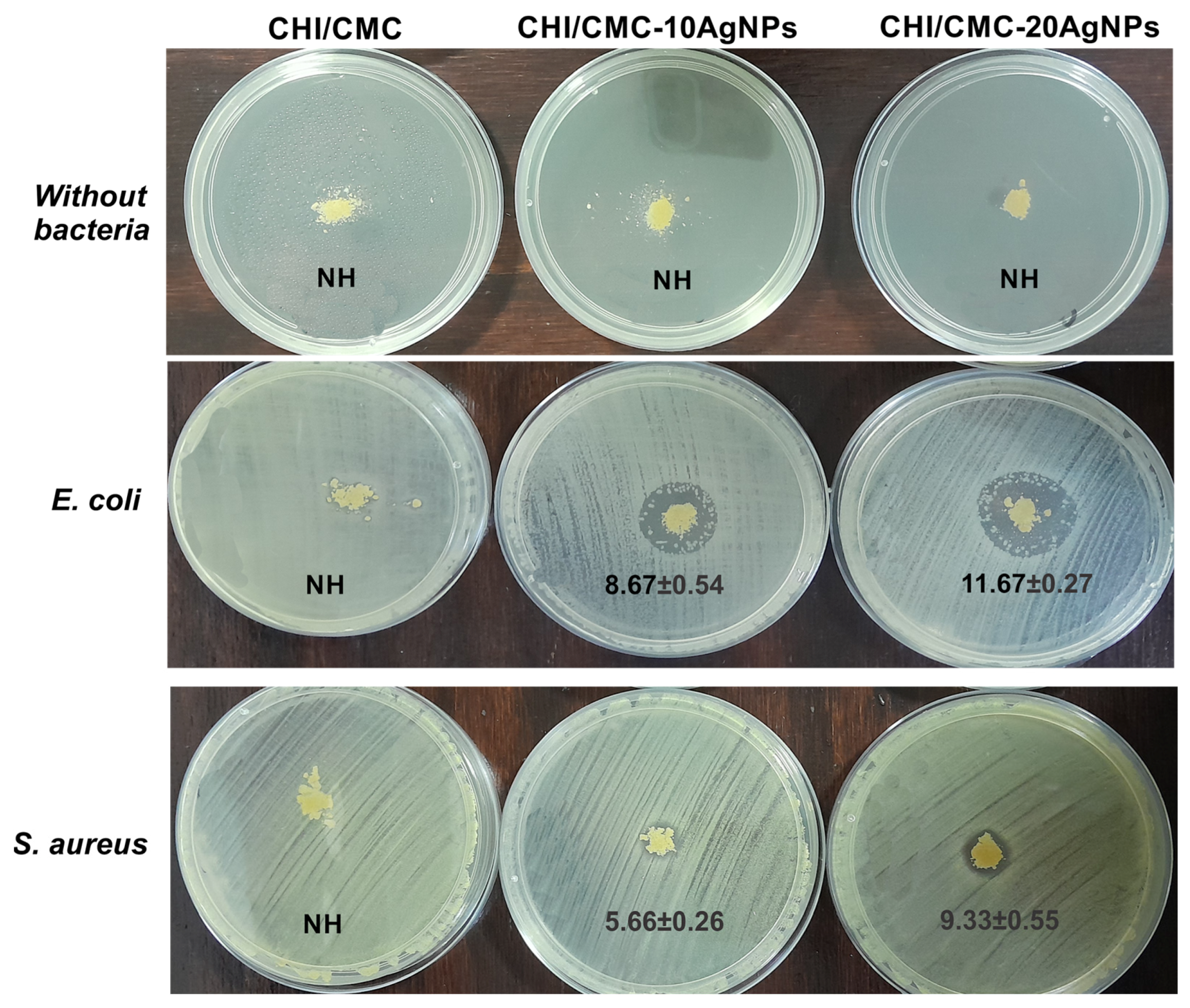
Bacterial suspensions of E. coli (ATCC 8739) and S. aureus (ATCC 6538) were adjusted to the 0.5 McFarland standard (~1 × 10⁸ CFU/mL) and uniformly spread onto the agar surface using sterile cotton swabs. Subsequently, 10 mg of each microgel formulation was carefully deposited directly onto the surface of the inoculated agar. Plates were incubated at 35 ± 2 °C for 24 h. Antibacterial activity was determined by measuring the diameter of the inhibition zones (in mm) using a digital caliper. Each test was performed in triplicate, and the results are expressed as mean ± SEM. NH denotes “no halo” of inhibition.

2.8. Hemostatic Assays

2.8.1. Hemolysis Test

The hemocompatibility of CHI/CMC and CHI/CMC-AgNPs microgels was evaluated by a hemolysis test (ASTM-F756-00) using red blood cells from voluntary donors. First, whole blood was centrifuged at 1500 rpm for 15 min to precipitate erythrocytes, and the supernatant was discarded. Immediately, erythrocytes were diluted at 2% v/v with physiological solution. Afterwards, 15 mg of each microgel was weighed and placed on a 24-well plate and 1.5 mL of erythrocyte solution was seeded to each well. Erythrocyte solutions without microgels were employed as the negative control (ABSn), while erythrocytes diluted at 2% with distilled water were used as the positive control (ABSp). All samples were incubated at 37 °C for 1 h to evaluate hemocompatibility. After that, the suspensions of microgels swelled with erythrocyte solutions were centrifuged at 500× g for 5 min. The supernatant was collected and used to determine the absorbance at 540 nm using the UV–Vis equipment (UNICO). Finally, hemolysis percentage was calculated using Equation (5):
H e m o l y s i s   % = ( A B S s A B S p ) A B S p A B S n   ×   100    
where ABSs is the absorbance of the microgels samples.

2.8.2. Blood Clotting Index

For blood clotting study, whole blood with sodium citrate a(1)s anticoagulant was used, based on previous protocols [43]. In brief, 10 mg of CHI/CMC, CHI/CMC-10AgNPs, and CHI/CMC-20AgNPs microgels were placed in Falcon tubes and 250 µL of whole blood was dropped slowly onto each sample. Following, 25 µL of 0.2 M CaCl2 solution was added to trigger blood coagulation, and the samples were incubated for 5, 10, 15, and 30 min in a water shaker bath at 37 °C. After that, 10 mL of distilled water was added very carefully, without disturbing the clot formed by the microgels and blood, for hemolyzing the unstrapped red blood cells in the clot. Next, the samples were centrifuged at 100× g for 2 min, and the supernatant was decanted into a tube with additional 40 mL of distilled water. The blood clotting index (BCI) was estimated to measure the absorbance at 540 nm of the hemoglobin in the unclotted blood, following Equation (6):
B C I   % = A B S s A B S w b   ×   100      
where ABSs is the absorbance for the microgel samples and ABSwb is the absorbance corresponding to 250 µL of whole blood diluted in 50 mL of distilled water.

2.8.3. Coagulation Time

The coagulation test allows for estimating the coagulation time taken by the microgel/blood aggregate to form clots [44]. For this purpose, 10 mg of CHI/CMC, CHI/CMC-10AgNPs, and CHI/CMC-20AgNPs microgels were weighed and placed into Eppendorf tubes, and then 1 mL of whole blood was added with CaCl2 (final concentration = 10 mM) to start the coagulation process. Eppendorf tubes were manually inverted every 1 min and set up vertically on the Eppendorf rack. This procedure was repeated until the blood/microgel aggregate completely ceased to flow (clotting process was finished), and the time was recorded. As a control group, the coagulation time of whole blood with CaCl2 and without microgels was measured. All samples were measured in triplicate to verify the data, and the outcomes were expressed as percentage relative to the control group.

2.9. Statistical Analysis and Data Processing

All trials described before were performed at least in triplicate to ensure the reliability of the results. The results are expressed as mean ± SEM and statistical differences were analyzed through ANOVA followed by the Multiple Range Test of Bonferroni using the SigmaPlot 12.0 software.

3. Results and Discussion

3.1. Silver Nanoparticles: Green Synthesis and Characterization

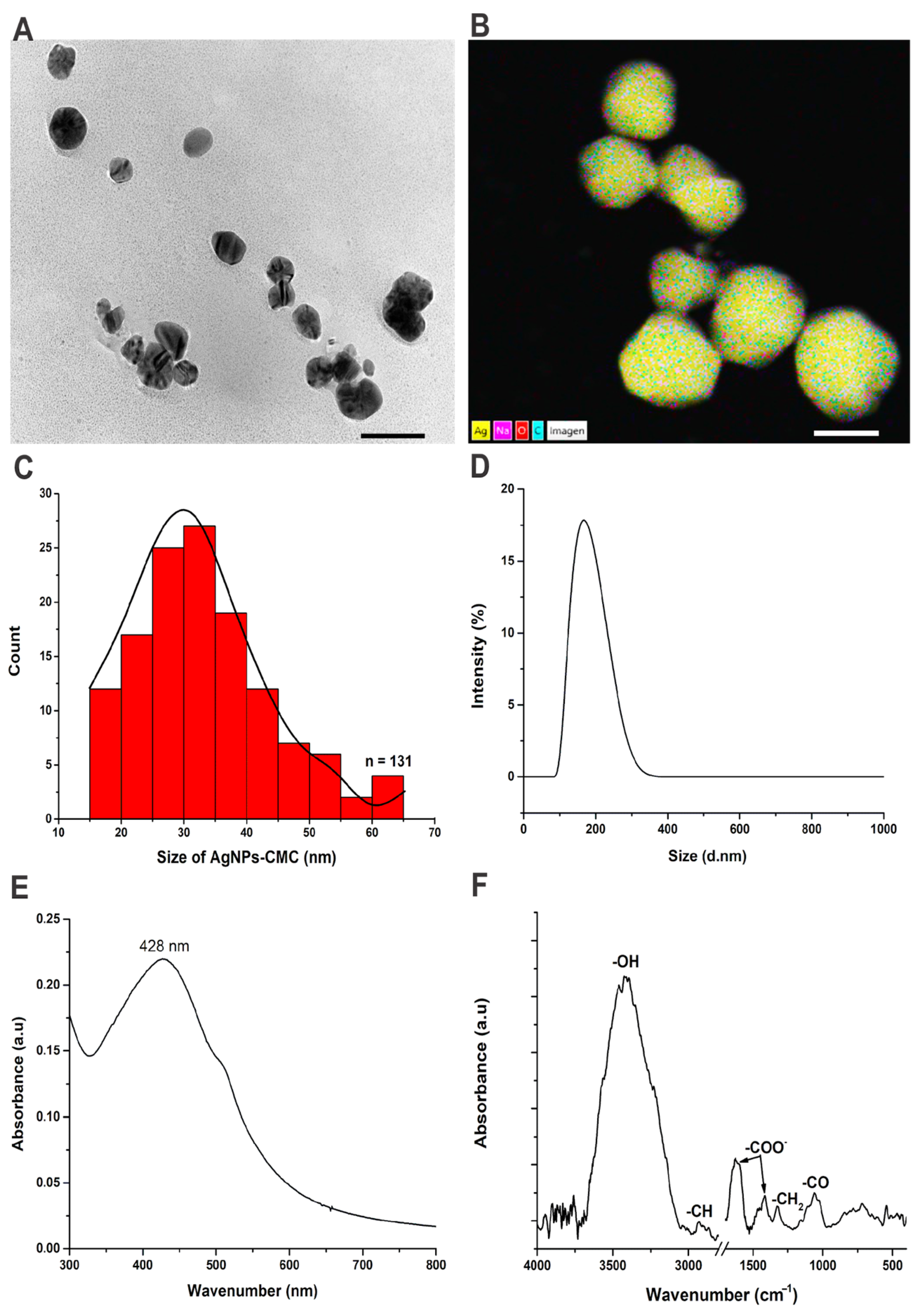
Synthesis of AgNPs by chemical reduction is the most common and conventional method to obtain them. However, in recent years, green nanotechnology has emerged as an ecofriendly process to fabricate nanoparticles, reducing the emergent risks of traditional nanotechnologies [45,46]. Green nanotechnology proposes replacing toxic and contaminant reagents by natural compounds such as phytocompounds and biopolymers to obtain AgNPs. The use of natural polymers including CHI and CMC to synthesize AgNPs has been widely reported as an easy, simple, and successful eco-friendly method [47,48,49]. The typical process involves the reduction and nucleation of Ag+ ions to form Ag0 nanostructures with different sizes and shapes using natural compounds as reducing and stabilizing agents. In this work, AgNPs with CMC as a natural reducing and stabilizing agent were successfully synthesized after 90 min of reaction at 95 ± 5 °C. The formation of ecofriendly AgNPs was detected by the changes in the color of the synthesis solution during the reaction: (1) after 15 min, the colloidal suspension takes a pale-yellow color, (2) next the color turned yellow until 30 min of reaction, to finally (3) acquire an orange-brown color (see Video S1 in Supplementary Information). The morphology and size distribution were evaluated by TEM images (Figure 2A–C), demonstrating that AgNPs-CMC has an oval-spheric form with a unimodal size distribution. The frequency histogram showed that AgNPs-CMC presented an average size of 33.50 ± 0.94 nm (Figure 2C). Also, for EDS analysis, signals of Ag (81.50 ± 1.00%), C (16.85 ± 1.50%), O (1.30 ± 0.20%), and Na (0.35 ± 0.05%) were detected in AgNPs-CMC (Figure 2B), indicating the presence of CMC as a capping agent. In addition, CMC as a stabilizer and capping agent was confirmed by the FTIR spectrum of AgNPs-CMC (Figure 2F). Typical peaks of carboxyl groups of CMC were detected at 1601 cm−1 (asymmetric stretch) and 1414 cm−1 (symmetric stretch), while the -OH, -CH, CH2, and -CO groups corresponding also to CMC were identified at 3425, 2927, 1324, and 1060 cm−1 respectively [50].
In addition, AgNPs-CMC showed an average hydrodynamic size of 168 ± 17 nm and 159 ± 14 nm, as determined by DLS intensity distribution and Z-average, respectively. These sizes are larger than the average size calculated by TEM, which can be explained by the fact that, as shown in Figure 2A, the AgNPs-CMC form clusters consisting of two or more nanoparticles. The intensity distribution profile was monomodal (Figure 2D), indicating that AgNPs-CMC had low size variability. This can be seen in the value of PDI (0.210 ± 0.025) since values close to 0.2–0.3 indicate nanoparticles with monodispersed size [51]. Additionally, the AgNPs-CMC colloidal suspension showed a negative surface charge determined by a negative zeta-potential value (−72.5 ± 3.6 mV). Therefore, it was possible to incorporate them into CMC solutions (also negative) to form PECs with CHI, as described in Section 3.2. The hydrodynamic size was also determined by NTA, with an average size of 135 ± 24 nm. This technique also allowed for the calculation of AgNPs concentration, which was (2.24 ± 0.01) × 1012 nanoparticles/mL. A video of AgNPs-CMC and their Brownian motion observed by NTA technique as provided in Supplementary Information (Video S2).
Finally, AgNPs-CMC presented a strong peak at 428 nm, as determined by UV-vis, which is attributed to the SPR phenomenon (Figure 2E). These results are consistent with other reports of green synthesis of AgNPs with CMC as a reducing agent [40,49,52].

3.2. CHI/CMC Microgel Formulation: Synthesis and Reaction Yield

Spray drying is a key and scalable method used to fabricate solid particles and microparticles in the pharmaceutical and food industry. Furthermore, this technique offers an efficient procedure to encapsulate natural compounds, essential oils, and other agents due to the spray dried process effectively minimizing evaporation and allowing for control of the encapsulation into the wall material employed [22]. The use of biopolymers such as CHI or CMC as wall material to engineer microparticles or microspheres by spray drying for drug delivery applications has been reported [53,54]. In addition, AgNPs are successfully encapsulated in CHI-microspheres via the spray drying technique [55]. Nevertheless, the specific production of powdered microgels based on PECs and AgNPs with hemostatic properties using the spray drying technique has only been investigated a little. In this work, PEC microgel powders of CHI/CMC, CHI/CMC-10AgNPs, and CHI/CMC-20AgNPs at pH 3.5 were successfully obtained by spray drying technique. Before spray drying, superficial charges of CHI, CMC and CMC-AgNPs were determined for zeta-potential values to evaluate the electrostatic interactions between them. It is known that CHI is a cationic linear polymer obtained after partial deacetylation of chitin, while CMC is an anionic polymer derived from cellulose [56]. Thus, CHI showed a positive charge of +20.5 ± 3.5 mV due to positive amine groups, while CMC presented a negative value of −13.6 ± 2.2 mV owing to carboxyl groups. The colloidal AgNPs-CMC suspension also exhibited a negative charge; thus, these were added into the CMC solution to form CHI/CMC-AgNPs microgels. The zeta-potential values for the CMC solution with AgNPs incorporated were also determined. In solutions of CMC-10AgNPs and CMC-20AgNPs (polymers + nanoparticles), the zeta-potential exhibited a slight increase (−14.8 ± 1.0 mV and −15.5 ± 1.5 mV, respectively) due to the AgNPs, compared to the CMC polymer solution (−13.6 ± 2.2 mV). Considering that the AgNPs-CMC had a concentration of (2.24 ± 0.01) × 1012 nanoparticles/mL, CHI/CMC-10AgNPs and CHI/CMC-20AgNPs were prepared with approximately 2.24 × 10¹³ and 4.48 × 10¹³ nanoparticles in total, respectively. Furthermore, after spray drying the different evaluated PECs, the reaction yield was theoretically calculated by Equation (1). CHI/CMC microgels exhibited a reaction yield of 32 ± 2%, while in the presence of AgNPs, the reaction yield experienced a slight increase (37 ± 5 and 36 ± 7% to CHI/CMC-10AgNPs and CHI/CMC-20AgNPs, respectively). Some authors suggest that a reaction yield of around 50% is an excellent result for spray drying [57]. Therefore, a reaction yield of around 32–37% for our microgels is an acceptable performance at the laboratory scale.

3.3. Morphological, Physicochemical, and Thermal Characterization of CHI/CMC and CHI/CMC-AgNPs Microgels

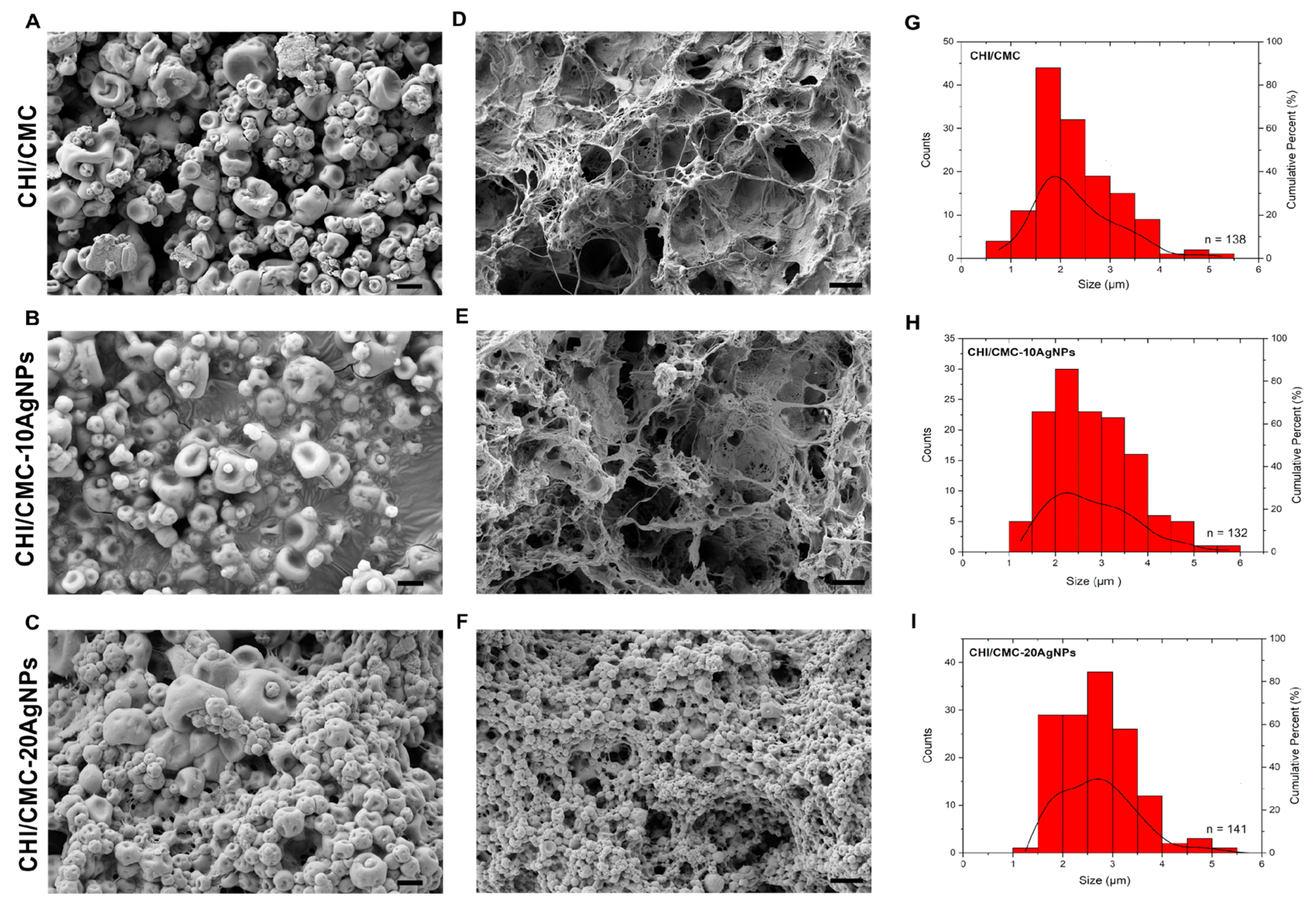
All the created microgels presented macroscopically a fine granular powder appearance (Figure S3). The CHI/CMC microgels displayed an ivory-white color, whereas the CHI/CMC-10AgNPs and CHI/CMC-20AgNPs microgels presented a faint orange color due to the presence of the nanoparticles. On the other hand, the shape and size distribution of the dried microgels were investigated by FE-SEM images (Figure 3). The results revealed that CHI/CMC and CHI/CMC-10AgNPs microgels presented irregular spherical shapes with the center flattened (Figure 3A,B), while CHI/CMC-20AgNPs samples exhibited oval-spherical shapes (Figure 3C). All engineered microgels showed a monodisperse size distribution with average sizes of 2.30 ± 0.07 µm (Figure 3G), 2.78 ± 0.08 µm (Figure 3H), and 2.69 ± 0.06 µm (Figure 3I) for CHI/CMC, CHI/CMC-10AgNPs, and CHI/CMC-20AgNPs, respectively. These results indicated that AgNPs-CMC slightly increased the average size of CHI/CMC microgels and they also raised the dispersal of microgel sizes, as seen in the frequency histogram plot for CHI/CMC-10AgNPs and CHI/CMC-20AgNPs samples (Figure 3H,I, respectively). Additionally, EDS-TEM chemical analysis reveals the presence of AgNPs-CMC in CHI/CMC-10AgNPs and CHI/CMC-20AgNPs microgels with a percentage of 0.60 ± 0.11% and 0.80 ± 0.13% per image, respectively.
On the other hand, microgel powders exhibit rapid swelling in aqueous media, so it was difficult to determine the sizes using swollen microgels in the particle size analyzers. However, the swollen microgels can be analyzed by FE-SEM images, after lyophilization. As observed in Figure 3D,E, swollen CHI/CMC and CHI/CMC-10AgNP microgels presented a complete modification of their structure due to the addition of water inside them. The irregular spherical shape of the dry microgels in contact with the liquid produced the union between them and formed solid microaggregates with the aspect of a big network with big pores capable of retaining liquid inside. Similar behavior was described to powdered microgels fabricated by spray drying using gelatin and chondroitin sulfate as PECs [25]. In contrast, the structure of the swollen CHI/CMC-20AgNP microgels did not show such noticeable changes, forming a less developed and porous network (Figure 3F). This difference likely results from the higher concentration of silver nanoparticles, which could hinder swelling and lead to a more compact microgel matrix. The SEM images for swollen CHI/CMC-20AgNPs microgels suggest that the increased nanoparticle load might reduce water uptake during swelling, thus affecting the internal structure and porosity observed.
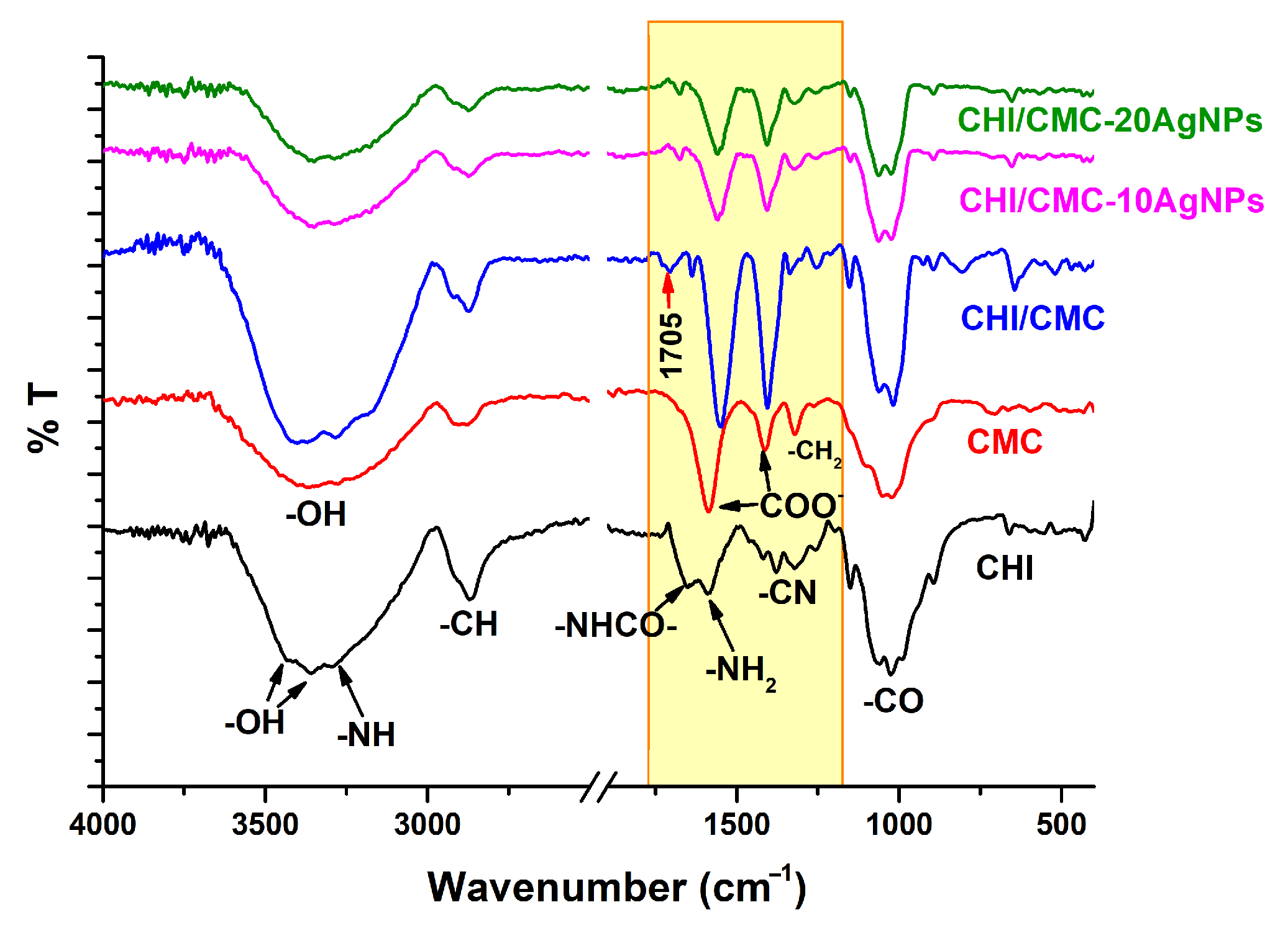
In addition, the analysis of the ATR-FTIR spectra of the CHI/CMC and CHI/CMC-AgNPs microgels demonstrates that the PEC microgels are formed by electrostatic interactions between amine groups of CHI and carboxyl groups of CMC (Figure 4). On the one hand, pure solid CHI presented typical peaks at 3450–3360, 3280, 2870, 1655, 1585, 1370, and 1060–980 cm−1 associated with -OH, -NH, -CH, -NHCO-, -NH2, -CN, and -CO functional groups, respectively; while pure solid CMC showed signals at 3340, 2890, 1595, 1414, 1320, and 1100–990 cm−1 corresponding to -OH, -CH, asymmetric COO-, symmetric COO-, -CH2, and -CO groups [5,50]. On the other hand, the spectra for CHI/CMC, CHI/CMC-10AgNPs, and CHI/CMC-20AgNPs microgels did not present significant changes in the signals associated with the -OH, -CH, and -CO groups. However, a shift in the signals of the amino groups of CHI and the carboxyl groups of CMC was observed, indicating the interaction of these functional groups to form PECs (Figure 4, yellow-shaded region), as also observed for CHI/CMC hydrogels and microparticles [53,58,59]. Detailly, in CHI/CMC, CHI/CMC-10AgNPs, and CHI/CMC-20AgNPs microgels, the signal at 1655 cm−1 (-NHCO) is very weak and the peak at 1585 cm−1 (-NH2) is missing, while the signals associated with the COO- group (1595 and 1414 cm−1) are shifted. This suggests that amine and carboxyl groups electrostatically interacted to form PEC microgels. In addition, in the CHI/CMC spectrum, a new peak at 1705 cm−1 was observed and this signal was previously attributed to the electrostatic interaction between CHI and CMC to form PEC hydrogels [53,58]. Nonetheless, the peak at 1705 cm−1 is missing in the CHI/CMC-10AgNPs and CHI/CMC-20AgNPs spectra, indicating that AgNPs-CMC affected the electrostatic interaction of the CHI/CMC matrix.
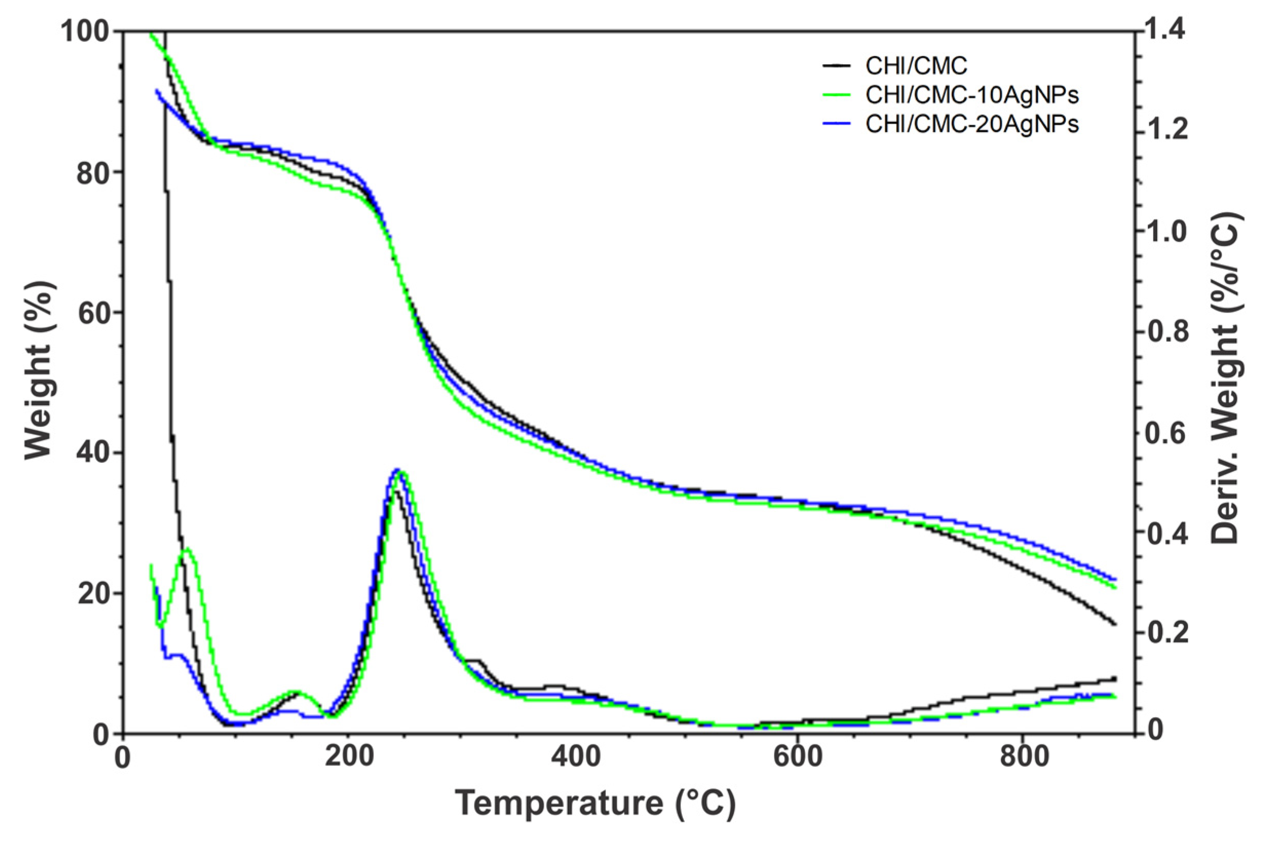
The thermal behavior was assessed by a TGA test, and the results are summarized in Table 1. All microgels experienced three or four stages of degradation (Figure 5); however, the maximum weight loss occurs at a maximum temperature determined as Tp. As observed in Table 1, AgNPs-CMC increased the Tp values, being noticeably higher in CHI/CMC-10AgNPs samples. Surprisingly, CHI/CMC-20AgNPs microgels exhibited similar Tp and weight loss values than CHI/CMC microgels, which can be explained due to the lower percentage of the gel fraction (cross-linking degree) determined by these samples (see Section 3.4). In addition, the residual mass percentage was measured and CHI/CMC showed higher degradation than CHI/CMC-10AgNPs and CHI/CMC-20AgNPs microgels, indicating that AgNPs-CMC increased the thermal stability and decreased the degradation of the microgels.

3.4. Swelling, Gel Fraction, and Biodegradation of Microgels

Hydrogels and microgels have a swelling capacity when they are hydrated in aqueous media. It is known that this swelling capacity is affected by the pH of the medium and polymer concentration [59]. The electrostatic interaction between the chains of polymers in PEC microgels or hydrogels also impacts the swelling; thus, the cross-linking between polycation and polyanion plays a crucial role [60]. To determine the cross-linking between CHI-CMC and CHI-CMC-AgNPs, a gel fraction test was performed. The results reveal that CHI/CMC, CHI/CMC-10AgNPs, and CHI/CMC-20AgNPs showed a GF% of 68.40 ± 1.48%, 83.38 ± 1.47%, and 55.49 ± 3.24%, respectively. These outcomes indicate that AgNPs modified the electrostatic interactions between CHI and CMC, as was also verified by the ATR-FTIR spectra. Additionally, these findings suggest that AgNPs-CMC at lower concentrations promote PEC microgel formation, possibly acting as nanoreinforcement and increasing the cross-linking and electrostatic interactions between CHI and CMC. At higher concentrations, however, AgNPs-CMC appears to interfere with these interactions, negatively impacting PEC microgel formation. On the other hand, to evaluate if GF% affects the swelling behavior of the microgels, swelling capacity was evaluated in PBS solutions adjusted to a physiological pH of 7.4 (mimicking plasma and blood conditions) and a pH of 6.0 (representative of the acidic microenvironment commonly observed in skin wounds) [41]. The swelling results are presented in Figure 6. At both pH values assessed, all samples experienced higher swelling from t = 1 min, reaching between 500 and 700%. The CHI/CMC and CHI/CMC-10AgNPs microgels showed similar swelling behavior at pH 7.4 and 6.0, attaining the maximum level of swelling at 30 min and then experiencing a slight decrease in swelling to 120 min. However, CHI/CMC-10AgNPs microgels showed a superior swelling capacity (approx. 100% more) than CHI/CMC samples in both pH values assessed in all time ranges evaluated. These outcomes demonstrated that AgNPs increased the swelling behavior of CHI/CMC, acting as nanoreinforcement to form composite micromaterials, according to the results of the GF% assay. On the other hand, CHI/CMC-20AgNPs samples exhibited a different performance. At pH 7.4, they reached maximum swelling at 15 min, but then experienced an abrupt drop in swelling, ending at 120 min with percentages lower than those reached initially (t = 1 min). This behavior showed that the CHI/CMC-20AgNPs microgels begin to de-swell immediately after reaching their maximum swelling, suggesting that they degrade rapidly, probably due to the low level of cross-linking demonstrated by their GF%. In contrast, at pH 6.0, the CHI/CMC-20AgNPs microgels showed stable swelling over time, but it was considerably lower compared to CHI/CMC and CHI/CMC-10AgNPs samples. The differences in swelling behavior at pH 7.4 and pH 6.0 of the CHI/CMC-20AgNPs microgels could be explained by the interactions between AgNPs and the polymer matrices, particularly their influence on the electrostatic interactions between CHI and CMC [61]. At pH 7.4, the increased concentration of AgNPs in CHI/CMC-20AgNPs appears to promote stronger interactions with the polymer chains, possibly through coordination or physical cross-linking, resulting in a more stabilized and less flexible network that resists water absorption and thus exhibits reduced swelling. Additionally, at this pH, chitosan’s amino groups are partially deprotonated, which diminishes electrostatic repulsion within the network and may further contribute to compaction. Conversely, at pH 6.0, more amino groups on CHI are protonated, increasing electrostatic repulsion and favoring higher swelling. The presence of AgNPs at this pH does not significantly hinder swelling, likely because their interactions with the polymer are weaker or different in nature, maintaining the network’s flexibility and allowing for water uptake to be similar to the unmodified CHI/CMC microgels. Overall, the effect of AgNPs on swelling is pH-dependent, with their capacity to reinforce or stabilize the network being more pronounced at neutral pH, whereas at acidic pH, increased protonation of functional groups diminishes these interactions, permitting higher swelling capacity [62].
For hemostatic agents, water absorption is a key parameter to be evaluated since faster swelling helps to concentrate clotting factors, forming blood clots quickly and enhancing hemostasis time [63]. Compared with other hemostatic materials like hydrogels and sponges, powdered microgels have a considerable surface area which increases their capacity to absorb water and also enhances adherence to the injury [64]. As we demonstrated, solid microgels immediately swell when immersed in PBS solutions and then gradually increase the swelling capacity over time toward the saturation or equilibrium rate. According to our results, similar behavior was previously reported for other types of microgels [5,44]. In summary, swelling results suggest that CHI/CMC and CHI/CMC-10AgNPs microgels presented an acceptable performance in both pH evaluated to be applied as hemostatic materials.
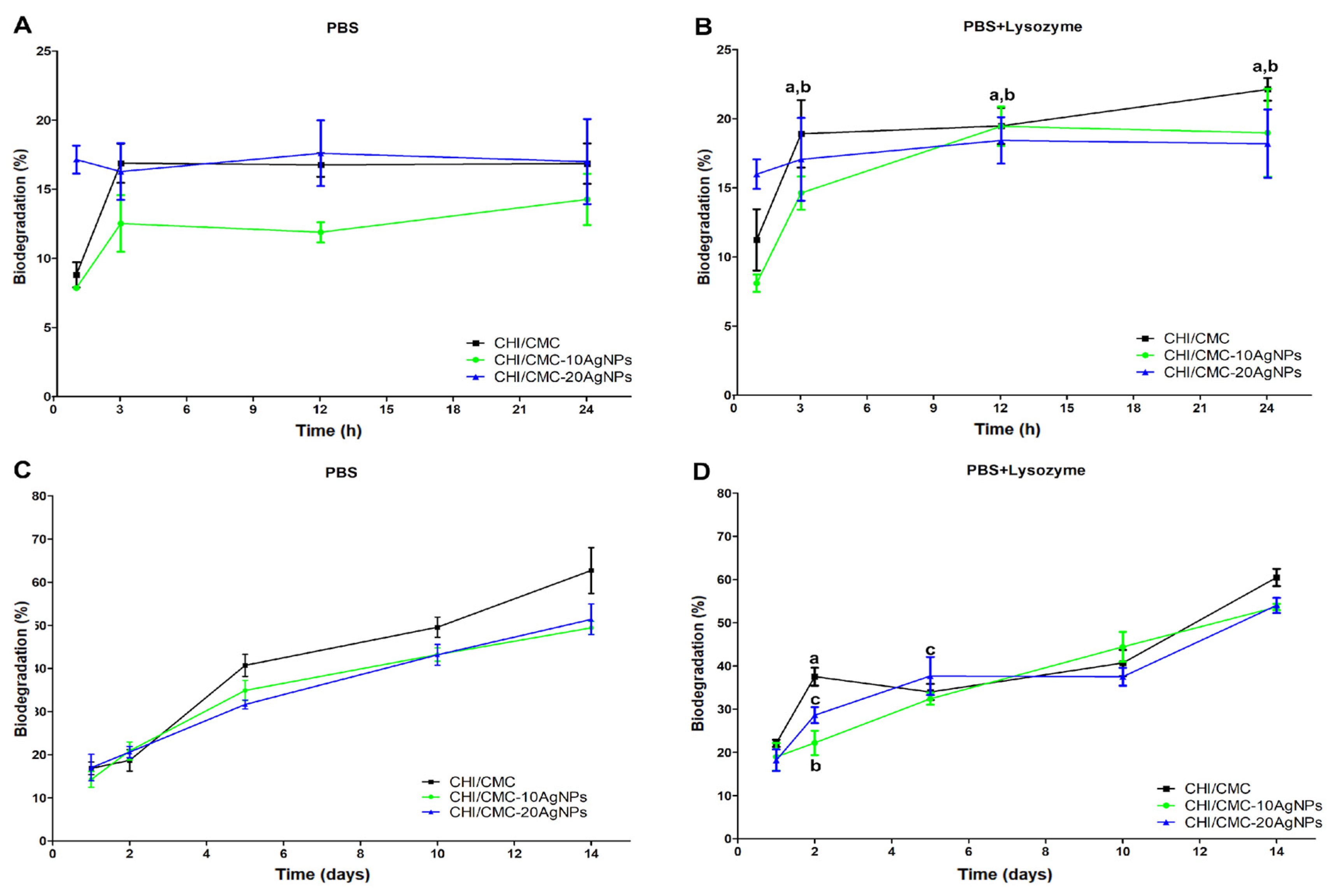
It is known that CHI and CMC are biodegradable and non-cytotoxic biopolymers; therefore, they are attractive to use to fabricate biomaterials. The in vitro biodegradation of hydrogels or microgels based on biopolymers is usually evaluated using a lysozyme as a catalytic agent [8]. In the human blood plasma, the concentration of lysozyme is in the range of 7–13 mg/L [65]. Thus, we evaluated the biodegradation of microgels in PBS solutions at physiological pH (7.4) without and with lysozyme at 13 mg/L (Figure 7). The biodegradation of the microgels was also assessed for a short time (1–24 h) and a long time (1–14 days). The results showed that all microgels began to degrade after 1 h of incubation; non-biodegradation was found at lower times. As seen in Figure 7A,B, CHI/CMC-20AgNPs microgels exhibited higher degradation compared to the other microgels in the short time period, according to the GF% and swelling results for these microgels. The presence of lysozyme increased the biodegradability of CHI/CMC and CHI/CMC-10AgNPs samples after 3 h of incubation (Figure 7B). In the long time period evaluation, all microgels presented continuous degradation over time in PBS solution without lysozyme, reaching at 14 days approximately 60 and 50% for CHI/CMC and both CHI/CMC-AgNPs samples, respectively (Figure 7C). As previously demonstrated by TGA, AgNPs-CMC reduced the degradation of microgels, suggesting that these nanomaterials enhanced physicochemical properties. As is consistent, when lysozyme was employed in long duration treatments, a significant increase in the biodegradability of all microgels was found at 2 days, but CHI/CMC microgels were the most affected (Figure 7D). However, at 14 days, similar lysozyme biodegradation was verified compared to the same samples in PBS without enzyme. All these results indicated that the microgels presented good biodegradation in physiological conditions, making them suitable to be applied in hemostatic treatments [8,66,67,68].

3.5. Antimicrobial Assays

AgNPs are recognized for their antibacterial activity against Gram-positive and Gram-negative bacteria [20,46,69]. The antimicrobial activity of AgNPs is attributed to their nanometric size and the capacity to release biocidal Ag+ in biological environments, which can trigger metabolic alterations and lead to bacterial death. Presently, AgNPs are considered the most promising nanomaterial to combat and kill multidrug-resistant bacteria that are unresponsive to traditional therapies [70]. In addition, their potent antibacterial activity is more efficient than conventional antibiotics, preventing microorganisms from colonizing wounds [18]. Consequently, hydrogels, films, and scaffolds designed by combining cross-linked polymers with AgNPs have emerged as a novel alternative of biomaterials for skin tissue engineering, drug delivery, and wound healing. In this work, we developed a novel powdered microgel loaded with green AgNPs as a new generation of hemostatic biomaterials. Their antibacterial activity was tested by measuring the zones of inhibition produced by the microgels on Petri dishes against S. aureus and E. coli, as representative models of Gram-positive and Gram-negative bacteria, respectively. The results are shown in Figure 8. First, the sterility of the microgels was assessed, and the outcomes revealed that they had no bacterial contamination due to their manufacturing and storage. On the other hand, CHI/CMC microgels did not show any zone of inhibition against either of the bacteria tested, indicating that they did not exhibit antibacterial activity. Nevertheless, CHI/CMC-10AgNPs and CHI/CMC-20AgNPs exhibited antimicrobial activity against both bacteria species, with E. coli being more susceptible to AgNPs-CMC than S. aureus. The average inhibition zone measured against S. aureus was 5.66 ± 0.26 mm for CHI/CMC-10AgNPs and 9.33 ± 0.55 mm for CHI/CMC-20AgNPs. The difference between groups was statistically significant (p < 0.05), indicating enhanced antimicrobial efficacy in CHI/CMC-20AgNPs. The average inhibition zone against E. coli was 8.33 ± 0.54 mm for CHI/CMC-10AgNPs microgels and 11.67 ± 0.27mm for CHI/CMC-20AgNPs. Statistical analysis showed a significant difference between the groups (p < 0.05), indicating greater antimicrobial activity in CHI/CMC-20AgNPs samples. Furthermore, CHI/CMC-20AgNPs produced a larger inhibition halo, which was associated with the higher amount of AgNPs incorporated into the microgel. Our results showed that E. coli was more susceptible to the action of AgNPs-CMC than S. aureus. The increased susceptibility of E. coli (Gram-negative) is likely due to its thinner peptidoglycan layer and more permeable outer membrane [71,72]. It is important to note that CHI/CMC-10AgNPs and CHI/CMC-20AgNPs microgels incubated with E. coli showed some bacterial colonies growing within the inhibition zone. Therefore, the inhibition measurements were reported, excluding areas where the colonies were present. The presence of isolated colonies within the inhibition zone of CHI/CMC-10AgNPs and CHI/CMC-20AgNPs may reflect a pseudo-halo effect, caused by limited local diffusion or insufficient AgNPs release from the solid matrix, an effect commonly observed in particulate antimicrobial systems [73]. Our outcomes confirmed the successful encapsulation of AgNPs in CHI/CMC microgels via the spray drying technique, and conferred antimicrobial activity on them, according to previous reports [42,55]. In addition, the results suggest that AgNPs are the dominant contributor to the observed inhibition halos, particularly due to the concentration-dependent increase in activity. CHI/CMC microgels without AgNPs did not produce any measurable inhibition zones under the same conditions, supporting the idea that AgNPs are the primary antimicrobial agents in this system. However, CHI may enhance the overall activity by disrupting bacterial membranes and facilitating AgNPs interaction with microbial cells, as was previously reported [74].

3.6. Hemostatic Capacity of Microgels

As mentioned before, CHI, CMC, and other biopolymers are attractive platforms to obtain hemostatic biomaterials. The abilities and advantages of polysaccharides to fabricate biodegradable and non-cytotoxic hemostatic materials such as hydrogels, sponges, and microspheres were exhaustively summarized by Pourshahrestani et al. [6], Guo et al. [7], and Ren et al. [4], respectively. Nonetheless, the specific formulation of the hemostatic powdered microgels based on CHI/CMC-PECs by the spray drying technique has not been studied yet. However, a previous report has demonstrated that the combination of CHI and CMC presented suitable hemostatic properties: Kim et al. [26] developed CHI/CMC blend nonwovens with enhanced hemostasis compared to commercial nonwoven-type hemostatic agents. To evaluate the hemocompatibility of CHI/CMC and CHI/CMC-AgNPs microgels fabricated via spray drying, hemolysis, coagulation time, and BCI were determined, and the results are summarized in Table 2. All microgels engineered decreased the coagulation time by 20% in comparison with whole blood, indicating that dried microgels accelerated hemostasis time. The BCI percentage serves to establish the temporal variation in antithrombogenic activity upon exposure of whole blood to the samples. This parameter establishes a relationship between antithrombogenic activity and the concentration of free hemoglobin in the unclotted blood, as determined by absorbance [43]. A more pronounced decrease in absorbance over time corresponds to reduced clotting time and enhanced hemostatic efficacy of the biomaterials. Thus, lower BCI values suggest more hemostatic efficacy of microgels and more hemocompatibility too [43]. As observed in Table 2, the microgels considerably decreased BCI values at 5 min, indicating that microgels accelerate the coagulation process. After 10 min, BCI results were similar between the microgels and whole blood. However, considering all intervals of time assessed, the BCI test suggests that CHI/CMC and CHI/CMC-10AgNPs presented suitable results to reduce hemostasis time. Furthermore, hemolysis test was determined according to ASTM F-756-00, and the results revealed that CHI/CMC and CHI/CMC-10AgNPs microgels exhibited hemocompatibility, associated with a lower percentage of hemolysis. In contrast, CHI/CMC-20AgNPs samples showed higher hemolysis values; thus, these microgels are not hemocompatible, and their use as hemostatic agents could compromise the biosecurity of the material. Representative images of the hemolysis test and coagulation time results are presented in Figure S4.

4. Conclusions

We demonstrate that powdered PEC microgels based on CHI and CMC (the most abundant polysaccharides) can be obtained through a scalable spray drying technique. In addition, AgNPs synthesized by ecofriendly methods with CMC were successfully added into CHI/CMC microgels by spray drying. CHI/CMC and CHI/CMC-10AgNPs microgels exhibited better performance than CHI/CMC-20AgNPs for use as hemostatic biomaterials. Briefly, CHI/CMC and CHI/CMC-10AgNPs showed higher swelling capacity, gel fraction, and thermal stability and lower biodegradability between 1 and 3 h (maximum time which microgels will be in contact with bleeding wounds as hemostatic material). Furthermore, by SEM images, swollen CHI/CMC and CHI/CMC-10AgNPs form solid microaggregates with the aspect of a big network with big pores capable of retaining liquid inside, while CHI/CMC-20AgNPs presented a poor network, which is related to their swelling capacity, gel fraction, and biodegradability. Also, CHI/CMC-20AgNPs microgels showed high hemolysis and, therefore, lower hemocompatibility. However, CHI/CMC and CHI/CMC-10AgNPs microgels revealed hemocompatibility due to their lower hemolysis and higher decrease in coagulation time. In conclusion, CHI/CMC and CHI/CMC-10AgNP powdered microgels have emerged as novel and appropriate new generations of dried micromaterials to be used as hemostatic agents with suitable physicochemical and hemostatic properties.

Supplementary Materials

The following supporting information can be downloaded at: https://www.mdpi.com/article/10.3390/polysaccharides6030084/s1, Figure S1. Relation between reduced viscosity (ƞred) and concentration of CHI. R2 and intrinsic viscosity values are provided. Figure S2. Relation between reduced viscosity (ƞred) and concentration of CMC. R2 and intrinsic viscosity values are provided. Figure S3. Macroscopical images of (A) CHI/CMC, (B) CHI/CMC-10AgNPs and (C) CHI/CMC-20AgNPs microgels. Figure S4. (A) Hemolysis test at t = 0 min, (B) Hemolysis test after 1 h of incubation at 37 °C, (C) Hemolysis results after centrifugation (free hemoglobin is in the supernatant) and (D) Coagulation time test: microgels/whole blood aggregated forming clots. Figure S5. Representative images of BCI assay for (A) whole blood, (B) CHI/CMC, (C) CHI/CMC-10AgNPs and (D) CHI/CMC- 20AgNPs microgels. Table S1. Viscosity results of CHI determined by Ubbelohde at 25 °C. Table S2. Viscosity results of CMC determined by Ubbelohde at 25 °C. Table S3. Absorbance values at 1655 and 3450 cm−1 determined by FTIR. S = n° sample. Table S4. DD% estimated for CHI by FTIR and titration methods. Video S1. Synthesis process of AgNPs-CMC. Video S2. NTA video of AgNPs-CMC.

Author Contributions

Conceptualization, A.G. and J.S.G.; methodology, A.G., M.F., L.G. and J.S.G.; formal analysis, A.G., M.F., J.S.G., L.G. and V.A.A.; investigation, A.G. and J.S.G.; writing—original draft preparation, A.G.; writing—review and editing, A.G., M.F., J.S.G. and V.A.A.; supervision, J.S.G. and V.A.A.; project administration, J.S.G.; funding acquisition, J.S.G. and V.A.A. All authors have read and agreed to the published version of the manuscript.

Funding

This research was funded by the Universidad Nacional de Mar del Plata under Grant ING 710/24; and CONICET under Grant 0638.

Institutional Review Board Statement

The study was carried out in accordance with the World Medical Association Declaration of Helsinki: Ethical principles for medical research involving human subjects. All procedures were performed in compliance with relevant laws and institutional guidelines. Ethical approval for this study was obtained from the Research Ethics Committee of the Interdisciplinary Thematic Program in Bioethics (PTIB), affiliated with the Secretariat of Science and Technology of the National University of Mar del Plata, on 8 July 2024. The Research Ethics Committee of the PTIB is registered in the Provincial Registry of Research Ethics Committees, under the Central Ethics Committee in Research of the Ministry of Health of the Province of Buenos Aires. The protocol was registered under the number NO-2024-27,503,325-GDEBA-DPEGSFFMSALGP from La Plata, Buenos Aires, Argentina.

Data Availability Statement

The original contributions presented in this study are included in the article/Supplementary Material. Further inquiries can be directed to the corresponding author(s).

DURC Statement

The authors recognize that the developed materials, including the antimicrobial hemostatic microgels incorporating silver nanoparticles, have potential applications that could be misused in military or other dual-use contexts. However, the primary aim of this research is to advance public health by providing innovative, safe, and effective solutions for controlling bleeding and preventing wound infections, which can significantly improve clinical outcomes and save lives. The research and applications described in this study are aligned with ethical standards intended to benefit society and do not support any harmful or malicious intent. The potential benefits of these materials—including rapid hemostasis, antimicrobial activity, and biocompatibility—significantly outweigh the risks associated with their dual-use potential.

Acknowledgments

The authors especially thank Andres Torres Nicolini and Tobias Salinas Larrecharte for their technical assistance.

Conflicts of Interest

The authors declare no conflicts of interest.

Abbreviations

The following abbreviations are used in this manuscript:
CHIChitosan
CMCCarboxymethyl cellulose
PECsPolyelectrolyte complexes
AgNPsSilver nanoparticles
MvAverage molecular weight
DSDegree of substitution
DD%Degree of deacetylation
AgNPs-CMCSilver nanoparticles obtained with carboxymethyl cellulose
SPRSurface plasmon resonance
GFGel fraction
PBSPhosphate-buffered saline
BCIBlood clotting index

References

  1. Dorgalaleh, A.; Daneshi, M.; Rashidpanah, J.; Roshani Yasaghi, E. An Overview of Hemostasis. In Congenital Bleeding Disorders: Diagnosis and Management; Dorgalaleh, A., Ed.; Springer International Publishing: Cham, Switzerland, 2018; ISBN 978-3-319-76723-9. [Google Scholar]
  2. Kalkwarf, K.J.; Drake, S.A.; Yang, Y.; Thetford, C.; Myers, L.; Brock, M.; Wolf, D.A.; Persse, D.; Wade, C.E.; Holcomb, J.B. Bleeding to Death in a Big City: An Analysis of All Trauma Deaths from Hemorrhage in a Metropolitan Area during 1 Year. J. Trauma Acute Care Surg. 2020, 89, 716–722. [Google Scholar] [CrossRef]
  3. Queirós, E.C.; Pinheiro, S.P.; Pereira, J.E.; Prada, J.; Pires, I.; Dourado, F.; Parpot, P.; Gama, M. Hemostatic Dressings Made of Oxidized Bacterial Nanocellulose Membranes. Polysaccharides 2021, 2, 80–99. [Google Scholar] [CrossRef]
  4. Ren, Z.; Wang, Y.; Wu, H.; Cong, H.; Yu, B.; Shen, Y. Preparation and Application of Hemostatic Microspheres Containing Biological Macromolecules and Others. Int. J. Biol. Macromol. 2024, 257, 128299. [Google Scholar] [CrossRef] [PubMed]
  5. Huang, H.; Chen, H.; Wang, X.; Qiu, F.; Liu, H.; Lu, J.; Tong, L.; Yang, Y.; Wang, X.; Wu, H. Degradable and Bioadhesive Alginate-Based Composites: An Effective Hemostatic Agent. ACS Biomater. Sci. Eng. 2019, 5, 5498–5505. [Google Scholar] [CrossRef] [PubMed]
  6. Pourshahrestani, S.; Zeimaran, E.; Kadri, N.A.; Mutlu, N.; Boccaccini, A.R. Polymeric Hydrogel Systems as Emerging Biomaterial Platforms to Enable Hemostasis and Wound Healing. Adv. Healthc. Mater. 2020, 9, 2000905. [Google Scholar] [CrossRef]
  7. Guo, Y.; Xie, X.; Li, J.; Yao, S. Recent Advances in Natural Polysaccharide-Based Hemostatic Sponges: A Review. Polysaccharides 2025, 6, 25. [Google Scholar] [CrossRef]
  8. Shen, Z.-S.; Cui, X.; Hou, R.-X.; Li, Q.; Deng, H.-X.; Fu, J. Tough Biodegradable Chitosan–Gelatin Hydrogels via in Situ Precipitation for Potential Cartilage Tissue Engineering. RSC Adv. 2015, 5, 55640–55647. [Google Scholar] [CrossRef]
  9. Peniche, C.; Argüelles-Monal, W.; Goycoolea, F.M. Chapter 25—Chitin and Chitosan: Major Sources, Properties and Applications. In Monomers, Polymers and Composites from Renewable Resources; Belgacem, M.N., Gandini, A., Eds.; Elsevier: Amsterdam, The Netherlands, 2008; pp. 517–542. ISBN 978-0-08-045316-3. [Google Scholar]
  10. Yang, J.; Wang, Y.; Wei, A.; Peng, K.; Huang, R.; Wang, Z.; Ma, X.; Tian, Q.; Chen, Y. Polyelectrolyte Composite Hydrogels Based on a Derivative of Functional Dietary Fiber for Long-Term Gastric Retention and Drug Delivery. Compos. Part B Eng. 2024, 272, 111194. [Google Scholar] [CrossRef]
  11. Muzzio, N.E.; Pasquale, M.A.; Diamanti, E.; Gregurec, D.; Moro, M.M.; Azzaroni, O.; Moya, S.E. Enhanced Antiadhesive Properties of Chitosan/Hyaluronic Acid Polyelectrolyte Multilayers Driven by Thermal Annealing: Low Adherence for Mammalian Cells and Selective Decrease in Adhesion for Gram-Positive Bacteria. Mater. Sci. Eng. C 2017, 80, 677–687. [Google Scholar] [CrossRef] [PubMed]
  12. Yang, D.; Gong, L.; Li, Q.; Fan, B.; Ma, C.; He, Y.C. Preparation of a Biobased Polyelectrolyte Complex from Chitosan and Sodium Carboxymethyl Cellulose and Its Antibacterial Characteristics. Int. J. Biol. Macromol. 2023, 227, 524–534. [Google Scholar] [CrossRef]
  13. Tsai, R.Y.; Chen, P.W.; Kuo, T.Y.; Lin, C.M.; Wang, D.M.; Hsien, T.Y.; Hsieh, H.J. Chitosan/Pectin/Gum Arabic Polyelectrolyte Complex: Process-Dependent Appearance, Microstructure Analysis and Its Application. Carbohydr. Polym. 2014, 101, 752–759. [Google Scholar] [CrossRef]
  14. Fang, Y.; Guo, W.; Ni, P.; Liu, H. Recent Research Advances in Polysaccharide-Based Hemostatic Materials: A Review. Int. J. Biol. Macromol. 2024, 271, 132559. [Google Scholar] [CrossRef]
  15. Li, J.; Wu, X.; Wu, Y.; Tang, Z.; Sun, X.; Pan, M.; Chen, Y.; Li, J.; Xiao, R.; Wang, Z.; et al. Porous Chitosan Microspheres for Application as Quick In Vitro and In Vivo Hemostat. Mater. Sci. Eng. C 2017, 77, 411–419. [Google Scholar] [CrossRef]
  16. Jin, J.; Ji, Z.; Xu, M.; Liu, C.; Ye, X.; Zhang, W.; Li, S.; Wang, D.; Zhang, W.; Chen, J.; et al. Microspheres of Carboxymethyl Chitosan, Sodium Alginate, and Collagen as a Hemostatic Agent In Vivo. ACS Biomater. Sci. Eng. 2018, 4, 2541–2551. [Google Scholar] [CrossRef]
  17. Bi, Y.-G.; Lin, Z.-T.; Deng, S.-T. Fabrication and Characterization of Hydroxyapatite/Sodium Alginate/Chitosan Composite Microspheres for Drug Delivery and Bone Tissue Engineering. Mater. Sci. Eng. C 2019, 100, 576–583. [Google Scholar] [CrossRef]
  18. Antezana, P.E.; Municoy, S.; Pérez, C.J.; Desimone, M.F. Collagen Hydrogels Loaded with Silver Nanoparticles and Cannabis Sativa Oil. Antibiotics 2021, 10, 1420. [Google Scholar] [CrossRef]
  19. Liu, R.; Dai, L.; Si, C.; Zeng, Z. Antibacterial and Hemostatic Hydrogel via Nanocomposite from Cellulose Nanofibers. Carbohydr. Polym. 2018, 195, 63–70. [Google Scholar] [CrossRef] [PubMed]
  20. Arshad, F.; Naikoo, G.A.; Hassan, I.U.; Chava, S.R.; El-Tanani, M.; Aljabali, A.A.; Tambuwala, M.M. Bioinspired and Green Synthesis of Silver Nanoparticles for Medical Applications: A Green Perspective. Appl. Biochem. Biotechnol. 2024, 196, 3636–3669. [Google Scholar] [CrossRef] [PubMed]
  21. Mardani, M.; Siahtiri, S.; Besati, M.; Baghani, M.; Baniassadi, M.; Nejad, A.M. Microencapsulation of Natural Products Using Spray Drying; an Overview. J. Microencapsul. 2024, 41, 649–678. [Google Scholar] [CrossRef] [PubMed]
  22. Mohammed, N.K.; Tan, C.P.; Manap, Y.A.; Muhialdin, B.J.; Hussin, A.S.M. Spray Drying for the Encapsulation of Oils—A Review. Molecules 2020, 25, 3873. [Google Scholar] [CrossRef]
  23. Liang, Y.; Xu, C.; Liu, F.; Du, S.; Li, G.; Wang, X. Eliminating Heat Injury of Zeolite in Hemostasis via Thermal Conductivity of Graphene Sponge. ACS Appl. Mater. Interfaces 2019, 11, 23848–23857. [Google Scholar] [CrossRef]
  24. Wang, L.; You, X.; Dai, C.; Tong, T.; Wu, J. Hemostatic Nanotechnologies for External and Internal Hemorrhage Management. Biomater. Sci. 2020, 8, 4396–4412. [Google Scholar] [CrossRef]
  25. Ferrante, M.; Gallo, M.B.; Gende, L.B.; Consolo, V.F.; Álvarez, V.A.; González, J.S. Synthesis and Characterization of Gelatin/Chondroitin Sulfate Microgels with NaCl: Preliminary Research toward Wound Healing Applications. Int. J. Biol. Macromol. 2025, 290, 138953. [Google Scholar] [CrossRef]
  26. Kim, G.H.; Im, J.N.; Kim, T.H.; Lee, G.D.; Youk, J.H.; Doh, S.J. Preparation and Characterization of Calcium Carboxymethyl Cellulose/Chitosan Blend Nonwovens for Hemostatic Agents. Text. Res. J. 2018, 88, 1902–1911. [Google Scholar] [CrossRef]
  27. Qian, J.; Wang, X.; Chen, Y.; Mo, C.; Liang, C.; Guo, H. The Correlation of Molecule Weight of Chitosan Oligomers with the Corresponding Viscosity and Antibacterial Activity. Carbohydr. Res. 2023, 530, 108860. [Google Scholar] [CrossRef] [PubMed]
  28. Czechowska-Biskup, R.; Wach, R.A.; Rosiak, J.M.; Ulański, P. Procedure for Determination of the Molecular Weight of Chitosan by Viscometry. Prog. Chem. Appl. Chitin Its Deriv. 2018, 23, 45–54. [Google Scholar] [CrossRef]
  29. Roberts, G.A.F.; Domszy, J.G. Determination of the Viscometric Constants for Chitosan. Int. J. Biol. Macromol. 1982, 4, 374–377. [Google Scholar] [CrossRef]
  30. Gómez-Díaz, D.; Navaza, J.M. Rheology of Aqueous Solutions of Food Additives: Effect of Concentration, Temperature and Blending. J. Food Eng. 2003, 56, 387–392. [Google Scholar] [CrossRef]
  31. Eremeeva, T.E.; Bykova, T.O. SEC of Mono-Carboxymethyl Cellulose (CMC) in a Wide Range of pH; Mark–Houwink Constants. Carbohydr. Polym. 1998, 36, 319–326. [Google Scholar] [CrossRef]
  32. Singh, R.K.; Khatri, O.P. A Scanning Electron Microscope Based New Method for Determining Degree of Substitution of Sodium Carboxymethyl Cellulose. J. Microsc. 2012, 246, 43–52. [Google Scholar] [CrossRef]
  33. Liu, D.; Wei, Y.; Yao, P.; Jiang, L. Determination of the Degree of Acetylation of Chitosan by UV Spectrophotometry Using Dual Standards. Carbohydr. Res. 2006, 341, 782–785. [Google Scholar] [CrossRef] [PubMed]
  34. Sánchez-Machado, D.I.; López-Cervantes, J.; Escárcega-Galaz, A.A.; Campas-Baypoli, O.N.; Martínez-Ibarra, D.M.; Rascón-León, S. Measurement of the Degree of Deacetylation in Chitosan Films by FTIR, 1H NMR and UV Spectrophotometry. MethodsX 2024, 12, 102583. [Google Scholar] [CrossRef]
  35. Duarte, M.L.; Ferreira, M.C.; Marvão, M.R.; Rocha, J. An Optimised Method to Determine the Degree of Acetylation of Chitin and Chitosan by FTIR Spectroscopy. Int. J. Biol. Macromol. 2002, 31, 1–8. [Google Scholar] [CrossRef]
  36. Dutta, J. Priyanka A Facile Approach for the Determination of Degree of Deacetylation of Chitosan Using Acid-Base Titration. Heliyon 2022, 8, e09924. [Google Scholar] [CrossRef]
  37. Kasaai, M.R. A Review of Several Reported Procedures to Determine the Degree of N-Acetylation for Chitin and Chitosan Using Infrared Spectroscopy. Carbohydr. Polym. 2008, 71, 497–508. [Google Scholar] [CrossRef]
  38. Domard, A.; Rinaudo, M. Preparation and Characterization of Fully Deacetylated Chitosan. Int. J. Biol. Macromol. 1983, 5, 49–52. [Google Scholar] [CrossRef]
  39. Capanema, N.S.V.; Mansur, A.A.P.; Carvalho, S.M.; Mansur, L.L.; Ramos, C.P.; Lage, A.P.; Mansur, H.S. Physicochemical Properties and Antimicrobial Activity of Biocompatible Carboxymethylcellulose-Silver Nanoparticle Hybrids for Wound Dressing and Epidermal Repair. J. Appl. Polym. Sci. 2018, 135, 45812. [Google Scholar] [CrossRef]
  40. Filipe, V.; Hawe, A.; Jiskoot, W. Critical Evaluation of Nanoparticle Tracking Analysis (NTA) by NanoSight for the Measurement of Nanoparticles and Protein Aggregates. Pharm. Res. 2010, 27, 796–810. [Google Scholar] [CrossRef]
  41. Proksch, E. pH in Nature, Humans and Skin. J. Dermatol. 2018, 45, 1044–1052. [Google Scholar] [CrossRef]
  42. Ferrante, M.; Alvarez, V.A.; Gende, L.B.; Guerrieri, D.; Chuluyan, E.; Gonzalez, J.S. Polyelectrolyte Complexes Hydrogels Based on Chitosan/Pectin/NaCl for Potentially Wound Dressing: Development, Characterization, and Evaluation. Colloid Polym. Sci. 2024, 302, 1231–1245. [Google Scholar] [CrossRef]
  43. Archana, D.; Dutta, J.; Dutta, P.K. Evaluation of Chitosan Nano Dressing for Wound Healing: Characterization, in Vitro and in Vivo Studies. Int. J. Biol. Macromol. 2013, 57, 193–203. [Google Scholar] [CrossRef]
  44. Behrens, A.M.; Sikorski, M.J.; Li, T.; Wu, Z.J.; Griffith, B.P.; Kofinas, P. Blood-Aggregating Hydrogel Particles for Use as a Hemostatic Agent. Acta Biomater. 2014, 10, 701–708. [Google Scholar] [CrossRef] [PubMed]
  45. Nasrollahzadeh, M.; Sajjadi, M.; Sajadi, S.M.; Issaabadi, Z. Chapter 5—Green Nanotechnology. In Interface Science and Technology; Nasrollahzadeh, M., Sajadi, S.M., Sajjadi, M., Issaabadi, Z., Atarod, M., Eds.; An Introduction to Green Nanotechnology; Elsevier: Amsterdam, The Netherlands, 2019; Volume 28, pp. 145–198. [Google Scholar]
  46. Ahmad, S.; Munir, S.; Zeb, N.; Ullah, A.; Khan, B.; Ali, J.; Bilal, M.; Omer, M.; Alamzeb, M.; Salman, S.M.; et al. Green Nanotechnology: A Review on Green Synthesis of Silver Nanoparticles—An Ecofriendly Approach. Int. J. Nanomed. 2019, 14, 5087–5107. [Google Scholar] [CrossRef]
  47. Thakore, S. Role of Biopolymers in Green Nanotechnology; IntechOpen: London, UK, 2012. [Google Scholar]
  48. Abdel-Halim, E.S.; Alanazi, H.H.; Al-Deyab, S.S. Utilization of Hydroxypropyl Carboxymethyl Cellulose in Synthesis of Silver Nanoparticles. Int. J. Biol. Macromol. 2015, 75, 467–473. [Google Scholar] [CrossRef]
  49. Dara, P.K.; Mahadevan, R.; Digita, P.A.; Visnuvinayagam, S.; Kumar, L.R.G.; Mathew, S.; Ravishankar, C.N.; Anandan, R. Synthesis and Biochemical Characterization of Silver Nanoparticles Grafted Chitosan (Chi-Ag-NPs): In Vitro Studies on Antioxidant and Antibacterial Applications. SN Appl. Sci. 2020, 2, 665. [Google Scholar] [CrossRef]
  50. Wang, R.; Shou, D.; Lv, O.; Kong, Y.; Deng, L.; Shen, J. pH-Controlled Drug Delivery with Hybrid Aerogel of Chitosan, Carboxymethyl Cellulose and Graphene Oxide as the Carrier. Int. J. Biol. Macromol. 2017, 103, 248–253. [Google Scholar] [CrossRef]
  51. Danaei, M.; Dehghankhold, M.; Ataei, S.; Hasanzadeh Davarani, F.; Javanmard, R.; Dokhani, A.; Khorasani, S.; Mozafari, M.R. Impact of Particle Size and Polydispersity Index on the Clinical Applications of Lipidic Nanocarrier Systems. Pharmaceutics 2018, 10, 57. [Google Scholar] [CrossRef] [PubMed]
  52. Hebeish, A.A.; El-Rafie, M.H.; Abdel-Mohdy, F.A.; Abdel-Halim, E.S.; Emam, H.E. Carboxymethyl Cellulose for Green Synthesis and Stabilization of Silver Nanoparticles. Carbohydr. Polym. 2010, 82, 933–941. [Google Scholar] [CrossRef]
  53. Cerchiara, T.; Abruzzo, A.; Parolin, C.; Vitali, B.; Bigucci, F.; Gallucci, M.C.; Nicoletta, F.P.; Luppi, B. Microparticles Based on Chitosan/Carboxymethylcellulose Polyelectrolyte Complexes for Colon Delivery of Vancomycin. Carbohydr. Polym. 2016, 143, 124–130. [Google Scholar] [CrossRef] [PubMed]
  54. Halahlah, A.; Piironen, V.; Mikkonen, K.S.; Ho, T.M. Polysaccharides as Wall Materials in Spray-Dried Microencapsulation of Bioactive Compounds: Physicochemical Properties and Characterization. Crit. Rev. Food Sci. Nutr. 2023, 63, 6983–7015. [Google Scholar] [CrossRef] [PubMed]
  55. Tokárová, V.; Kašpar, O.; Knejzlík, Z.; Ulbrich, P.; Štěpánek, F. Development of Spray-Dried Chitosan Microcarriers for Nanoparticle Delivery. Powder Technol. 2013, 235, 797–805. [Google Scholar] [CrossRef]
  56. Altam, A.A.; Zhu, L.; Huang, W.; Huang, H.; Yang, S. Polyelectrolyte Complex Beads of Carboxymethylcellulose and Chitosan: The Controlled Formation and Improved Properties. Carbohydr. Polym. Technol. Appl. 2021, 2, 100100. [Google Scholar] [CrossRef]
  57. Tontul, I.; Topuz, A. Spray-Drying of Fruit and Vegetable Juices: Effect of Drying Conditions on the Product Yield and Physical Properties. Trends Food Sci. Technol. 2017, 63, 91–102. [Google Scholar] [CrossRef]
  58. Zhu, X.; Bao, L.; Wei, Y.; Ma, J.; Kong, Y. Removal of Toxic Indigo Blue with Integrated Biomaterials of Sodium Carboxymethyl Cellulose and Chitosan. Int. J. Biol. Macromol. 2016, 91, 409–415. [Google Scholar] [CrossRef] [PubMed]
  59. Elliott, J.E.; Macdonald, M.; Nie, J.; Bowman, C.N. Structure and Swelling of Poly(Acrylic Acid) Hydrogels: Effect of pH, Ionic Strength, and Dilution on the Crosslinked Polymer Structure. Polymer 2004, 45, 1503–1510. [Google Scholar] [CrossRef]
  60. De Yao, K.; Liu, J.; Cheng, G.X.; Lu, X.D.; Tu, H.L.; Da Silva, J.A.L. Swelling Behavior of Pectin/Chitosan Complex Films. J. Appl. Polym. Sci. 1996, 60, 279–283. [Google Scholar] [CrossRef]
  61. De Piano, R.; Caccavo, D.; Barba, A.A.; Lamberti, G. Swelling Behavior of Anionic Hydrogels: Experiments and Modeling. Gels 2024, 10, 813. [Google Scholar] [CrossRef]
  62. Wang, R.; Cheng, C.; Wang, H.; Wang, D. Swollen Hydrogel Nanotechnology: Advanced Applications of the Rudimentary Swelling Properties of Hydrogels. ChemPhysMater 2024, 3, 357–375. [Google Scholar] [CrossRef]
  63. Neuffer, M.C.; McDivitt, J.; Rose, D.; King, K.; Cloonan, C.C.; Vayer, J.S. Hemostatic Dressings for the First Responder: A Review. Mil. Med. 2004, 169, 716–720. [Google Scholar] [CrossRef][Green Version]
  64. Sun, X.; Fang, Y.; Tang, Z.; Wang, Z.; Liu, X.; Liu, H. Mesoporous Silica Nanoparticles Carried on Chitosan Microspheres for Traumatic Bleeding Control. Int. J. Biol. Macromol. 2019, 127, 311–319. [Google Scholar] [CrossRef]
  65. Pankratov, T.A.; Gannesen, A.V.; Nikolaev, Y.A. Regulation of Lysozyme Activity by Human Hormones. Iran Biomed. J. 2023, 27, 58–65. [Google Scholar] [CrossRef] [PubMed]
  66. Sahiner, M.; Yilmaz, A.S.; Ayyala, R.S.; Sahiner, N. Carboxymethyl Chitosan Microgels for Sustained Delivery of Vancomycin and Long-Lasting Antibacterial Effects. Gels 2023, 9, 708. [Google Scholar] [CrossRef]
  67. Correia, C.R.; Moreira-Teixeira, L.S.; Moroni, L.; Reis, R.L.; van Blitterswijk, C.A.; Karperien, M.; Mano, J.F. Chitosan Scaffolds Containing Hyaluronic Acid for Cartilage Tissue Engineering. Tissue Eng. Part C Methods 2011, 17, 717–730. [Google Scholar] [CrossRef]
  68. Chen, C.; Liu, M.; Lii, S.; Gao, C.; Chen, J. In Vitro Degradation and Drug-Release Properties of Water-Soluble Chitosan Cross-Linked Oxidized Sodium Alginate Core–Shell Microgels. J. Biomater. Sci. Polym. Ed. 2012, 23, 2007–2024. [Google Scholar] [CrossRef]
  69. Franci, G.; Falanga, A.; Galdiero, S.; Palomba, L.; Rai, M.; Morelli, G.; Galdiero, M. Silver Nanoparticles as Potential Antibacterial Agents. Molecules 2015, 20, 8856–8874. [Google Scholar] [CrossRef]
  70. Omoteso, O.A.; Fadaka, A.O.; Walker, R.B.; Khamanga, S.M. Innovative Strategies for Combating Multidrug-Resistant Tuberculosis: Advances in Drug Delivery Systems and Treatment. Microorganisms 2025, 13, 722. [Google Scholar] [CrossRef]
  71. Peiris, M.M.K.; Fernando, S.S.N.; Jayaweera, P.M.; Arachchi, N.D.H.; Guansekara, T.D.C.P. Comparison of Antimicrobial Properties of Silver Nanoparticles Synthesized from Selected Bacteria. Indian J. Microbiol. 2018, 58, 301–311. [Google Scholar] [CrossRef]
  72. Maher, C.; Hassan, K.A. The Gram-Negative Permeability Barrier: Tipping the Balance of the in and the Out. mBio 2023, 14, e0120523. [Google Scholar] [CrossRef]
  73. Khaldoun, K.; Khizar, S.; Saidi-Besbes, S.; Zine, N.; Errachid, A.; Elaissari, A. Synthesis of Silver Nanoparticles as an Antimicrobial Mediator. J. Umm Al-Qura Univ. Appl. Sci. 2025, 11, 274–293. [Google Scholar] [CrossRef]
  74. Laanoja, J.; Sihtmäe, M.; Vija, H.; Kurvet, I.; Otsus, M.; Šmits, K.; Kahru, A.; Kasemets, K. Particle-Driven Synergistic Antibacterial Effect of Silver–Chitosan Nanocomposites Against Escherichia coli, Pseudomonas aeruginosa, and Staphylococcus aureus. ACS Omega 2025, 10, 27904–27919. [Google Scholar] [CrossRef] [PubMed]
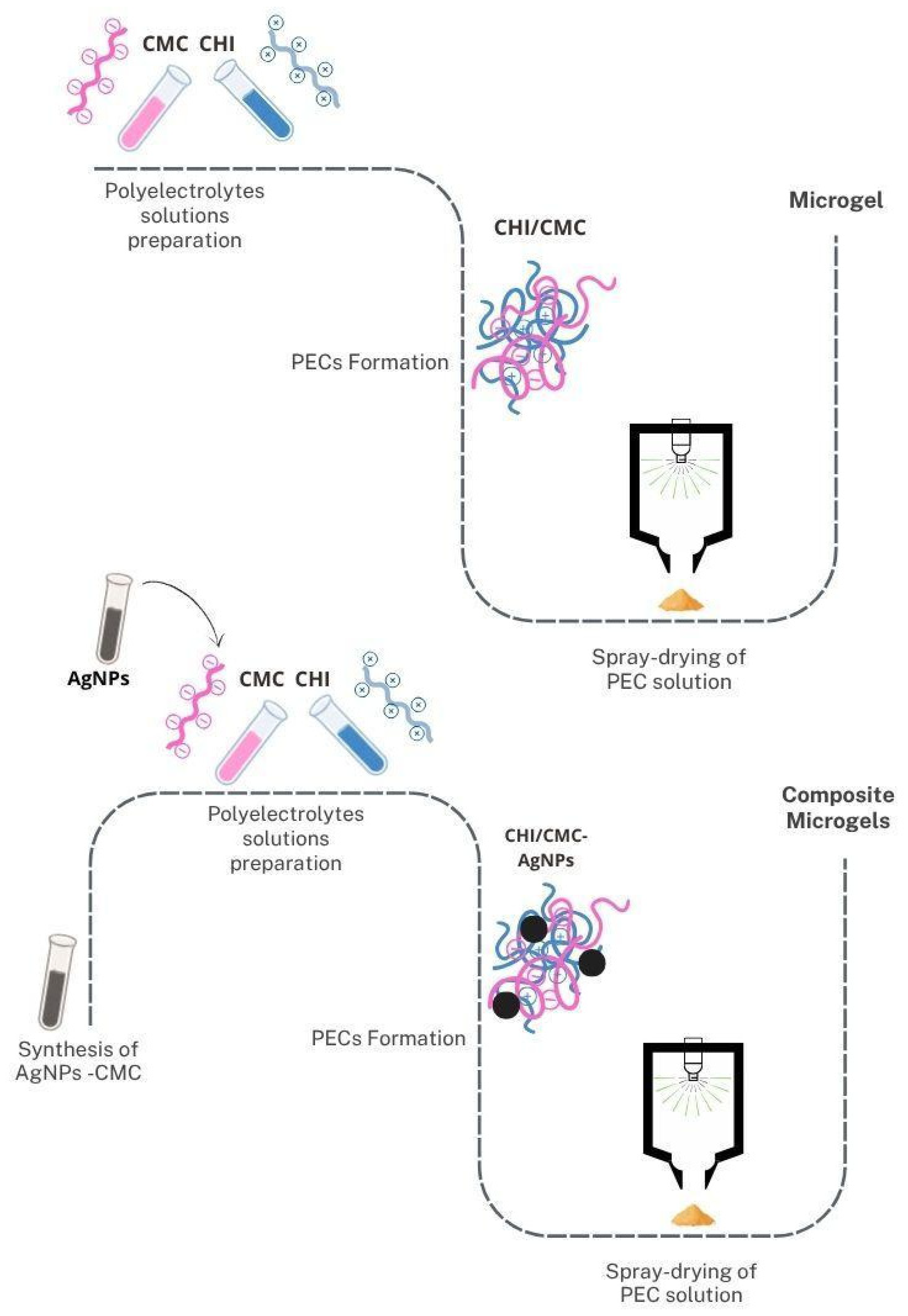
Figure 1. Representative diagram of CHI/CMC microgel and CHI/CMC-AgNPs composite microgel formulations.
Polysaccharides 06 00084 g001
Figure 2. AgNPs-CMC characterization. (A) TEM image, scale bar 50 nm; (B) TEM image with chemical composition verified by EDS (Ag: yellow; Na: magenta; O: red; C: sky blue), scale bar 20 nm; (C) frequency histogram of size distribution determined by TEM images; (D) intensity distribution profile determined by DLS; (E) UV-vis and (F) FTIR spectra.
Polysaccharides 06 00084 g002
Figure 3. SEM analysis of (A) powder of CHI/CMC samples, scale bar 3 µm; (B) powder of CHI/CMC-10AgNPs samples, scale bar 3 µm; (C) powder of CHI/CMC-20AgNPs samples, scale bar 3 µm; (D) hydrated and lyophilized CHI/CMC samples, scale bar 10 µm; (E) hydrated and lyophilized CHI/CMC-10AgNPs samples, scale bar 10 µm; (F) hydrated and lyophilized CHI/CMC-20AgNPs samples, scale bar 10 µm; (GI) histograms of size distribution of each powdered microgel.
Polysaccharides 06 00084 g003
Figure 4. ATR-FTIR spectra obtained by pure CHI (black), pure CMC (red), CHI/CMC microgels (blue), CHI/CMC-10AgNPs (magenta), and CHI/CMC-20AgNPs (green). Principal functional groups are indicated. Light-yellow shaded region indicates interaction zone between CHI and CMC to form microgels.
Polysaccharides 06 00084 g004
Figure 5. TGA curves. Residual mass (%) as function of temperature.
Polysaccharides 06 00084 g005
Figure 6. Swelling capacity of microgels at pH 7.4 (A) and 6.0 (B). * indicates significant differences with CHI/CMC samples (p < 0.05).
Polysaccharides 06 00084 g006
Figure 7. Biodegradation of microgels: (A) between 1 and 24 h in PBS, (B) between 1 and 24 h in PBS with lysozyme, (C) between 1 and 14 days in PBS, and (D) between 1 and 14 days in PBS with lysozyme. a indicates significant differences between CHI/CMC in PBS and CHI/CMC in PBS+lysozyme; b indicates significant differences between CHI/CMC-10AgNPs in PBS and CHI/CMC-10AgNPs in PBS+lysozyme; c indicates significant differences between CHI/CMC-20AgNPs in PBS and CHI/CMC-20AgNPs in PBS+lysozyme (p < 0.05).
Polysaccharides 06 00084 g007
Figure 8. Antibacterial activity of microgels against E. coli and S. aureus. Microbial inhibition halos are expressed in mm. NH: no halo of inhibition observed.
Polysaccharides 06 00084 g008
Table 1. Thermogravimetric behavior of microgels determined by TGA test. * Indicates significant differences with CHI/CMC microgels (p < 0.05).
CHI/CMCCHI/CMC-10AgNPsCHI/CMC-20AgNPs
Degradation temperature (Tp, °C)241.26 ± 1.00246.93 ± 1.50 *242.55 ± 1.10
Weight loss at Tp (%)32.43 ± 0.6035.28 ± 0.5032.78 ± 1.00
Residual Mass at 880 °C (%)15.64 ± 1.2020.94 ± 1.20 *21.90 ± 1.30 *
Table 2. Hemostatic activity of powdered microgels. * Indicates significant differences with whole blood, a indicates significant differences with CHI/CMC microgels, and b indicates significant differences with CHI/CMC-10AgNPs microgels (p < 0.05).
SampleHemolysis (%)Coagulation Time (%)BCI (%)
5 min10 min15 min30 min
Whole Blood-100.00 ± 0.3936.47 ± 0.605.17 ± 0.220.52 ± 0.020.38 ± 0.04
CHI/CMC3.08 ± 0.4682.49 ± 2.45 *10.50 ± 0.14 *4.70 ± 0.220.52 ± 0.110.33 ± 0.08
CHI/CMC-10AgNPs5.11 ± 0.36 a80.84 ± 2.83 *15.20 ± 0.20 *3.61 ± 0.19 *0.55 ± 0.130.39 ± 0.03
CHI/CMC-20AgNPs13.06 ±1.37 a,b81.35 ± 0.40 *14.32 ± 0.25 *5.75 ± 0.11 a,b1.05 ± 0.26 a,b,*0.78 ± 0.07 a,b,*
Disclaimer/Publisher’s Note: The statements, opinions and data contained in all publications are solely those of the individual author(s) and contributor(s) and not of MDPI and/or the editor(s). MDPI and/or the editor(s) disclaim responsibility for any injury to people or property resulting from any ideas, methods, instructions or products referred to in the content.