Article

Chitosan Mixtures from Marine Sources: A Comparative Study of Biological Responses and Practical Applications

by
Verginica Schröder
1,
Gabriela Mitea
1,*,
Ileana Rău
2,*,
Manuela Rossemary Apetroaei
3,
Irina Mihaela Iancu
1 and
Miruna-Maria Apetroaei
4
1
Faculty of Pharmacy, Ovidius University of Constanta, 6 Capt. Aviator Al. Șerbănescu Street, Campus C, 900470 Constanta, Romania
2
Faculty of Chemical Engineering and Biotechnologies, National University of Science and Technology Politehnica Bucharest, 011061 Bucharest, Romania
3
Mircea cel Batran Naval Faculty of Marine Engineering, “Mircea cel Bătrân” Naval Academy of Constanta, 1 Fulgerului Street, 900218 Constanta, Romania
4
Faculty of Pharmacy, Carol Davila University of Medicine and Pharmacy, 6 Traian Vuia Street, 020956 Bucharest, Romania
*
Authors to whom correspondence should be addressed.
Polysaccharides 2025, 6(3), 80; https://doi.org/10.3390/polysaccharides6030080
Submission received: 30 June 2025 / Revised: 3 August 2025 / Accepted: 3 September 2025 / Published: 5 September 2025

Abstract

Chitosan, a biopolymer with molecular variability, continues to demonstrate promising potential for biomedical and biotechnological applications. In this study, mixtures of β-oligochitosan, with a low molar mass (MM) of 1.5 kDa (CH1), α-oligochitosan, MM = 26.39 kDa (CH2), and α-chitosan, MM = 804.33 kDa (CH3) were analyzed. The tested solutions, chitosan alone and mixtures (CH1:CH2 and CH1:CH3), prepared in different mass ratios (1:1, 2:1, 3:1), were characterized in terms of MM and degree of deacetylation (DDA). The antimicrobial activity on S. aureus, E. coli, and C. parapsilosis was evaluated. The fractional inhibitory concentration index (FICI) was also calculated for mixtures. Using the Brine Shrimp Lethality Assay (BSLA), the in vivo interactions, which involve the internalization of chitosan in the cells, were assessed. The results showed that α-β chitosan mixtures exhibited an in vitro antimicrobial antagonistic effect (FICI > 1) for all samples. In contrast, significantly improved larval survival (%), development, and motility (p < 0.0001), with a close correlation between cellular inclusions and naupliar stages (R = 0.94), were detected in vivo testing. These data support the strategic use of chitosan mixtures with variable characteristics in biotechnological applications, with potential for optimizing intake, biological activity, and controlling cytotoxicity.

1. Introduction

Chitosan is a derivative obtained by the deacetylation process of chitin extracted from various sources. Chitin is a polysaccharide (β-(1-4)-linked N-acetyl-D-glucosamine) [1], which is an important component of the cell wall structure in fungi and yeasts [2,3,4,5] or forms complex structures, which have a protective role in aquatic or terrestrial invertebrates (crustaceans, mollusks, and insects), but are absent in higher forms of organisms [6].
Chitin and chitosan are among the most extensively studied natural polymers due to their structural composition based on glucosamine monomers, which confer molecular adaptability through the presence of reactive functional groups, including hydroxyl (-OH), amino (-NH2), and N-acetyl (-CH3CONH-) moieties [7]. Among these, the amino groups (-NH2) are primarily responsible for interactions with biological systems [8,9]. Response differences and controversial effects related to the molecular versatility and toxicity, or biocompatibility of chitosan, have raised questions about the optimal utilization of this polymer in various applications [7]. The deacetylated variant from fungal cell walls, along with β-chitosan extracted from mollusks [10,11,12] has expanded the scope for new investigations and applications [6,13]. The minimized chemical extraction process leads to decreased waste generation and eliminates allergenic substances.
Although their chemical attributes make chitosan one of the most promising polymers with applications in various fields [12,14,15,16,17,18,19,20,21,22], from a chemical point of view, there are aspects that reduce their use, such as solubility, viscosity, or molar mass (MM), which are also valid for chitin [23]. Chitosan can be categorized into three types according to its MM: low MM chitosan, which has a mass lower than 100 kDa; medium MM chitosan, with a mass ranging from 100 to 1000 kDa; and high MM chitosan, which exceeds 1000 kDa [24].
The understanding of the physicochemical properties of biopolymers and the methods for optimizing their production [12,25,26,27,28] has guided investigations into their interactions with biological systems [18,24,29,30,31,32,33,34].
The functionalization of chitosan is a topic of interest for applications in the medical field [24], including the development of biomaterials [1], delivery systems [28,30], and treatments for cancer, inflammation, infections, and fungal diseases [8,24,26,32].
In cancer therapy, chitosan and oligochitosan have been investigated for their antitumor activity, which is, at least in part, mediated through modulation of the phosphatidylinositol 3-kinase (PI3K)/AKT signaling pathway [35]. Oligochitosan has several pharmacological properties, but the most promising is its antitumor activity [36], which has been hypothesized to be closely related to its solubility in solutions and its ability to interact at the molecular level, targeting proteins [37,38]. Similarly, the study of chitosan distribution in vivo (fluorescent labels) allowed the observation of increased penetration into tumor cells after 24–48 h [39].
Furthermore, chitosan molecules under various experimental conditions (nanoparticles, oligochitosan molecules, chitosan derivatives, membranes) have been extensively studied as drug delivery systems for antitumor drugs [40,41,42,43]. In addition, a number of applications have been linked to the preparation and utilization of nanoparticles with chitosan [44,45,46], forms that are attracting the attention of modern pharmaceutical developments [47] and even in veterinary medicine applications [48,49].
The rapid development of effective antimicrobial agents is necessary in response to emergencies and concerns regarding multidrug-resistant pathogens. Chitosan is the subject of numerous approaches in this regard, as it is capable of interacting with microorganisms in a variety of ways [50]. Recently, the role of chitosan in modulating pathogenic bacteria during intestinal inflammation has been investigated, with particular attention to its prebiotic effects [51]. This study highlighted how chitosan’s antagonistic activity influences the gut microbiota, with differential effects observed depending on microbial species.
Recent research has focused on the impact of chitosan on developing new nutraceuticals and fortified foods [8], as well as its wide applications in agriculture [27,52,53], and food safety [54,55,56]. The methods employed are diverse and involve cross-linking, chelation, and modification of the molecular characteristics of chitosan [8,9,32,57].
The behavior of chitosan is influenced by the distribution of co-monomers as well as by the polymorphism of the structures, respectively, the arrangement of the chains, which can be α (antiparallel), β (parallel arrangement), and γ (combined) [7]. These chain arrangements imprint different inter- and intramolecular forces, as well as the versatility of interactions with molecules of various chemical species in solutions [7,58]. Notably, β-chitosan compared to α-chitosan possesses a higher potential in the biomedical field [10,11,12]. The functionalization, understanding of the mode of action, and increased efficiency of chitosan were achieved by creating mixtures with other natural (collagen) or synthetic (polyacrylamide, polycaprolactone) [59] or plant compounds [60].
Furthermore, our earlier investigation revealed that exposing cancer cells to solutions of α-chitosan (derived from shrimp shells) and β-chitosan (sourced from Rapana venosa egg capsules) [61], either individually or in combination, yielded significant findings. The mixture of these two chitosan types elicited distinct biological responses compared to their individual forms [12], with the effects attributed to the reorientation of the polymer chains [17,19,62]. In the examined cancer cells, SK-MEL-28 melanocytes, the impact of the mixture resulted in decreased viability and also influenced the synthesis of YKL-40-like proteins, which are directly involved in carcinogenesis [63,64,65,66]. Accordingly, the findings could lead to a novel approach to targeted therapies [65].
Based on previous findings that chitosan variants exhibit differential biological effects depending on molecular features such as MM and DDA, we hypothesized that binary mixtures of α- and β-chitosan types can modulate antimicrobial activity and cytocompatibility in a predictable manner. The objective of this investigation was to assess the cytological activity of α-chitosan and β-chitosan mixtures by utilizing two distinct test systems to observe the molecular and biological versatility of chitosan. Combinations of α-chitosan extracted from shrimps (MM = 804.33 kDa), α-oligochitosan (MM = 26.39 kDa), and β-oligochitosan isolated from Rapana venosa egg capsules (MM = 1.5 kDa) were analyzed both in vitro and in vivo. In order to prove our hypothesis, two complementary systems (an in vitro assay targeting microbial growth on S. aureus, E. coli, and C. parapsilosis, and an in vivo model on Artemia salina larvae) were employed to assess toxicity, ingestion, and developmental effects. We further posited that the molecular association between polymeric and oligomeric chitosan enhances biological versatility without increasing cytotoxicity. Additionally, we aimed to modulate the biological activity of chitosan through binary mixtures of polymers and oligomers, aiming to enhance their applicability in medical and environmental contexts. The novelty lies in demonstrating that simple molecular associations between chitosan variants can alter biological responses in a predictable manner. The study draws attention to the differences in biological response in contact with chitosan from different natural sources. The in vitro and in vivo responses are significantly influenced by the addition of β-chitosan, which enables the application of mixtures in a variety of formulations and for a variety of applications, including aquaculture, nutrition, drug bioavailability, and supplements. Modeling antimicrobial activity can be deemed beneficial in the context of comprehending and identifying new opportunities for the development of alternative methods with a wide range of practical applications. Furthermore, the in vivo data contribute to a deeper understanding of the cellular mechanisms modulated by chitosan-based formulations.
Based on the principles of the circular economy, which aim to prevent waste generation by obtaining products based on the efficiency of chemical processes with minimal impact on the environment, the chitosan used in this study is in line with current sustainable environmental policies [67]. Thus, the sources of chitosan in this study were biological waste based on shrimp exoskeletons from the seafood industry and R. venosa egg capsules recovered from the beach after sea storms. This strategy yielded biopolymers with characteristics suitable for various applications, aiming both at the sustainable use of resources with minimal environmental impact and at adding economic value to waste from the food industry.

2. Materials and Methods

2.1. Materials

In this study, materials from natural marine sources were used. The α-chitosan samples were obtained by processing shrimp shells (from seafood waste), and the β-oligochitosan sample was extracted from the egg capsules of the species Rapana venosa.
The reagents used included: 37% hydrochloric acid solution (for demineralization) from Chemical Company S.A., Iași, Romania; NaOH (analytical grade) in pellet form, from ChimReactiv S.R.L., Bucharest, Romania (used for deproteinization and deacetylation); acetone and ethyl alcohol (analytical grade) from Sigma-Aldrich, USA (for sample decolorization); glacial acetic acid from Sigma-Aldrich, St. Louis, MO, USA, used for the preparation of 1% acetic acid solutions necessary for the solubilization of chitosan; commercial chitosan from Sigma-Aldrich (product code CS-448877), with average molar mass, was used as a control sample.

2.2. Chitosan Types

In order to obtain α-chitosan from shrimps, the processing steps of the shrimp exoskeletons included: demineralization (treatment with 4% HCl solution at room temperature for 50 min, in a ratio of acid solution/powder mass of 13:1); deproteinization (treatment with 5% NaOH solution at a temperature of 65 °C ± 1 °C for 2 h, at an alkaline solution/demineralized powder ratio of 16:1); deacetylation (performed with 35% NaOH solutions for α-oligochitosan and 45% for polymeric α-chitosan, at T = 95 °C), for different times, at different chitin mass/alkali volume ratios [68]. Table 1 presents the values of the chitin mass/NaOH solution ratios that were selected based on the α-chitin deacetylation procedures from our previous studies on similar types of marine biological waste [68,69]. These protocols were optimized for each specific source (shrimp exoskeletons and Rapana venosa egg capsules) and validated under controlled experimental conditions. It is important to note that the MM and DDA values reported here are not newly determined in this study but derive from previously optimized and validated extraction protocols [68,69], where all determinations were performed in triplicate. The present work used these pre-characterized chitosan samples without re-analysis, as the study focused on investigating their biological effects rather than re-evaluating their physicochemical reproducibility.
In order to obtain β-oligochitosan from Rapana venosa, the egg capsules of Rapana venosa were subjected to alkaline extraction with NaOH 6.5%, in a ratio of alkaline solution volume to capsule mass of 40:1, for 120 min at 90 °C, according to the method described by [69].

2.3. Determination of Physicochemical Characteristics

The MM of the chitosan samples (g/mol) used in the study was determined by the viscometric method, measuring the intrinsic viscosity (mL/g) of weakly acidic chitosan solutions using the Mark–Houwink–Sakurada equation (Equation (1)), knowing K and α, which depend on the nature of the solvent, temperature, and chemical structure of the polymer [68,70]. The measurements were carried out at 25 ± 1 °C, using a micro-Ostwald viscometer with a capillary tube inner diameter of Øi = 0.43 mm, in acidic solutions of chitosan (2% acetic acid with 0.1 M KCl), as described in the study by [70,71]:
[ η ]   =   K × M v α
where [η] is the intrinsic viscosity (mL/g), Mv is the viscosity-average molar mass (g/mol), K is a constant dependent on the polymer–solvent system (13.8 × 10⁻³ mL/g), and α is the Mark–Houwink exponent (0.85), both specific to chitosan dissolved in 2% acetic acid with 0.1 M KCl at 25 ± 1 °C.
The degree of deacetylation (DDA) of chitosan samples was determined based on data obtained by potentiometric pH titration, applying Equations (2) and (3), according to the previously published methodology [68,70]:
DDA   ( % )   =   203 · Q 1 + 42 · Q
where Q represents the number of moles of amino groups per gram of sample, determined by pH potentiometric titration; 203 represents the molar mass of chitin (g/mol), and 42 is the molar mass of the acetyl group (g/mol).
Q = C M × V m
where CM is the molar concentration of the NaOH solution used for the titration (mol/L); ΔV is the volume difference between the two inflection points (L); m is the mass of the chitosan tested (g).
The values reported for MM and DDA represent the averages of triplicate determinations.

2.4. Preparation of Chitosan Solutions and Binary Mixtures

The following powders were used to prepare the diluted acidic chitosan solutions: CH1 (β-oligochitosan, MM = 1.5 kDa, DDA = 70%); CH2 (α-oligochitosan, MM = 26.39 kDa, DDA = 75.5%); CH3 (α-chitosan, MM = 804.33 kDa, DDA = 86.65%).
Each chitosan sample was dissolved in 1% acetic acid under medium stirring at 50 °C for 3 h, forming solutions with a concentration of 7 mg/mL. Two sets of binary mixtures were subsequently prepared: series C (C1–C3), consisting of CH1:CH2, and series K (K1–K3), consisting of CH1:CH3. These combinations were formulated in mass-to-mass ratios (m/m) of 1:1 (7 mg/7 mg), 2:1 (14 mg/7 mg), and 3:1 (21 mg/7 mg) of CH1 to CH2 or CH3, respectively. The total mass was dissolved in 1% acetic acid and maintained under continuous moderate stirring for 24 h at 30 °C. The reference samples were individual solutions of chitosan (CH1, CH2, and CH3) in 1% acetic acid.

2.5. MM and DDA Calculation of the Mixtures

The molar mass of the mixtures was calculated according to Equation (4):
MM mix = m 1 + m 2 m 1 M M 1 + m 2 M M 2
where m1 is the mass of CH1 (mg), m2 is the mass of CH2 or CH3 (mg), MM1 is the molar mass of CH1 (g/mol), MM2 is the molar mass of CH2 or CH3 (g/mol).
The degree of deacetylation (DDA) of the mixtures was calculated according to Equation (5):
DDA mix   ( % )   =   = D D A α c h · m α c h + 2 D D A β c h · m β c h m α c h + 2 m β c h
where DDAαch is the DDA of αchitosan sample (CH2 or CH3) from the mixture (%); mαch is the mass of αchitosan (CH2 or CH3) from the mixture (mg); DDAβch is the DDA of β-chitosan from the mixture (%); and mβch is the mass of β-chitosan from the mixture (mg).

2.6. In Vitro Assessment

2.6.1. Preparation of Bacterial Suspensions

In this study, two bacterial strains were analyzed: Staphylococcus aureus ATCC 23235 (Gram-positive) and Escherichia coli ATCC 25922 (Gram-negative), as well as a fungal strain, Candida parapsilosis, ATCC 22019. Bacterial inoculum was prepared using the direct suspension method (CLSI). Thus, bacterial colonies selected from a Tryptic Soy Agar (TSA) plate at 35–37 °C, cultured 24 h before, were suspended in Mueller-Hinton medium (MHB) according to the 0.5 McFarland standard, measured with a densitometer (Biomerieux, Marcy-l’Étoile, France) at a bacterial density of approximately 1.5 × 10⁸ CFU/mL. The inoculum of Candida parapsilosis was prepared using the same method, but the suspension was made in RPMI 1640 medium with 3 × 10⁸ CFU/mL. The positive control, for bacterial growth, was prepared with 90 μL of culture medium and 10 μL of bacterial suspension, while the negative control was represented by culture medium (100 μL). For comparison, solutions of the antibiotic, ceftriaxone, and the antifungal, fluconazole, were used.
Plates were sealed and incubated at 37 ± 1 °C for bacteria and 35 ± 2 °C for fungi for 24 h. Each well was evaluated visually based on sample opacity. Additionally, resazurin, a conventional dye, was used for one set of samples. The evaluation was based on the color change from blue to pink, a reaction induced by the microbial enzyme dehydrogenase, when the bacteria are active (alive) [72].

2.6.2. Micro-Well Dilution Assay

The minimum inhibitory concentration (MIC) was assessed using the microdilution technique in Mueller-Hinton (MH) culture medium. For each test solution, seven serial dilutions were prepared, resulting in final concentrations within the wells ranging from 78 µg/mL to 5000 µg/mL. 96-well microplates were utilized. The test solution, culture medium, and inoculum were introduced into these wells, resulting in a final volume of 100 µL in the test wells. The experiment was conducted in duplicate. The minimum inhibitory concentration (MIC) is defined as the lowest concentration at which visible bacterial inhibition occurs. The lowest concentration of an antimicrobial agent that eliminates 99.9% of both bacterial and fungal populations is referred to as the minimum bactericidal concentration (MBC) and the minimum fungicidal concentration (MFC) [73]. The MIC and MBC were confirmed in the test vessels, which visually demonstrated inhibition in small volumes (0.1 µL) of the culture media (TSA and Sabouraud) after incubation for 24 h. The absorbances measured on the microplate reader (OD 630 nm) are also analyzed to confirm results against the antibiotic control.
For the evaluation of the bactericidal/fungicidal or bacteriostatic/fungistatic effect, the MBC/MIC ratio was calculated. A bactericidal/fungicidal effect is considered if the ratio MBC/MIC = 1 or 2, and when MBC/MIC = 4 or 16, the induced effect is considered bacteriostatic/fungistatic [74].

2.6.3. Evaluation of the Fractional Inhibitory Index of Chitosan in the Mixtures

The fractional inhibitory concentration index (FICI) serves as a technique for evaluating the interaction among multiple antimicrobial agents, focusing on whether their combined effects are synergistic, additive, or antagonistic. The calculation involves summing the fractional inhibitory concentrations of each substance within the combination (Equation (6)) [75].
FICI = M I C   c o m p o u n d   ( i n m i x t u r e ) M I C   α c h i t o s a n + M I C   c h i t o s a n   ( i n m i x t u r e ) M I C β c h i t o s a n
where MIC refers to the minimum inhibitory concentration (mg/mL) of each compound, either alone or in combination.
The mixtures are considered to show: (a) synergistic effect if FICI ≤ 0.5; (b) additive effect or indifferent when FICI > 0.5 and < 1, and (c) antagonistic effect if FICI > 1 [76].

2.7. In Vivo Testing—Brine Shrimp Lethality Assay (BSLA) Testing Protocol

For the in vivo assessment, stage I–III larvae were obtained in the laboratory according to the Artoxkit protocol (ARTOXKIT M: 24 h mortality test based on the anostracan crustacean Artemia salina). BSLA assessments are commonly used in cytotoxicity studies [77,78,79] as they provide information on how the tested substances influence aquatic organisms. The test allows for the assessment of several parameters, including behavior, anatomical changes, growth assessment, and sublethal and lethal effects.

2.7.1. Experimental Design for Larvae

The larvae were obtained by incubating dehydrated cysts (Artemia Brine Shrimp Eggs). Incubation of the cysts was carried out in saline water (35 ppt), continuous aeration and illumination, for 24–30 h, at a temperature of 22–23 °C. After hatching, the nauplius-type larvae (instar I) were separated and placed in test tanks. Saline solutions were prepared with sea salt (Artemia salt, Dohse Aquaristik GmbH & Co., Gelsdorf, Germany) in distilled water, filtered through filter paper (Filtres Fioroni S.A.S., Ingre, France, diameter 110 mm, thickness 0.16 mm) to remove impurities.
The assessment was carried out in sterile Plexiglas cuvettes with 1 mL wells (NunclonTM Surface, sterile, Roskilde, Denmark). Larvae hatched for approximately 20–24 h were placed in the test cuvettes (on average, 10 specimens/cuvette). Sorting and separation were performed under an Optika binocular microscope at 4× or 10× magnification. To avoid concentration errors, organisms were pipetted in saline in controlled volumes (50 µL).
The test solutions of chitosan and oligochitosan in 1% acetic acid were prepared to minimize the acetic acid-induced effect; the concentrations of chitosan (alone or in mixtures) tested were 35 µg/mL and 70 µg/mL. For each concentration, the test was carried out in triplicate. The number of larvae/test cell was a minimum of 10. The effects were evaluated at 10 h, 24 h, and 48 h. Water control, 1% acetic acid control, and commercial chitosan purchased from Sigma Aldrich were also evaluated for control. Table 2 illustrates Artemia salina larval sampling and distribution across test conditions in vivo.

2.7.2. Structural Modifications and Ingestion Assessment of Polymers

Microscopic observations were performed with an Optika binocular microscope in order to assess the developmental stage of the larvae, their behavior, and any locomotor changes. To conduct a comprehensive evaluation of morphological development, microscope slides were prepared to assess larval structure, the digestive tract (including peristaltic movements), and the cellularity of the intestinal epithelium.
An epifluorescence microscope was used to investigate the penetration of chitosan at the cellular level. Chitosan was labeled with fluorescein isothiocyanate (FITC, 1 mg/mL in DMSO), and its detection was performed using blue light illumination [80,81]. The presence of chitosan was detected by the emission of green color. Following a 24 h period after the addition of chitosan solutions, the organisms were subjected to FITC solutions for 10 min and subsequently examined using UV epifluorescence microscopy with an excitation wavelength of 495 nm and an emission wavelength of 515 nm. The negative control consisted of larval specimens kept solely in seawater. Throughout this interval, the larvae did not receive food. Dye removal was conducted using saline water during the preparation of microscope slides.

2.7.3. Larval Behavior and Observations on Growth and Development

Survival was determined by subtracting the number of dead larvae from the total number of larvae collected in the study for each cell. A larva is considered dead if it fails to exhibit any antennae movements for 10 s. For confirmation, control samples consisting of freshly hatched specimens were placed in artificial seawater with a salinity of 35 ppt, as determined using an automatic temperature-compensated refractometer (ATAGO, Co. Ltd., Tokyo, Japonia, Japan).
Microscopic quantification of larval development and anatomical changes was conducted through slide-lamella preparations, without fixation or staining of the larvae. Measurements were performed for each analyzed larva, leading to the establishment of three categories based on size and morphological characteristics: stage I larvae (L I), naupliar stage larvae exhibiting visible segmentation and forming appendicular buds (L II), and stage larvae with elongated appendicular extensions (L III). Appendicular organogenesis was assessed based on cell lineage alterations and categorized on a scale from 1 to 3.
Larvae were periodically evaluated, and locomotor behavior was assessed using a binocular microscope at regular intervals for each sample goblet. Under normal conditions, larvae perform circular movements between the bottom of the bowl and the surface, a behavior referred to as circular swimming. When organisms remain aggregated at the bottom of the bowl with restricted movement, sublethal effects are assessed. The speed of movement execution was evaluated on a scale ranging from 1 (specimens exhibiting minimal movement) to 10 (specimens demonstrating rapid and orderly movement execution).
In this study, biocompatibility was operationally defined as the absence of lethal or sublethal effects following exposure to chitosan solutions, including survival rate, normal motility patterns, developmental progression (larval stage classification), and cellular integrity. Motility was used as a behavioral marker for sublethal systemic toxicity, as reduced or disorganized movement is indicative of physiological stress or neuromuscular dysfunction.

2.8. Statistical Interpretation

Statistical analysis was performed using StatPlus:mac v8 software (AnalystSoft Inc., Brandon, FL, USA). To evaluate the significance of the differences between the tested groups, a two-way ANOVA was applied, followed by Tukey’s test for multiple comparisons, in order to compare the results obtained between the control samples and those with chitosan formulations in the mixture. The results were expressed as median values with 95% confidence intervals, based on three biological replicates, and differences were considered significant at a threshold of p < 0.0001.
For the comparative analysis of the effects induced by chitosan in the two biological systems (in vitro and in vivo), a principal component analysis (PCA) was applied. This method enabled the identification of variables with the highest contribution to the variability of the tested system and highlighted the relationships between them. PCA was performed based on the average values of the following parameters: larval viability at two concentrations (35 and 70 µg/mL), naupliar stages, organogenesis score, mobility, degree of cell inclusions, and MBC/MIC and MFC/MIC ratios. The PCA results were represented graphically in a loadings plot, on the first two principal components (PC1 and PC2), which cumulatively explained 82.94% of the total variability. Correlations between parameters were calculated using the pairwise deletion method, and Pearson correlation coefficients (R) were interpreted according to statistical significance at a 95% confidence level. The variables were treated as potential predictors for understanding the relationship between the physicochemical characteristics of the formulations and the observed biological effects.

3. Results

3.1. Characteristics of the Chitosan Samples Used

The individual chitosan samples (CH1–CH3) had different MM and DDA, as shown in Table 3.
Thus, the β-oligochitosan sample (CH1) had a very low MM (1.5 kDa) and a DDA of 70%, while α-oligochitosan (CH2) had an MM of 26.39 kDa and a DDA of 75.5%. The highest MM was recorded for the polymeric α-chitosan (CH3), with a value of 804.33 kDa and a DDA of 86.65%. These values confirm the optimization of the deacetylation and fractionation processes applied to obtain biopolymer samples (Table 3) with different MM and DDA.
Moreover, the MM values of the two sets of binary mixtures C1–C3 (CH1:CH2) and K1–K3 (CH1:CH3) ranged between 1.96 and 2.99 kDa, reflecting the influence of β-oligochitosan (MM = 1.5 kDa) in decreasing the total MM of the mixtures. The decreasing trend of the molar mass in mixtures with increasing proportions of CH1 is evident in both series C (CH1:CH2) and series K (CH1:CH3). Thus, these results confirm the applicability of the calculation formula used, which considered the actual masses and MM of the components (Equation (4)).
The DDA of mixtures C1–C3 and K1–K3 was calculated based on the mass proportions and individual values of the component samples. The values obtained ranged between 70.79% and 75.55%, depending on the composition (Table 3). It was observed that mixtures containing α-chitosan with high molecular weight (CH3) had higher DDA values compared to those obtained from α-oligochitosan (CH2).
These results were subsequently used to evaluate how the MM of chitosan mixtures influenced the physicochemical properties of the tested solutions, including their molecular interactions with biological systems.

3.2. In Vitro Antibacterial and Antifungal Activity

Table 4 shows the inhibitory, bactericidal, or fungicidal effects induced by chitosan solutions in mixture, compared to the control samples of β-oligochitosan, α-oligochitosan, and α-chitosan, as well as the MBC/MIC and MFC/MIC ratios.
The observed antimicrobial activity indicates different responses depending on the evaluated strains. Thus, it can be seen that for S. aureus, the bactericidal effect is recorded at concentrations of 5 mg/mL (MBC) in the α-β chitosan mixture samples (C1–C3), while the values are halved (MBC = 2.50 mg/mL) in the tests performed with the mixtures in series K (K1–K3). A remarkable effect is observed at a low concentration of 0.32 mg/mL (MBC) for the CH1 solution. Compared to the control sample, ceftriaxone (MBC = 0.30 mg/mL), it can be concluded that CH1 is as effective as the antibiotic in inhibiting S. aureus.
Regarding the E. coli strain, the effect is equivalent between samples C1–C3 and K1–K3. A lower effect can be observed in the case of β-oligochitosan (CH1), MBC = 5 mg/mL and MIC = 2.5 mg/mL, compared to its effect on the S. aureus strain.
The fungicidal activity was determined at a concentration of 5 mg/mL, and the MIC recorded was 2.50 mg/mL for all α-chitosan samples and α-combinations with β-oligochitosan. When testing β-oligochitosan (CH1), effects were observed at low concentrations, 0.31 mg/mL (MIC) and 0.31 mg/mL (MFC), with an MFC/MIC ratio of 1, illustrating the fungicidal effect of this type of chitosan. These results suggest that regardless of the physicochemical characteristics of the biopolymers tested, fungicidal effects are obtained in separate samples (chitosan alone) from CH1, CH2, CH3 at lower concentrations (CH1 = 0.31 mg/mL, CH2 = 3 mg/mL, CH3 = 1.70 mg/mL) than in combinations, where MFC in all samples is recorded at 5 mg/mL (Table 4).
The calculation of the MBC/MIC and MFC/MIC ratios for α-chitosan and β-oligochitosan enables the identification of the bactericidal or fungicidal effect of chitosan forms separately or in combination. It was also observed that oligochitosan forms (CH1 and CH2) induce effects at lower concentrations than the polymeric form with average MM (CH3). Thus, these values are close to the concentrations of ceftriaxone (MIC = 0.16, MBC = 0.30) and fluconazole (MIC = 0.62 mg/mL, MFC = 1.25 mg/mL) used as positive controls (Table 4).
The FICI index indicates that chitosan CH1 in the mixtures demonstrates an antagonistic effect on all analyzed solutions, regardless of the specific characteristics of the molecules in the tested samples (Table 5).
FICI values exceed 1 for all tested combinations across the microorganisms examined. Consequently, it can be concluded that the actions of chitosan vary in combinations, irrespective of the concentrations employed, negatively impacting the anticipated antibacterial effect, while being correlated with the type of chitosan (α or β). Oligochitosan demonstrates significant efficacy at low concentrations, exhibiting a MIC that is 15.6 times lower than that of mixture 1 in antibacterial assays against the S. aureus strain.

3.3. In Vivo Testing (BSLA)

3.3.1. Larval Survival 24 h After Exposure

Survival rates were statistically compared using two-way ANOVA, confirming the biocompatibility of the mixtures, particularly at lower concentrations and with increasing β-oligochitosan proportion. Figure 1 illustrates the statistical analysis of larval survival 24 h after exposure.
At a concentration of 35 µg/mL, larval survival rates were nearly 100% in samples containing β-oligochitosan (CH1) and α-oligochitosan (CH2). Survival significantly declined at a chitosan concentration of 70 µg/mL exclusively in the α-oligochitosan (CH2) sample (Figure 1A). In the mixtures C1-C3, larval survival reached 100% at both concentrations, demonstrating high statistical significance (p < 0.0001) when compared to control samples (CH1 and CH2). The analysis of results for each sample in mixture 1 (C1, C2, C3) at the tested concentrations of 35 µg/mL and 70 µg/mL indicates minimal differences in survival, which lack statistical significance (p > 0.1). The samples containing α-type chitosan may exhibit variations in response due to molecular morphological differences rather than differences in physicochemical characteristics (MM or DD).
In mixture 2, a viability reduction of 6–10% was observed at a concentration of 35 µg/mL for samples K1–K3, compared to the control (CH1). Organisms exposed to α-chitosan, CH3, at a concentration of 70 µg/mL exhibit observable differences. In K1, a concentration of 70 µg/mL results in a 27% reduction in survival rate (Figure 1B), whereas K2 and K3 exhibit survival rates of 97% and 100%, respectively. In K1, the ratio of α-chitosan to β-oligochitosan was 1:1. The enhancement in survival rates in K2 and K3 correlates with the incorporation of β-oligochitosan and the adjustment of the α-chitosan/β-oligochitosan ratio. The 1:2 ratio in K2 and the 1:3 ratio in K3 mitigated the negative effects caused by exposure to α-chitosan at a concentration of 70 µg/mL, compared to the control CH3, with a statistically significant difference observed (p < 0.0001).

3.3.2. Incorporation and Accumulation of Chitosan Particles in the Body of Larvae, 48 h After Exposure

A significant effect, measured following the opening of the digestive tract, is ingestion, as illustrated in Figure 2.
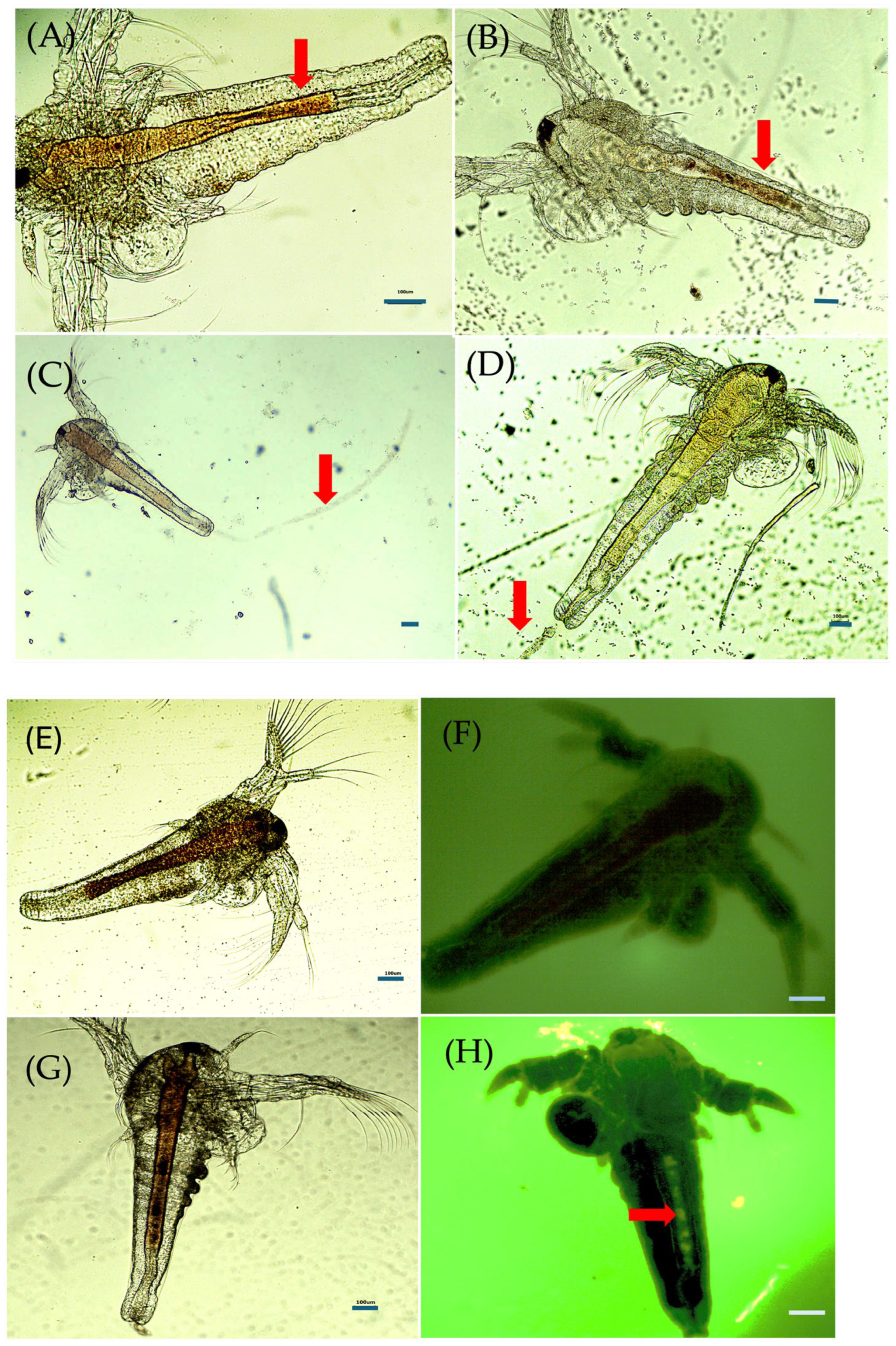
The larvae filter chitosan particles from the water, which are observable as contents within the digestive tract (Figure 2). According to microscopic observations, chitosan is in transit through the digestive tract. Periodic elimination shows agglomerated particles attached to the anal area of the larva (Figure 2C) or can be distinguished as waste at the bottom of the experimental tanks (pellets). Fluorescence microscopy confirms the presence of chitosan particles in the digestive tract of the larvae in the exposed samples (Figure 2G,H), compared to the control specimens (Figure 2E,F).
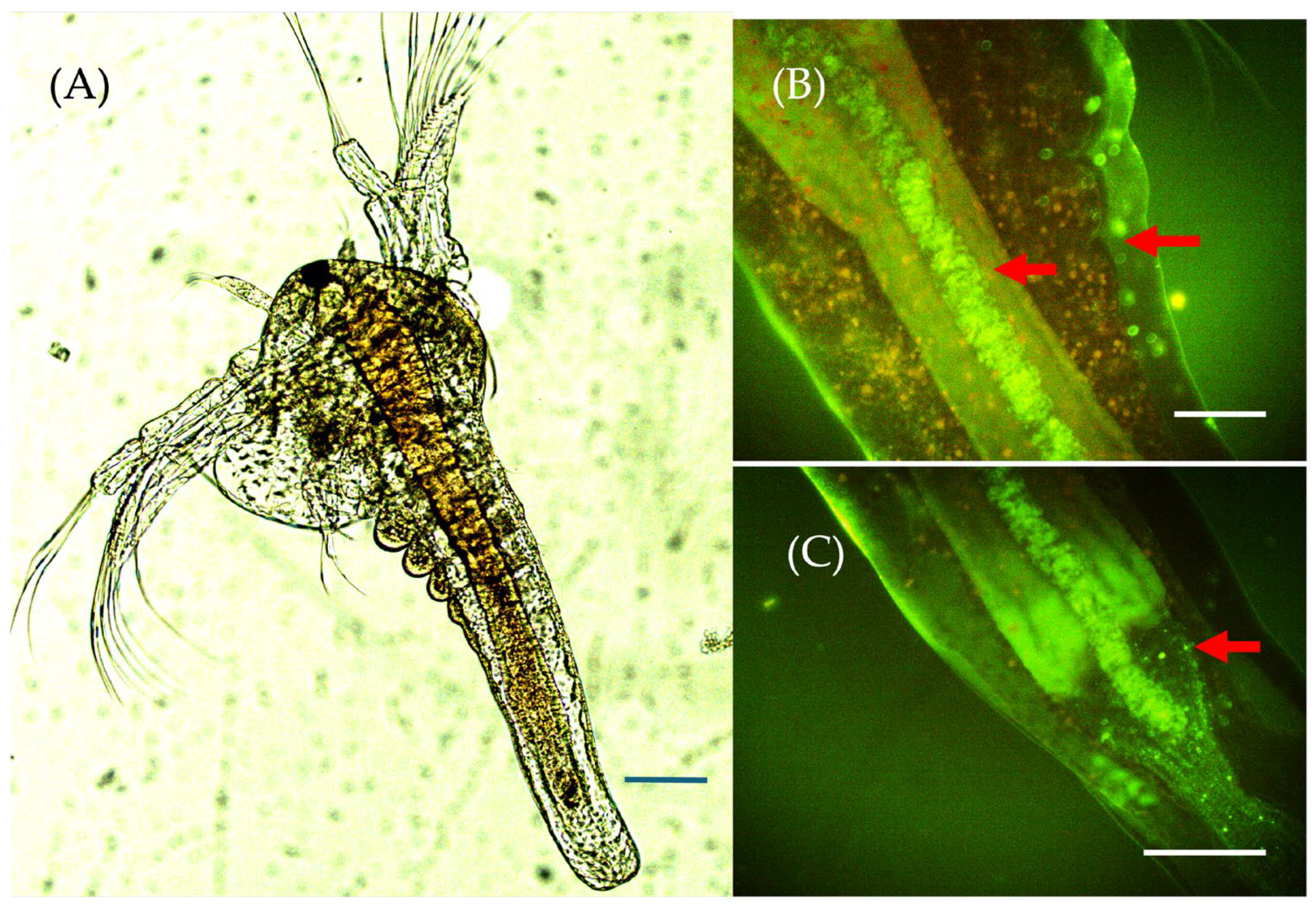
After 48 h of exposure to chitosan solutions, the presence of chitosan can be identified at the microscopic level (Figure 3), with positive green fluorescence in cells lining the digestive tract (Figure 3D–F), in epithelial cells and structures in the antennae, or in subcutaneous structures, which confirms the passage of polymers at the cellular level (Figure 3B,C).
Larvae in stages II and III, after opening the mouth, can ingest significant amounts of particles from the water through the filtration process. Part of the ingested chitosan passes through the epithelium of the digestive tract (Figure 4B,C), which explains the presence of these emissions in fluorescence in cells from different structures (antenna, cuticle, epithelium) (Figure 3A–F and Figure 4B,C).
Additionally, abnormal effects were reported in dead specimens (Figure 5). It was found that some larvae died before entering stage II (Figure 5A), while others died due to blockage of the digestive tract (Figure 5B–D). Nauplius I larvae account for 50% of dead specimens after exposure to α-oligochitosan at a concentration of 70 µg/mL (CH2). This large number of deceased larvae in stage I can be explained by the fact that their mortality was most likely caused by an obstruction during molting. Molting is an essential component of the development process.
Chitosan molecules may prevent the transition from stage I to stage II by forming electrostatic contacts with the larvae’s surfaces or cell membranes. Chitosan particles were found throughout the larva, adhering to it and creating an exterior, branching layer with a fluffy appearance.

3.3.3. Larval Behavior and Observations on Growth and Development

Normal behavior was evaluated based on orderly movements and a balanced rhythm of movements, using a scale of 1 to 10, where the maximum score indicates rapid and orderly antenna movements (Figure 6A). All organisms subjected to low concentrations exhibited consistent development, and the larvae demonstrated organogenesis activity (Figure 6B). All samples with chitosan mixtures are distinguished by stimulating larval development (score 3), and compared to the samples of chitosan alone, score 1 (CH2) and score 2 (CH1 si CH3, allowing for the microscopical details (Figure 6B), the values of the scores are used for quantification according to the observations.
Reduced and saccadic movements, as indicated by 5 motility values (Figure 6A), were reported 20–22 h after exposure in larvae from vessels containing α-chitosan solutions (CH2 and CH3), which were considered sublethal.
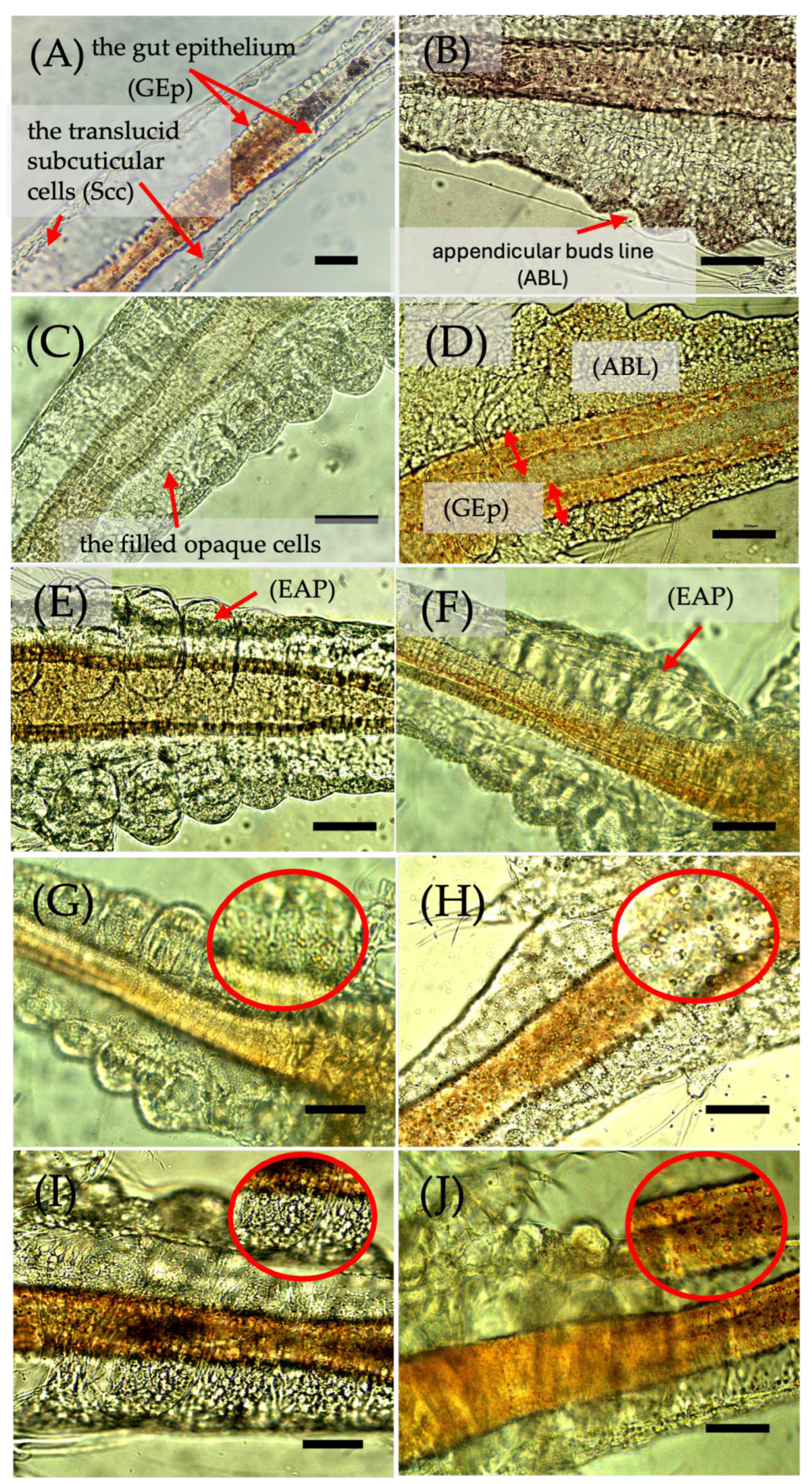
In all experimental vessels, except those with β-oligochitosan CH1 and α-oligochitosan CH2 samples, particles eliminated from the digestive tract (fecal pellets) are evident. These were very abundant in samples with chitosan mixtures. In these samples with chitosan mixtures, the larvae also developed more rapidly, reaching larval stage II (characterized by clear, contoured cell lines indicating appendicular organogenesis) and III (with elongated appendicular appendages) compared to the control samples (Figure 7A,B).
Observations show that larval development and growth in size can be correlated with the incorporation of chitosan, a process that also ensures the energy supply necessary for division and molting.
Appendicular development is present in larvae exposed to mixtures (Figure 7C,D), with very prominent buds visible in microscopic preparations made with larvae from samples C1 and K1 that had an α-CH:β-oligoCH ratio of 1:1 (Figure 7 E–G,J).

3.3.4. Cytological Effects

Several effects were observed in microscopic preparations with lamellae, allowing for observations on live specimens without the need for staining (Figure 7).
Intense cytological activity was observed, with epithelial cells appearing swollen and opaque (Figure 7C–E), compared to the control, where they were transparent (Figure 7A). Embryonic lipid accumulations were identified in subcuticular cells (Figure 7H) and epithelial cells of the digestive tract (Figure 7I). Cytoplasmic inclusions (Figure 7C) were more pronounced in samples with mixture 1, with α-oligochitosan and β-oligochitosan (C1, C2) (Figure 7C,D,G,H), probably due to the accumulation of chitosan particles. It is interesting to note the chitosan accumulations that occur upon exposure to CH1 at a concentration of 70 µg/mL of β-oligochitosan in the extracellular space (Figure 7J).
The formation of appendicular buds was barely visible in the control (Figure 7B), but more prominent in the samples with chitosan mixture (Figure 7C–G). Additionally, the specimens analyzed from all tanks with chitosan mixtures showed an increase in the thickness of the epithelium lining the digestive tract (Figure 7C–F).

3.3.5. Comparative Analysis of the Effects Induced by Chitosan on the Two Biological Systems

According to PCA, the eight variables involved were grouped into principal components (PC), which include the variability of the data set in descending order (eigenvalue and proportion) and comprise a possible grouping of samples. Components with eigenvalues > 1.0 were retained based on the Kaiser criterion, a standard approach to ensure interpretability and minimize overfitting. The first two PCs (PC1—55.46% and PC2—27.48%) explained a cumulative 82.94% of the total variance, while inclusion of PC3 (13.07%) increased the cumulative variance to 95.99%, which is above the conventional 70–80% threshold for biological datasets and ensures that the retained components adequately capture the dominant patterns in the data while reducing dimensionality. The first two components were used for plotting due to their dominant explanatory power and clear separation of biological test systems, while the representation of the samples in PC1-PC2 coordinates shows that the in vitro samples are separated from the in vivo samples.
Analyzing the variables included (loading) in PC1 and PC2, it can be seen that 55.46% are in PC1 compared to 27.48% associated with PC2.
Analyzing variables from PC1 and PC2, the highest association in PC1 corresponds to naupliar stages (98%), which varied significantly between samples, followed by larval behavior (motility, 85%), organogenesis (85%), and cellular inclusions (88%). Similarly, organogenesis has loading in PC2 (50%). In PC3, there is loading for viability 35 (81%) corresponding to the emergence of effects induced by the minimum tested concentration (35 µg/mL), which is predictable given the slightly variable responses.
Quantified cellular parameters (cellular inclusions), appendicular organogenesis, as well as viability and motility are grouped in PC1, where the naupliar stage represents 98%, being the significant component (Figure 8). The larval stage III is recorded in all mixed samples as well as in the CH1 control.
This analysis demonstrates that the differentiation between samples is achieved by modifying the response due to the mixture and the ratios between the types of chitosan and, to a considerable extent, due to the different effects in the biological system (cellular changes, motility changes, viability).
Table 6 illustrates the correlation coefficients between morphological parameters and antimicrobial efficacy tested in vitro and in vivo.
There is a high correlation between larval development (naupliar stage) and organism viability at high chitosan concentrations (viability parameter 70) (R = 0.94 and p-value < 0.0001), which confirms that combinations of different forms of chitosan can cause significant changes related to the growth and development of the organisms tested in vivo. The “cell inclusions” parameter is associated with oligochitosan particles and exhibits a substantial correlation with naupliar development processes (R = 0.78, p-value = 0.01), influencing larval viability. No statistically significant correlations were found between the analyzed parameters and the MBC/MIC ratio, p-value (at the 1% level).
Thus, according to the data obtained, the in vivo test reveals multiple interferences, including intracellular accumulation (cellular inclusions, organogenesis), impaired survival or behavior (motility), and naupliar stages. According to these observations, any of the parameters analyzed in vivo is highly significant for monitoring the effects induced by chitosan forms. The correlation coefficient (R = 0.94; R = 0.81) indicates that the changes quantified at the morphological level (in vivo model) are correlated with the survival rate (p-value < 0.005) but cannot be associated with the results obtained from the in vitro model (p-value > 0.1).

4. Discussions

Chitin, found in the exoskeletons of crustaceans, is the source of chitosan, a biopolymer derived from the marine environment. Chitosan is readily soluble in diluted solutions of various inorganic and organic acids. Due to its natural properties, it is biocompatible, biodegradable, and hematocompatible [82,83,84]. Chito-oligosaccharides, which can be obtained from chitosan, are becoming increasingly popular as drug delivery vehicles due to their mucoadhesive, biodegradable, and biocompatible properties. By enhancing their stability, targeted delivery, and controlled release, grafting—the chemical modification of chitosan to chito-oligosaccharides—has been utilized to increase their drug delivery capabilities [85]. In this study, a relevant sustainability issue is addressed. The use of chitosan obtained from marine waste, particularly shrimp exoskeletons and Rapana venosa egg capsules, aids in reducing environmental impact through waste revalorization and in enhancing the identification of the most effective chemical extraction process. This approach minimizes the consumption of chemical reagents and energy, aligning with circular economy principles, while also producing chitosan types with specific characteristics for diverse applications [86].
Thus, in our study, it can be observed that mixtures containing α-chitosan with high molar mass (CH3) had higher DDA values compared to those obtained from α-oligochitosan (CH2). This variation can be attributed to the higher content of amino groups available for reactions in the CH3 samples, as well as to a more favorable distribution of β-oligochitosan fractions in the structure, according to the structural hypothesis formulated in our previous study [12]. The calculated MM and DDA values of the mixtures confirm that the method used for preparing the binary formulations (CH1:CH2 and CH1:CH3) was reproducible and effective. All values obtained are averages of triplicate determinations. A direct correlation was found between the molar mass and the proportion of CH1, while the impact of the structural reactivity of β-oligochitosan is particularly evident in mixtures C1–C3. The DDA results obtained for the mixtures of α-oligochitosan and β-oligochitosan (C1–C3) and α-chitosan and β-chitosan (K1–K3) formed in diluted acetic acid solutions confirmed the structural influence of β-oligochitosan chains in each combination formed. Thus, it is important to highlight that the creation of chitosan-based mixtures represents a novel approach in the development of biodegradable and biocompatible formulations with enhanced functionalities [87,88].
The antibacterial activity of chitosan is significantly influenced by its MM, which additionally impacts how it interacts with bacteria, viruses, fungi, and other microbes. The antimicrobial action associated with MM can be attributed to electrostatic mechanisms, which involve the interaction between the reactive groups of chitosan and the anionic active sites on the surfaces of microorganisms. The chemical structure of chitosan has three types of reactive groups: the amino group at C2 of the glucosidic ring, and two types of hydroxyl groups, one primarily active at C6 and the other secondarily active at C3 [89].
These electrostatic interactions due to the presence of reactive groups on the glycosidic ring of chitosan would be explained by the good solubility of the biopolymer in acidic environments, but its efficacy is limited at pH values above 6 [90]. Chang et al. demonstrated in their study how the zeta potential and antibacterial activity of chitosan are influenced by pH levels and the MM of chitosan [91]. Thus, biopolymers with MM ranging from 3.3 to 300 kDa were obtained at acidic and neutral pH levels, whose antibacterial activity was correlated with their MM, pH levels, and zeta potential values. At acidic pH levels, the antibacterial activity of chitosan is directly dependent on its MM, in contrast to the behavior observed at neutral pH levels. Depending on the pH value and zeta potential, significant variations were observed. Thus, at acidic pH values, an increase in MM was associated with a proportional increase in zeta potential. In contrast, a decrease in MM led to higher zeta potential values. In contrast, a significant reduction in zeta potential towards negative values was observed for chitosan with high MM at neutral pH values, negatively affecting its antibacterial activity. Currently, particular emphasis is placed on the charge density of chitosan nanoparticles (NPC). Studies in the literature have shown that, depending on the charge density of NPCs, anionic or cationic nanoparticles can cause stronger interactions with bacterial cells, causing significant mitochondrial damage and increased cell absorption, especially those characterized by a positive charge density [92].
These innovative chitosan-based formulations, derived from various sources and possessing distinct functional characteristics, may be regarded as hierarchical assemblies designed through the self-assembly of functionally selective natural biopolymer macromolecules via physical, non-covalent hydrogen bonding [84,93]. The amino groups of the glycosidic ring, which are protonated in a mildly acidic medium, are primarily responsible for the development of intermolecular bonds within each supramolecular system [94]. The formation of hydrogen bonds and electrostatic interactions among polymer chains (α- and β-oligochitosan, α-chitosan) with varying MM, DDA, and rigidity could provide insight into the selective modulation observed in the tested mixtures, which exhibited notable antibacterial activity, low toxicity in blended forms, and high solubility. Both β-oligochitosan and α-oligochitosan are copolymers composed of β (1→4) linked D-glucosamine and N-acetyl D-glucosamine units [94]. Their self-association in dilute, weakly acidic acetic acid solutions leads to the formation of new structures (C1–C3), which vary according to the mass ratio of β-oligochitosan. Even though the two components of the C1–C3 mixtures are different types of oligochitosan, raising the β-oligochitosan ratio facilitates the development of configurations with high molecular mobility, which makes it easier for membranes to pass through and allows for quick contact with cell surfaces. We hypothesize that in the K1–K3 mixtures, the short, parallel chains of β-oligochitosan form intermolecular hydrogen-bond interactions with the long, antiparallel, and stiff chains of the α-chitosan. These novel structures generate electrostatic interactions that may promote membrane blocking, thereby enhancing the antibacterial activity. Nevertheless, the present study may be limited by the fact that the varieties of chitosan and their combinations were compared based on equal mass (mg) rather than equimolar proportions. This analysis does not provide an equivalent number of polymer chains or functional groups between the samples studied due to the substantial differences in molar mass between β-oligochitosan (CH1), α-oligochitosan (CH2), and high molar mass α-chitosan (CH3). Nevertheless, the present study may be limited by the fact that the varieties of chitosan and their combinations were compared based on equal mass (mg) rather than equimolar proportions. This analysis does not provide an equivalent number of polymer chains or functional groups between the samples studied due to the substantial differences in MM between β-oligochitosan (CH1), α-oligochitosan (CH2), and high molar mass α-chitosan (CH3).
The bactericidal effect against the S. aureus strain was observed at low concentrations of oligochitosan, suggesting that this type of chitosan exhibits rapid cellular penetration. The cell wall structure in E. coli and C. parapsilosis likely obstructs the processes observed in S. aureus, resulting in cell inhibition and cell death through the disruption of membrane exchanges.
The samples with CH1 exhibited distinct behavior, recording the lowest MIC values across the three species of microorganisms, which may be attributed to the rapid penetration of β-oligochitosan. The diminished bacterial inhibition observed in C1–C3 and K1–K3 samples indicates a molecular interaction between various forms of chitosan, which affects the rate of cellular penetration. Therefore, the biological effects observed may reflect not only structural differences (e.g., DDA, MM, polymer architecture), but also differences in the relative number of active molecules present in the solution.
The antibacterial effect of various forms of chitosan (alkylated, nanoparticles) was proposed by Mirbagheri et al. [95] through experimental studies on E. coli MG1655 and in silico analyses. The authors indicated a change in cell permeability at chitosan concentrations of 200 µg/mL. These mechanisms include membrane, intracellular, and gene activation. In contrast, alkylated and nanoparticle forms of chitosan induced increased membrane permeability at 20, 50, and 750 µg/mL [95]. On the other hand, in our study, we observed differences in response depending on the type of chitosan and MM, suggesting that the mechanisms might be similar.
Chemical modifications of chitosan, involving the insertion of radicals or enhanced interactions via additional groups, alter its antibacterial properties [8,24,48,49,50,51,96,97,98,99]. The significance of potential connections in modeling antibacterial activity has been analyzed across 28 chitosan derivatives [100]. The analysis of an N-ethyl chitosan derivative (DA 9.8%, DS 52.8, MM 50 kDa, pH 7), which maintains the same physicochemical parameters as chitosan but features induced chemical modifications with varying degrees of substitution, reveals significant changes in the antibacterial behavior across different species [100]. The observed effects were at concentrations of 32 µg/mL for S. aureus, 64 µg/mL for E. coli, and 128 µg/mL for P. aeruginosa. The N-dodecyl chitosan derivative exhibits similar molecular characteristics; however, achieving the same effect in identical microorganism species necessitates a doubling of chitosan concentration (64 µg/mL for S. aureus, 128 µg/mL for E. coli, and over 258 µg/mL for P. aeruginosa). In this direction, it is noteworthy that chitooligosaccharides are capable of penetrating the bacterial wall, leading to various interferences at the cellular level, including DNA replication and protein synthesis [99].
In another investigation, a synergistic effect was observed when chitosan conjugated with various phytochemical compounds (caffeic acid, ferulic acid, and sinapic acid) was combined with unconjugated chitosan [101], highlighting the versatility of macromolecular structures. Moreover, Gaunieri et al. conducted an investigation on the antimicrobial activity of chitosan extracted from insects (Hermetia illucens) [102], revealing a significant effect at concentrations of 0.30 mg/mL and 0.15 mg/mL (MIC values) against E. coli and Mycobacterium flavum. Those results suggest that substantial variations may arise based on the chitosan source and the developmental stage of the analyzed insects (larvae, pupal exuviae, and adults).
Consequently, the findings from our study indicate that the reduction in antibacterial and antifungal activities in the mixtures is significantly influenced by the source of chitosan and the alterations caused by the combinations within the test system. Khan et al. [103] conducted a study on the antimicrobial activity of specific particles (biogenic silver nanoparticles, chitosan-coated Mentha spicata) and standard antibiotics. The addition of chitosan to the mixture resulted in an antagonistic effect on the species S. aureus, which was comparable to our study. The synergistic effect of chitosan was identified in Pseudomonas aeruginosa, Serratia marcescens, and Klebsiella pneumonia in the same study. Escherichia coli and Streptococcus pyogenes exhibited an additive effect under identical conditions. These observations indicate significant disparities in response to various microorganism species [103].
In our study, the observed antagonistic antimicrobial effects suggest potential avenues for further research into formulating combinations that can sustain a balance of microorganisms, such as in probiotics, or specifically target pathogenic forms using chitosan combinations. This antagonistic phenomenon has also been observed in other studies; for example, Kipkoech et al., using chitosan extracted from insects, demonstrated that chitosan significantly increased Lactobacillus spp., but decreased the activity of S. typhimurium. Chitosan and oligochitosan generate these antimicrobial effects against pathogens, while probiotic organisms, in the presence of chitosan, grow rapidly [51]. In addition, in vitro and in vivo studies related to these significant changes in the microbiota have been extensively reported in the study [99,104].
In the case of the antifungal effect, interaction with cell membranes may explain the mechanism of action. Palma-Guerrero et al., in their study, showed that chitosan binds to negatively charged phospholipids, modifies the fluidity of the plasma membrane, and thus induces membrane permeabilization [105]. Studies conducted on mutant strains of Neurospora crassa, a fatty acid desaturase mutant, have shown that modification of the membrane barrier leads to sensitivity of filamentous fungi to chitosan, thus explaining the mechanisms of antifungal action [106].
In our study, both forms of oligochitosan (β-chitosan and α-chitosan) induced minimal inhibitory effects at low concentrations, MIC = 0.31 mg/mL (CH1) and MIC = 0.80 mg/mL (CH2), which are significant compared to the control, fluconazole, which induced effects at MIC = 0.60 mg/mL. These data give hope for the use of these chitosan variants as a therapeutic alternative. These observations may support the application strategies of antifungal therapies, given the molecular characteristics of chitosan. Mixtures with limited effect and respective antagonists, α-chitosan with MM = 804.33 kDa, are probably the ones that modify the interaction with the membranes. Data from the literature suggest that, as in the case of fungi, oligochitosan can penetrate membranes, affect nucleic acids, and fungicidal effects may also be related to action on mitochondria [106].
Antimicrobial activity, examined as part of a comprehensive evaluation, is categorized distinctly from effects observed in vivo, indicating variability in response based on the biological system employed [1,62,96,107,108]. The literature extensively reports that the advantages of chitosan are associated with its antimicrobial activity, along with its bioavailability and biodegradability. Antimicrobial activity is measured using standard strains, which allows for reproducibility.
In vivo systems, various factors influence the effects, including the test model [109], growth stage, engulfing capacity [110], interactions with membranes and enzymes in the digestive tract, and the presence of associated microorganisms [111].
Compatibility and biodegradability are challenging to study due to molecular versatility, with relatively little data available compared to the multiple indications and applications of chitosan. A study by Richardson et al. (1999) [112] draws attention to differences in the biodistribution of chitosan depending on molecular weight (MW). Thus, it shows that chitosan with MW < 5 kDa is found in the blood after 1 h (30% of the dose), while chitosan with MW = 5–10 kDa and MW > 10 kDa is found in smaller proportions, with recovery of approximately 8% and 4%, respectively. It was also observed that the accumulation of large molecules increases in the liver [112].
Also, the biocompatibility of chitosan and its cytotoxic effects were studied on different types of cells in vitro: human colorectal adenocarcinoma-derived cell line—Caco-2 cells (IC50 = 0.23–0.67 mg/mL, 87% DD, 20, 45, 200, 460 kDa MM) [113], hepatocytes (IC50 > 200 mg/mL, 97% DD, 18.7 kDa MM) [114], fibroblasts, endothelial cells [59], epithelial cells [115]. Thus, various interactions and effects related to the molecular characteristics of chitosan were highlighted [39].
Another element that is highlighted by in vivo investigations of A. salina is the potential for remediation of larval survival in the setting of cytotoxic alterations (which may be caused by pollution or environmental antibiotics in various cultures of these organisms). The issue of microplastic pollutants and their impact on biological systems in the natural aquatic environment has garnered considerable attention recently [116,117,118]. Various environmental risk assessment studies for emerging contaminants employ Artemia as a model [119], and toxicity studies are performed to evaluate synthetic [120] or chitosan-based nanoparticles [23,121]. Therefore, the analysis of the experimental effects obtained within this study indicates that an increase in the weight of β-chitosan in the mixtures and the formation of supramolecular assemblies result in decreased cytotoxicity in A. salina. This reduction is attributed to high solubility and low aggregation, which are influenced by the low MM and medium DDA. These characteristics facilitate particle engulfment, with the larvae exhibiting filtration capabilities and potential enzymatic processing.
However, the transition to naupliar stages I and II implies significant biochemical alterations. A study conducted by Lopalcio et al. indicated that in larval stages I and II, triacylglycerols, free fatty acids, and cholesteryl esters are predominant, comprising high percentages (around 50%) [122]. In this direction, our results suggest that cytologic changes, including dilatation and thickening of the epithelial layers in the digestive tract, as well as an abundance of cytoplasmic inclusions (Figure 7C–E,H,I), may be caused by the interaction of chitosan particles with lipid biomolecules. This hypothesis is based on the fact that chitosan has been found to interact with lipids through electrostatic and hydrophobic interactions, thereby affecting lipid digestion and absorption. These can lead to chitosan binding to dietary fats, potentially reducing their absorption in the gastrointestinal tract [123].
Chitosan is capable of navigating intestinal epithelial cells; however, its bioavailability is reduced as MM increases [99]. In vivo studies using fluorescein as a marker indicate that oligochitosan with a MM under 10 kDa can enhance intestinal absorption by approximately 25 times [124]. The effects observed in samples containing β-oligochitosan (MM = 1.5 kDa) were both rapid and visible (Figure 3). FITC labeling demonstrated a clear loading of the digestive tract and entrapment in the subcuticular space in organisms exposed to oligochitosan (Figure 4). The chito-oligosaccharide variants derived from the degradation of chitin and chitosan are emerging as alternatives in biological applications. These variants consist of 2–20 units of β-(1-4)-N-acetyl-D-glucosamine and predominantly D-glucosamine residues, which reach a low MM (less than 3900 Da). Chito-oligosaccharides exhibit a better solubility profile in water than polymers, a characteristic that has generated research interest in understanding their biological actions [25,48,51].
In the analysis of the six supramolecular mixtures, each consisting of two components, α-oligochitosan-β-oligochitosan and β-oligochitosan-α-chitosan, dissolved in weakly acidified solutions, the length of the polymer chains, their conformation, MM, and DDA were considered, both for the individual components and for the mixtures obtained. Our results demonstrated that mixtures with a higher β-oligochitosan content exhibited significant antibacterial activity, along with reduced cytotoxicity, effects that can be attributed to the protonated amino groups in a weakly acidic environment, which favored the formation of intermolecular bonds between the two polymers, generating electrostatic interactions between the new formulations and the cell membranes. Moreover, MM and DDA are also critical factors in interpreting the phenomena observed in both in vivo and in vitro tests. A low MM (C1–C3) facilitated the penetration of biopolymers through cell membranes, influencing various intracellular processes (Figure 7). In contrast, the increase in MM (K1–K3) inhibited membrane penetration, an effect that might be attributed to the polymer’s ability to form chains on the surface of membranes and in the digestive tract. Furthermore, the influence of the DDA is also closely related to the high number of amino groups, which are protonated in a weakly acidic environment, thereby favoring electrostatic interactions with the negative charges at the cell membrane level. This mechanism was also observed in the increased drug uptake of HCT116 cells treated with doxorubicin-coupled chitosan oligosaccharide. This combination (chitosan-drug) significantly increased the inhibitory effect of the drug on cell proliferation at low doses [36].
Thus, it can be concluded that a higher DDA leads to stronger electrostatic interactions and an increase in affinity for the cell surface. These effects were more pronounced in sample C1 compared to C2 and C3. In A. salina, the mixtures characterized by a low MM (C3) and a high DDA (K1) proved favorable for intracellular accumulation and for intensifying interactions with the lipid structures of the membranes, compared to the control sample.
It is also important to mention that the PCA results indicated that the biological parameters in vivo, such as larval viability at 70 µg/mL exposure, naupliar developmental stage, mobility, and organogenesis, are closely correlated, being significantly grouped in the first principal component (PC1), which explains over 55% of the variation in the tested system. This association highlights the complexity of the biological response identified in organisms exposed to chitosan, indicating that the progression of larval development and cell integrity significantly influences survival. The parameter “cell inclusions”, associated with the presence of β-oligochitosan in the tested formulations, showed a significant positive correlation with the naupliar stages, confirming the ability of this type of chitosan to penetrate intracellularly and stimulate cell division and differentiation in Artemia larvae. Additionally, it is noteworthy that the weak or negative correlations between the variables obtained in the in vitro test and those obtained in the in vivo test are observed. Thus, antimicrobial efficacy does not directly predict the degree of biocompatibility or support for larval development, which highlights the need for a differentiated and customized approach in choosing formulations depending on the intended application. Furthermore, the tested mixtures generated morphological profiles similar to the control sample CH1, while having a preserved or even improved viability at high concentrations, which could imply that the combination of chitosan with different properties (structural and functional) can favorably modulate the biological response, possibly by creating intermolecular networks that balance the potential toxicity of the polymer forms. These observations provide significant empirical evidence for integrating different forms of chitosan mixtures into bioactive formulations, particularly where a balance between antimicrobial activity and biocompatibility is required, such as in topical therapies or controlled-release pharmaceutical forms [125,126]. Consequently, it becomes possible to adapt the treatment response to the biological target.

5. Conclusions

This study aimed to obtain chitin derivatives with a defined MM and DDA through the optimization of the deacetylation process of α-chitin derived from shrimp waste, as well as the valorization of β-oligochitosan sourced from R. venosa egg capsules. Our results emphasize the importance of the comprehensive cellular-level analysis of the effects induced by biopolymers, which are generally regarded as biologically harmless. Notable differences in response were observed between prokaryotic and eukaryotic cellular systems, influenced by the type of chitosan.
Our results demonstrated that chitosan mixtures combining β-oligochitosan (MM = 1.5 kDa, DDA = 70%) with either α-oligochitosan or high-molecular-weight α-chitosan can modulate biological responses in a ratio-dependent manner. While mixtures showed reduced antimicrobial efficacy compared to β-oligochitosan alone, they significantly improved biocompatibility in the Artemia salina model. Larval survival exceeded 97% in mixed formulations, with enhanced developmental progression (stage III) and motility scores (≥8/10) at 35–70 µg/mL concentrations.
These results corroborate the hypothesis that the molecular tuning of chitosan mixtures allows for an acceptable compromise between antimicrobial potency and cytological safety, with the potential to tailor formulations based on the specific application. The strategy is especially pertinent for biomedical, pharmaceutical, nutraceutical, and aquaculture systems, where sustained biological interaction and reduced toxicity are needed. Future research should focus on the exploration of equimolar formulations and the investigation of mechanistic underpinnings in order to refine structure–activity relationships.

Author Contributions

Conceptualization, V.S. and M.R.A.; methodology, V.S. and M.-M.A., software, V.S., G.M.; validation, and I.R.; formal analysis, V.S. and M.-M.A.; investigation, V.S. and M.R.A.; resources, V.S. and M.R.A., data curation, G.M.; writing—original draft preparation, V.S., M.-M.A. and M.R.A.; writing—review and editing, V.S., G.M., M.R.A. and I.M.I.; visualization, G.M. and I.M.I.; supervision, I.R.; project administration, I.R.; funding acquisition, I.R. All authors have read and agreed to the published version of the manuscript.

Funding

This work was supported by a grant from the Romanian Ministry of Education and Research, CNCS—UEFISCDI, project number PN-III-P4-ID-PCE-2020-2243.

Institutional Review Board Statement

Not applicable.

Data Availability Statement

The original contributions presented in the study are included in the article; further inquiries can be directed to the corresponding author.

Conflicts of Interest

The authors declare no conflicts of interest.

References

  1. Ul-Islam, M.; Alabbosh, K.F.; Manan, S.; Khan, S.; Ahmad, F.; Ullah, M.W. Chitosan-Based Nanostructured Biomaterials: Synthesis, Properties, and Biomedical Applications. Adv. Ind. Eng. Polym. Res. 2024, 7, 79–99. [Google Scholar] [CrossRef]
  2. Banks, I.R.; Specht, C.A.; Donlin, M.J.; Gerik, K.J.; Levitz, S.M.; Lodge, J.K. A Chitin Synthase and Its Regulator Protein Are Critical for Chitosan Production and Growth of the Fungal Pathogen Cryptococcus Neoformans. Eukaryot. Cell 2005, 4, 1902–1912. [Google Scholar] [CrossRef] [PubMed]
  3. Araki, Y.; Ito, E. A Pathway of Chitosan Formation in Mucor Rouxii: Enzymatic Deacetylation of Chitin. Eur. J. Biochem. 1974, 55, 71–78. [Google Scholar] [CrossRef]
  4. Baker, L.G.; Specht, C.A.; Donlin, M.J.; Lodge, J.K. Chitosan, the Deacetylated Form of Chitin, Is Necessary for Cell Wall Integrity in Cryptococcus Neoformans. Eukaryot. Cell 2007, 6, 855–867. [Google Scholar] [CrossRef]
  5. Adams, D.J. Fungal Cell Wall Chitinases and Glucanases. Microbiology 2004, 150, 2029–2035. [Google Scholar] [CrossRef]
  6. Elsoud, M.M.A.; El Kady, E.M. Current Trends in Fungal Biosynthesis of Chitin and Chitosan. Bull. Natl. Res. Cent. 2019, 43, 59. [Google Scholar] [CrossRef]
  7. Bellich, B.; D’agostino, I.; Semeraro, S.; Gamini, A.; Cesàro, A. “The Good, the Bad and the Ugly” of Chitosans. Mar. Drugs 2016, 14, 99. [Google Scholar] [CrossRef]
  8. Li, B.; Elango, J.; Wu, W. Recent Advancement of Molecular Structure and Biomaterial Function of Chitosan from Marine Organisms for Pharmaceutical and Nutraceutical Application. Appl. Sci. 2020, 10, 4719. [Google Scholar] [CrossRef]
  9. Piekarska, K.; Sikora, M.; Owczarek, M.; Jóźwik-Pruska, J.; Wiśniewska-Wrona, M. Chitin and Chitosan as Polymers of the Future—Obtaining, Modification, Life Cycle Assessment and Main Directions of Application. Polymers 2023, 15, 793. [Google Scholar] [CrossRef]
  10. Jung, H.S.; Kim, M.H.; Shin, J.Y.; Park, S.R.; Jung, J.Y.; Park, W.H. Electrospinning and Wound Healing Activity of β-Chitin Extracted from Cuttlefish Bone. Carbohydr. Polym. 2018, 193, 205–211. [Google Scholar] [CrossRef]
  11. Krishnan, S.; Chakraborty, K.; Dhara, S. Biomedical Potential of β-Chitosan from Cuttlebone of Cephalopods. Carbohydr. Polym. 2021, 273, 118591. [Google Scholar] [CrossRef]
  12. Schröder, V.; Gherghel, D.; Apetroaei, M.R.; Gîjiu, C.L.; Isopescu, R.; Dinculescu, D.; Apetroaei, M.-M.; Enache, L.E.; Mihai, C.-T.; Rău, I.; et al. α-Chitosan and β-Oligochitosan Mixtures-Based Formula for In Vitro Assessment of Melanocyte Cells Response. Int. J. Mol. Sci. 2024, 25, 6768. [Google Scholar] [CrossRef] [PubMed]
  13. De Lima Batista, A.C.; De Souza Neto, F.E.; De Souza Paiva, W. Review of Fungal Chitosan: Past, Present and Perspectives in Brazil. Polímeros 2018, 28, 275–283. [Google Scholar] [CrossRef]
  14. Kozma, M.; Acharya, B.; Bissessur, R. Chitin, Chitosan, and Nanochitin: Extraction, Synthesis, and Applications. Polymers 2022, 14, 3989. [Google Scholar] [CrossRef] [PubMed]
  15. Vaz, L.M.; Branco, R.; Morais, P.V.; Guiomar, A.J. Sterilized Polyhexanide-Releasing Chitosan Membranes with Potential for Use in Antimicrobial Wound Dressings. Membranes 2023, 13, 877. [Google Scholar] [CrossRef]
  16. Abdelsalam, K.M.; Shaltout, N.A.; Ibrahim, H.A.; Tadros, H.R.Z.; Aly-Eldeen, M.A.E.; Beltagy, E.A. A Comparative Study of Biosynthesized Marine Natural-Product Nanoparticles as Antifouling Biocides. Oceanologia 2022, 64, 35–49. [Google Scholar] [CrossRef]
  17. Lengyel, M.; Kállai-Szabó, N.; Antal, V.; Laki, A.J.; Antal, I. Microparticles, Microspheres, and Microcapsules for Advanced Drug Delivery. Sci. Pharm. 2019, 87, 20. [Google Scholar] [CrossRef]
  18. Leung, S.W.; Cheng, P.C.; Chou, C.M.; Lin, C.; Kuo, Y.C.; Lee, Y.L.A.; Liu, C.Y.; Mi, F.L.; Cheng, C.H. A Novel Low-Molecular-Weight Chitosan/Gamma-Polyglutamic Acid Polyplexes for Nucleic Acid Delivery into Zebrafish Larvae. Int. J. Biol. Macromol. 2022, 194, 384–394. [Google Scholar] [CrossRef]
  19. Solairaj, D.; Rameshthangam, P.; Arunachalam, G. Anticancer Activity of Silver and Copper Embedded Chitin Nanocomposites against Human Breast Cancer (MCF-7) Cells. Int. J. Biol. Macromol. 2017, 105, 608–619. [Google Scholar] [CrossRef]
  20. Bhoopathy, S.; Inbakandan, D.; Rajendran, T.; Chandrasekaran, K.; Kasilingam, R.; Gopal, D. Curcumin Loaded Chitosan Nanoparticles Fortify Shrimp Feed Pellets with Enhanced Antioxidant Activity. Mater. Sci. Eng. C 2021, 120, 111737. [Google Scholar] [CrossRef]
  21. Darwesh, O.M.; Sultan, Y.Y.; Seif, M.M.; Marrez, D.A. Bio-Evaluation of Crustacean and Fungal Nano-Chitosan for Applying as Food Ingredient. Toxicol. Rep. 2018, 5, 348–356. [Google Scholar] [CrossRef]
  22. Iancu, I.M.; Schröder, V.; Apetroaei, M.-R.; Crețu, R.M.; Mireșan, H.; Honcea, A.; Iancu, V.; Bucur, L.A.; Mitea, G.; Atodiresei-Pavalache, G. Biocompatibility of Membranes Based on a Mixture of Chitosan and Lythri Herba Aqueous Extract. Appl. Sci. 2023, 13, 8023. [Google Scholar] [CrossRef]
  23. Panneer, D.S.; Tirunavukkarasu, S.; Sadaiyandi, V.; Rajendiran, N.; Mohammad, F.; Oh, W.C.; Sagdevan, S. Antiproliferative Potentials of Chitin and Chitosan Encapsulated Gold Nanoparticles Derived from Unhatched Artemia Cysts. Chem. Phys. Lett. 2022, 790, 139345. [Google Scholar] [CrossRef]
  24. Ardean, C.; Davidescu, C.M.; Nemeş, N.S.; Negrea, A.; Ciopec, M.; Duteanu, N.; Negrea, P.; Duda-Seiman, D.; Musta, V. Factors Influencing the Antibacterial Activity of Chitosan and Chitosan Modified by Functionalization. Int. J. Mol. Sci. 2021, 22, 7449. [Google Scholar] [CrossRef] [PubMed]
  25. Masselin, A.; Rousseau, A.; Pradeau, S.; Fort, L.; Gueret, R.; Buon, L.; Armand, S.; Cottaz, S.; Choisnard, L.; Fort, S. Optimizing Chitin Depolymerization by Lysozyme to Long-Chain Oligosaccharides. Mar. Drugs 2021, 19, 320. [Google Scholar] [CrossRef]
  26. Manimohan, M.; Rahaman, M.; Pandiaraj, S.; Thiruvengadam, M.; Pugalmani, S. Exploring Biological Activity and In-Vitro Anticancer Effects of a New Biomaterial Derived from Schiff Base Isolated from Homarus americanus (Lobster) Shell Waste. Sustain. Chem. Pharm. 2024, 37, 101363. [Google Scholar] [CrossRef]
  27. Kandasamy, G.; Manisekaran, R.; Arthikala, M.K. Chitosan Nanoplatforms in Agriculture for Multi-Potential Applications—Adsorption/Removal, Sustained Release, Sensing of Pollutants & Delivering Their Alternatives—A Comprehensive Review. Environ. Res. 2024, 240, 117447. [Google Scholar]
  28. Alvarez-Lorenzo, C.; Blanco-Fernandez, B.; Puga, A.M.; Concheiro, A. Crosslinked Ionic Polysaccharides for Stimuli-Sensitive Drug Delivery. Adv. Drug Deliv. Rev. 2013, 65, 1148–1171. [Google Scholar] [CrossRef]
  29. Polk, A.E.; Amsden, B.; Scarratt, D.J.; Gonzal, A.; Okhamafe, A.O.; Goosen, M.F. Oral Delivery in Aquaculture: Controlled Release of Proteins from Chitosan-Alginate Microcapsules. Aquac. Eng. 1994, 13, 311–323. [Google Scholar] [CrossRef]
  30. Stie, M.B.; Thoke, H.S.; Issinger, O.G.; Hochscherf, J.; Guerra, B.; Olsen, L.F. Delivery of Proteins Encapsulated in Chitosan-Tripolyphosphate Nanoparticles to Human Skin Melanoma Cells. Colloids Surf. B Biointerfaces 2019, 174, 216–223. [Google Scholar] [CrossRef]
  31. Zhou, W.; He, Y.; Liu, F.; Liao, L.; Huang, X.; Li, R.; Zou, Y.; Zhou, L.; Zou, L.; Liu, Y.; et al. Carboxymethyl Chitosan-Pullulan Edible Films Enriched with Galangal Essential Oil: Characterization and Application in Mango Preservation. Carbohydr. Polym. 2021, 256, 117579. [Google Scholar] [CrossRef]
  32. Mahmoud, D.E.; Billa, N. Physicochemical Modifications in Microwave-Irradiated Chitosan: Biopharmaceutical and Medical Applications. J. Biomater. Sci. Polym. Ed. 2024, 35, 898–915. [Google Scholar] [CrossRef] [PubMed]
  33. Saleem, M.S.; Anjum, M.A.; Naz, S.; Ali, S.; Hussain, S.; Azam, M.; Sardar, H.; Khaliq, G.; Canan, İ.; Ejaz, S. Incorporation of Ascorbic Acid in Chitosan-Based Edible Coating Improves Postharvest Quality and Storability of Strawberry Fruits. Int. J. Biol. Macromol. 2021, 189, 160–169. [Google Scholar] [CrossRef] [PubMed]
  34. Indumathi, M.P.; Sarojini, K.S.; Rajarajeswari, G.R. Antimicrobial and Biodegradable Chitosan/Cellulose Acetate Phthalate/ZnO Nano Composite Films with Optimal Oxygen Permeability and Hydrophobicity for Extending the Shelf Life of Black Grape Fruits. Int. J. Biol. Macromol. 2019, 132, 1112–1120. [Google Scholar] [CrossRef] [PubMed]
  35. Amirani, E.; Hallajzadeh, J.; Asemi, Z.; Mansournia, M.A.; Yousefi, B. Effects of Chitosan and Oligochitosans on the Phosphatidylinositol 3-Kinase-AKT Pathway in Cancer Therapy. Int. J. Biol. Macromol. 2020, 164, 456–467. [Google Scholar] [CrossRef]
  36. Yi, G.; Ling, J.; Jiang, Y.; Lu, Y.Q.; Yang, L.Y.; Ouyang, X.K. Fabrication, Characterization, and in Vitro Evaluation of Doxorubicin-Coupled Chitosan Oligosaccharide Nanoparticles. J. Mol. Struct. 2022, 1268, 133688. [Google Scholar] [CrossRef]
  37. Ye, L.; Chen, H. Characterization of the Interactions between Chitosan/Whey Protein at Different Conditions. Food Sci. Technol. 2019, 39, 163–169. [Google Scholar] [CrossRef]
  38. Morin-Crini, N.; Lichtfouse, E.; Torri, G.; Crini, G. Applications of Chitosan in Food, Pharmaceuticals, Medicine, Cosmetics, Agriculture, Textiles, Pulp and Paper, Biotechnology, and Environmental Chemistry. Environ. Chem. Lett. 2019, 17, 1667–1692. [Google Scholar] [CrossRef]
  39. Kean, T.; Thanou, M. Biodegradation, Biodistribution and Toxicity of Chitosan. Adv. Drug Deliv. Rev. 2010, 62, 3–11. [Google Scholar] [CrossRef]
  40. Jafernik, K.; Ładniak, A.; Blicharska, E.; Czarnek, K.; Ekiert, H.; Wiącek, A.E.; Szopa, A. Chitosan-Based Nanoparticles as Effective Drug Delivery Systems—A Review. Molecules 2023, 28, 1963. [Google Scholar] [CrossRef]
  41. Dhavale, R.P.; Sahoo, S.C.; Kollu, P.; Jadhav, S.U.; Patil, P.S.; Dongale, T.D.; Chougale, A.D.; Patil, P.B. Chitosan Coated Magnetic Nanoparticles as Carriers of Anticancer Drug Telmisartan: PH-Responsive Controlled Drug Release and Cytotoxicity Studies. J. Phys. Chem. Solids 2021, 148, 109749. [Google Scholar] [CrossRef]
  42. Rostami, E. Progresses in Targeted Drug Delivery Systems Using Chitosan Nanoparticles in Cancer Therapy: A Mini-Review. J. Drug Deliv. Sci. Technol. 2020, 58, 101813. [Google Scholar] [CrossRef]
  43. Huang, S.-J.; Wang, T.-H.; Chou, Y.-H.; Wang, H.-M.D.; Hsu, T.-C.; Yow, J.-L.; Tzang, B.-S.; Chiang, W.-H. Hybrid PEGylated Chitosan/PLGA Nanoparticles Designed as PH-Responsive Vehicles to Promote Intracellular Drug Delivery and Cancer Chemotherapy. Int. J. Biol. Macromol. 2022, 210, 565–578. [Google Scholar] [CrossRef] [PubMed]
  44. Agnihotri, S.A.; Mallikarjuna, N.N.; Aminabhavi, T.M. Recent Advances on Chitosan-Based Micro- and Nanoparticles in Drug Delivery. J. Control. Release 2004, 100, 5–28. [Google Scholar] [CrossRef]
  45. Garg, U.; Chauhan, S.; Nagaich, U.; Jain, N. Current Advances in Chitosan Nanoparticles Based Drug Delivery and Targeting. Adv. Pharm. Bull. 2019, 9, 195–204. [Google Scholar] [CrossRef]
  46. Sreekumar, S.; Goycoolea, F.M.; Moerschbacher, B.M.; Rivera-Rodriguez, G.R. Parameters Influencing the Size of Chitosan-TPP Nano- and Microparticles. Sci. Rep. 2018, 8, 4695. [Google Scholar] [CrossRef]
  47. Wu, T.; Wu, H.; Wang, Q.; He, X.; Shi, P.; Yu, B.; Cong, H.; Shen, Y. Current Status and Future Developments of Biopolymer Microspheres in the Field of Pharmaceutical Preparation. Adv. Colloid Interface Sci. 2024, 334, 103317. [Google Scholar] [CrossRef]
  48. Shah, B.R.; Dvořák, P.; Velíšek, J.; Mráz, J. Opening a New Gateway towards the Applications of Chitosan Nanoparticles Stabilized Pickering Emulsion in the Realm of Aquaculture. Carbohydr. Polym. 2021, 265, 118096. [Google Scholar] [CrossRef]
  49. Carvalho, S.G.; Silvestre, A.L.P.; dos Santos, A.M.; Fonseca-Santos, B.; Rodrigues, W.D.; Gremião, M.P.D.; Chorilli, M.; Villanova, J.C.O. Polymeric-Based Drug Delivery Systems for Veterinary Use: State of the Art. Int. J. Pharm. 2021, 604, 120756. [Google Scholar] [CrossRef]
  50. Matica, M.A.; Aachmann, F.L.; Tøndervik, A.; Sletta, H.; Ostafe, V. Chitosan as a Wound Dressing Starting Material: Antimicrobial Properties and Mode of Action. Int. J. Mol. Sci. 2019, 20, 5889. [Google Scholar] [CrossRef]
  51. Kipkoech, C.; Kinyuru, J.N.; Imathiu, S.; Meyer-Rochow, V.B.; Roos, N. In Vitro Study of Cricket Chitosan’s Potential as a Prebiotic and a Promoter of Probiotic Microorganisms to Control Pathogenic Bacteria in the Human Gut. Foods 2021, 10, 2310. [Google Scholar] [CrossRef]
  52. Elsherbiny, A.S.; Galal, A.; Ghoneem, K.M.; Salahuddin, N.A. Novel Chitosan-Based Nanocomposites as Ecofriendly Pesticide Carriers: Synthesis, Root Rot Inhibition and Growth Management of Tomato Plants. Carbohydr. Polym. 2022, 282, 119111. [Google Scholar] [CrossRef]
  53. Gao, K.; Zhan, J.; Qin, Y.; Liu, S.; Xing, R.; Yu, H.H.; Chen, X.; Li, P. Synthesis and Effects of the Selective Oxidation of Chitosan in Induced Disease Resistance against Botrytis cinerea. Carbohydr. Polym. 2021, 265, 118073. [Google Scholar] [CrossRef]
  54. Delgado-Cedeño, A.; Hernández-Martínez, S.P.; Ramos-Zayas, Y.; Marroquín-Cardona, A.G.; Méndez-Zamora, G.; Franco-Molina, M.A.; Kawas, J.R. Insoluble Chitosan Complex as a Potential Adsorbent for Aflatoxin B1 in Poultry Feed. Front. Mater. 2022, 9, 1044495. [Google Scholar] [CrossRef]
  55. Flórez, M.; Guerra-Rodríguez, E.; Cazón, P.; Vázquez, M. Chitosan for Food Packaging: Recent Advances in Active and Intelligent Films. Food Hydrocoll. 2022, 124, 107328. [Google Scholar] [CrossRef]
  56. Gutiérrez-Martínez, P.; Ramos-Guerrero, A.; Rodríguez-Pereida, C.; Coronado-Partida, L.; Angulo-Parra, J.; González-Estrada, R. Chitosan for Postharvest Disinfection of Fruits and Vegetables. In Postharvest Disinfection of Fruits and Vegetables; Elsevier: Amsterdam, The Netherlands, 2018; pp. 231–241. [Google Scholar]
  57. Khanmohammadi, M.; Elmizadeh, H.; Ghasemi, K. Investigation of Size and Morphology of Chitosan Nanoparticles Used in Drug Delivery System Employing Chemometric Technique. Iran. J. Pharm. Res. IJPR 2015, 14, 665–675. [Google Scholar] [PubMed]
  58. Jang, M.-K.; Kong, B.-G.; Jeong, Y.-I.; Lee, C.H.; Nah, J.-W. Physicochemical Characterization of α-Chitin, β-Chitin, and γ-Chitin Separated from Natural Resources. J. Polym. Sci. Part A Polym. Chem. 2004, 42, 3423–3432. [Google Scholar] [CrossRef]
  59. Lu, P.; Ruan, D.; Huang, M.; Tian, M.; Zhu, K.; Gan, Z.; Xiao, Z. Harnessing the Potential of Hydrogels for Advanced Therapeutic Applications: Current Achievements and Future Directions. Signal Transduct. Target. Ther. 2024, 9, 166. [Google Scholar] [CrossRef]
  60. Wang, C.; Song, Z.; Ning, X.; Wang, Y.; Xiao, J. Solid Lipid–Threshold Guided Chitosan Coating of NLCs for Improved Gastrointestinal Stability and Curcumin Bioavailability. Colloids Surf. A Physicochem. Eng. Asp. 2025, 723, 137388. [Google Scholar] [CrossRef]
  61. Schröder, V.; Rău, I.; Dobrin, N.; Stefanov, C.; Mihali, C.V.; Pădureţu, C.C.; Apetroaei, M.R. Micromorphological Details and Identification of Chitinous Wall Structures in Rapana venosa (Gastropoda, Mollusca) Egg Capsules. Sci. Rep. 2020, 10, 14550. [Google Scholar] [CrossRef]
  62. Supernak, M.; Makurat-Kasprolewicz, B.; Kaczmarek-Szczepańska, B.; Pałubicka, A.; Sakowicz-Burkiewicz, M.; Ronowska, A.; Wekwejt, M. Chitosan-Based Membranes as Gentamicin Carriers for Biomedical Applications—Influence of Chitosan Molecular Weight. Membranes 2023, 13, 542. [Google Scholar] [CrossRef] [PubMed]
  63. Suzuki, H.; Boki, H.; Kamijo, H.; Nakajima, R.; Oka, T.; Shishido-Takahashi, N.; Suga, H.; Sugaya, M.; Sato, S.; Miyagaki, T. YKL-40 Promotes Proliferation of Cutaneous T-Cell Lymphoma Tumor Cells through Extracellular Signal–Regulated Kinase Pathways. J. Investig. Dermatol. 2020, 140, 860–868.e3. [Google Scholar] [CrossRef]
  64. Chang, M.C.; Chen, C.T.; Chiang, P.F.; Chiang, Y.C. The Role of Chitinase-3-like Protein-1 (YKL40) in the Therapy of Cancer and Other Chronic-Inflammation-Related Diseases. Pharmaceuticals 2024, 17, 307. [Google Scholar] [CrossRef] [PubMed]
  65. Böckelmann, L.C.; Felix, T.; Calabrò, S.; Schumacher, U. YKL-40 Protein Expression in Human Tumor Samples and Human Tumor Cell Line Xenografts: Implications for Its Use in Tumor Models. Cell. Oncol. 2021, 44, 1183–1195. [Google Scholar] [CrossRef]
  66. Mazur, M.; Zielińska, A.; Grzybowski, M.M.; Olczak, J.; Fichna, J. Chitinases and Chitinase-like Proteins as Therapeutic Targets in Inflammatory Diseases, with a Special Focus on Inflammatory Bowel Diseases. Int. J. Mol. Sci. 2021, 22, 6966. [Google Scholar] [CrossRef]
  67. Maraksa, K.; Suyotha, W.; Cheirsilp, B. Production of Alpha-and Beta-Chitin, Chitosan and Protein Hydrolysate from Seafood Processing Wastes Using an Integration of Lactic Acid and Digestive Protease from Fish Viscera as Alternative Green Extraction. Biocatal. Agric. Biotechnol. 2025, 64, 103496. [Google Scholar] [CrossRef]
  68. Dinculescu, D.D.; Apetroaei, M.R.; Gîjiu, C.L.; Anton, M.; Enache, L.; Schröder, V.; Isopescu, R.; Rău, I. Simultaneous Optimization of Deacetylation Degree and Molar Mass of Chitosan from Shrimp Waste. Polymers 2024, 16, 170. [Google Scholar] [CrossRef]
  69. Dinculescu, D.; Gîjiu, C.L.; Apetroaei, M.R.; Isopescu, R.; Rău, I.; Schröder, V. Optimization of Chitosan Extraction Process from Rapana Venosa Egg Capsules Waste Using Experimental Design. Materials 2023, 16, 525. [Google Scholar] [CrossRef]
  70. Pădurețu, C.-C.; Isopescu, R.; Rău, I.; Apetroaei, M.R.; Schröder, V. Influence of the Parameters of Chitin Deacetylation Process on the Chitosan Obtained from Crab Shell Waste. Korean J. Chem. Eng. 2019, 36, 1890–1899. [Google Scholar] [CrossRef]
  71. Apetroaei, M.; Manea, A.M.; Tihan, G.; Zgârian, R.; Schroder, V.; Rǎu, I. Improved Method of Chitosan Extraction from Different Crustacean Species of Romanian Black Sea Coast. UPB Sci. Bull. Ser. B Chem. Mater. Sci. 2017, 79, 25–36. [Google Scholar]
  72. Berkow, E.L.; Lockhart, S.R.; Ostrosky-Zeichner, L. Antifungal Susceptibility Testing: Current Approaches. Clin. Microbiol. Rev. 2020, 33, 10–1128. [Google Scholar] [CrossRef] [PubMed]
  73. Rodríguez-Melcón, C.; Alonso-Calleja, C.; García-Fernández, C.; Carballo, J.; Capita, R. Minimum Inhibitory Concentration (MIC) and Minimum Bactericidal Concentration (MBC) for Twelve Antimicrobials (Biocides and Antibiotics) in Eight Strains of Listeria Monocytogenes. Biology 2021, 11, 46. [Google Scholar] [CrossRef]
  74. Hazen, K.C. Fungicidal versus Fungistatic Activity of Terbinafine and Itraconazole: An in Vitro Comparison. J. Am. Acad. Dermatol. 1998, 38, S37–S41. [Google Scholar] [CrossRef] [PubMed]
  75. Jalalifar, S.; Razavi, S.; Mirzaei, R.; Irajian, G.; Bagheri, K.P. A Hope for Ineffective Antibiotics to Return to Treatment: Investigating the Anti-Biofilm Potential of Melittin Alone and in Combination with Penicillin and Oxacillin against Multidrug Resistant-MRSA and -VRSA. Front. Microbiol. 2024, 14, 1269392. [Google Scholar] [CrossRef] [PubMed]
  76. White, R.L.; Burgess, D.S.; Manduru, M.; Bosso, J.A. Comparison of Three Different in Vitro Methods of Detecting Synergy: Time-Kill, Checkerboard, and E Test. Antimicrob. Agents Chemother. 1996, 40, 1914–1918. [Google Scholar] [CrossRef]
  77. Manfra, L.; Savorelli, F.; Di Lorenzo, B.; Libralato, G.; Comin, S.; Conti, D.; Floris, B.; Francese, M.; Gallo, M.L.; Gartner, I.; et al. Intercalibration of Ecotoxicity Testing Protocols with Artemia Franciscana. Ecol. Indic. 2015, 57, 41–47. [Google Scholar] [CrossRef]
  78. Libralato, G.; Prato, E.; Migliore, L.; Cicero, A.M.; Manfra, L. A Review of Toxicity Testing Protocols and Endpoints with Artemia spp. Ecol. Indic. 2016, 69, 35–49. [Google Scholar] [CrossRef]
  79. Apetroaei, M.R.; Pădureţu, C.; Rău, I.; Schroder, V. New-Chitosan Characterization and Its Bioassay in Different Salinity Solutions Using Artemia Salina as Bio Tester. Chem. Pap. 2018, 72, 1853–1860. [Google Scholar] [CrossRef]
  80. Hassan, U.A.; Hussein, M.Z.; Alitheen, N.B.; Ariff, S.A.Y.; Masarudin, M.J. In Vitro Cellular Localization and Efficient Accumulation of Fluorescently Tagged Biomaterials from Monodispersed Chitosan Nanoparticles for Elucidation of Controlled Release Pathways for Drug Delivery Systems. Int. J. Nanomed. 2018, 13, 5075–5095. [Google Scholar] [CrossRef]
  81. Kuen, C.Y.; Masarudin, M.J. Chitosan Nanoparticle-Based System: A New Insight into the Promising Controlled Release System for Lung Cancer Treatment. Molecules 2022, 27, 473. [Google Scholar] [CrossRef]
  82. Roy, S.; Chakraborty, T.; Begum, J.; Hasnain, M.S.; Nayak, A.K. Chitosan. In Chitosan in Biomedical Applications; Elsevier: Amsterdam, The Netherlands, 2022; pp. 1–11. [Google Scholar]
  83. Popescu, R. Development and Preliminary Evaluation of Intranasal Hydrocolloidal Systems Based on Chitosan and PVA with Insulin, for Central Nervous System-Associated Diseases. Farmacia 2024, 72, 963–974. [Google Scholar] [CrossRef]
  84. Liu, A.; Song, H.; Jia, P.; Lin, Y.; Song, Q.; Gao, J. Supramolecular Assembly and Reversible Transition and of Chitosan Fluorescent Micelles by Noncovalent Modulation. Adv. Polym. Technol. 2021, 2021, 5175473. [Google Scholar] [CrossRef]
  85. Mohite, P.; Shah, S.R.; Singh, S.; Rajput, T.; Munde, S.; Ade, N.; Prajapati, B.G.; Paliwal, H.; Mori, D.D.; Dudhrejiya, A.V. Chitosan and Chito-Oligosaccharide: A Versatile Biopolymer with Endless Grafting Possibilities for Multifarious Applications. Front. Bioeng. Biotechnol. 2023, 11, 1190879. [Google Scholar] [CrossRef]
  86. Apetroaei, M.-M. Integrating Nutraceuticals in the One Health Framework: A Path to Holistic Health Solutions. Farmacia 2024, 72, 719–729. [Google Scholar] [CrossRef]
  87. Merz, C.R. Physicochemical and Colligative Investigation of α (Shrimp Shell)- and β (Squid Pen)-Chitosan Membranes: Concentration-Gradient-Driven Water Flux and Ion Transport for Salinity Gradient Power and Separation Process Operations. ACS Omega 2019, 4, 21027–21040. [Google Scholar] [CrossRef] [PubMed]
  88. Seidi, F.; Yazdi, M.K.; Jouyandeh, M.; Dominic, M.; Naeim, H.; Nezhad, M.N.; Bagheri, B.; Habibzadeh, S.; Zarrintaj, P.; Saeb, M.R.; et al. Chitosan-Based Blends for Biomedical Applications. Int. J. Biol. Macromol. 2021, 183, 1818–1850. [Google Scholar] [CrossRef] [PubMed]
  89. Frank, L.A.; Onzi, G.R.; Morawski, A.S.; Pohlmann, A.R.; Guterres, S.S.; Contri, R.V. Chitosan as a Coating Material for Nanoparticles Intended for Biomedical Applications. React. Funct. Polym. 2020, 147, 104459. [Google Scholar] [CrossRef]
  90. Yan, D.; Li, Y.; Liu, Y.; Li, N.; Zhang, X.; Yan, C. Antimicrobial Properties of Chitosan and Chitosan Derivatives in the Treatment of Enteric Infections. Molecules 2021, 26, 7136. [Google Scholar] [CrossRef]
  91. Chang, S.-H.; Lin, H.-T.V.; Wu, G.-J.; Tsai, G.J. PH Effects on Solubility, Zeta Potential, and Correlation between Antibacterial Activity and Molecular Weight of Chitosan. Carbohydr. Polym. 2015, 134, 74–81. [Google Scholar] [CrossRef]
  92. Akdaşçi, E.; Duman, H.; Eker, F.; Bechelany, M.; Karav, S. Chitosan and Its Nanoparticles: A Multifaceted Approach to Antibacterial Applications. Nanomaterials 2025, 15, 126. [Google Scholar] [CrossRef]
  93. Pădurețu, C.-C.; Apetroaei, M.R.; Rău, I.; Schroder, V. Characterization of chitosan extracted from different romanian black sea crustaceans. Chem. Mater. Sci. 2018, 80, 13–24. [Google Scholar]
  94. Petroni, S.; Tagliaro, I.; Antonini, C.; D’Arienzo, M.; Orsini, S.; Mano, J.; Brancato, V.; Borges, J.; Cipolla, L. Chitosan-Based Biomaterials: Insights into Chemistry, Properties, Devices, and Their Biomedical Applications. Mar. Drugs 2023, 21, 147. [Google Scholar] [CrossRef] [PubMed]
  95. Mirbagheri, V.S.; Alishahi, A.; Ahmadian, G.; Petroudi, S.H.H.; Ojagh, S.M.; Romanazzi, G. Toward Understanding the Antibacterial Mechanism of Chitosan: Experimental Approach and in Silico Analysis. Food Hydrocoll. 2024, 147, 109382. [Google Scholar] [CrossRef]
  96. No, H.K.; Park, N.Y.; Lee, S.H.; Meyers, S.P. Antibacterial Activity of Chitosans and Chitosan Oligomers with Different Molecular Weights. Int. J. Food Microbiol. 2002, 74, 65–72. [Google Scholar] [CrossRef] [PubMed]
  97. Abu-Sbeih, K.A.; Al-Mazaideh, G.M.; Al-Zereini, W.A. Production of Medium-Sized Chitosan Oligomers Using Molecular Sieves and Their Antibacterial Activity. Carbohydr. Polym. 2022, 295, 119889. [Google Scholar] [CrossRef]
  98. Fernández-Pan, I.; Maté, J.I.; Gardrat, C.; Coma, V. Effect of Chitosan Molecular Weight on the Antimicrobial Activity and Release Rate of Carvacrol-Enriched Films. Food Hydrocoll. 2015, 51, 60–68. [Google Scholar] [CrossRef]
  99. Guan, Z.; Feng, Q. Chitosan and Chitooligosaccharide: The Promising Non-Plant-Derived Prebiotics with Multiple Biological Activities. Int. J. Mol. Sci. 2022, 23, 6761. [Google Scholar] [CrossRef]
  100. Sahariah, P.; Másson, M. Antimicrobial Chitosan and Chitosan Derivatives: A Review of the Structure–Activity Relationship. Biomacromolecules 2017, 18, 3846–3868. [Google Scholar] [CrossRef]
  101. Kim, S. Competitive Biological Activities of Chitosan and Its Derivatives: Antimicrobial, Antioxidant, Anticancer, and Anti-Inflammatory Activities. Int. J. Polym. Sci. 2018, 2018, 1708172. [Google Scholar] [CrossRef]
  102. Guarnieri, A.; Triunfo, M.; Scieuzo, C.; Ianniciello, D.; Tafi, E.; Hahn, T.; Zibek, S.; Salvia, R.; De Bonis, A.; Falabella, P. Antimicrobial Properties of Chitosan from Different Developmental Stages of the Bioconverter Insect Hermetia Illucens. Sci. Rep. 2022, 12, 8084. [Google Scholar] [CrossRef]
  103. Khan, H.; Andleeb, S.; Nisar, T.; Latif, Z.; Raja, S.A.; Awan, U.A.; Maqbool, K.; Khurshid, S. Interactions of Chitosan-Coated Green Synthesized Silver Nanoparticles Using Mentha Spicata and Standard Antibiotics against Bacterial Pathogens. Curr. Pharm. Biotechnol. 2023, 24, 203–212. [Google Scholar] [CrossRef] [PubMed]
  104. Sung, Y.K.; Kim, S.W. Recent Advances in Polymeric Drug Delivery Systems. Biomater. Res. 2020, 24, 12. [Google Scholar] [CrossRef] [PubMed]
  105. Palma-Guerrero, J.; Lopez-Jimenez, J.A.; Pérez-Berná, A.J.; Huang, I.-C.; Jansson, H.-B.; Salinas, J.; Villalaín, J.; Read, N.D.; Lopez-Llorca, L.V. Membrane Fluidity Determines Sensitivity of Filamentous Fungi to Chitosan. Mol. Microbiol. 2010, 75, 1021–1032. [Google Scholar] [CrossRef] [PubMed]
  106. Robles-Martínez, L.; Guerra-Sánchez, M.G.; Hernández-Lauzardo, A.N.; Pardo, J.P.; Velázquez-del Valle, M.G. Effects of Chitosan and Oligochitosan on Development and Mitochondrial Function of Rhizopus stolonifer. J. Basic Microbiol. 2014, 54, S42–S49. [Google Scholar] [CrossRef]
  107. Balusamy, S.R.; Rahimi, S.; Sukweenadhi, J.; Sunderraj, S.; Shanmugam, R.; Thangavelu, L.; Mijakovic, I.; Perumalsamy, H. Chitosan, Chitosan Nanoparticles and Modified Chitosan Biomaterials, a Potential Tool to Combat Salinity Stress in Plants. Carbohydr. Polym. 2022, 284, 119189. [Google Scholar] [CrossRef]
  108. Moenne, A.; González, A. Chitosan-, Alginate- Carrageenan-Derived Oligosaccharides Stimulate Defense against Biotic and Abiotic Stresses, and Growth in Plants: A Historical Perspective. Carbohydr. Res. 2021, 503, 108298. [Google Scholar] [CrossRef]
  109. Motta, C.M.; Simoniello, P.; Arena, C.; Capriello, T.; Panzuto, R.; Vitale, E.; Agnisola, C.; Tizzano, M.; Avallone, B.; Ferrandino, I. Effects of Four Food Dyes on Development of Three Model Species, Cucumis Sativus, Artemia Salina and Danio Rerio: Assessment of Potential Risk for the Environment. Environ. Pollut. 2019, 253, 1126–1135. [Google Scholar] [CrossRef]
  110. Soltanian, S.; François, J.M.; Dhont, J.; Arnouts, S.; Sorgeloos, P.; Bossier, P. Enhanced Disease Resistance in Artemia by Application of Commercial β-Glucans Sources and Chitin in a Gnotobiotic Artemia Challenge Test. Fish Shellfish. Immunol. 2007, 23, 1304–1314. [Google Scholar] [CrossRef]
  111. Riddle, M.R.; Baxter, B.K.; Avery, B.J. Molecular Identification of Microorganisms Associated with the Brine Shrimp Artemia franciscana. Aquat. Biosyst. 2013, 9, 7. [Google Scholar] [CrossRef]
  112. Richardson, S.; Kolbe, H.V.; Dunca, R. Potential of Low Molecular Mass Chitosan as a DNA Delivery System: Biocompatibility, Body Distribution and Ability to Complex and Protect DNA. Int. J. Pharm. 1999, 178, 231–243. [Google Scholar] [CrossRef]
  113. Opanasopit, P.; Aumklad, P.; Kowapradit, J.; Ngawhiranpat, T.; Apirakaramwong, A.; Rojanarata, T.; Puttipipatkhachorn, S. Effect of Salt Forms and Molecular Weight of Chitosans on In Vitro Permeability Enhancement in Intestinal Epithelial Cells (Caco-2). Pharm. Dev. Technol. 2007, 12, 447–455. [Google Scholar] [CrossRef] [PubMed]
  114. Zhang, C.; Qu, G.; Sun, Y.; Yang, T.; Yao, Z.; Shen, W.; Shen, Z.; Ding, Q.; Zhou, H.; Ping, Q. Biological Evaluation of N-Octyl-O-Sulfate Chitosan as a New Nano-Carrier of Intravenous Drugs. Eur. J. Pharm. Sci. 2008, 33, 415–423. [Google Scholar] [CrossRef] [PubMed]
  115. Patil, S.V.; Nanduri, L.S.Y. Interaction of Chitin/Chitosan with Salivary and Other Epithelial Cells—An Overview. Int. J. Biol. Macromol. 2017, 104, 1398–1406. [Google Scholar] [CrossRef] [PubMed]
  116. Au, S.Y.; Lee, C.M.; Weinstein, J.E.; van den Hurk, P.; Klaine, S.J. Trophic Transfer of Microplastics in Aquatic Ecosystems: Identifying Critical Research Needs. Integr. Environ. Assess. Manag. 2017, 13, 505–509. [Google Scholar] [CrossRef]
  117. Bucci, K.; Tulio, M.; Rochman, C.M. What is Known and Unknown about the Effects of Plastic Pollution: A Meta-Analysis and Systematic Review. Ecol. Appl. 2020, 30, e02044. [Google Scholar] [CrossRef]
  118. Granek, E.F.; Brander, S.M.; Holland, E.B. Microplastics in Aquatic Organisms: Improving Understanding and Identifying Research Directions for the next Decade. Limnol. Oceanogr. Lett. 2020, 5, 1–4. [Google Scholar] [CrossRef]
  119. Fogliano, C.; Carotenuto, R.; Agnisola, C.; Motta, C.M.; Avallone, B. Impact of Benzodiazepine Delorazepam on Growth and Behaviour of Artemia salina Nauplii. Biology 2024, 13, 808. [Google Scholar] [CrossRef]
  120. Ahmadzadeh, P.; Naeemi, A.S.; Mansouri, B. Toxicity of Polystyrene Nanoplastic and Copper Oxide Nanoparticle in Artemia Salina: Single and Combined Effects on Stress Responses. Mar. Environ. Res. 2025, 203, 106831. [Google Scholar] [CrossRef]
  121. Lima, L.R.; Andrade, F.K.; Alves, D.R.; de Morais, S.M.; Vieira, R.S. Anti-Acetylcholinesterase and Toxicity against Artemia salina of Chitosan Microparticles Loaded with Essential Oils of Cymbopogon flexuosus, Pelargonium × ssp. and Copaifera officinalis. Int. J. Biol. Macromol. 2021, 167, 1361–1370. [Google Scholar] [CrossRef]
  122. Lopalco, P.; Lobasso, S.; Lopes-Dos-Santos, R.M.A.; Van Stappen, G.; Corcelli, A. Lipid Profile Changes During the Development of Artemia franciscana, from Cysts to the First Two Naupliar Stages. Front. Physiol. 2019, 9, 1872. [Google Scholar] [CrossRef]
  123. Wydro, P.; Krajewska, B.; Ha̧c-Wydro, K. Chitosan as a Lipid Binder: A Langmuir Monolayer Study of Chitosan−Lipid Interactions. Biomacromolecules 2007, 8, 2611–2617. [Google Scholar] [CrossRef]
  124. Chae, S.Y.; Jang, M.-K.; Nah, J.-W. Influence of Molecular Weight on Oral Absorption of Water Soluble Chitosans. J. Control. Release 2005, 102, 383–394. [Google Scholar] [CrossRef]
  125. Zhao, L.; Chen, J.; Bai, B.; Song, G.; Zhang, J.; Yu, H.; Huang, S.; Wang, Z.; Lu, G. Topical Drug Delivery Strategies for Enhancing Drug Effectiveness by Skin Barriers, Drug Delivery Systems and Individualized Dosing. Front. Pharmacol. 2024, 14, 1333986. [Google Scholar] [CrossRef]
  126. Simion, D.; Apetroaei, M.; Gaidau, C.; Simion, M.; Vasile, G.; Cruceru, L.; Pascu, L.; Ciprian, C.; Apetroaei, M.; Schroder, V. Advanced Biotechnologies for Obtaining Biodegradable Collagen Based “Core-Shell/Hollow” Structural Nano—SiO2 Composite and Its Applications for Drug. In Proceedings of the International Multidisciplinary Scientific GeoConference Surveying Geology and Mining Ecology Management, Albena, Bulgaria, 17–26 June 2014; SGEM: Vienna, Austria, 2014; Volume 1. [Google Scholar]
Figure 1. Survival of A. salina larvae (%) after exposure to pure chitosan solutions and mixtures (A) samples C1, C3, C4 compared to control samples (CH1 and CH3) and (B) samples K1, K2, K3 compared to control samples (CH1, CH3); The results represent averages of 3 replicates (median with 95% CI); the data were analyzed using a two-way ANOVA test with Tukey multiple comparisons test; **** p-value < 0.0001.
Polysaccharides 06 00080 g001
Figure 2. Microscopic observations of larvae and highlighting the incorporation and accumulation of biopolymer particles in the digestive tract of exposed larvae compared to unexposed organisms: (A) larva in water, control; (B) larva exposed to mixture 1; (C) larva in CH3 solutions (70 µg/mL) × 100; (D) larva in mixture 1, concentration C3; (E) larva in the control sample, observations in normal light; (F) larva in the control sample, observations in fluorescent light, (G) larva after exposure to CH2, normal light, accumulations of chitosan in the digestive tract; (H) larva after exposure to CH2, accumulations of chitosan evident in the digestive tract under fluorescent light, staining with FITC (10 min); magnification 500× (F,H); red arrow indicates the digestive tract and the elimination of chitosan pellets (C,D); scale bar = 100 µm.
Polysaccharides 06 00080 g002
Figure 3. Details on the distribution of chitosan incorporated into different structures organisms exposed for 24 h in solutions with oligochitosan; (A) larvae with digestive tract, observations in normal light, 100×; (B,C) antennae and accumulation of chitosan at the cellular level; 400× and 600×; (D) digestive tract loaded with chitosan particles, 400×; (E,F) chitosan elimination, detail in the terminal abdominal level; 200× and 400× (stained with fluorescein 1 mg, in DMSO); red arrow indicates the chitosan accumulation; scale bar = 100 µm.
Polysaccharides 06 00080 g003
Figure 4. Details on the distribution of chitosan incorporated into structures from digestive tract and details at the cellular level; organisms exposed for 48 h in solutions with oligochitosan (CH1); (A) larvae observations in normal light; (B) abdominal details in fluorescence, anterior section, 400×; (C) abdominal details in fluorescence, posterior section, 500×; (stained with fluorescein 1 mg, in DMSO); red arrow indicates the chitosan accumulation; scale bar = 200 µm.
Polysaccharides 06 00080 g004
Figure 5. Morphological changes and effects of particle accumulation in the digestive tract of organisms exposed to α-oligochitosan (CH2), concentration 70 µg/mL; (A) first larval stage, 200×; (B) second larval stage changes in digestive tract transit, 200×; (C) curvature of the longitudinal axis of the larva due to excess particle accumulation, 200×; (D) details of the digestive tract in the terminal region, where embedded particles form agglomerates that block the digestive tract; 400×; red arrow indicates the chitosan accumulation; scale bar = 100 µm.
Polysaccharides 06 00080 g005
Figure 6. Effects quantified at 48 h after exposure (A) changes in mobility (scale from 1 to 10 was used) and larval stage level (L I, L II, L III), identified in samples; (B) organogenesis score (quantification of 1–segmentation, cells forming appendicular buds; 2–evident appendicular buds (thoracopod buds); 3–evident appendicular extensions (long undifferentiated thoracopods). The data represent the values (scores) used for quantification, with a sample size of n = 30 (minimum number of organisms evaluated).
Polysaccharides 06 00080 g006
Figure 7. Microscopic observations of the larval abdomen (anterior region); (A) control sample, terminal region, image showing the digestive tract in the sagittal plane and details of the subcuticular epithelial (Scc) with translucid cells, (200×); (B) control sample, details of the subcuticular epithelial (Scc), region with appendicular buds cell lines (ABL); (C) epithelial cells with cytoplasmic accumulations, whitish-opaque appearance (C3); (D) details with cells aligned in the area of appendicular bud formation and highlighting of the exacerbated epithelium lining the digestive tract (C2); (E) elongated appendages (EAP) in samples with a mixture of α oligochitosan and β oligochitosan (C1); (F) elongated appendages (EAP) in samples with a mixture of α oligochitosan and β oligochitosan (K1); (G) very fine inclusions (VFI) in epithelial cells, prominent appendicular buds (K1); (H) evident lipid inclusions in subcuticular epithelial cells upon exposure to solutions with β oligochitosan (CH1); (I) lipid inclusions especially in the epithelium lining the digestive tract, sample CH1 (concentration 35 µg/mL); (J) accumulations in the extracellular space of chitosan (CH1) β oligochitosan at a concentration of 70 µg/mL; (400×); the red circle show detailed areas; scale bar = 100 µm.
Polysaccharides 06 00080 g007
Figure 8. Principal Component Analysis and Loading plots for PC1 and PC2, grouping the effects measured in in vivo systems (viability, motility, organogenesis, naupliar stages, cell inclusions) and in vitro systems (MBC/MIC and MFC/MIC).
Polysaccharides 06 00080 g008
Table 1. Deacetylation process parameters.
SampleCNaOH (%)Chitin Mass/NaOH Solution VolumeTime (min)MM (kDa)DDA (%)
α-oligoCH3501:1315026.3975.50
α-CH (polymer)4501:18120804.3386.65
Table 2. In vivo sampling.
SamplesCH1CH2CH3Mixture 1
CH1:CH2
Mixture 2
CH1:CH3
Control
C1C2C3K1K2K3M
Chitosan typeβ-oligoCHα-oligo CHα-CH1:12:13:11:12:13:1Water
(35 ppt)
Total larvae896761767885100948045
Average number of larvae/cuvette 14101010101114111110
Table 3. Physicochemical characteristics of chitosan samples and their binary mixtures.
SamplesCH1CH2 CH3Mixture 1Mixture 2
CH1:CH2CH1:CH3
C1C2C3K1K2K3
Chitosan typeβ-oligoCH α-oligoCHα-CH1:12:13:11:12:13:1
MM (kDa)1.526.39804.332.842.191.962.992.252
DDA (%)7075.586.6571.8371.170.7975.5573.3372.38
Table 4. Inhibitory, bactericidal, or fungicidal effects (mg/mL) induced by chitosan solutions in mixture (C1–C3 and K1–K3) compared to control samples CH1 (β-oligochitosan), CH2 (α-oligochitosan), CH3 (α-chitosan), MBC/MIC, and MFC/MIC ratio values.
SampleStaphylococcus aureus ATCC 23235 (Gram-Positive)Ratio ValueEscherichia coli ATCC 25922 (Gram-Negative)Ratio ValueCandida parapsilosis ATCC 22019Ratio Value
MICMBCMBC/MICMICMBCMBC/MICMICMFCMFC/MIC
CH10.160.322.002.505.002.000.310.311.00
CH20.501.002.000.605.002.000.801.702.00
CH30.801.702.001.703.502.001.703.002.00
C12.505.002.002.502.501.002.505.002.00
C22.505.002.002.502.501.002.505.002.00
C32.505.002.002.502.501.002.505.002.00
K11.252.502.002.502.501.002.505.002.00
K21.252.502.002.502.501.002.505.002.00
K31.252.502.002.502.501.002.505.002.00
ceftriaxone0.160.302.000.160.161.00 -2.00
fluconazole - - 0.621.252.00
MIC: minimum inhibitory concentration; MBC: minimum bactericidal concentration; MFC: minimum fungicidal concentration.
Table 5. Establishing the mode of action of chitosan-oligochitosan combinations based on the fractional inhibitory concentration index (FICI).
MicroorganismsMixture 1Mixture 2
FICICH2FICICH1 FICI *FICCH3FICCH1FICI **
Staphylococcus aureus515.620.61.67.89.4
Escherichia coli415.21.512.5
Candida parapsilosis3.18.111.21.58.19.5
Legend: FICI > 1 indicates an antagonistic effect between the components of the mixture. The control sample CH2 represents α-oligochitosan, and CH1 corresponds to β-oligochitosan, both of which are used in Mixture 1. In Mixture 2, CH3 is the control for α-chitosan. Values marked with * represent the average FICI index for all samples in series C1, C2, and C3. Values marked with ** correspond to series K1, K2, and K3.
Table 6. Correlation coefficients (pairwise deletion) between the main morphological elements and quantified effects in the tested biological systems.
VAR vs. VARRNp-Value
Naupliar stages vs. Survival 700.9490.000068
Organogenesis vs. Naupliar stages0.8990.000608
Motility vs. Naupliar stages0.8890.000783
Organogenesis vs. Survival 700.8190.003800
Organogenesis vs. Motility0.890.004580
Cellular inclusions vs. Naupliar stages0.7890.006800
Cellular inclusions vs. Survival 35−0.7890.006850
Cellular inclusions vs. Survival 700.7790.007280
Motility vs. Survival 700.7290.01
Cellular inclusions vs. Organogenesis0.6090.04
Cellular inclusions vs. Motility0.5590.06
Naupliar stages vs. Survival 35−0.3990.15
Organogenesis vs. Survival 35−0.3990.15
MFC/MIC m vs. Cellular inclusions−0.3590.18
Survival 70 vs. Survival 35−0.3190.21
MFC/MIC m vs. Organogenesis0.2990.23
Motility vs. Survival 35−0.2890.23
MFC/MIC m vs. Survival 70−0.2390.28
MFC/MIC m vs. Naupliar stages−0.1890.32
MFC/MIC m vs. Motility−0.1290.37
MFC/MIC m vs. Survival 35−0.0290.48
Legend: The parameter m represents the value associated with antimicrobial activity. Naupliar stages (L I, L II, L III) reflect the developmental stages of Artemia larvae. Viability 35 and Viability 70 indicate the survival percentage of larvae exposed to concentrations of 35 µg/mL and 70 µg/mL, respectively, for all samples tested. Motility refers to the level of locomotor activity of the larvae, rated on a scale from 1 (low activity) to 10 (intense and orderly activity). Organogenesis refers to the level of appendicular development, quantified as follows: 1—presence of segmented cell lines, 2—appearance of appendicular buds (thoracopod buds), 3—elongated appendicular extensions, still undifferentiated. Cellular inclusions are interpreted as the level of visible accumulation of particles in larval cells and are classified into three grades: 1—low, 2—medium, 3—high. MFC/MIC is the ratio between the minimum fungicidal concentration (MFC) and the minimum inhibitory concentration (MIC), determined for each formulation tested.
Disclaimer/Publisher’s Note: The statements, opinions and data contained in all publications are solely those of the individual author(s) and contributor(s) and not of MDPI and/or the editor(s). MDPI and/or the editor(s) disclaim responsibility for any injury to people or property resulting from any ideas, methods, instructions or products referred to in the content.